Hindawi Publishing CorporationInternational Journal of NephrologyVolume 2012, Article ID 239476, 7 pagesdoi:10.1155/2012/239476
Clinical Study
Serum Hepcidin Levels and Reticulocyte HemoglobinConcentrations as Indicators of the Iron Status of PeritonealDialysis Patients
Aya Eguchi,1 Takahiro Mochizuki,2 Misao Tsukada,1 Koji Kataoka,2 Yukio Hamaguchi,3
Shinichiro Oguni,4 Kosaku Nitta,1 and Ken Tsuchiya1
1 Department of Medicine IV, Tokyo Women’s Medical University, Kawada-cho 8-1, Shinjuku, Tokyo 162-8666, Japan2 Department of Nephrology, Kameda Medical Center, Higashi-machi 929, Kamogawa, Chiba 296-8602, Japan3 Immunology & Chemistry Product Engineering, ICH Business Unit, Sysmex Corporation, Wakihama, Kaigandori 1-5-1,Chuou-ku, Koube, Hyogo 651-0073, Japan
Hepcidin is the key mediator of renal anemia, and reliable measurement of serum hepcidin levels has been made possible by theProteinChip system. We therefore investigated the iron status and serum hepcidin levels of peritoneal dialysis (PD) patients whohad not received frequent doses of an erythrocytosis-stimulating agent (ESA) and had not received iron therapy. In addition to theusual iron parameters, the iron status of erythrocytes can be determined by measuring reticulocyte hemoglobin (RET-He). Themean serum hepcidin level of the PD patients (n = 52) was 80.7 ng/mL. Their serum hepcidin levels were significantly positivelycorrelated with their serum ferritin levels and transferrin saturation (TSAT) levels, but no correlations were found between theirserum hepcidin levels and RET-He levels, thereby suggesting that hepcidin has no effect on the iron dynamics of reticulocytes.Since low serum levels of CRP and IL-6, biomarkers of inflammation, were not correlated with the serum hepcidin levels, there islikely to be a threshold for induction of hepcidin expression by inflammation.
1. Introduction
Anemia is one of the major problems in the managementof complications that occurs in peritoneal dialysis (PD)patients who have neither received frequent doses of anerythropoiesis-stimulating agent (ESA) nor received irontherapy. Several factors unique to PD patients, includingexposure to PD solution, episodes of peritoneal infection(peritonitis), and biological changes in the peritoneum, inaddition to a basic deficiency of intrinsic erythropoietin anddysregulation of iron metabolism, may be involved in thepathogenesis of the anemia. Clinical and subclinical chronicinflammation may contribute to the etiology of the renalanemia that also sometimes develops in PD patients.
Hepcidin expression is stimulated by inflammation andby iron loading, and hepcidin is the key mediator of renal
anemia [1]. Human hepcidin is a 25-amino acid peptidesynthesized by hepatocytes, and it may be a mediator ofinnate immunity and the long-sought iron-regulatoryhormone [2]. Hepcidin expression is greatly stimulatedby inflammation and by iron overload, and hepcidinmaintains iron homeostasis. Hepcidin activity is alsopartially responsible for the iron sequestration seen in theanemia of chronic disease [3], and serum hepcidin levelsare elevated in chronic kidney disease (CKD) patients[4]. Reliable serum hepcidin measurements have beenmade possible by the ProteinChip system [4], but no clear,direct correlations between serum hepcidin levels and ironparameters have been found. In addition to being able tomeasure the usual iron parameters by the routine methods,it has recently become possible to determine the iron statusof erythrocytes by measuring reticulocyte hemoglobin [5, 6].
2 International Journal of Nephrology
The conventional method of diagnosing iron deficiencyinvolves measuring serum iron levels, ferritin levels, andtransferrin saturation (TSAT) levels, but they are indirectmarkers. The ideal method of evaluating iron status would beone that directly measures the iron content of erythrocytes,particularly of newly produced erythrocytes. Reticulocytehemoglobin content (RET-He) can now be measured bya flow cytometric technique [7]. RET-He is a reticulocyteparameter that is thought to reflect hemoglobin synthesis byerythrocytes newly formed in the bone marrow in real time.
In order to clarify the relationship between the iron statusand serum hepcidin levels of PD patients, in this study, weinvestigated the iron status and serum hepcidin levels of PDpatients who had neither received frequent doses of ESA norfrequent iron therapy.
2. Subjects and Methods
2.1. Patients. The protocol of this study and the informedconsent form were approved by the hospital’s institutionalreview board, and the study was carried out according to theprinciples of the Declaration of Helsinki. Informed consentwas obtained from all of the subjects.
Table 1 indicates the partition of the patients. Fifty-twopatients who were undergoing PD at Tokyo Women’s MedicalUniversity or Kameda Medical Hospital were enrolled.Anemia was defined as a Hb concentration <10 g/dL. Irondeficiency was defined as a TSAT level <20% and a serumferritin level <100 ng/mL. The data were cross-sectionallysampled in the patients. We excluded patients in whom therehad been any change in rHuEPO or iron supplementation,any bleeding episodes or blood transfusions, evident inflam-mation, diagnosis of an infectious disease, or diagnosis ofmalignancy in the 4 weeks prior to the commencement ofthe study.
2.2. Samples. Blood specimens were collected during out-patient visits. Whole blood for the blood counts wascollected by venipuncture into tubes containing trisodiumEDTA. Serum samples were prepared immediately afterthe specimen was collected and stored at −80◦C until themeasurements were made. The serum was later thawed andused to measure serum iron, ferritin, total iron bindingcapacity (TIBC), transferrin, and TSAT. The TSAT level wascalculated and the serum ferritin level measured as indicatorsof iron metabolism. The serum ferritin levels were measuredwith a Roche MODULAR Analytics analyzer. TSAT wascalculated after measurement of the serum iron level andtotal iron binding capacity with a Hitachi automatic analyzer(model 7700, Nitoroso PSAP). Serum hepcidin levels weremeasured by surface-enhanced laser desorption ionizationtime-of-flight mass spectrometry (SELDI-TOF-MS), and IL-6 (interleukin-6) was measured by enzyme immunoassay.
2.3. Measurement of Reticulocyte Hemoglobin Content (RET-He). Conventional erythrocyte parameters and RET-Hewere measured with a blood cell count analyzer (model XE-2100) and upgraded software (XE RET master, Sysmex).
Table 1: The profile of the patients.
Number 52
Sex F/M 22/30
DM n (%) 13 (25)
Age (year) 64.0± 15.8
Duration of PD (month) 38.4± 35.2
BUN (mg/dL) 54.5± 13.5
Cr (mg/dL) 9.1± 3.9
TP (g/dL) 6.2± 0.6
Kt/V 2.1± 0.5
Weekly CCr 75.6± 29.8
Urine volume (mL/day) 881.1± 559.4
Anemia n (%) 28 (53.8)
Iron deficiency n (%) 4 (7.7)
Hb (g/dL) 9.9± 1.5
Ht (%) 30.6± 4.6
Fe (µg/dL) 84.1± 36.7
TIBC (µg/dL) 287.0± 155.0
TSAT (%) 32.3± 16.1
Ferritin (ng/mL) 245.8± 169.2
Ret (%) 7.7± 4.5
RET-He (pg) 32.3± 2.2
Hepcidin-25 (ng/mL) 80.7± 59.4
CRP (mg/dL) 0.3± 0.6
IL-6 (pg/mL) 5.6± 4.0
ESA use n (%) 48 (92.3)
I Epoetin beta n (%) 16 (30.8)
I Epoetin beta (U/month) 13875± 6469
I Dalbepoetin alpha n (%) 32 (61.5)
I Dalbepoetin alpha (µg/month) 133± 77
RET-He is measured by a fluorescent flow cytometry tech-nique which in the reticulocyte channel, using a polymethinedye, and also measures the mean value of the forward lightscatter intensity of mature erythrocytes and reticulocytes [8].
2.4. Statistical Analysis. Pearson’s correlation coefficientswere calculated by using the Dr SPSS II software program(SPSS Inc.). The significance of intergroup differences wastested by analysis of variance. P values < 0.05 were regardedas statistically significant. Two-tailed P < 0.05 were consid-ered to indicate a statistically significant difference.
3. Results
3.1. Patient Profile. Table 1 summarizes the baseline data foreach parameter analyzed. The mean age of the subjects as awhole was 64.0 ± 15.8 years old, and 13 of them (25%) haddiabetes mellitus (DM). The subjects had been undergoingPD for 38.4 ± 35.2 (months). Anemia was present in 53%,and their mean Hb concentration and mean Ht were 9.9 ±1.5 g/dL and 30.6 ± 4.6%, respectively. Their mean ironparameter values were above iron deficiency levels (TSAT;32.3±16.1%, ferritin 245.8±169.2 ng/mL). The mean serum
International Journal of Nephrology 3
y = −12.758x + 202.75
R2 = 0.1026
0
50
100
150
200
250
5 10 15
Hep
cidi
n-2
5 (n
g/m
L)
P = 0.020
Hb (g/dL)
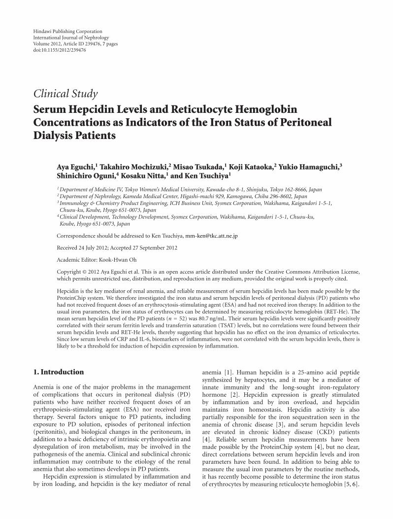
Figure 1: Correlation between serum hepcidin-25 levels andhemoglobin (Hb) concentrations.
hepcidin level of the PD patients (n = 52) was 80.7 ±59.4 ng/mL, and it was higher than the mean value reportedfor healthy subjects (10.8 ng/mL) [9]. The mean RET-Helevel of the PD patients was 32.3 ± 2.2 pg, and the normalmean that we previously reported in regular hemodialysispatients was 32.4± 4.0 pg [7].
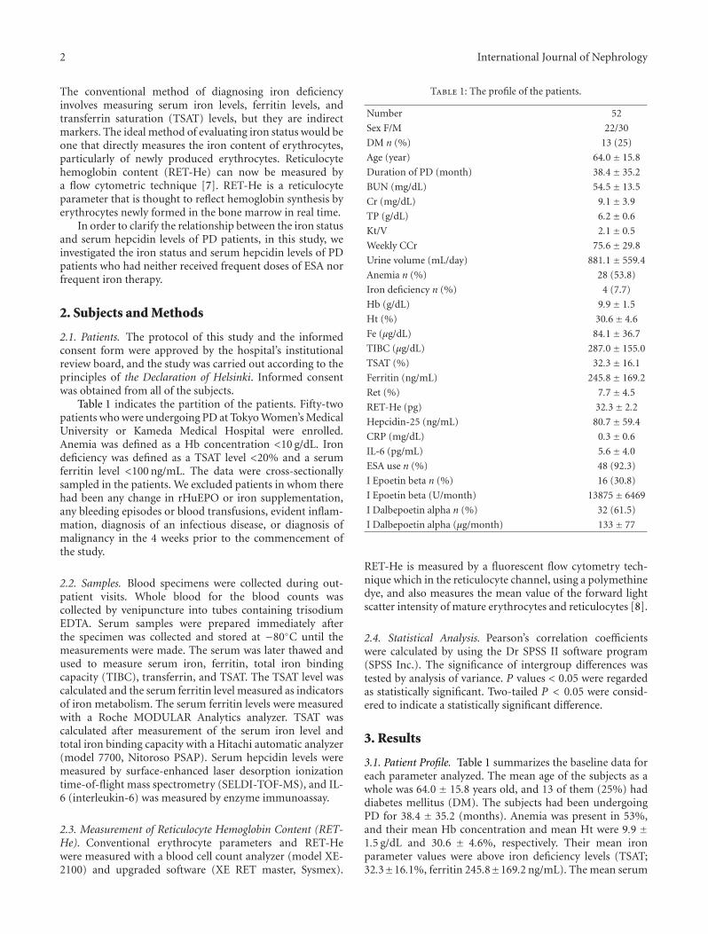
3.2. Correlation between Serum Hepcidin Levels and HbConcentrations. As shown in Figure 1, the serum hepcidinlevels were negatively correlated with the Hb concentrations.However, as to iron parameter, especially the direct ironmarker of reticulocyte, there was no correlation betweenRET-He level and Hb level (Figure 2).
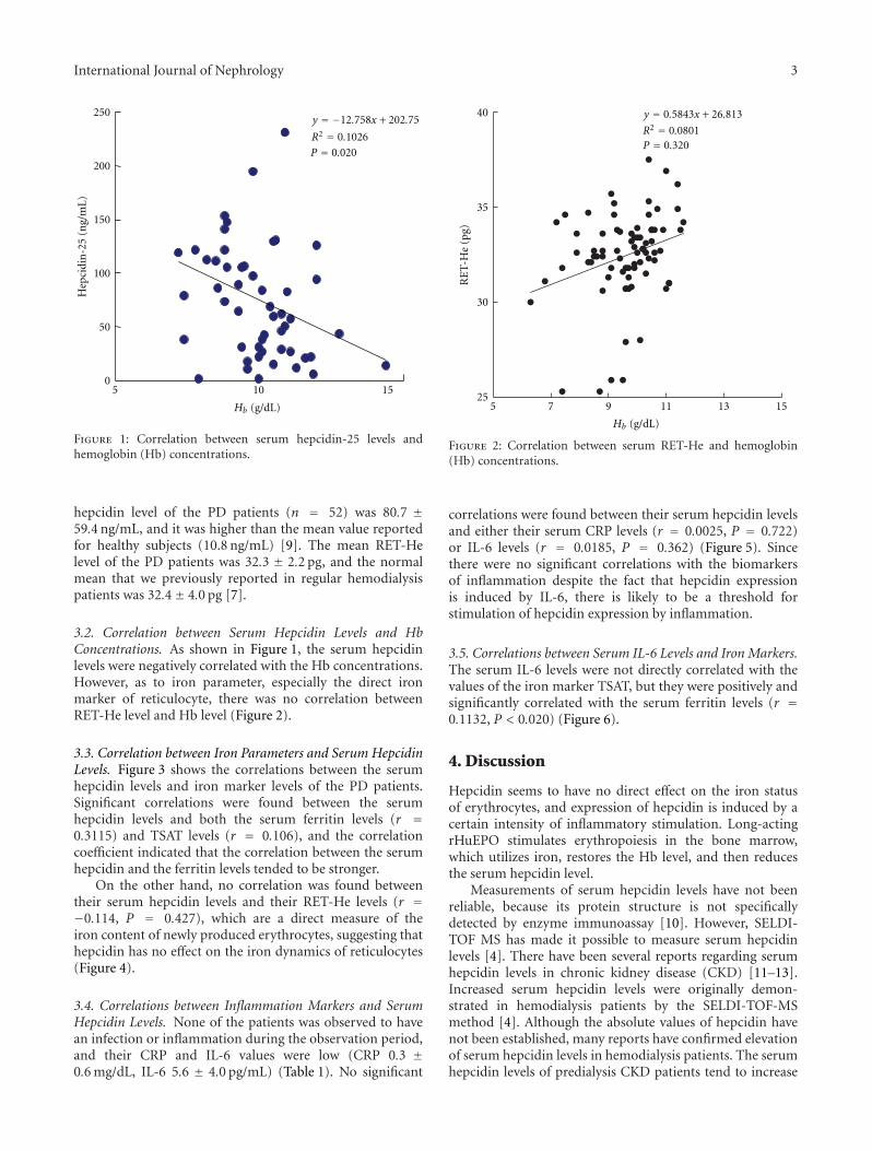
3.3. Correlation between Iron Parameters and Serum HepcidinLevels. Figure 3 shows the correlations between the serumhepcidin levels and iron marker levels of the PD patients.Significant correlations were found between the serumhepcidin levels and both the serum ferritin levels (r =0.3115) and TSAT levels (r = 0.106), and the correlationcoefficient indicated that the correlation between the serumhepcidin and the ferritin levels tended to be stronger.
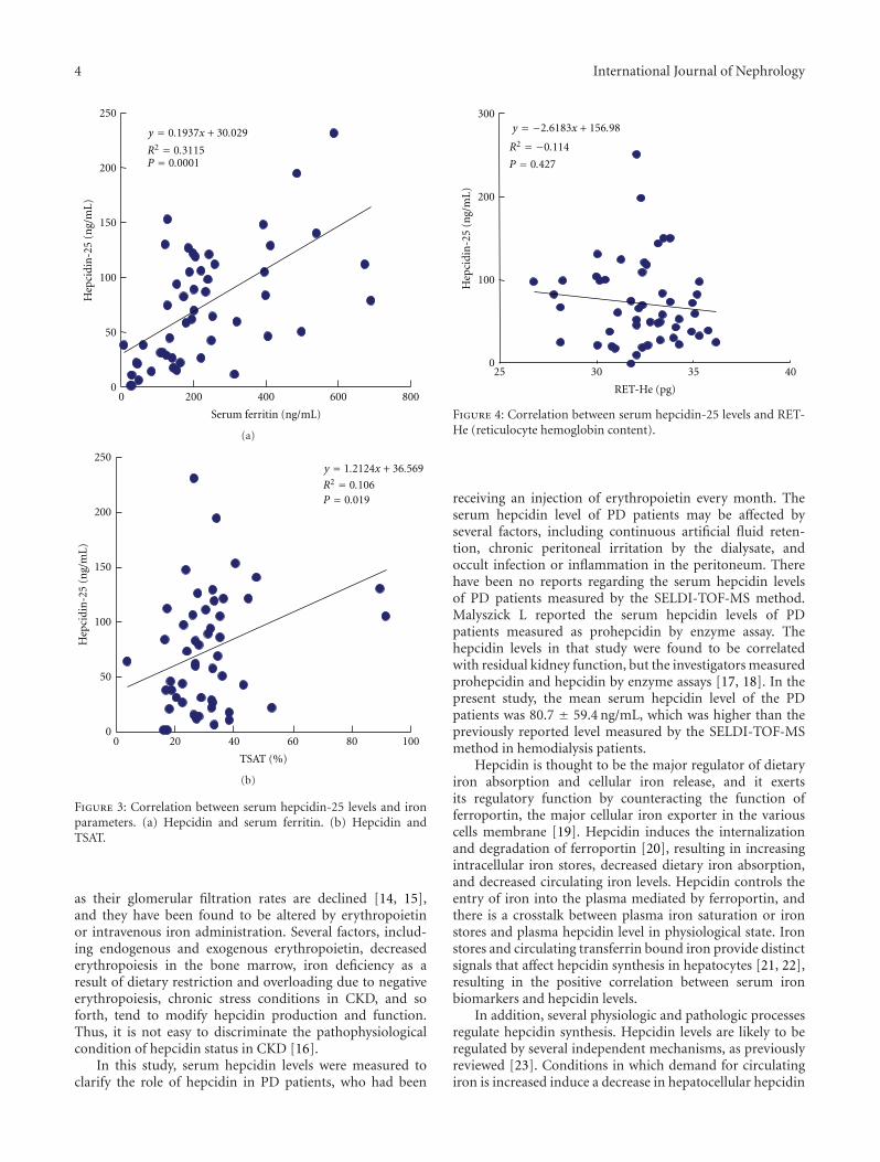
On the other hand, no correlation was found betweentheir serum hepcidin levels and their RET-He levels (r =−0.114, P = 0.427), which are a direct measure of theiron content of newly produced erythrocytes, suggesting thathepcidin has no effect on the iron dynamics of reticulocytes(Figure 4).
3.4. Correlations between Inflammation Markers and SerumHepcidin Levels. None of the patients was observed to havean infection or inflammation during the observation period,and their CRP and IL-6 values were low (CRP 0.3 ±0.6 mg/dL, IL-6 5.6 ± 4.0 pg/mL) (Table 1). No significant
y = 0.5843x + 26.813
R2 = 0.0801P = 0.320
40
35
30
255 7 9 11 13 15
RE
T-H
e (p
g)
Hb (g/dL)
Figure 2: Correlation between serum RET-He and hemoglobin(Hb) concentrations.
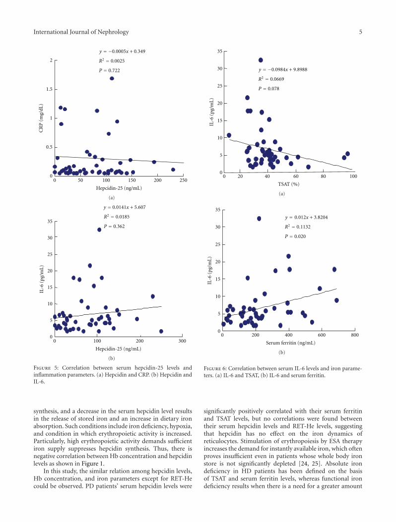
correlations were found between their serum hepcidin levelsand either their serum CRP levels (r = 0.0025, P = 0.722)or IL-6 levels (r = 0.0185, P = 0.362) (Figure 5). Sincethere were no significant correlations with the biomarkersof inflammation despite the fact that hepcidin expressionis induced by IL-6, there is likely to be a threshold forstimulation of hepcidin expression by inflammation.
3.5. Correlations between Serum IL-6 Levels and Iron Markers.The serum IL-6 levels were not directly correlated with thevalues of the iron marker TSAT, but they were positively andsignificantly correlated with the serum ferritin levels (r =0.1132, P < 0.020) (Figure 6).
4. Discussion
Hepcidin seems to have no direct effect on the iron statusof erythrocytes, and expression of hepcidin is induced by acertain intensity of inflammatory stimulation. Long-actingrHuEPO stimulates erythropoiesis in the bone marrow,which utilizes iron, restores the Hb level, and then reducesthe serum hepcidin level.
Measurements of serum hepcidin levels have not beenreliable, because its protein structure is not specificallydetected by enzyme immunoassay [10]. However, SELDI-TOF MS has made it possible to measure serum hepcidinlevels [4]. There have been several reports regarding serumhepcidin levels in chronic kidney disease (CKD) [11–13].Increased serum hepcidin levels were originally demon-strated in hemodialysis patients by the SELDI-TOF-MSmethod [4]. Although the absolute values of hepcidin havenot been established, many reports have confirmed elevationof serum hepcidin levels in hemodialysis patients. The serumhepcidin levels of predialysis CKD patients tend to increase
4 International Journal of Nephrology
y = 0.1937x + 30.029
R2 = 0.3115P = 0.0001
Serum ferritin (ng/mL)
0 200 400 600 800
250
200
150
100
50
0
Hep
cidi
n-2
5 (n
g/m
L)
(a)
y = 1.2124x + 36.569
R2 = 0.106P = 0.019
TSAT (%)
0 20 40 60 80 100
250
200
150
100
50
0
Hep
cidi
n-2
5 (n
g/m
L)
(b)
Figure 3: Correlation between serum hepcidin-25 levels and ironparameters. (a) Hepcidin and serum ferritin. (b) Hepcidin andTSAT.
as their glomerular filtration rates are declined [14, 15],and they have been found to be altered by erythropoietinor intravenous iron administration. Several factors, includ-ing endogenous and exogenous erythropoietin, decreasederythropoiesis in the bone marrow, iron deficiency as aresult of dietary restriction and overloading due to negativeerythropoiesis, chronic stress conditions in CKD, and soforth, tend to modify hepcidin production and function.Thus, it is not easy to discriminate the pathophysiologicalcondition of hepcidin status in CKD [16].
In this study, serum hepcidin levels were measured toclarify the role of hepcidin in PD patients, who had been
y = −2.6183x + 156.98
P = 0.427
Hep
cidi
n-2
5 (n
g/m
L)
300
200
100
025 30 35 40
RET-He (pg)
R2 = −0.114
Figure 4: Correlation between serum hepcidin-25 levels and RET-He (reticulocyte hemoglobin content).
receiving an injection of erythropoietin every month. Theserum hepcidin level of PD patients may be affected byseveral factors, including continuous artificial fluid reten-tion, chronic peritoneal irritation by the dialysate, andoccult infection or inflammation in the peritoneum. Therehave been no reports regarding the serum hepcidin levelsof PD patients measured by the SELDI-TOF-MS method.Malyszick L reported the serum hepcidin levels of PDpatients measured as prohepcidin by enzyme assay. Thehepcidin levels in that study were found to be correlatedwith residual kidney function, but the investigators measuredprohepcidin and hepcidin by enzyme assays [17, 18]. In thepresent study, the mean serum hepcidin level of the PDpatients was 80.7 ± 59.4 ng/mL, which was higher than thepreviously reported level measured by the SELDI-TOF-MSmethod in hemodialysis patients.
Hepcidin is thought to be the major regulator of dietaryiron absorption and cellular iron release, and it exertsits regulatory function by counteracting the function offerroportin, the major cellular iron exporter in the variouscells membrane [19]. Hepcidin induces the internalizationand degradation of ferroportin [20], resulting in increasingintracellular iron stores, decreased dietary iron absorption,and decreased circulating iron levels. Hepcidin controls theentry of iron into the plasma mediated by ferroportin, andthere is a crosstalk between plasma iron saturation or ironstores and plasma hepcidin level in physiological state. Ironstores and circulating transferrin bound iron provide distinctsignals that affect hepcidin synthesis in hepatocytes [21, 22],resulting in the positive correlation between serum ironbiomarkers and hepcidin levels.
In addition, several physiologic and pathologic processesregulate hepcidin synthesis. Hepcidin levels are likely to beregulated by several independent mechanisms, as previouslyreviewed [23]. Conditions in which demand for circulatingiron is increased induce a decrease in hepatocellular hepcidin
International Journal of Nephrology 5
y = −0.0005x + 0.349
R2 = 0.0025
P = 0.722
Hepcidin-25 (ng/mL)
2
1.5
1
0.5
00 50 100 150 200 250
CR
P (
mg/
dL)
(a)
y = 0.0141x + 5.607
R2 = 0.0185
P = 0.362
Hepcidin-25 (ng/mL)
35
30
25
20
15
10
5
00 100 200 300
IL-6
(pg
/mL
)
(b)
Figure 5: Correlation between serum hepcidin-25 levels andinflammation parameters. (a) Hepcidin and CRP. (b) Hepcidin andIL-6.
synthesis, and a decrease in the serum hepcidin level resultsin the release of stored iron and an increase in dietary ironabsorption. Such conditions include iron deficiency, hypoxia,and condition in which erythropoietic activity is increased.Particularly, high erythropoietic activity demands sufficientiron supply suppresses hepcidin synthesis. Thus, there isnegative correlation between Hb concentration and hepcidinlevels as shown in Figure 1.
In this study, the similar relation among hepcidin levels,Hb concentration, and iron parameters except for RET-Hecould be observed. PD patients’ serum hepcidin levels were
y = −0.0984x + 9.8988
R2 = 0.0669
P = 0.078
35
30
25
20
15
10
5
00 20 40 60 80 100
IL-6
(pg
/mL
)
TSAT (%)
(a)
y = 0.012x + 3.8204
R2 = 0.1132
P = 0.020
35
30
25
20
15
10
5
00 200 400 600 800
IL-6
(pg
/mL
)
Serum ferritin (ng/mL)
(b)
Figure 6: Correlation between serum IL-6 levels and iron parame-ters. (a) IL-6 and TSAT, (b) IL-6 and serum ferritin.
significantly positively correlated with their serum ferritinand TSAT levels, but no correlations were found betweentheir serum hepcidin levels and RET-He levels, suggestingthat hepcidin has no effect on the iron dynamics ofreticulocytes. Stimulation of erythropoiesis by ESA therapyincreases the demand for instantly available iron, which oftenproves insufficient even in patients whose whole body ironstore is not significantly depleted [24, 25]. Absolute irondeficiency in HD patients has been defined on the basisof TSAT and serum ferritin levels, whereas functional irondeficiency results when there is a need for a greater amount
6 International Journal of Nephrology
of iron to support erythropoiesis than can be supplied. Thus,the conventional methods of estimating iron stores, suchas serum ferritin and TSAT measurements, are inadequateto evaluate functional iron deficiency. A strong correlationbetween serum ferritin and TSAT levels and serum hepcidinlevels has been confirmed, but there is no information aboutthe relation between hepcidin and reticulocyte hemoglobin.No correlation was found between the serum hepcidin levelsand reticulocyte hemoglobin levels in this study, suggestingthat hepcidin does not directly regulate iron metabolism innewly produced erythrocytes.
The primary mediator of inflammation seems to beIL-6, which causes the signal transducer and activator oftranscription-3 to bind to the hepcidin promoter, increasingits activity [26]. Previous studies have shown markedlyincreased serum hepcidin levels in humans with chronicinfections and severe inflammatory diseases, suggesting thatelevated serum hepcidin levels play a key role in theanemia of inflammation and reticuloendothelial blockade[27]. Correlations between serum hepcidin levels and serumlevels of inflammatory markers, including IL-6, IL-1, andhigh sensitive CRP, have been found in several studies [4, 15].However, several studies have not necessarily shown therelationship between serum hepcidin levels and the levelsof these inflammatory markers [11, 12]. Since low levelsof CRP and IL-6, biomarkers of inflammation, were notcorrelated with the serum hepcidin levels, there is likelyto be a threshold for stimulation of hepcidin induction byinflammation.
5. Conclusion
SELDI-TOF-MS measurements showed that the PD patientsin this study had high serum hepcidin levels, nevertheless incase of peritonitis or in high levels of biomarker indicatinginflammation. Good correlations were found between thePD patients’ serum hepcidin levels and both their TSATferritin levels, the same as reported previously, but hepcidinwas found to have no direct effect on erythrocyte iron status.In inflammatory conditions, the primary mediator seems tobe IL-6 levels and induces hepcidin expression; there hasbeen no definite causal relationship in the regular status ofPD patients.
Conflict of Interests
All authors declared they have no conflict of interests.
Acknowledgment
This study was supported in part by the Biomarker Society.
References
[1] R. Deicher and W. H. Horl, “Hepcidin: a molecular linkbetween inflammation and anaemia,” Nephrology DialysisTransplantation, vol. 19, no. 3, pp. 521–524, 2004.
[2] G. Nicolas, C. Chauvet, L. Viatte et al., “The gene encodingthe iron regulatory peptide hepcidin is regulated by anemia,
hypoxia, and inflammation,” Journal of Clinical Investigation,vol. 110, no. 7, pp. 1037–1044, 2002.
[3] G. Weiss and L. T. Goodnough, “Anemia of chronic disease,”The New England Journal of Medicine, vol. 352, no. 10, pp.1011–1023, 2005.
[4] N. Tomosugi, H. Kawabata, R. Wakatabe et al., “Detection ofserum hepcidin in renal failure and inflammation by usingProteinChip System,” Blood, vol. 108, no. 4, pp. 1381–1387,2006.
[5] S. Fishbane, C. Galgano, R. C. Langley Jr., W. Canfield,and J. K. Maesaka, “Reticulocyte hemoglobin content in theevaluation of iron status of hemodialysis patients,” KidneyInternational, vol. 52, no. 1, pp. 217–222, 1997.
[6] C. Brugnara, B. Schiller, and J. Moran, “Reticulocyte hemo-globin equivalent (Ret He) and assessment of iron-deficientstates,” Clinical and Laboratory Haematology, vol. 28, no. 5, pp.303–308, 2006.
[7] N. Miwa, T. Akiba, N. Kimata et al., “Usefulness of measuringreticulocyte hemoglobin equivalent in the management ofhaemodialysis patients with iron deficiency,” InternationalJournal of Laboratory Hematology, vol. 32, no. 2, pp. 248–255,2010.
[8] L. Thomas, S. Franck, M. Messinger, J. Linssen, M. Thome,and C. Thomas, “Reticulocyte hemoglobin measurement—comparison of two methods in the diagnosis of iron-restrictederythropoiesis,” Clinical Chemistry and Laboratory Medicine,vol. 43, no. 11, pp. 1193–1202, 2005.
[9] T. Uehata, N. Tomosugi, T. Shoji et al., “Serum hepcidin-25 levels and anemia in non-dialysis chronic kidney diseasepatients: a cross-sectional study,” Nephrology Dialysis Trans-plantation, vol. 27, no. 3, pp. 1076–1083, 2012.
[10] I. C. Macdougall, J. Malyszko, R. C. Hider, and S. S. Bansal,“Current status of the measurement of blood hepcidin levelsin chronic kidney disease,” Clinical Journal of the AmericanSociety of Nephrology, vol. 5, no. 9, pp. 1681–1689, 2010.
[11] D. R. Ashby, D. P. Gale, M. Busbridge et al., “Plasma hepcidinlevels are elevated but responsive to erythropoietin therapy inrenal disease,” Kidney International, vol. 75, no. 9, pp. 976–981,2009.
[12] A. Kato, T. Tsuji, J. Luo, Y. Sakao, H. Yasuda, and A.Hishida, “Association of prohepcidin and hepcidin-25 witherythropoietin response and ferritin in hemodialysis patients,”American Journal of Nephrology, vol. 28, no. 1, pp. 115–121,2007.
[13] L. Valenti, D. Girelli, G. F. Valenti et al., “HFE mutationsmodulate the effect of iron on serum hepcidin-25 in chronichemodialysis patients,” Clinical Journal of the American Societyof Nephrology, vol. 4, no. 8, pp. 1331–1337, 2009.
[14] H. P. E. Peters, C. M. M. Laarakkers, D. W. Swinkels, and J. F.M. Wetzels, “Serum hepcidin-25 levels in patients with chronickidney disease are independent of glomerular filtration rate,”Nephrology Dialysis Transplantation, vol. 25, no. 3, pp. 848–853, 2010.
[15] J. Zaritsky, B. Young, H. J. Wang et al., “Hepcidin—a potentialnovel biomarker for iron status in chronic kidney disease,”Clinical Journal of the American Society of Nephrology, vol. 4,no. 6, pp. 1051–1056, 2009.
[16] J. L. Babitt and H. Y. Lin, “Molecular mechanisms of hepcidinregulation: implications for the anemia of CKD,” AmericanJournal of Kidney Diseases, vol. 55, no. 4, pp. 726–741, 2010.
[17] J. Malyszko, J. S. Malyszko, K. Pawlak, L. Drozdowska-Rams,S. Brzosko, and M. Mysliwiec, “Hepcidin Is linked to anemiaand inflammation in peritoneal dialysis patients,” PeritonealDialysis International, vol. 28, no. 4, pp. 418–421, 2008.
International Journal of Nephrology 7
[18] J. Malyszko, J. S. Malyszko, P. Kozminski, and M. Mysliwiec,“Type of renal replacement therapy and residual renal func-tion may affect prohepcidin and hepcidin,” Renal Failure, vol.31, no. 10, pp. 876–883, 2009.
[19] T. Ganz, “Molecular control of iron transport,” Journal of theAmerican Society of Nephrology, vol. 18, no. 2, pp. 394–400,2007.
[20] E. Nemeth, M. S. Tuttle, J. Powelson et al., “Hepcidin regulatescellular iron efflux by binding to ferroportin and inducing itsinternalization,” Science, vol. 306, no. 5704, pp. 2090–2093,2004.
[21] M. W. Hentze, M. U. Muckenthaler, B. Galy, and C. Cam-aschella, “Two to tango: regulation of mammalian iron metab-olism,” Cell, vol. 142, no. 1, pp. 24–38, 2010.
[22] D. W. Coyne, “Hepcidin: clinical utility as a diagnostic tooland therapeutic target,” Kidney International, vol. 80, no. 3, pp.240–244, 2011.
[23] B. Young and J. Zaritsky, “Hepcidin for clinicians,” ClinicalJournal of the American Society of Nephrology, vol. 4, no. 8, pp.1384–1387, 2009.
[24] I. C. Macdougall, R. D. Hutton, I. Cavill, G. A. Coles, and J. D.Williams, “Poor response to treatment of renal anaemia witherythropoietin corrected by iron given intravenously,” BritishMedical Journal, vol. 298, no. 6692, pp. 157–158, 1989.
[25] D. B. Van Wyck, J. C. Stivelman, J. Ruiz, L. F. Kirlin, M. A. Katz,and D. A. Ogden, “Iron status in patients receiving erythropoi-etin for dialysis-associated anemia,” Kidney International, vol.35, no. 2, pp. 712–716, 1989.
[26] G. Papanikolaou, M. Tzilianos, J. I. Christakis et al., “Hepcidinin iron overload disorders,” Blood, vol. 105, no. 10, pp. 4103–4105, 2005.
[27] E. Nemeth, S. Rivera, V. Gabayan et al., “IL-6 mediateshypoferremia of inflammation by inducing the synthesis ofthe iron regulatory hormone hepcidin,” Journal of ClinicalInvestigation, vol. 113, no. 9, pp. 1271–1276, 2004.


![7976].pdf · Design and specifications may be subject to change due to further product development. SYSMEX CORPORATION 1-5-1, Wakinohama-Kaigandori. Chuo-ku …](https://static.documents.pub/doc/80x56/5b85404b7f8b9ab7618d60c9/7976pdf-design-and-specifications-may-be-subject-to-change-due-to-further.jpg)


