UNIVERSITA’ DEGLI STUDI DI MILANO Facoltà di Medicina e Chirurgia Dipartimento di Scienze Biomediche per la Salute Scuola di Dottorato in Scienze Morfologiche e Fisiologiche XXV ciclo SGLT-1: A NEW THERAPEUTIC STRATEGY TO MAINTAINS INTESTINAL EPITHELIAL INTEGRITY AND BARRIER FUNCTION Settore Disciplinare BIO16 Tutor: Prof. Cristiano Rumio Coordinatore: Prof.ssa Laura Vizzotto Tesi di Dottorato di Ricerca di Diego Cardani Matr. R08679 Anno Accademico 2012-2013
Transcript
UNIVERSITA’ DEGLI STUDI DI MILANO
Facoltà di Medicina e Chirurgia
Dipartimento di Scienze Biomediche per la Salute
Scuola di Dottorato in Scienze Morfologiche e Fisiologiche
XXV ciclo
SGLT-1: A NEW THERAPEUTIC STRATEGY
TO MAINTAINS INTESTINAL EPITHELIAL INTEGRITY
AND BARRIER FUNCTION
Settore Disciplinare BIO16
Tutor: Prof. Cristiano Rumio
Coordinatore: Prof.ssa Laura Vizzotto
Tesi di Dottorato di Ricerca di
Diego Cardani
Matr. R08679
Anno Accademico 2012-2013
2
INDEX
1 INTRODUCTION……………………………………………………………………3
2 MATHERIALS AND METHODS…………………………………………………21
3 RESULTS…………………………………………………………………………..31
4 DISCUSSION………………………………………………………………………53
5 CONCLUSIONS…………………………………………………………………....60
6 REFERENCES……………………………………………………………………..61
3
1. INTRODUCTION
1.1 EPITHELIAL BARRIER: THE IMPORTANCE OF BEING IN TACT
Epithelial barrier and its role in intestinal homeo stasis maintenance
Pluricellular organisms interface with their external environments at multiple sites,
including mucosae of the airway, oral cavity, digestive tract and genitourinary tract, and the
skin. Although the skin is the most visible site of interface, the combined area of the
mucosal surface is much greater than that of the skin. Epithelial cells that define the
interface between the organism and external world forms barriers that are essential to life.
This is particularly true in the intestine, where the epithelial barrier supports nutrient and
water transport while preventing microbial contamination of the interstitial tissue.
Gut epithelial surfaces are composed of cells lineages that arise from a pluripotent stem
cell progenitor: absorptive enterocytes, globet cells, enteroendocrine cells and Paneth
cells (1). All cellular types contribute in a unique way to mucosal defense and the
maintenance of barrier integrity. Goblet cells, both the small and large intestines, secrete
mucus, which is composed mainly of highly glycosylated proteins that form a protective
layer on the epithelium surface. Mucus acts trapping the vast majority of antigens that, in
the intestinal tract, are cleared by peristalsis. By keeping microbes moving, these physical
factors limit the time available for adherence to the epithelia and thus restrict invasion. The
importance of mucus gel hydration is shown by cystic fibrosis, in which the production of
hyper-viscous mucus contributes to pulmonary, pancreatic and intestinal disease (2).
Defective mucus production has also been reported in various immune-mediated
diseases, and spontaneous colitis develops in mice that lack specific mucin genes (3).
In the small intestine, Paneth cells are the key effectors of antimicrobial defense. These
specialized epithelial cells are situated at the base of small intestinal crypts and harbor
secretory granules containing several microbicidal proteins including defensins, lysozyme,
and phospholipase A2. Paneth cells sense bacterial proximity and react by discharging
4
their microbicidal granule contents into the gut lumen (4). Only the microbes that can by-
pass innate defenses have the ability to infect and cause disease.
The epithelial and sub-epithelial region of mucosal surfaces contains abundant
immunocytes of many varieties. Indeed, in a healthy human adult, the mucosal immune
system contributes almost 80% of all immunocytes (5). The number of immunoglobulin
(Ig)-secreting cells in this population exceeds by several fold the number of Ig-secreting
cells in all other lymphoid organs (spleen, bone marrow, and lymph nodes) combined (6).
Importantly, these cells also have the ability to directly sense danger through expression of
surface receptors specific for conserved microbial products. Under the influence of signals
from the epithelium, stimulation through these receptors induces these cells to secrete
cytokines and other modulatory factors. These factors work in concert with the epithelial
signals to alert and dictate adaptive responses (7). In this way, cooperation of leukocytes
and epithelial cells extends the definition of the innate immune system to include all cells
of the body (8).
Enteroendocrine cells (EEC) form the largest endocrine system in the body. They secrete
multiple regulatory molecules which control physiological and homeostatic functions,
particularly postprandial secretion and motility. Their key purpose is to act as sensors of
luminal contents, either in a classical endocrine fashion, or by a paracrine effect on
proximate cells, notably vagal afferent fibers (9). They also play a pivotal role in the control
of food intake, and emerging data add roles in mucosal immunity and repair.
The primary responsibility for mucosal barrier function resides in the epithelial cell plasma
membrane, which is impermeable to most hydrophilic solutes (in the absence of specific
transporters). Accordingly, direct epithelial cell damage, such as that induced by mucosal
irritants or cytotoxic agents, including surfactants and some drugs used for cancer
chemotherapy, results in a marked loss of barrier function. However, in the presence of an
intact epithelial cell layer, the paracellular pathway between cells must be sealed or
5
modulated in physiological conditions. This function is mediated by an apical junctional
complex, formed by tight junction and adherents junctions (10; 11) (Figure 1).
Figure 1: Distinct subpopulations of IEC cells. IECs are integrated into a layer presenting two main regions, apical and basolateral. Enterocytes, in the small intestine, and colonocytes in the large intestine, as well as specialized Paneth cells in the bases of small intestinal crypts continually senses the microbiota to induce the production of antimicrobial peptides (AMPs). Goblet cells produce mucus, organized into a dense gel that forms an IEC-adherent inner mucous layer, and a less dense outer mucous layer. The outer layer is highly colonized by constituents of the microbiota. The inner mucous layer is largely impervious to bacterial colonization or penetration due to its high concentration of bactericidal AMPs.
Junctional systems complexes are key players of the epithelial barrier effect
The elements of the complexes, identified as zonula occludens (tight junction), zonula
adhaerens (intermediary junction), and macula adherens (desmosome), occupy a
juxtaluminal position and succeed each other in the order given in an apical-basal
direction.
The zonula occludens (tight junction) is characterized by fusion of the adjacent cell
membranes resulting in closure of the intercellular space at variable distances. Within the
6
closed region, the dense outer leaflets of the adjoining cell membranes converge to form a
single intermediate line. A diffuse layer of dense cytoplasmic material is often associated
with this junction and its development varies from one epithelium to another.
The zonula adhaerens (intermediate junction) is characterized by the presence of an
intercellular space (~200 Å) occupied by homogeneous, apparently amorphous material of
low density; by strict parallelism of the adjoining cell membranes over distances of 0.2 to
0.5 µm; and by conspicuous bands of dense material located in the subjacent cytoplasmic
matrix.
The desmosome or macula adhaerens is also characterized by the presence of an
intercellular space (~240 Å) which, in this case, contains a central disc of dense material;
by discrete cytoplasmic plaques disposed parallel to the inner leaflet of each cell
membrane; and by the presence of bundles of cytoplasmic fibrils converging on the
plaques (12-14) (Figure 2).
Figure 2: scheme of adherens junctions main components
7
Tight junctions are multi-protein complexes composed of transmembrane proteins,
peripheral membrane (scaffolding) proteins and regulatory molecules including kinases
(Figure 3).
Tight junctions limit solute flux along the paracellular pathway, which is typically more
permeable than the transcellular pathway. Tight junctions have also the role of rate-limiting
step in transepithelial transport and are the principal determinant of mucosal permeability.
Thus, it is important to understand the specific barrier properties of the tight junction, which
can be defined in terms of size selectivity and charge selectivity.
A second pathway is characterized by small pores that are thought to be defined by tight
junction-associated claudin proteins, which are also primary determinants of charge
selectivity. These pores have a radius that excludes molecules larger than 4 Å.
The expression of claudins varies between organs and even within different regions of a
single organ and, as detailed below, can be modified by external stimuli, such as
cytokines. Thus, tight junctions show both size selectivity and charge selectivity, and these
properties may be regulated individually or jointly by physiological or pathophysiological
stimuli.
8
Figure 3: Tight Junctions structure and mayor components
At present, the most well-studied tight junction proteins are the claudins, a large family that
includes at least 24 members (15-16). These proteins have four transmembrane helices
with a very short intracellular amino-terminal sequence and a somewhat longer C-terminal
tail. The first extracellular loop is approximately 50 residues long, although there is some
variation among claudin family members. More importantly, sequence variation within the
first extracellular loop determines tight junction charge selectivity (17), consistent with the
view that the array of claudin proteins expressed in a given cell type defines the
paracellular pore (18; 19) through which ions and perhaps nonionic solutes travel. The
second extracellular loop of claudins is smaller than the first, ranges from 16 to 33 amino
acids in length, and is poorly characterized. The cytoplasmic C-terminal tail varies in length
from 21 to 63 residues and is the least conserved region of claudin proteins, suggesting
that the tail may also be a site of functional regulation. Importantly, almost all claudin
proteins present a three-aminoacid terminal motif that binds to PDZ domains (Figure 4).
Tight junction proteins panel include ZO-1 as well as the related proteins ZO-2 and ZO-3
(20); each of these proteins contains three PDZ domains within the amino-terminal portion
9
of the protein. PDZ domains, which were initially identified in PSD95, DlgA, and ZO-1, are
specialized for protein interactions. In the case of ZO-1, ZO-2, and ZO-3, the most
aminoterminal PDZ domain binds to the C-terminal tail of claudins (21). This is functionally
critical because deletion of the PDZ-binding motif prevents efficient claudin targeting to the
tight junction. Moreover, ZO-1 and ZO-2 are each able to direct claudins to developing
tight junctions (22); simultaneous elimination of ZO-1 and ZO-2 expression blocks claudin
recruitment and tight junction formation with consequent lost of barrier function (23).
Several other groups of proteins, including transmembrane, cytoskeletal, and signaling
proteins (24) complete the tight junction complex; in particular actin and myosin are
essential structural elements of the tight junction (25).
Figure 4: PDZ domain interactions and the regulation of EMT and Wnt signalling. Schematic diagram of some of the critical regulators involved in the regulation of the canonical and non-canonical Wnt signalling pathways. Interlinked with this are PDZ interactions important in the regulation of EMT. Proteins possessing PDZ domains are shown in red and those possessing PBMs are shown in blue. β-CAT, β-catenin; CK2, casein kinase 2; GSK-3β, glycogen synthase kinase 3β; PCP, planar cell polarity; TCF, T-cell factor; WGEF, weak-similarity GEF.
10
The best-characterized example of tight junctions regulation in response to physiological
stimuli is represented by the reversible increase in intestinal paracellular permeability
induced by apical Na+/nutrient co-transport (26; 27); increase in paracellular permeability is
thought to allow passive paracellular flux of nutrients, along with water. Ultrastructural
analyses of tight junctions showed that activation of Na+/nutrient co-transport induced
condensation of perijunctional microfilaments (28), interpreted as an indicator of
actomyosin contraction. Unfortunately no tools needed to study these biochemical events
in intact tissue are still developed. To solve problem, an in vitro model that simulate
Na+/glucose co-transport–induced tight junction regulation was developed through use of
cultured intestinal epithelial cell monolayers (29). Based on the ultrastructural data,
phosphorylation of myosin II regulatory light chain (MLC), a trigger for actomyosin
contraction, was examined and shown to increase following activation of Na+/glucose co-
transport (30). Moreover, MLC-kinase (MLCK) inhibition prevented both MLC-
phosphorylation and barrier regulation induced by Na+/glucose co-transport in cultured
monolayers and also blocked barrier regulation in intact intestinal mucosa (31). These data
suggest that MLCK is a critical physiological regulator of tight junction permeability.
Furthermore, in vitro studies of epithelial cells with inducible expression of constitutively
active MLCK demonstrate that enzymatic MLCK activation is sufficient to trigger
downstream events necessary for barrier regulation (32).
Immune stimulation and barrier regulation: the fund amental role of apoptotic
signals
The ability of cytokines, such as TNF and IFNγ, to regulate the function of the tight junction
(33) barrier was first described 20 years ago. Since then, increased tight junction protein
transcription, vesicular removal of proteins from the tight junction, tight junction protein
degradation, kinase activation and cytoskeletal modulation have all been proposed to
mediate cytokine-induced loss of tight junction barrier function (34). Although extensive
11
apoptosis of epithelial cells may also cause barrier loss, the relevance of single-cell
apoptosis to barrier dysfunction remains controversial owing to differing results in diverse
experimental systems.
TNF and IFNγ modify tight junction barrier function in intestinal, renal, pulmonary and
salivary gland epithelia as well as between endothelial cells. The effects of TNF on barrier
integrity have been best studied in the gut (35), where this cytokine has a central role in
many diseases associated with intestinal epithelial barrier dysfunction, including
inflammatory bowel disease, intestinal ischaemia and graft-versus host disease. For
example, although the effect of therapy with TNF-specific antibodies may be largely due to
the overall reduction in inflammation. Increased mucosal TNF production may also
contribute to increased intestinal permeability and susceptibility to colitis in mice with
defective mucin biosynthesis. MLCK has been shown to have a central role in TNF
induced epithelial and endothelial barrier deregulation, both in vitro and in vivo (36).
Similar to Na+/nutrients co-transport, TNF-induced MLCK activation seems to increase
paracellular flux through the leak pathway. Similarly, MLCK expression and activity are
increased in intestinal epithelial cells of patients with inflammatory bowel disease.
1.2 BARRIER BREAKDOWN: THE PANDORA'S BOX OPENED
Chemotherapy-induced gastrointestinal mucositis
Many anti-cancer therapeutic strategies based on radiotherapy and chemotherapy are
effective in the treatment of malignant disease but, conversely, present a plethora of side
effects including oral and gastrointestinal mucositis. There is no approved therapy to
prevent or treat chemotherapy-induced mucositis at present and the development of an
effective intervention is seen as a high priority in oncological supportive care (37-39).
Severe mucositis is especially common among patients who receive aggressive
myeloablative chemotherapy and in patients who receive radiation therapy as treatment for
cancers of the oral cavity, oropharynx, nasopharynx and salivary glands. Patients with
12
severe mucositis develop ulcerations that penetrate fully into the submucosa and cause
severe pain, which routinely necessitates narcotic analgesia.
In addition to the symptoms of mucositis and its impact on quality of life, mucositis
adversely affects a variety of other health and economic outcomes. Among patients who
have received chemotherapic treatments, the presence of mucositis results in lengthened
hospital stays, increased use of resources and higher costs. Mucositis also threatens the
efficacy of treatment plans by necessitating breaks in chemotherapy, reductions in doses
of drugs used in chemotherapy and modifications in the selection of anti-neoplastic agents.
Anthracyclines like doxorubicin are among the most widely used and effective
chemotherapeutic agents but also the use of which often causes the onset of mucositis
(especially in combination with 5-fluorouracil) (40-42).
One of the most important event that compromise the intestinal epithelium biological
functions (like nutrients absorptions and barrier effect against pathogens invasion) is the
lack of the correct permeability caused by alterations of the junctional systems (43; 44) of
epithelial cells in response to apoptotic events promoted by chemotherapeutic agents,
events like apoptosis and inflammation, which affect the tunica mucosa, the vascular
endothelium and the whole body, contribute to the onset of mucositis. One of the most
important apoptotic pathways involves caspases; in particular caspase-3 that is connected
with junctional damages; two target proteins for Tight Junctions and Adherent Junctions,
zonula occludens-1 (ZO-1) and beta-catenin respectively, are down-regulated in presence
of chemotherapy-induced apoptosis. (45; 46) Because the gastrointestinal tract present a
vast array of microorganisms, loss of epithelial integrity, especially when patients undergo
myeloablation, markedly increases the risk of bacteraemia, fungaemia and sepsis (47; 48)
Inflammatory Bowel Diseases (IBD)
Idiopathic IBDs such as Crohn disease and ulcerative colitis occur in clinically
immunocompetent individuals whose characteristic symptoms and signs arise from a
13
robust, cytokine-driven (yet noninfectious) inflammation of the gut (49). Crohn disease is
associated with excess IL-12/IL-23 and IFN-γ/IL-17 production that affects the small bowel
and colon with discontinuous ulceration and full thickness bowel wall inflammation often
including granulomas. Patients report gastrointestinal symptoms of abdominal pain,
diarrhea, and rectal bleeding as well as systemic symptoms of weight loss, fever, and
fatigue. Crohn disease patients can also develop obstructing structures of the bowel and
inflammatory connections (fistulae) (Figure 5) between segments of bowel or between the
bowel and skin and other organs. In comparison, ulcerative colitis is associated with
excess IL-13 production, primarily affecting the colon, with a continuous inflammation of
the mucosa nearly always involving the rectum and extending proximally (50).
It has been proposed that three main features should be present for IBD to occur: a
genetically susceptible mucosal immune system; an antigen, or pro-inflammatory
compound, which reaches the gut and can trigger the susceptible immune system; and an
alteration in gut barrier function which allows antigens to have contact with the mucosal
immune system (51).
Since the studies about Crohn's disease have shown immunological factors underlying
onset of this disease, the conventional treatments for IBD as corticosteroids, mesalamine,
and immunosuppressants, provide mostly to block downstream inflammatory events.
Medical therapy relies on classic anti-inflammatory and immunosuppressant drugs:
corticosteroids, mesalamine compounds, azathioprine, and derivatives of the latter. (52-
54). Newer biological drugs such as anti–TNF-α antibodies targeting the general
inflammatory cytokine, TNF-α, have added greatly to our ability to control IBD, but even
this therapy is limited by lack or loss of efficacy and associated toxicities (55). Emerging
therapies for IBD are focusing on major effector cytokines as they are identified in ongoing
investigations, for instance using an anti–IL-12p40 antibody to neutralize the effects of IL-
12 and IL-23 in Crohn disease (56).
14
Increased paracellular permeability was reported in patients with Crohn’s disease (CD)
over 25 years ago (57). Although such barrier loss may be caused by erosions that occur
in active disease, some approaches used to measure permeability partially excluded this
possibility by normalizing absorption of the inert sugar lactulose to that of the smaller, and
more easily absorbed, inert sugar mannitol (58) .
In recent years, several lines of evidence suggested that an increased intestinal
permeability play a central role in the pathogenesis of IBD. In different animal models of
Crohn's disease, an increased small intestinal permeability has been shown, even before
disease expression, and the reversal of this barrier defect can attenuate the disease,
implying that the increased permeability is not simply an epiphenomenon but rather is an
important etiological event (59-61). In the human condition it is also evident that increased
small intestinal permeability is commonly observed in populations at high risk of
developing Crohn's disease (62; 63). This would suggest that there is a subtle alteration of
function, independent of inflammation, which can be visible as an increase in paracellular
permeability. This raises the possibility that restoration of barrier function may be
therapeutic in CD Consistent with this hypothesis, emerging data indicate that inhibition of
cytoskeletally mediated barrier dysfunction may be able to prevent disease progression.
Barrier restoration may, therefore, represent a non-immunosuppressive approach to
achieving or maintain disease remission.
15
Figure 5: Classical representation of Crohn Disease lesions
1.3 SODIUM-DEPENDENT GLUCOSE TRANSPORTER-1
SGLT1: classical transporter and novel receptor. Tw o faces of the same medal
The primary functions of the gastrointestinal (GI) tract have traditionally been perceived to
be limited to the digestion and absorption of nutrients and electrolytes and to water
homeostasis but the intestinal epithelial barrier also controls the equilibrium between
tolerance and immunity to non-self-antigens by regulating antigen trafficking both through
the transcellular and paracellular pathways.
Luminal content influences intestinal epithelium function and the components of diet
certainly play a role in the modulation of enterocytes response. The large surface of the
gut is a wide area to protect and constitutes an easy way to the whole organism for
pathogen bacteria. On the other hand a so large line on external environment is necessary
to an appropriate absorption of the nutrients. Often the absorption is a transport against
gradient because the concentration of nutrient molecules is lower in the lumen than inside
the cells. For this reason a wide range of transporters are evolved in order to guarantee
16
the absorption of water and electrolytes, minerals and vitamins, sugars, fatty acids, amino-
acids and small peptides. The same molecules probably exert different effects on the role
of IECs in mucosal immunity. D-glucose, in particular, is prevalently considered as
energetic substrate and other possible roles are poorly analyzed.
Glucose is among the more abundant monosaccharide in the diet, and its absorption in the
intestine is mediated by the high-affinity, sodium-dependent glucose cotransporter (SGLT-
1), located in the apical membrane of enterocytes.
Recently SGLT-1 has been described as involved in the repair of plasma membrane
integrity and tight junction (TJ) integrity injured by heat stress or cisplatin chemo-treatment
(45; 64) In our recent papers we demonstrate the protective effect of the activation of
sodium-dependent glucose transporter-1 (SGLT-1) on damages induced by TLRs ligands
in intestinal epithelial cells, in a murine model of septic shock and in LPS-induced liver
injury, as well as liver injury and death induced by an overdose of acetaminophen (65; 66).
Intestinal glucose absorption and the role of SGLT- 1
D-glucose is the major source of energy in mammals and it is absorbed by the epithelial
cells lining the surface of the small intestine. The transport of glucose from lumen to
enterocytes in the small intestine is principally mediated by Na+-dependent glucose co-
transporters (SGLTs, members of a larger family of Na+-dependent transporters, gene
name SLC5A) that use the Na+-electrochemical gradient at the generated by the Na+/K+-
ATPase to transport glucose into cells (Figure 6)
SGLT-1 is the most important active glucose transporter at intestinal level and it is found in
brush border membrane of mature enterocytes in the small intestine. During the process of
intestinal sugar transport, on the luminal side of the brush border membrane, two Na+ ions
bind to SGLT-1 and produce a conformational change that permits sugar binding. Another
conformational change allows the substrates to enter the enterocyte. The sugar, followed
by the Na+, dissociates from SGLT-1 because the affinity of the cytosolic sites is low, and
17
also because the intracellular concentration of Na+ is low (10 vs 140 mEq/L). The Na+/K+-
ATPase in the basolateral membrane is responsible for maintaining the Na+ and K+
electrochemical gradients across the cell membrane (67-70).
Figure 6: schematic model of intestinal sugar transport. SGLT1 is the sodium dependent glucose/galactose transporter on the brush border membrane. The Na+K+-ATPase on the basolateral membrane maintains the gradient necessary for the functioning of SGLT1. GLUT2 transports glucose, galactose and fructose out of the cell. IF images of SGLT-1 in villus brush border.
SGLT-1 as a new player in epithelial homoeostasis m aintenance. Engagement with
natural and synthetic molecules.
Since 2005 SGLT-1 assumes a novel physiological role. SGLT-1 activation loads to a local
and systemic anti-inflammatory and cytoprotective response in different inflammatory
conditions. Firstly the effect of glucose has been analysed in enterocytes exposed to
agonists of TLR4 and TLR9. The encouraging in vitro results prompted us to analyze the
effects of glucose on a systemic inflammatory response syndrome (endotoxic shock) in
mice (Figure 7).
18
Figure 7: schematic representation of SGLT-1 activation pathways in different cases of tissue damage
Oral ingestion of glucose was found to protect 100% of mice from lethal endotoxic shock
induced by intraperitoneal LPS administration (65); protection was only observed when
glucose was administered orally, not by i.p. route, suggesting the important role of
intestinal epithelial cells. Subsequently the study investigated the possibility that orally
administered D-glucose exerts a systemic anti-inflammatory activity on hepatic liver failure.
In this study (66) that D-glucose prevents LPS-induced liver injury, as well as liver injury
and death induced by acetaminophen overdosing. In both of these models, physiological
liver morphology is maintained and organ protection was confirmed also by unchanged
levels of circulating markers of hepatotoxicity, such as ALT or LDH. In addition, D-glucose
was found to protect liver from alpha-amanitin intoxication. In this case, however, a second
signal had to be present in addition to glucose in order to achieve protective efficacy.
In addition, we observed that the in vivo protection depends from a systemic increase of
anti-inflammatory cytokine IL- 10. The milestone of the observed immunomodulatory
effects resides in activation of SGLT-1; in fact, the glucose analogue 3-OMG, which
induces the transporter activity but is not metabolized, exerted the same effects as glucose
both in vitro and in vivo. Unfortunately 3-OMG is active at the same high concentration of
19
D-glucose and in order to solve this problem we decided to screen a library of synthetic
glucose analogues as ligands for our experiments.
BLF501: origin of the synthetic compound
In the search for non-metabolizable glucoderivatives able to “activate” the SGLT-1
transporter at pharmacological concentrations, the chemical researcher team generated a
small library of naphthyl C-glycoside and glycitol derivatives, mostly without the use of
protecting groups. We tested in vitro anti-inflammatory activity of different compounds,
among them, we have selected a synthetic SGLT-1 ligand, named BLF501 (figure 8),
which binds to the transporter with high potency, i.e. at concentrations 5 orders of
magnitude lower than glucose.
O
HO
HO
OH
HO
NH
S OO
NMe2
OOMe
HO
HO
OH
HO
O
HO
HO
O
HO
OH
O
HO
HO
OH
HO
NH2
1 2
BLF501
a b c
1
23
4
5
6
1'
2'
Figure 8: Synthesis and Structure of BLF501
BLF501, a member of the C-glycoside class, was designed to resist glucose metabolism
and to retain hydrolytic stability in the acidic and basic conditions found in the
gastrointestinal tract. Stability of BLF501 in different pH conditions was tested in samples
prepared by dissolving 2.3 mg in 550 µl of D2O and adjusting the pH to 1.2 through DCl
addition (acidic conditions) or by dissolving 3 mg of BLF501 in 550 µl of D2O and adjusting
the pH to 12.5 through NaOD addition (basic conditions). Both samples were kept at room
temperature until 1H-NMR spectra were recorded at 10 min, 4.5 h and 24 h. As shown in
Figure 2, neither sample evidenced degradation in 24 h. BLF501 matches with the
fundamental characteristic of absence of metabolic activity. In fact in the field of C-
glycosides like BLF501 no relevant bioactivity has been reported until now (71), allowing
us to consider BLF501 a non-metabolizable glucose derivate.
20
3 mg pH 12.5 (D20, NaOD) 2.3 mg pH 1.2 (D20, DCl)
Figure 9. 1H-NMR spectra of BLF501 at pH 12.5 and at pH 1.2.
1.4 AIM OF THE STUDY:
The aim of this work is to demonstrate that SGLT-1 engagement via orally administered
ligands, in particular the synthetic one BLF501, could activate protective mechanisms at
intestinal epithelial level, preserving correct epithelial morphology and permeability and
accelerating recovery of homeostatic conditions.
ppm (f1)
2.03.04.05.06.07.08.0
10 min
4.5 h
24 h
ppm (f1)
2.03.04.05.06.07.08.0
10 min
4.5 h
24 h
ppm (f1)
2.03.04.05.06.07.08.0
10 min
4.5 h
24 h
ppm (f1)
2.03.04.05.06.07.08.0
10 min
4.5 h
24 h
21
2. MATERIALS AND METHODS:
2.1 SGLT-1 ACTIVATION IN GIM MODELS:
Chronic and acute in vivo experiments were performed on mice strain BALB/C purchased
from Charles River Italy. The mice used for the different treatments were housed in
specific aseptic conditions, with a constant temperature and humidity and received food
and water ad libitum. The animals were divided randomly into groups and assigned to two
treatment protocols.
Chronic treatment:
15 animals formed the control group; 15 animals formed the group treated with doxorubicin
and 5-fluorouracil, 15 animals formed the group treated with doxorubicin and 5-fluorouracil
and BLF501 25 µg/Kg; 15 animals formed the group treated with doxorubicin and 5-
fluorouracil and BLF501 2.5 µg/Kg; 15 animals formed the group treated with doxorubicin
and 5-fluorouracil and BLF501 0.25 µg/Kg and 15 animals formed the group treated with
(Euroclone). For Caco-2/bbe/SGLT1 cells, 250 µg/ml G418 gentamicin bisulfate salt
solution (Sigma-Aldrich) was added as antibiotic agent; for normal Caco-2/bbe
penicillin/streptamicin solution (Euroclone) was added.
A431 cells were cultured in RPMI 1640 medium (Euroclone) supplemented with 10% FBS
(Euroclone), 1% glutamine (Euroclone) and 1% penicillin/streptamicin solution (Euroclone).
Cell treatments
For viability assays 1 x 10⁵ Caco-2 cells were plated on 96 flat-bottom well plates and
treated after 24 hours with doxorubicin (100 µM) with or without BLF501 11.4 µM. Cell
viability was evaluated after 48 h with Neutral Red assay.
2.2 SGLT-1 ACTIVATION IN IBD MODELS
Mice and in vivo treatments
C57BL/6 female mice were purchased from Charles River, Italy (Head Office Wilmington,
MA). Mice were housed under specific pathogen-free conditions, maintained at constant
temperature and humidity, with food and water given ad libitum, and used at 8-12 weeks of
age. Experimental protocols were approved by the Ethics Committee for Animal
27
Experimentation of Istituto Nazionale Tumori, Milano, and carried out according to
guidelines of the United Kingdom Co-ordinating Committee on Cancer Research for
animal welfare in experimental neoplasia (1998).
Induction of acute and chronic colitis
Mice weighting 20-22 g received 2% dextran sodium salt (m. m. 40 kD, MP Biomedicals,
Irvine, CA) ad libitum in filter-purified drinking water for 7 days. For treatments studies
mice undergoing DSS from day 4 at day 7 received oral administration of BLF501 250
µg/kg or 25 µg/kg or 2.5 µg/kg once day. BLF501 was administrated with a sterile gastric
tube In other mice groups received only BLF501 250 µg/kg or 25 µg/kg or 2,5 µg/kg or
glucose 2,5 g/kg. One group received only DSS 2% in drinking water.
Day 1 Day 2 Day 3 Day 4 Day 5 Day 6 Day 7 Day 8 DSS 2% DSS 2% DSS 2% DSS 2%
+BLF501 DSS 2% +BLF501
DSS 2% +BLF501
DSS 2% +BLF501
Sacrifice
Table 2 . Schematic presentation of different treatments in the time. Mice received DSS 2% in drinking water. BLF501 250 µg/kg or 25 µg/kg or 2,5 µg/kg were administrated (diluted in physiological solution) once per day at day 4, 5, 6, 7 and at 8th mice were sacrificed. Groups Treatment 1 Untreated 2 DSS 2% only 3 DSS 2% + BLF501 250 µg/kg 4 DSS 2% + BLF501 25 µg/kg 5 DSS 2% + BLF501 2,5 µg/kg 6 BLF501 250 µg/kg 7 BLF501 25 µg/kg 8 DSS 2% + physiological solution
To induce chronic colitis four groups of 10 mice/groups mice received three cycles of DSS
2%. Each cycle consists of 2% DSS dissolved in filter-purified drinking water for 7 days,
followed by a 14 days interval with normal water administration. After ten days from the
last DSS cycle we started different treatments.
The first group of animals (control group) received normal drinking water only, during the
whole time of the experiment. The second group of 12 mice received only three cycles of
28
treatment with DSS, as previously described, and no any further treatment. Four mice of
this group were sacrificed one week after DSS cycle completion; four mice of this group
were sacrificed two weeks after DSS cycle completion; and 4 mice were sacrificed after
three weeks.
After DSS cycles completion, third mice group received oral administration of 25 µg/kg
BLF501 four times/week for one week; forth received 25 µg/kg BLF501 four times/week for
two consecutive weeks; and fifth group received 25 µg/kg BLF501 four times/week for
three consecutive weeks.
Groups Treatments 1 Untreated 2 DSS 2% +physiological solution 3 DSS 2%+ BLF501 25 µg/kg for 1 week 4 DSS 2% + BLF501 25 µg/kg for two week 5 DSS 2% + BLF501 25 µg/kg for three weeks
Table 4. Schematic representation mice groups. 12 mice in group 1. In goups 2, 3, 4 and 5 10 mice/group.
TREAT.
DSS
H2O
DSS
H2O
DSS
H2O
BLF501 25 µg/kg
Death Group3/2
BLF501 25 µg/kg
Death Group4/2
BLF501 25 µg/kg
Death Group5/2
N days 7 14 7 14 7 10 7 X 7 X 7 X
Table 5 Schematic presentation of different treatments time. N days is for number days of water DSS 2% added or water only administration. After ten days of the last DSS cycle BLF501 was orally administrated 4 times/week for 1 or 2 or 3 consecutive week. X is for sacrifice day.
Evaluation of intestinal inflammation in acute and chronic colitis
Acute colitis was scored daily using standard parameters that includes evaluation of body
weight and stool consistence. Body weight of mice was registered daily at 5.00 p.m.. Stool
consistence was evaluated daily, but comparative analysis between different groups was
conducted at the end of experiments.
Score Stool consistence Blood
0 Normal No visibile 1 Soft but still formed No visibile 2 Very soft Blood traces in stool visible 3 Diarrea Rectal bleeding
Table 6. Scoring system for the comparative analysis of stool consistence.
29
At the end of treatments the colon of mice was cut close to the ileo-cecal valve and
rectum, and the length was measured with and .for acute colitis experiment photos were
captured.
Ussing Chamber analysis..
In order to measure the colon permeability in 5 mice/group, organ segments were
mounted between the two chambers of a Ussing System (0.125 cm2 opening). Two
calomel voltage-sensitive electrodes and two Ag-AgCl current passing electrodes (EVC-
4000 World Precision Instrument Inc., Sarasota, FL) were connected to the Ussing
chamber via agar bridges. Both the mucosal and serosal sides of the chamber were
connected to sterilized circulating reservoirs containing 10 ml of oxygenated Krebs buffer
(115 mM NaCl, 8 mM KCl, 1.25 mM CaCl2, 1.2 mM MgCl2, 2 mM KH2PO4, and 225 mM
NaHCO3; pH 7.35). The buffers were maintained at 37°C by a heated water jacket and will
circulate by a gas lift column of 95% oxygen/5% CO2. Glucose (5.5 mM) was added to the
serosal and mucosal sides. Colonic membrane mounted in the Ussing chamber, the
system was allowed to stabilize for 20 minutes, in order to test the system functionality and
the integrity of the colonic mucosal membrane. Trans-epithelial electrical potential
difference in millivolt across the mucosal membrane was measured directly, while the
trans-membrane resistance was calculated indirectly as ohms x cm2, using Ohm's law.
Histological evaluation of colitis severity.
Murine colon specimens of 5 mice/group will be fixed in 10% neutral buffered formalin,
embedded in paraffin, sectioned at 4 µm and collected on xilanized slides.
Histopathological analysis using hematoxylin-eosin-stained sections of distal colon
samples of mice were performed. Samples were observed with a Nikon Eclipse 80i
microscope equipped with a digital Nikon DS-L1 camera. To quantify/evaluate acute and
chronic colitis-associated histological alteration in the colon we used a scoring system,
provided in table 7.
30
Score Histologic changes 0 No evidence of inflammation 1 Low level of inflammation with scattered infiltranting mononuclear cells 2 Moderate inflammation with multiple foci 3 High level of inflammation with increate vascular density and marked wall thickening 4 Maximal severity of inflammation with transmural leukocyte infiltration and loss of globet cells.
Table 7. Scoring system for inflammation associated histological changes in the colon
Immunofluorescence analysis.
Murine colon specimens were fixed in 10% neutral buffered formalin, embedded in
paraffin, sectioned at 4 µm and collected on xilanized slides; samples were deparaffinized,
rehydrated, and incubated for 10 minutes at 37 °C i n a humidified chamber with protease
type XIV 2 mg/ml (from Sigma) in Tris/HCl; samples were then incubated with glycine 0,1
M for 20 minutes at room temperature, washed with Tris/HCl + Triton X-100 0.01%,
incubated with NaBH4 0.5mg/ml for 20 minutes at room temperature, washed with Tris/HCl
+ Triton X-100 0.01%, incubated with Image-IT FX signal enhancer (Invitrogen) for 30
minutes, blocked with 2% goat serum for 20 minutes at room temperature and incubated
with rabbit anti-occludin or anti-ZO-1 antibody 4 µg/ml (both from Invitrogen). Incubation
with secondary antibody 546 Alexa conjugated goat anti-rabbit and staining of the nuclei
with DAPI were performer. Samples were observed with a Nikon Eclipse 80i microscope
equipped with a digital Nikon DS-L1 camera.
31
3 RESULTS
3.1 SGLT-1 activation exerted by BLF501 accelerates recovery from the alterations
of the small intestine induced by a single administ ration of DXR.
The effect of BLF501 on alterations of the small intestine induced by DXR was evaluated
in mice treated with DXR alone (20mg/kg i.p., n=14); DXR plus BLF501 (25ug/kg BLF501
n=12); BLF501 alone (n=14) or left untreated (n=7). Half of the mice were sacrificed after
48 hours and the others after 72 hours.
Macroscopic examination of small intestine samples collected at 48 or 72 hours did not
reveal morphological alterations. On the other hand, determination of the proliferation rate
of crypt cells rate, performed by immunofluorescence BrdU assay, revealed a significant
reduction of proliferation in DXR-treated mice (1.3 ± 0.2 % at 48 h; 0.8 ± 0.6 % at 72h,
untreated 2.3 ± 2.2 % (72h); p=0.0086 and p=0.0229 respectively), that was absent in
mice treated simultaneously with DXR and BLF501 (DXR + BLF501 3.3 ± 0.5 % and 2.2 ±
0.5 % at 48 and 72 h respectively; p=0.004 and p=0.028 vs DXR at 48h and 72h,
respectively) (Figure 10). No modification of the proliferation of intestinal epithelial cells
was observed in samples from mice treated with BLF501 alone.
3.3 BLF501 reduces the expression of specific marke rs for epithelial damages
connected with chemotherapy-induced mucositis.
It’s possible to appreciate that BLF 501 could restore the proper expression of a panel of
proteins involved in cellular response after treatment with chemotherapic agents like
doxorubicin and 5-fluorouracil. Akt is involved in cellular response events after a generic
damage and, in the specific case, after treatment with 5-FU. Western Blots for Akt and P-
Akt indicated that this protein is strongly expressed after treatments with doxorubicin and
5-fluorouracil. BLF501 treatment reduced Akt/ P-Akt levels in a dose-dependent manner.
Caspase-3 is a marker for apoptosis and its expression is increased by chemotherapic
treatment. BLF501 co-administration only at 25 µg/Kg dosage was effective to reduce
Caspase 3 expression. Ezrin is a protein involved in tight junctions and adherent junctions
remodelling and functional management; over-expression of Ezrin induces TJ and AJ
opening and an alteration of intercellular permeability. Doxorubicin and 5-fluorouracil
chronic administration induced Ezrin over-expression and BLF501 restored low levels of
this protein in dose-dependent manner (Figure 15). BLF501 at 0.25 µg/Kg dose was
completely ineffective (data not shown). Beta-actin assay was performed to assess
uniformity of proteins concentration.
38
Figure 15 : western blot assay for epithelial damage markers.
3.4 Oral administration of BLF501 does not interfer e with the antineoplastic activity
of DXR.
Overexpression of SGLT-1 is one survival strategy put in place by several tumoral cellular
types, in particular EGFR-positive ones (31). To evaluate whether oral administration of
BLF501 could interfere with the antitumor activity of the chemotherapeutics, we performed
experiments in athymic mice that were injected subcutaneously with A431 cells, which
strongly express both EGFR and SGLT-1 (32). Tumor growth rate in mice co-administered
with DXR and BLF501 was similar to that in mice treated with DXR alone (p=0.1836). This
shows that oral administration of BLF501 does not interfere with the antitumor activity of
DXR (Figure 7). Interestingly, while DXR-treated mice showed an average reduction of 1g
of body weight at the end of the experiment, a slight increase in the weight (~0.4 g) was
observed in the group treated with DXR and BLF501. No differences in tumor growth rate
were observed between untreated mice and mice treated with BLF501 alone.
39
Figure 16. Evaluation of tumor growth rate and body weight rate in simulated chemotherapy experiments.
3.5 SGLT-1 activation in stably transfected Caco-2/ bbe cells is fundamental for
cytoprotection after doxorubicin plus BLF501 challe nge.
BLF501 is a selective ligand for SGLT-1 (71) and in vivo experiments confirm the
biological activity of the synthetic molecule. Despite this animal model is a complex system
and BLF501 interaction with others targets could be not excluded. In vitro experiments
were performed to assess SGLT-1 activation ability to maintain normal cells viability rates
after DXR treatment and to further demonstrate the unique and selective functional
interaction between SGLT-1 and BLF501. Caco-2/bbe cells stably transfected with SGLT-1
were treated with DXR with or without BLF501 co-administration for 48 hours.
Dose-response Neutral Red cell viability assays were performed in order to determine
DXR cytotoxic dose and BLF501 effective concentration (data not shown). Selected doses
for DXR and BLF501 were 100 µM and 11.5 µM respectively and they maintained used for
all in vitro experiments. Not-transfected Caco-2/bbe cells were used as negative control, in
order to confirm and clarify the pivotal role of SGLT-1 in protective action.
40
Neutral red viability assay was performed 48 hours after treatments; not-transfected Caco-
2 cells resulted sensitive to DXR (viability rate reduced of 74% vs untreated cells) and
BLF501 co-administration was ineffective to preserve cells viability (viability rate reduced
of 75% vs untreated cells). Stably transfected Caco-2/bbe cells resulted sensitive to DXR
treatment (viability rate reduced of 29% vs untreated cells) and BLF501 co-administration
preserves cells viability.
Figure 17. Neutral Red viability assays on transfected and not-transfected Caco2 cells
41
3.6 BLF501 protection against inflammatory bowel di sease in vivo. Oral
administration of BLF-501 protects mice from DSS-in duced diarrhoea.
In vitro and vivo results in GIM model lead us to hypothesize that SGLT-1 may be an
important target in intestinal diseases involving alteration of epithelial barrier effect. We
tested BLF501’s activity in a chemically induced mouse model of acute and chronic
intestinal inflammation choosing a dextran sodium sulphate (DSS)-induced colitis model
for its simplicity and reproducibility of the colonic lesions. DSS polymers are directly toxic
to the gut epithelial cells, affecting firstly the integrity of the mucosal barrier and then
stimulating local inflammation as a secondary phenomenon. For this reason we can
improve BLF.501 activity in intestinal barrier protection, which is thought to be the initial
inciting event in many intestinal disorders, including IBD.
A DSS concentration of 2% (w/v) addicted in the drinking water for 7 days induced strong
colitis. To evaluate the effects of BLF-501 in this model, mice (10/group) were treated in
the drinking water for 7 days with DSS 2% concomitantly or not with oral administration of
BLF-501 (250 or 25 µg/kg) once a day in the last four days (as described in materials and
methods). DSS intake did not differ in different groups of mice. Colitis in the DSS model is
usually associated with wasting disease. Determination of weight every day to get a rough
idea of colitis severity and is often indicative of differences in colitis development between
experimental groups. Body weight significantly decreased in DSS group compared to
control mice. The oral treatment with BLF-501 at doses of 250 µg/kg and 25 µg/kg
recovers DSS-induced weight loss; the 2.5 µg/kg dose appeared to be ineffective.
42
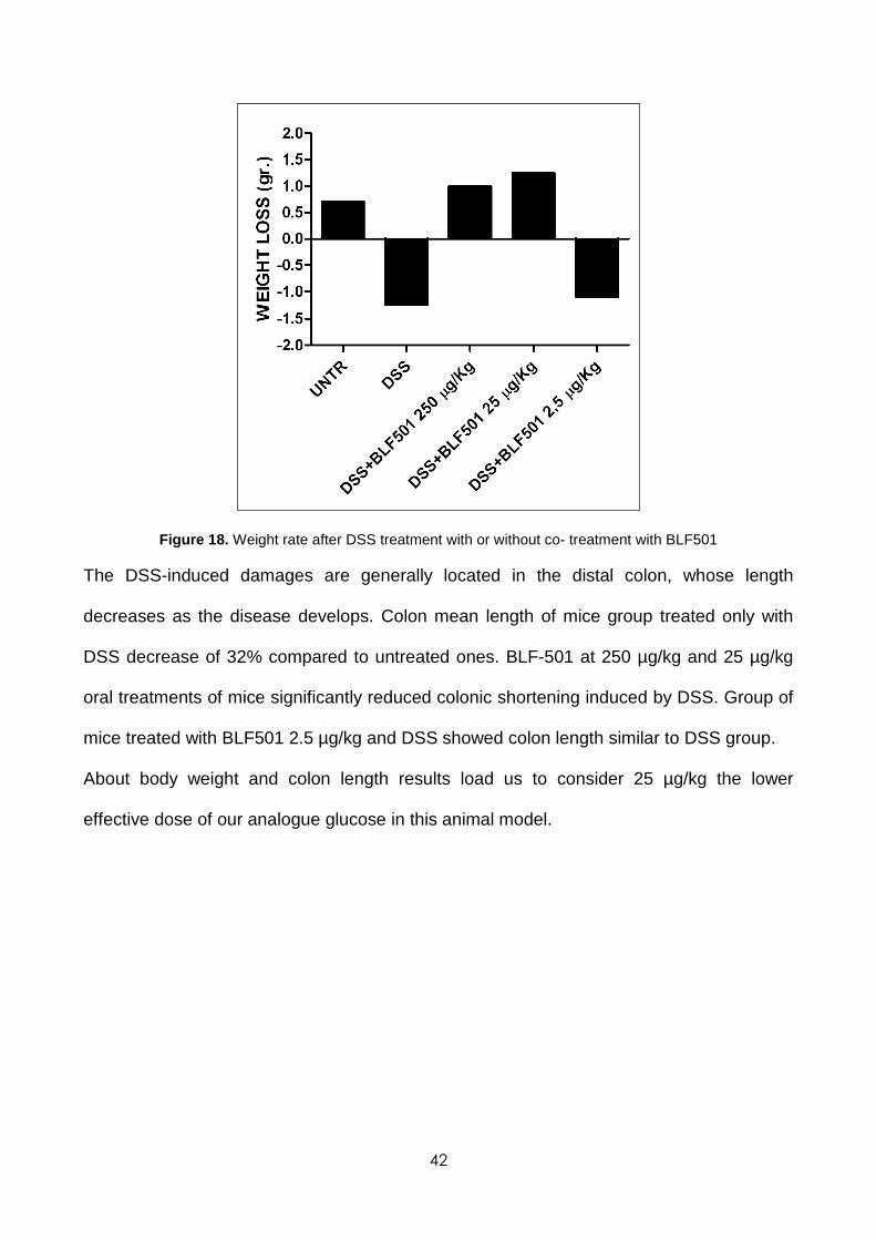
Figure 18. Weight rate after DSS treatment with or without co- treatment with BLF501
The DSS-induced damages are generally located in the distal colon, whose length
decreases as the disease develops. Colon mean length of mice group treated only with
DSS decrease of 32% compared to untreated ones. BLF-501 at 250 µg/kg and 25 µg/kg
oral treatments of mice significantly reduced colonic shortening induced by DSS. Group of
mice treated with BLF501 2.5 µg/kg and DSS showed colon length similar to DSS group.
About body weight and colon length results load us to consider 25 µg/kg the lower
effective dose of our analogue glucose in this animal model.
43
Figure 19. Colon length evaluation after DSS treatment with or without co- treatment with BLF501
After evaluation of colon length, colitis level of different group was scored using stool
consistence as a standard parameter. DSS treated animals group have very soft stools
with a 1,9 mean score, whereas untreated group shows normal stools with mean score 0
These results showed that oral treatment with BLF501 250 or 25 µg/kg inhibit water loss,
maintaining normal stool consistence.
* *
44
Figure 20. Stool score after DSS treatment with or without co- treatment with BLF501
The most important damage DSS-induced is characterized by changes in colon epithelium
showing loss of crypts and reduction of globet cells, signs of surface epithelial
regeneration, focal ulcerations, infiltration of inflammatory cells into the mucosa, and
oedema in the sub-mucosa. Histopathological analysis using hematoxylin-eosin staining of
colon section showed that DSS treatment induced crypt damage, ulceration, and infiltration
of inflammatory cells in the distal colon. A histological colitis score was calculated for 10
slides for each mouse of different groups; score increasing has associated with
inflammation develop (mean score of DSS group = 3.5 vs untreated = 0). Scoring system
of colon section of DSS-treated group plus BLF-501 250 or 25 µg/kg shows protective role
of SGLT-1 in this animal model of acute colitis.
*
*
45
A
B
a b c
A
A
46
Figure 21. (A) Rapresentative colonic H&E section from mice receiving water without DSS . a) Lamina mucosa; b)Lamina submucosa; c)Circular and longitudinal mussle. In magnification arrow shows colon crypte with normal morphology and black arrowhead indicates globet cells. (B) Colon histology from DSS-treated group. DSS induces thickening of the colon wall, globet cells and crypt loss in large areas (in magnification arrow). Infiltration reaching the lamina submucosa (withe arrow). (C) H&E of BLF501 250 µg/kg and (D) BLF501 25 µg/kg, histologically comparable with control group.
C
D
47
Figure 22. Histological scores of the colon section stained with H&E. Results are representative of 10 section for each animal of different groups. DSS mean score shows high level of inflammation with transmurall leukocite infiltration and loss of globet cells. BLF501 250 and 25 µg/kg oral treatment prevents and protects from DSS-induced mucosal damage.
DSS is directly toxic to gut epithelial cells of mucosal epithelium and induces a alteration of
the mucosal barrier, with increase of antigens from colon lumen to lamina propria. In this
animal model intestinal permeability alteration is the first step in colitis aetiology/onset.
Previous data have already described that increased colon permeability is associated with
colitis development after administration of DSS. Ussing Chamber analysis of the colon of
DSS-treated mice presented very low transmembrane resistance (25 ± 2,24 Ωcm2 vs 53.4
±2,73 Ωcm2 for untreated mice), indicating an increased permeability, whereas in mice
group treated with DSS and oral administration of glucose and BLF501 250 and 25 µg/kg
the transmembrane resistance remained similar to untreated mice (respectively 49,6 ±
1,52 Ω cm 2 or 50,4 ± 1,52 or 50,2 ± 1,2 vs 53.4 ±2,73 Ω cm2 in untreated mice). This
suggests that the increase of colon permeability DSS-induced is reverted by BLF501 oral
administration.
48
Figure 23. Colon resistence. Values represent the mean of 5 animals/group. Data are the means (±SD). *, p < 0,01 vs DSS.
3.7 BLF501 oral administration protects against che mically-induced mouse model of
chronic intestinal inflammation.
SGLT-1.activation by BLF501 protects against DSS-induced acute colitis with important
recovery of epithelial barrier intestinal barrier function. Several data in literature show that
in different animal model of Crohn disease increased intestinal permeability has been
shown before inflammation expression and the reversal of this defect can attenuate the
disease, implying that the increased permeability is not simply epiphenomena but rather is
a important etiological event. The administration of DSS at a concentration of 2% in the
drinking water for three cycles is a validated chemically-induced mouse model of chronic
intestinal inflammation. To evaluate the effect of BLF-501 in this model we valuated
different parameters with attention for epithelial permeability function and morphology.
After chronic colitis induction in DSS administered mice the colon length was significantly
reduced if compared to samples from untreated mice. A significant decrease in the
* *
49
shortening of the colon length was observed in mice that had received BLF501 almost for
3 week compared with DSS-administered mice (Figure 24).
Figure 24. Distal colon length. Values represent the mean of 5 animals/group. Data are the means (±SD). *, p < 0,01 vs DSS
From Histopathological analysis of hematoxylin/eosin-stained colon section is observable
in DSS-treated mice samples the loss of crypts and reduction of globet cells, focal
ulcerations, moderate infiltration of inflammatory cells to the mucosa, and oedema in
submucosa. A significant decrease in histological score was observed in DSS plus
BLF501 treated mice compared with those who received only DSS (Figure 25)