23
Shaobin Guo 11/20/2012
| Date post: | 23-Dec-2015 |
| Category: |
Documents |
| Upload: | jessica-glenn |
| View: | 217 times |
| Download: | 0 times |

Shaobin Guo11/20/2012
various types of Single-molecule force spectroscopy
•Optical tweezers
•Magnetic tweezers
•Atomic force microscopy (AFM)
•Micro-needle manipulation
•Biomembrane force probe
•Flow-induced stretching

Single-molecule manipulation capacity
•Length: 10-10 - 10-4 m / measurement of RNA polymerase advancing a single base pair to manipulation of cells
•Force: 10-14 - 10-8 Newton / assaying nucleic acid folding kinetics to mechanical disruption of covalent bonds

Optical tweezers (Trap)
•Lasers: near-infrared wavelengths (800-1100 nm)
•High numerical aperture (NA) microscope objective (at least 1.2)
•An approximate linear spring for small displacements (~150 nm)
•Position detection: back-focal plane (BFP) interferometry
•Calibration: position detector and trap stiffness

applications
Interaction assay
Tethered assay
Infrared laser
Infrared laser
Infrared laser
silica bead
kinesin molecule
microtubule
RNA polymerase molecule
Dumbbell assay
Limitations and Drawbacks
•Require optically homogeneous preparations and highly purified samples for high resolution trapping.
•Lack selectivity and exclusivity
•Local heating
•Optical damage
•Limited range of applied force (0.1-100 pN) and range of displacement (~150 nm or less)

magnetic tweezers
•A pair of permanent magnets
• Inverted microscope with a charge-coupled device (CCD)
•Able to rotate super-paramagnetic beads ranging from 0.5 to 5 um
•Force-clamp property: an effective stiffness on the order of 10-6 pN/nm
•Free from sample heating, photodamage and other problems related to optical tweezers
•Drawbacks: not versatile, unable to directly measure rotational torque generation, limited to slow and large displacements
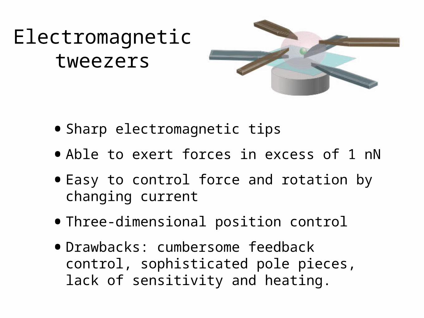
Electromagnetic tweezers
•Sharp electromagnetic tips
•Able to exert forces in excess of 1 nN
•Easy to control force and rotation by changing current
•Three-dimensional position control
•Drawbacks: cumbersome feedback control, sophisticated pole pieces, lack of sensitivity and heating.
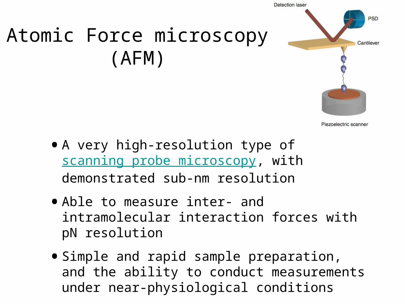
Atomic Force microscopy (AFM)
•A very high-resolution type of scanning probe microscopy, with demonstrated sub-nm resolution
•Able to measure inter- and intramolecular interaction forces with pN resolution
•Simple and rapid sample preparation, and the ability to conduct measurements under near-physiological conditions
applications
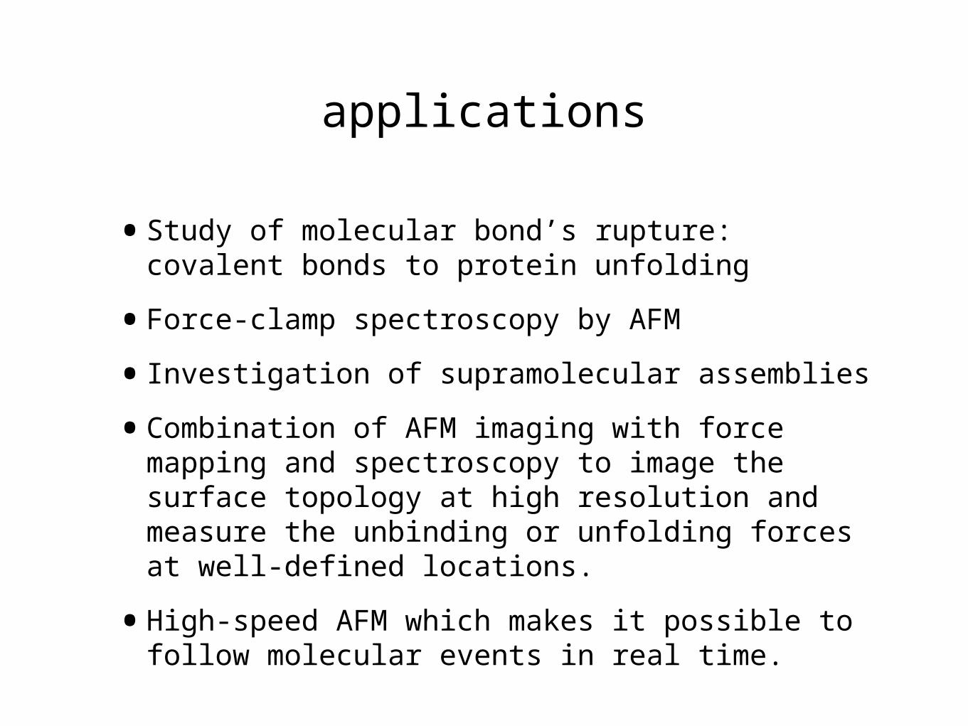
•Study of molecular bond’s rupture: covalent bonds to protein unfolding
•Force-clamp spectroscopy by AFM
•Investigation of supramolecular assemblies
•Combination of AFM imaging with force mapping and spectroscopy to image the surface topology at high resolution and measure the unbinding or unfolding forces at well-defined locations.
•High-speed AFM which makes it possible to follow molecular events in real time.
Limitations and Drawbacks
•Narrow useful force range resulting from large size and relative high stiffness of the cantilevers
•Lack of specificity

Background
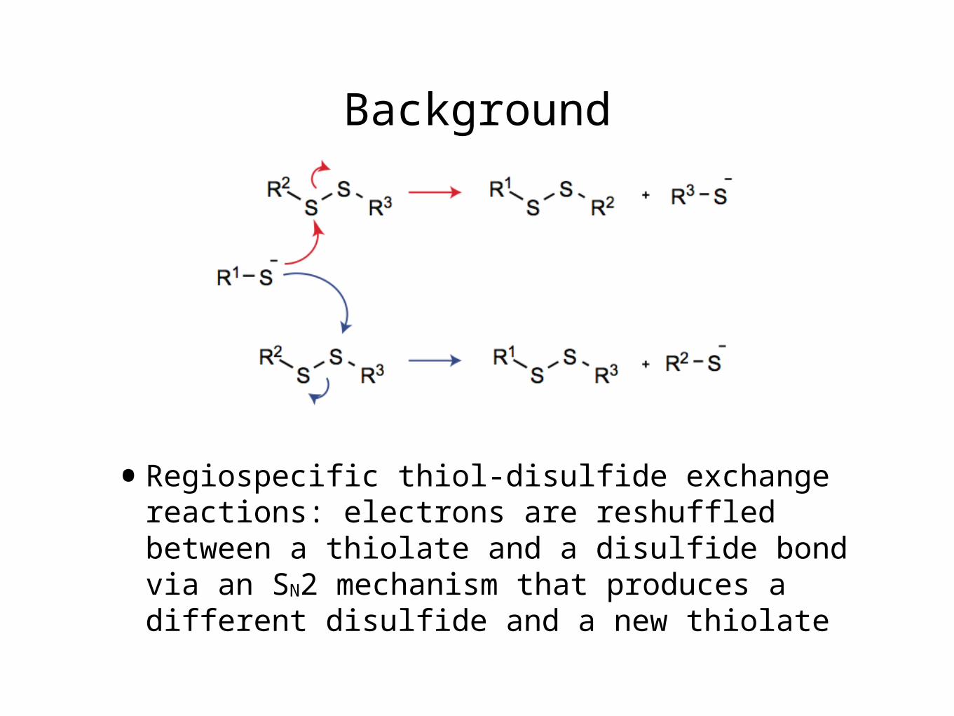
•Regiospecific thiol-disulfide exchange reactions: electrons are reshuffled between a thiolate and a disulfide bond via an SN2 mechanism that produces a different disulfide and a new thiolate

problems with bulk techniques
•Inability to differentiate disulfide isomers
•Variations among different techniques used to analyze reactions and subsequent divergent interpretations
•Interference from the reverse reaction

experiment set-up
Cantilever
Tip
Piezoelectric scanner
Force: 250 pN
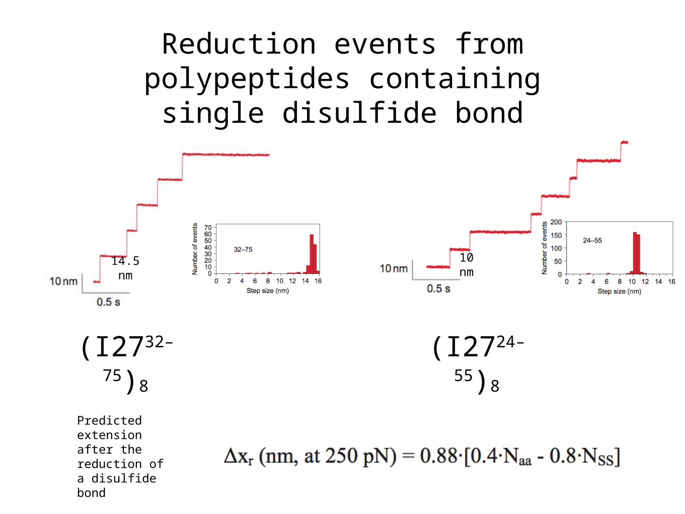
Reduction events from polypeptides containing
single disulfide bond
14.5 nm
10 nm
(I2732–
75)8
(I2724–
55)8
Predicted extension after the reduction of a disulfide bond
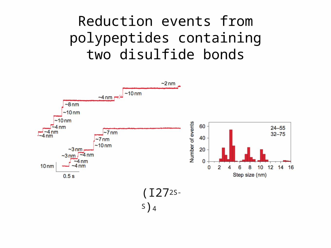
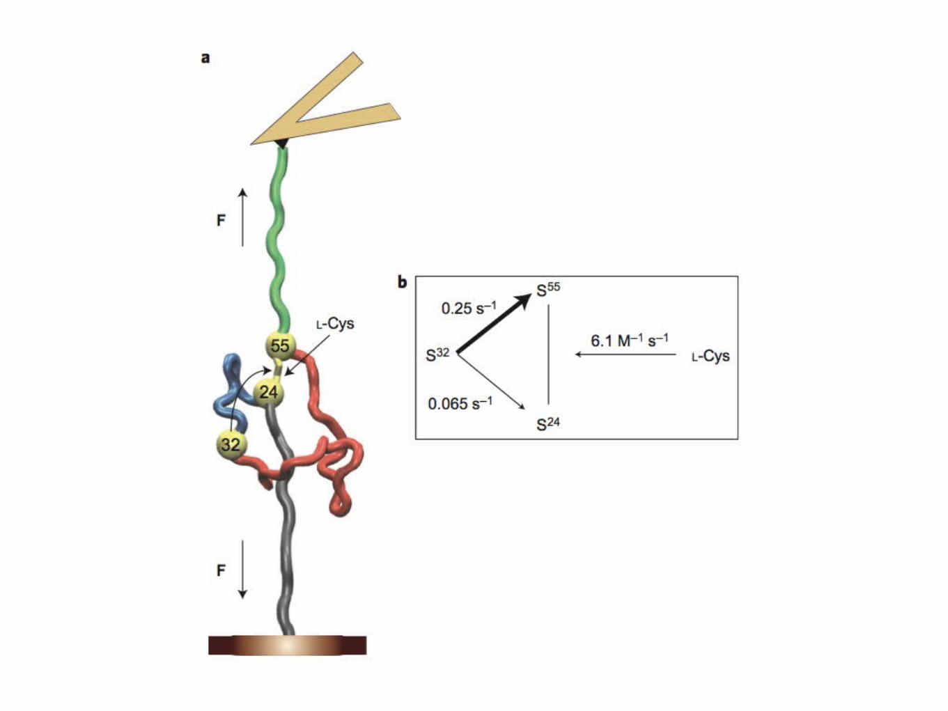
Reduction events from polypeptides containing two
disulfide bonds
(I272S-S)4
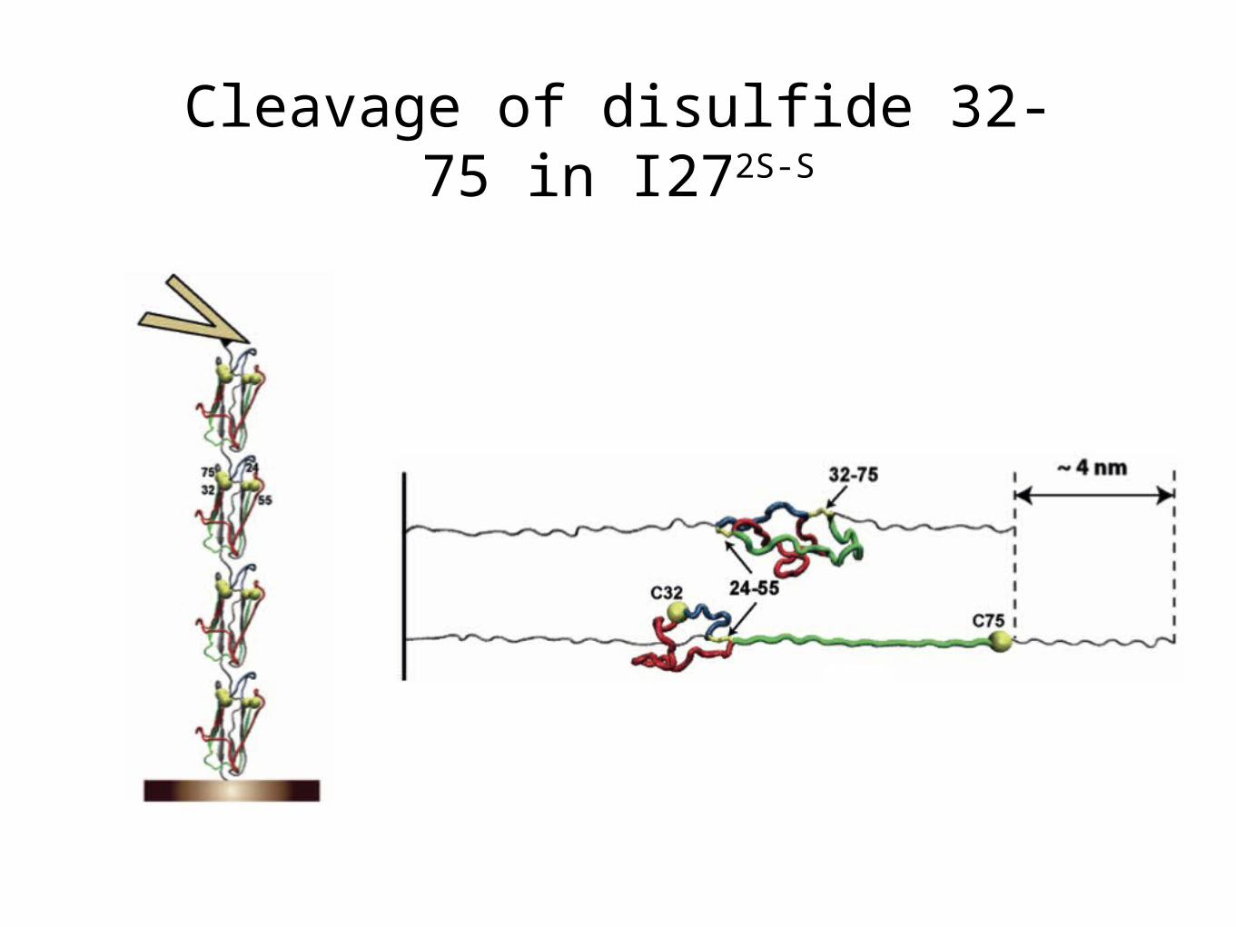
Cleavage of disulfide 32-75 in I272S-S

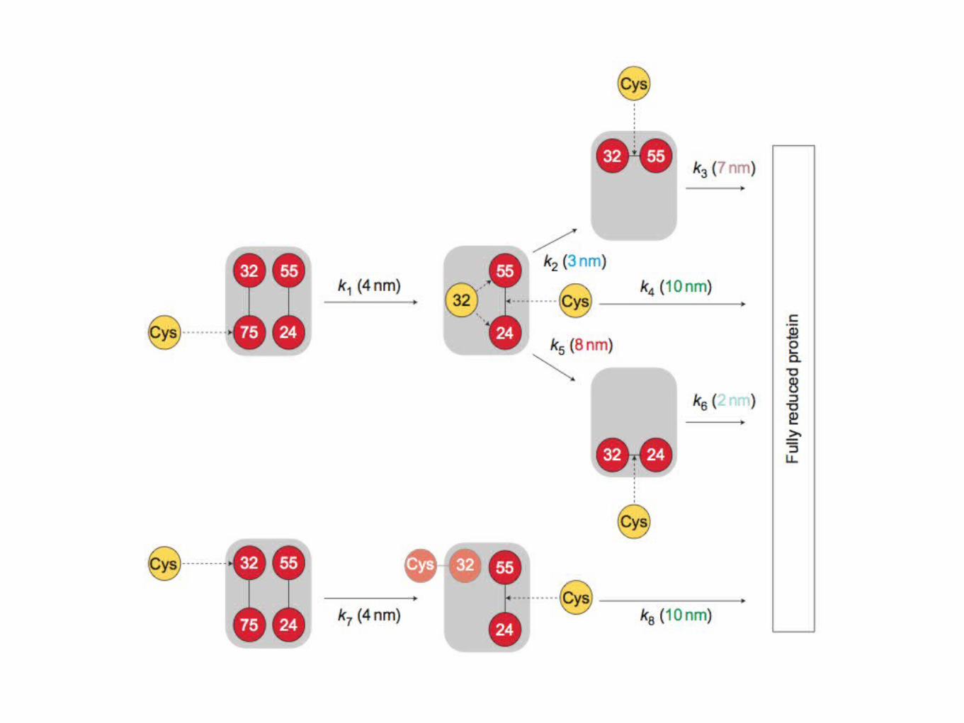
Pathways available for the reduction of disulfide 24–55 after the reduction of disulfide 32–75
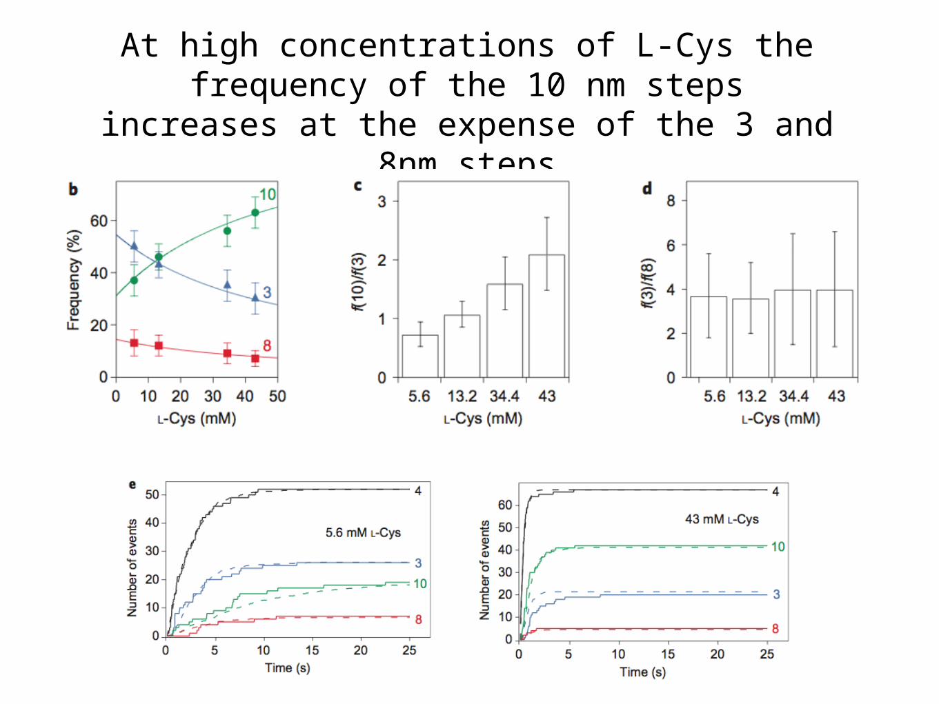
At high concentrations of L-Cys the frequency of the 10 nm steps increases at
the expense of the 3 and 8nm steps

Thanks!Questions?


![OPT Optical Tweezers - U of T Physicsphy326/opt/opt.pdf · Hooke’s Law [4]. Trapping only occurs when the gradient force is stronger than the scattering force. 2.2.2 – Modeling](https://static.documents.pub/doc/80x56/5e9a22e624933472d45c2499/opt-optical-tweezers-u-of-t-physics-phy326optoptpdf-hookeas-law-4-trapping.jpg)


