SIZE AND SHAPE OF PLEUROPNEUMONIA-LIKE ORGANISMS GROWN IN LIQUID MEDIA C. WEIBULL1 AND BRITT-MARIE LUNDIN Central Bacteriological Laboratory of Stockholmti City, Stockholm, Sweden Received for publication April 19, 1962 ABSTRACT WEIBULL, C. (Central Bacteriological Labora- tory of Stockholm City, Stockholm, Sweden), AND BRITT-MARIE LUNDIN. Size and shape of pleuropneumonia-like organisms grown in liquid media. J. Bacteriol. 84:513-519. 1962.-Samples of liquid cultures containing mainly nonaggre- gated cells of Mycoplasma agalactiae or M. laidlawii were transferred to agar blocks containing the same medium as the liquid cultures. By use of a phase-contrast microscope, photomicrographs were made of the slide cultures immediately after they had been prepared, and the dimensions of a large number of pleuropneumonia-like organ- isms (PPLO) were measured. These measure- ments indicated that, in young cultures (incu- bated for 24 to 48 hr), the size of the cells did not vary much more than that of ordinary bacteria; 95% of the cells had a width of 0.2 to 0.6 ,u. The growth of individual PPLO was followed during incubation of the slide cultures. It was found that 80 to 100% of the cells present in liquid over- night cultures divided and gave rise to micro- colonies within a few hours. Rod-shaped, el- lipsoidal, and spherical cells were seen in these cultures. Liquid cultures incubated for several days contained mainly spherical cells. Fewer than 5% of the cells in these cultures showed any indication of growth during incubation in slide cultures for 5 days. Photomicrographs of cells of M. agalactiae moving freely in liquid me- dium were taken with an electronic flash as the light source. The photographs thus obtained directly demonstrated the existence of rod-shaped cells. Several studies on the growth of pleuropneu- monia-like organisms (PPLO) in slide cultures have been reported (Bartmann and H6pken, 1 Present address: Department of Bacteriology, Medical School, University of UmeA, UmeA, Sweden. 1955; Kandler and Kandler, 1955a; Liebermeis- ter, 1960). Detailed observations concerning the morphology, mode of division, and colony forma- tion of PPLO were made in these investigations. In our study, special attention was paid to the relationship between the size and shape of the PPLO cells, on the one hand, and the viability of these cells, on the other. Aggregates consisting of several individual cells are often found in liquid cultures of PPLO. In this study, organisms and media were chosen in such a way that nonaggregated cells were in the majority in the cultures investigated. MATERIALS AND METHODS Organisms. Mycoplasma laidlawii, strain A (Laidlaw and Elford, 1936), was obtained from E. A. Freundt, State Serum Institute, Copen- hagen. The 311. agalactiae strain used was ob- tained from E. Klieneberger-Nobel, Lister Insti- tute of Preventive Medicine, London. It had been isolated from a goat ill with agalactia. Growth conditions. 71. agalactiae was grown at 37 C in meat broth supplemented with 10% inac- tivated horse serum. Mll. laidlauii was grown at 30 C in broth supplemented with 1% serum. Erlenmeyer flasks (200-ml), each containing 50 ml of medium, were used as culture vessels. The M. laidlawii cultures were incubated on a rotary shaker (100 rev/min). Subcultures of the PPLO were made daily. Preparation of slide cultures and microscopy. The methods described by Weibull and Lundin (1962) were used in most experiments. A few photomicrographs of PPLO moving freely in liquid medium between slide and cover slip were taken with a Leitz Multiblitz-Mikro 300-w electronic flash as the light source. The box camera used in other photographic studies could not be used in these experiments, since insuffi- cient light reached the photographic plate. In- stead, a microcamera equipped with 35-mm Gevaert Duplo Ortho film was used. The techni- 513 on August 9, 2019 by guest http://jb.asm.org/ Downloaded from
Transcript
SIZE AND SHAPE OF PLEUROPNEUMONIA-LIKE ORGANISMS GROWN INLIQUID MEDIA
C. WEIBULL1 AND BRITT-MARIE LUNDIN
Central Bacteriological Laboratory of Stockholmti City, Stockholm, Sweden
Received for publication April 19, 1962
ABSTRACT
WEIBULL, C. (Central Bacteriological Labora-tory of Stockholm City, Stockholm, Sweden),AND BRITT-MARIE LUNDIN. Size and shape ofpleuropneumonia-like organisms grown in liquidmedia. J. Bacteriol. 84:513-519. 1962.-Samplesof liquid cultures containing mainly nonaggre-gated cells of Mycoplasma agalactiae or M. laidlawiiwere transferred to agar blocks containing thesame medium as the liquid cultures. By use of aphase-contrast microscope, photomicrographswere made of the slide cultures immediatelyafter they had been prepared, and the dimensionsof a large number of pleuropneumonia-like organ-isms (PPLO) were measured. These measure-ments indicated that, in young cultures (incu-bated for 24 to 48 hr), the size of the cells did notvary much more than that of ordinary bacteria;95% of the cells had a width of 0.2 to 0.6 ,u. Thegrowth of individual PPLO was followed duringincubation of the slide cultures. It was found that80 to 100% of the cells present in liquid over-night cultures divided and gave rise to micro-colonies within a few hours. Rod-shaped, el-lipsoidal, and spherical cells were seen in thesecultures. Liquid cultures incubated for severaldays contained mainly spherical cells. Fewerthan 5% of the cells in these cultures showedany indication of growth during incubation inslide cultures for 5 days. Photomicrographs ofcells of M. agalactiae moving freely in liquid me-dium were taken with an electronic flash as thelight source. The photographs thus obtaineddirectly demonstrated the existence of rod-shapedcells.
Several studies on the growth of pleuropneu-monia-like organisms (PPLO) in slide cultureshave been reported (Bartmann and H6pken,
1 Present address: Department of Bacteriology,Medical School, University of UmeA, UmeA,Sweden.
1955; Kandler and Kandler, 1955a; Liebermeis-ter, 1960). Detailed observations concerning themorphology, mode of division, and colony forma-tion of PPLO were made in these investigations.In our study, special attention was paid to therelationship between the size and shape of thePPLO cells, on the one hand, and the viability ofthese cells, on the other.
Aggregates consisting of several individualcells are often found in liquid cultures of PPLO.In this study, organisms and media were chosenin such a way that nonaggregated cells were inthe majority in the cultures investigated.
MATERIALS AND METHODS
Organisms. Mycoplasma laidlawii, strain A(Laidlaw and Elford, 1936), was obtained fromE. A. Freundt, State Serum Institute, Copen-hagen. The 311. agalactiae strain used was ob-tained from E. Klieneberger-Nobel, Lister Insti-tute of Preventive Medicine, London. It hadbeen isolated from a goat ill with agalactia.
Growth conditions. 71. agalactiae was grown at37 C in meat broth supplemented with 10% inac-tivated horse serum. Mll. laidlauii was grown at 30C in broth supplemented with 1% serum.Erlenmeyer flasks (200-ml), each containing 50ml of medium, were used as culture vessels. TheM. laidlawii cultures were incubated on a rotaryshaker (100 rev/min). Subcultures of the PPLOwere made daily.
Preparation of slide cultures and microscopy.The methods described by Weibull and Lundin(1962) were used in most experiments. A fewphotomicrographs of PPLO moving freely inliquid medium between slide and cover slip weretaken with a Leitz Multiblitz-Mikro 300-welectronic flash as the light source. The boxcamera used in other photographic studies couldnot be used in these experiments, since insuffi-cient light reached the photographic plate. In-stead, a microcamera equipped with 35-mmGevaert Duplo Ortho film was used. The techni-
cal quality of the photomicrographs thus obtainedwas slightly inferior to that obtained with thebox camera. The final magnification of all photo-graphs was 2,600 X.
Measurements of respiration. The conventionalWarburg technique was used for these determina-tions. To obtain measurable oxygen consumption,the bacterial cultures were concentrated abouttenfold by centrifugation at 78,000 X g for 60 min
and the sedimented cells were resuspended infresh growth medium.
RESULTS
Occurrence of cell aggregates and nonaggregatedcells in liquid cultures of M. agalactiae and M.laidlawii. When M. laidlawii was grown in brothsupplemented with 10% horse serum, cell ag-gregates were predominant in the cultures. How-
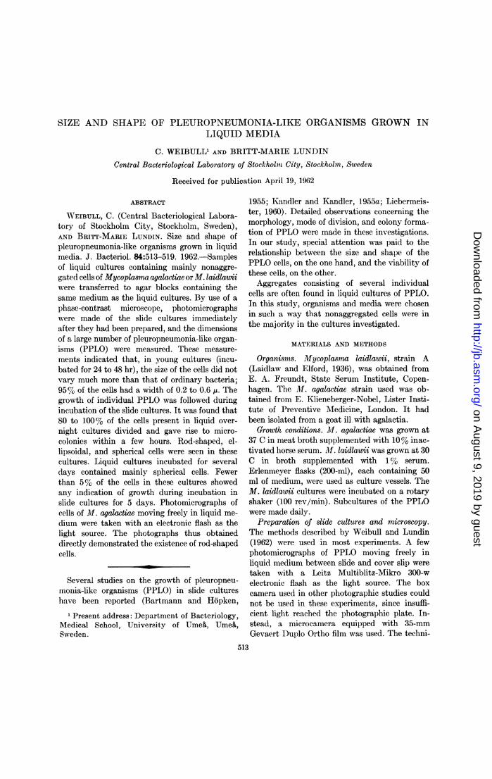
FIG. 1. (top) Slide cultures of Mycoplasma agalactiae, photographed immediately after inoculation withcells from 24-hr (left) and 250-hr (right) liquid cultures of this organism.
FIG. 2. (bottom) Slide cultures of Mycoplasma laidlawii, photographed immediately after inoculationwith cells from 24-hr (left) and 100-hr (right) liquid cultures of this organism.
diminished, the number of nonaggregated cellsincreased. Thus, in a medium containing 1 %serum, one to three cell aggregates per ten non-
aggregated cells were usually found. Each aggre-
gate contained three to ten individual cells.Agitation of the culture vessels during incubationdid not diminish appreciably the number of cellaggregates in a culture of M. laidlawuti.
In liquid cultures of M. agalactiae, a maximumof one cell aggregate per ten nonaggregated cellswas found.
Morphology of PPLO in liquid cultures ofvarious ages. Photomicrographs of typical cellsfrom 24-hr liquid cultures of M. agalactiae and M.laidlawii are shown in Fig. 1 and 2. It can beseen that these cells were spherical, ellipsoidalor rod-shaped. Most of the rods had swollenends. Several cells consisted of two rodlike seg-
ments, forming an angle with each other (suchcells are indicated by arrows in Fig. 2).
In PPLO cultures incubated for several days,almost all cells were spherical (Fig. 1 and 2).
Filtration experiments have been carried outto establish the size of the smallest viable ele-ments of PPLO (Elford, 1938; Sabin, 1941; Kel-lenberger, Liebermeister, and Bonifas, 1956;Klieneberger-Nobel, 1956). It was deemed ofinterest to compare the results of these experi-ments with the dimensions of PPLO as deter-mined microscopically. Therefore, the maximalwidth (in the case of spherical cells, the diameter)of a large number of M. agalactiae and M.laidlauii cells was determined by measurements
(-)ur
uL 300
co20
2:
z 10I
0.2 0.4 0.6 0.8 1.0 0.2 0.4 0.6 0.8
MAXIMAL WIDTH OF CELLS 0A1.0
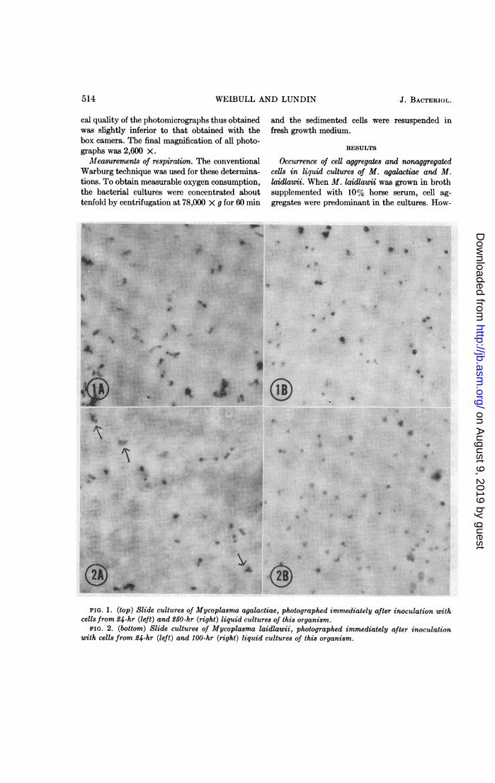
FIG. 3. Maximal width of cells present in liquidcultures of Mycoplasma agalactiae. The cells uweretransferred to agar blocks, photographed, and thewi(lth of the cells was measuredfrom the photographicprints. (A) Cells from 24-hr cultures. Pooled datafrom two different cultures. (B) Cells from 250-hrcultures. Pooled data from two different cultures.
40
0.2 0.4 0.6 0.8 1.0 0.2 0.4 0.6 0.8 1.0
MAXIMAL WIDTH OF CELLS (p)
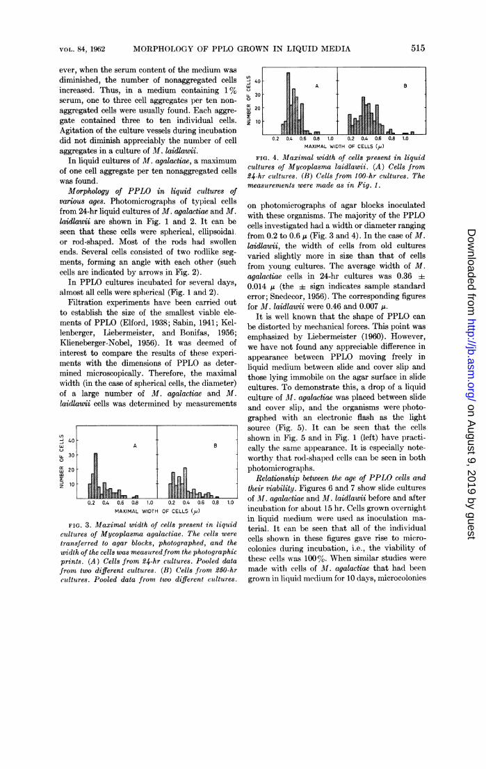
FIG. 4. Maximal width of cells present in liquid
cultures of Mycoplasma laidlawii. (A) Cells from
24-hr cultures. (B) Cells from 100-hr cultures. The
measurements were made as in Fig. 1.
On photomicrographs of agar blocks inoculated
with these organisms. The majority of the PPLO
cells investigated had a width or diameter ranging
from 0.2 to 0.6 ,u' (Fig. 3 and 4). In the case of IlI.laidlawii, the width of cells from old cultures
varied slightly more in size than that of cells
from young cultures. The average width of M.
agalactiae cells in 24-hr cultures was 0.36 i0.014 ,uo (the i sign indicates sample standard
error; Snedecor, 1956). The corresponding figures
for M. laidlawii were 0.46 and 0.007 ,u..It is well known that the shape of PPLO can
be distorted by mechanical forces. This point was
emphasized by Liebermeister (1960). However,
we have not found any appreciable difference in
appearance between PPLO moving freely in
liquid medium between slide and cover slip and
those lying immobile on the agar surface in slide
cultures. To demonstrate this, a drop of a liquid
culture of M1. agalactias was placed between slide
and cover slip, and the organisms were photo-
graphed with an electronic flash as the light
source (Fig. 5). It can be seen that the cells
shown in Fig. 5 and in Fig. 1 (left) have practi-
cally the same appearance. It is especially note-
worthy that rod-shaped cells can be seen in both
photomicrographs.
Relationship between the age of PPLO cells and
their viability. Figures 6 and 7 show slide cultures
of M1. agalactiae and M. laidlawii before and after
incubation for about 15 hr. Cells grown overnight
in liquid medium were used as inoculation ma-
terial. It can be seen that all of the individual
cells shown in these figures gave rise to micro-
colonies during incubation, i.e., the viability of
these cells was 100%7. When simlilar studies were
made with cells of Ml. agalactiae that had been
grown inI liquid medium for 10 days, microcolonies
FIG. 5. (top, left) Freely moving cells in a 24-hr liquid culture of Mycopla-sma agalactiae. A drop of theculture was placed between slide and cover- slip, and the cells were photographed with electronic flash as thelight source.
FIG. 6. (top, center and right) Slide culture of Mycoplasmia agalactiae before (center) and after (right) 12hr of incubation at 37 C. The slide cultutre was inoculated u'ith cclls from a 24-hr liquid culture of M. aga-lactiae.
FIG. 7. (bottom) Slide culture of Mycoplasmia laidlawii before (left) and after (right) 20 hr of incubationat 30 C. The slide culture was inoculated with cells from an 18-hr- liquid culture of M. laidlawii.
were only occasionally found in the mnicroscopic confirmed by counting experiments. Cells fromifields of view. Most of the individual PPLO cells liquid PPLO cultures of various ages were used todid not change their appearance during incuba- prepare slide cultures of M. agalactiae and 31.tion for 2 days in slide cultures. Sim-ilar results laidlawii. Before incubation, the number of cellswere obtained with cells of Ml. laidlauii grown in in several microscopic fields of view was deter-liquid medium for 4 to 5 days. mined. The slide cultures were then incubatedThe photographic studies described above were for 18 to 120 hr. The number of microcolonies
4 M. laidlawii 24 16.3 4 0.8 17.3 ±4 0.8 185 M. laidlawii 48 16.9 4d 1.1 17.1 i1 0.8 186 M. laidlawii 72 19.9 i 0.9 11.4 4- 0.6 247 M. laidlawii 72 19.4 ±4 1.1 4.2 4- 0.3 248 M. laidlawii 96 24.1 i1: 1.1 0.7 4 0.1 24
9 M. laidlawii 250 9.9:1:
0.6 {0.33 4 0.06 480154 0.06 120
* Agar blocks were inoculated with samples of liquid PPLO cultures. The inoculated blocks wereused for preparation of slide cultures, which were studied microscopically before and after incubation.The numbers of cells and colonies given below are the average nunmbers found per microscopic field ofview. The A= sign indicates sample standard error.
t Number of spherical cells.t Number of rod-shaped cells.
formed during incubation was determined in thesame way as the number of cells in the unincu-bated slide cultures. The average size of themicrocolonies was about 10 ,u. Table 1 shows theresults of these counting experiments. It can beseen that the viability of the young PPLO cellsstudied was 80 to 100%. On the other hand, cellsfrom cultures that had been grown for severaldays in liquid medium before transfer to theslide cultures displayed a viability of less than5%. The figures obtained from experiments 2and 3 (Table 1) suggest that only rod-shapedcells are viable in old cultures of M. agalactiae.It could be argued that the semianaerobic condi-tions prevailing in the slide cultures might inhibitto some extent the growth of old PPLO. To testthis possibility, samples of liquid cultures ofPPLO were spread uniformly on agar plates. Aportion of each plate was used to prepare a slideculture. The slide cultures and the plates were
then incubated in parallel for the desired periodof time (between 1 and 5 days). A second set ofslide cultures was then prepared from the plates.Immediately afterwards, the number of coloniesin each of the pairs of slide cultures was countedand compared. No significant difference was re-
vealed in the number of colonies in the initialand final slide cultures in each pair. This showsthat the cells of M. agalactiae and Al. laidlawii
TABLE 2. Respiration of PPLO cells grownfor various times in liquid medium*
* The liquid cultures were concentrated abouttenfold by centrifugation and resuspension of thesediment in fresh growth medium. The figuresindicate the uptake of 02 (,liters/hr) by 1 mnl ofthe original culture. Each figure represents amean value of data from two independent experi-ments.
formed colonies equally easily in the semianaero-bic conditions of the slide cultures and in thefully aerobic agar plates.
Respiration experiments. Kandler and Kandler(1955b) published data showing that cells fromold cultures of a PPLO strain isolated from micerespired much less vigorously than cells fromyoung cultures. Table 2 shows that the same isalso true from M. agalactiae and M. laidlaunvi.
Thus, the loss in viability of the PPLO describedin the preceding paragraphs is accompanied by amarked decrease in respiratory activity.
DISCUSSION
The pleomorphism of the PPLO has been em-phasized by most workers who have studied theseorganisms. (The PPLO have recently been revisedby Klieneberger-Nobel, 1962.) The PPLO investi-gated by us were studied under conditions thatshould minimize this pleomorphism. Firstly,cells grown in liquid media were used as thestarting material for our investigations. Thisimplies that all organisms in a lparticular culturewere grown under practically identical environ-mental conditions. (This is never the case withorganisms grown on solid media.) Secondly,mainly viable organisms were studied, sinceaging and degenerating forms of PPLO often arehighly pleomorphic. Thirdly, media were chosenin which the nonaggregated cells outnumberedthe cell aggregates.
According to our results, the diimiensions of thePPLO studied do not vary much more than thoseof many ordinary bacteria under comparableconditions. The maximal width of the thinnestviable PPLO photographed by us was about 0.2,u (M. agalactiae) or 0.3 A (Al. laidlauii). Thesefigures are slightly larger than those obtainedfrom filtration experiments designed to determinethe smallest reproductive units of PPLO, namely,0.12 to 0.25 ,t (Elford, 1938; Sabin, 1941; Kliene-berger-Nobel, 1956; Kellenberger, Liebermeisterand Bonifas, 1956). The agreement can, however,be regarded as rather close, considering that thePPLO cells possess a certain degree of plasticity(Liebermeister, 1960). These cells might thuspass through filter pores in the form of thread-like structures having a smaller diameter thanthe PPLO in unfiltered cultures. On the otherhand, our photomicrographs of 311. agalactiaetaken with an electronic flash as the light sourceshow that cells of this organism are not de-formed so easily that they will change theirshape appreciably when they are transferredfrom a liquid culture to the agar block of a slideculture.Cuckow and Klieneberger-Nobel (1956) and
Klieneberger-Nobel (1956, 1962) published elec-tron micrographs of several PPLO, among themAl. agalactiae and Ml. laidlawii. Particles ofvarious sizes were seen in the )hotographs. The
authors concluded that the smallest of theseparticles, which were approximately sphericaland had a diameter of approximately 0.1 A,represented minimal reproductive units. How-ever, it was not clearly proven that a significantnumber of these particles were viable beforebeing fixed. In addition, the washing and fixingprocedures used for the preparation of the elee-tron-microscope specimens may have changedthe size and shape of the PPLO particles presentin the original cultures.To our knowledge, comparisons between viable
and total counts of PPLO cultures, other thanours, have not been made so far. In cultures ofM. agalactiae and M. laidlawii grown for 24 to 48hr, 80 to 100%o of the cells were found to beviable. Our counting experiments, and l)hoto-graphs taken of slide cultures of PPLO beforeand after incubation, indicate that the PPLOstudied do not possess any submicroscopic repro-ductive units. Some viable cells were spherical,but the majority of them were rod-shaped orellipsoidal. In cultures several days old, most ofthe cells were spherical and few of them dividedwhen transferred to slide cultures. Probably theonly viable cells in these cultures were the rod-shaped ones. This suggests that the sphericalcells l)resent in PPLO cultures several days olddo not represent significant reproductive units.Bartmann and Hopken (1955) drew the sameconclusion from their photomnierographs.
Weibull and Lundin (1962) studied the sizeand shape of elements found in 24-hr cultures of astable Proteus L form. AMainly spherical bodieswere found, their diameters ranging from theresolving limit of the microscope used (about 0.2Iu) to about 1.5 ,u. Only elements having a diame-ter > 0.6 to 0.7 ,u were able to grow in slidecultures. Thus, the elements of this L formexhibited an inhomogeneity, with respect toboth size and viability, that was not encounteredin the )resent study on PPLO cells from young(24 to 48 hr) liquid cultures. It should also beemphasized that the small, nonviable L elements(diameter < 0.6 ,u) respired vigorously; thus,there was a dissociation between respiratory andreproductive capabilities in these bodies. In thisrespect, too, the PPLO studied by us behaveddifferently; viable PPLO cells respired vigorouslyand nonviable cells showed at the most a weakrespiratory activity. Thus, these results andthose reported by Weibull and Lundin (1962) are
in accordance with the view that very signifi-cant dissimilarities exist between the PPLO andthe bacterial L forms.
ACKNOWLEDGMENTS
The authors wish to thank Mrs. K. Hammar-berg for skillful assistance in part of the experi-ments.
This investigation was part of a program on theSubmicroscopic structure of the bacterial cellfinancially supported by the Swedish NaturalScience Research Council.
LITERATURE CITED
BARTMANN, K., AND W. HOPKEN. 1955. Phasen-kontrastmikroskopische Beobachtungen zurMorphologie der peripneumonieiihnlichen Or-ganismen (PPLO). Zentr. Bakteriol. Para-sitenk., Abt. I. Orig. 177:319-332.
CIJCKOW, F. W., AND E. KLIENEBERGER-NOBEL.1955. Further studies of organisms of thepleuropneumonia group by electron micros-copy. J. Gen. Microbiol. 13:149-154.
ELFORD, W. J. 1938. The sizes of virus and bacte-riophages and methods for their determination,p. 126-176. In R. Doerr and C. Hallauer[ed.], Handbuch der Virusforschung, vol. 1,first part. Julius Springer, Vienna.
KANDLER, G., AND 0. KANDLER. 1955a. Unter-suchungen uber die Morphologie und dieVermehrung der pleuropneumonie-iahnlichenOrganismen und der L-Phase der Bakterien.Arch. Mikrobiol. 21:178-201.
KANDLER, G., AND 0. KANDLER. 1955b. Ernah-rungs- und stoffwechsel-physiologische Unter-suchungen an pleuropneumonie-iihnlichenOrganismen und der L-Phase der Bakterien.Zentr. Bakteriol. Parasitenk., Abt. II.108:383-397.
KELLENBERGER, E., K. LIEBERMEISTER, AND V.BONIFAS. 1956. Studien zur L-Form derBakterien. II. Dimension und Filtrierbarkeitder globtilaren Form. Z. Naturforsch. 11b:206-215.
KLIENEBERGER-NOBEL, E. 1956. tUber die Wesen-verschiedenheit der peripneumonieiihnlichenOrganismen und der L-Phase der Bakterien.Zentr. Bakteriol. Parasitenk., Abt. I. Orig.165:329-343.
KLIENEBERGER-NOBEL, E. 1962. Pleuropneu-monia-like organisms (PPLO). Mycoplasma-taceae. Academic Press, Inc., New York.
LAIDLAW, P. P., AND W. J. ELFORD. 1936. A newgroup of filterable organisms. Proc. Roy. Soc.(London), Ser. B 120:292-303.
LIEBERMEISTER, K. 1960. Morphology of thePPLO and L-forms of Proteus. Ann. N.Y.Acad. Sci. 79:326-343.
SABIN, A. B. 1941. The filterable microorganismsof the pleuropneumonia group. Bacteriol.Rev. 5:1-66.
SNEDECOR, G. W. 1956. Statistical methods appliedto experiments in agriculture and biology,p. 43. The Iowa State College Press, Ames.
WEIBULL, C., AND B.-M. LUNDIN. 1962. Growth ofelements of various sizes found in cultures ofa stable Proteus L form. J. Gen. Microbiol.27:21-248.