25
Short Announcements 1 st Quiz today Homework #2 on web. Due next Monday. Chpt 2 Reading Due next Wednesday (NOT Monday) Today’s Lecture: Protein Folding
| Date post: | 29-Dec-2015 |
| Category: |
Documents |
| Upload: | gervais-abner-dean |
| View: | 216 times |
| Download: | 0 times |

Short Announcements1st Quiz todayHomework #2 on web. Due next Monday.Chpt 2 Reading Due next Wednesday (NOT Monday)Today’s Lecture: Protein Folding
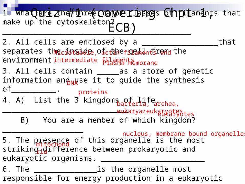
Quiz #1 (covering Chpt 1, ECB)
1. What are the three major classes of filaments that make up the cytoskeleton?__________________________________________
2. All cells are enclosed by a _________________that separates the inside of the cell from the environment.
3. All cells contain _____as a store of genetic information and use it to guide the synthesis of__________.
4. A) List the 3 kingdoms of life. ____________________________
B) You are a member of which kingdom? __________________
5. The presence of this organelle is the most striking difference between prokaryotic and eukaryotic organisms. _______________________
6. The ______________is the organelle most responsible for energy production in a eukaryotic cell.
Microtubule, actin filaments and intermediate filaments
Plasma membrane
bacteria, archea, eukarya/eukaryotes
eukaryotes
mitochondria
nucleus, membrane bound organelles
DNA
proteins
The Protein (Free) Energy Landscape
Largely from Martin Gruebele, Chemistry, Physics UIUC
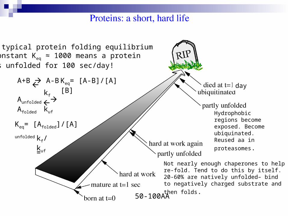
A typical protein folding equilibriumconstant Keq ≈ 1000 means a proteinis unfolded for 100 sec/day!
day
50-100AA
Not nearly enough chaperones to help re-fold. Tend to do this by itself. 20-60% are natively unfolded– bind to negatively charged substrate
and then folds.
Hydrophobic regions become exposed. Become ubiquinated. Reused aa in
proteasomes.
A+B A-B Keq= [A-B]/[A][B]
Keq= [Afolded]/[A] unfolded
=
Aunfolded Afolded
kf
kuf
kf/ kuf
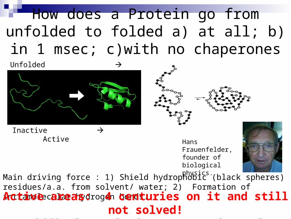
How does a Protein go from unfolded to folded a) at all; b) in 1 msec; c)with no chaperones
Hans Frauenfelder, founder of biological physics.
Unfolded Folded
Inactive Active
Main driving force : 1) Shield hydrophobic (black spheres) residues/a.a. from solvent/ water; 2) Formation of intramolecular hydrogen bonds.
Active areas: 4 centuries on it and still not solved!Difficulty relating to experimental observations.
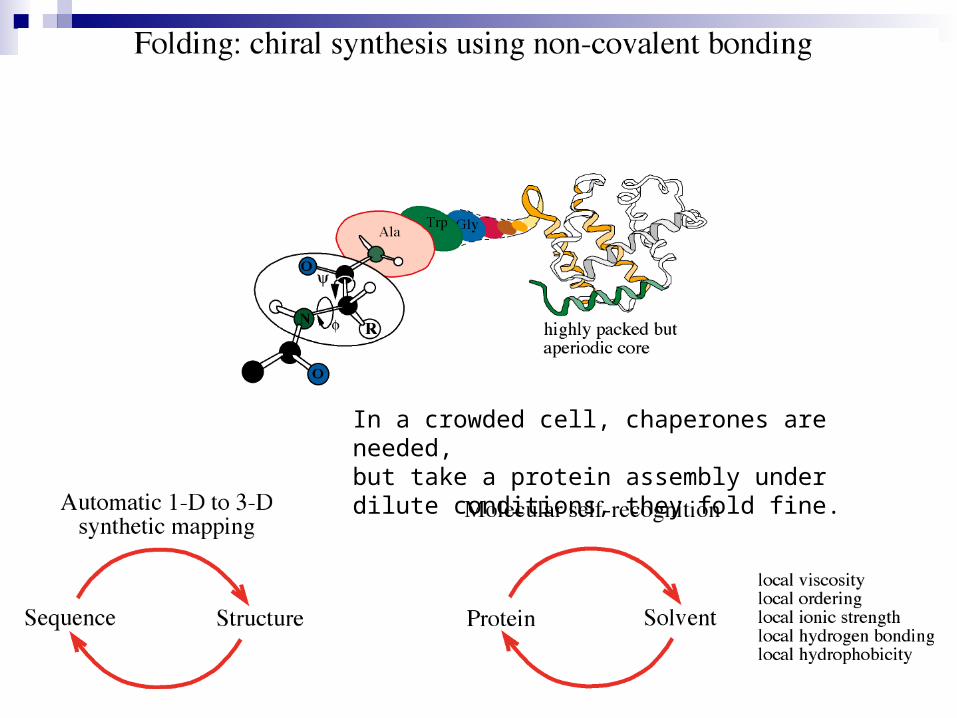
In a crowded cell, chaperones are needed, but take a protein assembly under dilute conditions, they fold fine.
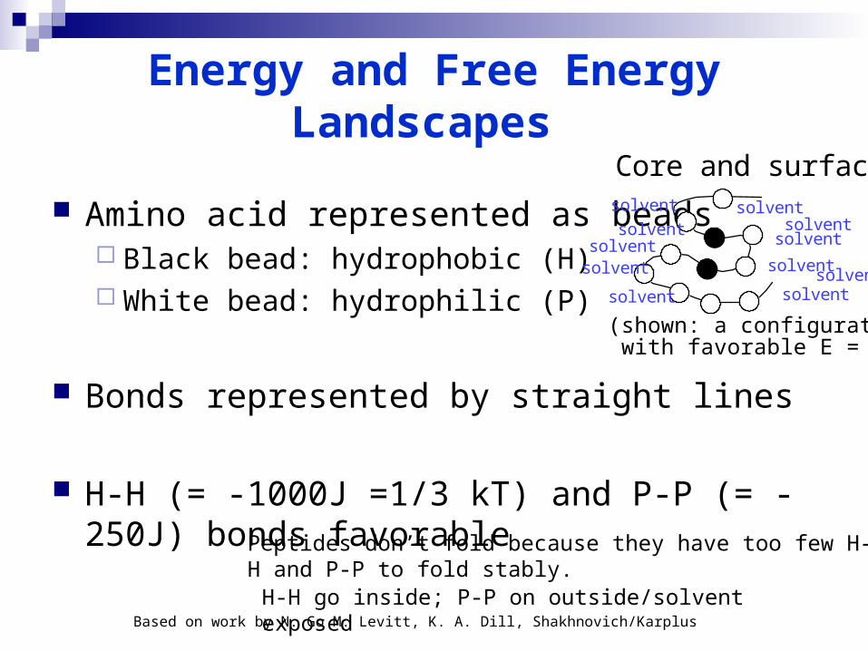
Energy and Free Energy Landscapes
Amino acid represented as beads Black bead: hydrophobic (H) White bead: hydrophilic (P)
Bonds represented by straight lines
H-H (= -1000J =1/3 kT) and P-P (= -250J) bonds favorable
Based on work by N. Go M. Levitt, K. A. Dill, Shakhnovich/Karplus
a
Core and surface
(shown: a configuration with favorable E = <H>)
solvent
solventsolvent
solvent
solvent
solvent
solventsolvent
solvent
solvent
solvent
H-H go inside; P-P on outside/solvent exposed
Peptides don’t fold because they have too few H-H and P-P to fold stably.
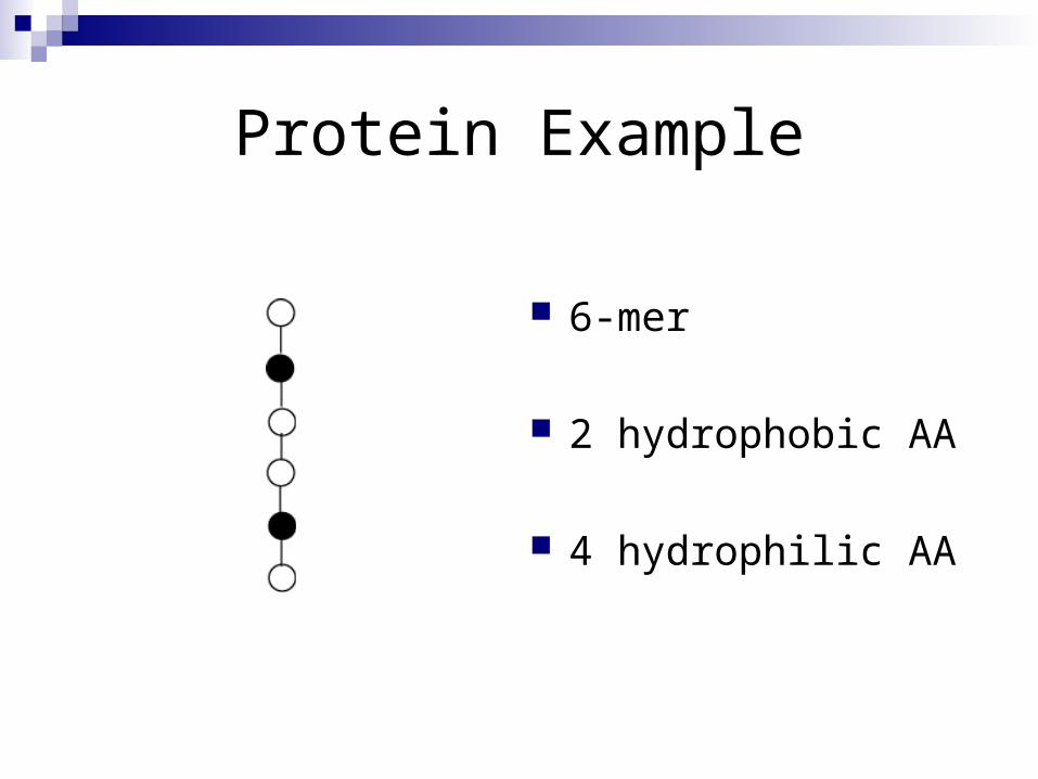
Protein Example
6-mer
2 hydrophobic AA
4 hydrophilic AA
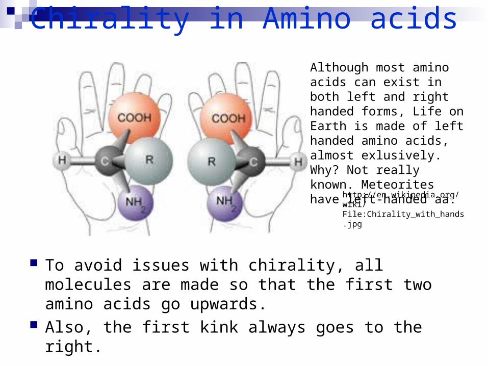
Chirality in Amino acids
To avoid issues with chirality, all molecules are made so that the first two amino acids go upwards.
Also, the first kink always goes to the right.
Although most amino acids can exist in both left and right handed forms, Life on Earth is made of left handed amino acids, almost exlusively. Why? Not really known. Meteorites have left-handed aa.
http://en.wikipedia.org/wiki/File:Chirality_with_hands.jpg
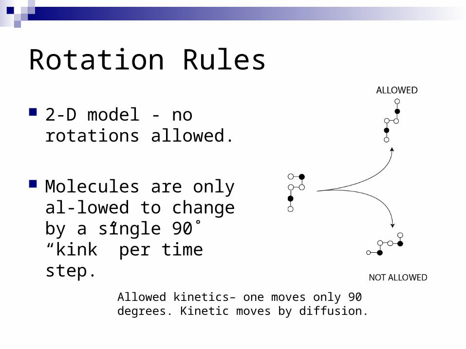
Rotation Rules
2-D model - no rotations allowed.
Molecules are only al-lowed to change by a single 90˚ “kink” per time step.
Allowed kinetics– one moves only 90 degrees. Kinetic moves by diffusion.

The Journey
A trap!
Direct folding!Direct folding!
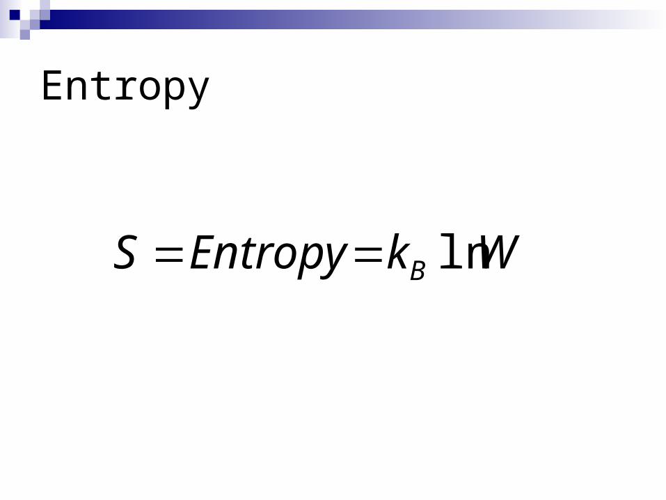
Entropy
WkEntropyS B ln
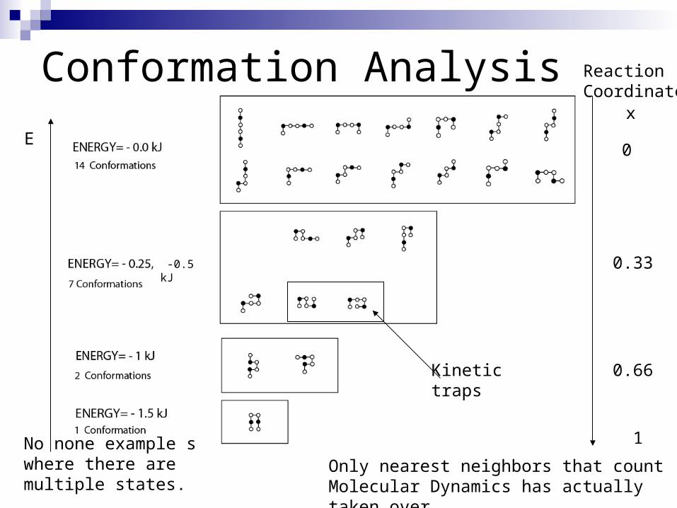
Conformation Analysis
E
ReactionCoordinate
1
0
0.33
0.66Kinetictraps
-0.5 kJ
x
Only nearest neighbors that countMolecular Dynamics has actually taken over
No none example s where there are multiple states.
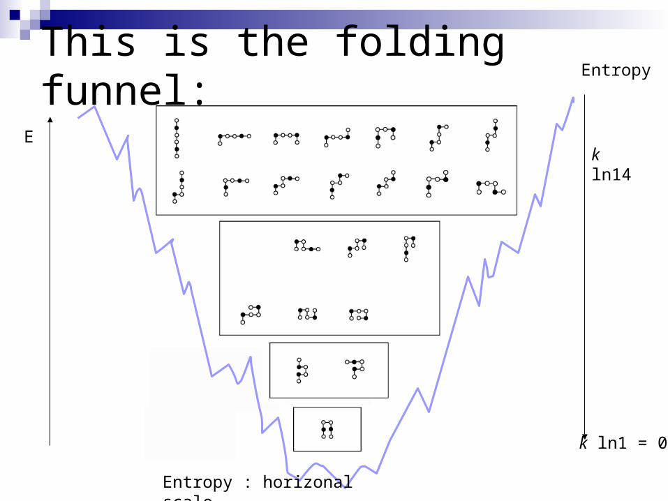
This is the folding funnel:
E
Entropy
k ln1 = 0
k ln14
Entropy : horizonal scale
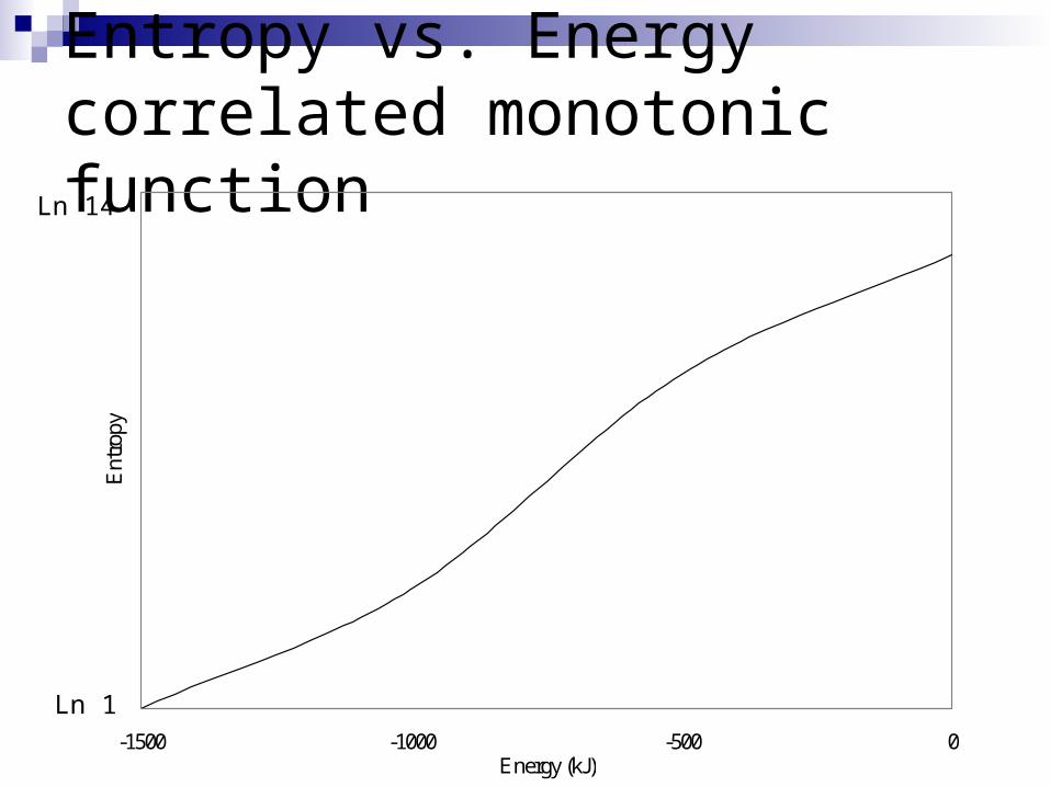
Entropy vs. Energycorrelated monotonic function
-1500 -1000 -500 0Energy (kJ)
Ent
ropy
Ln 14
Ln 1
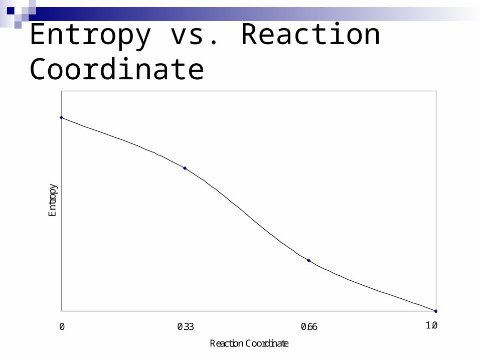
Entropy vs. Reaction Coordinate
0 0.33 0.66 0.99
Reaction Coordinate
Ent
ropy
1.0
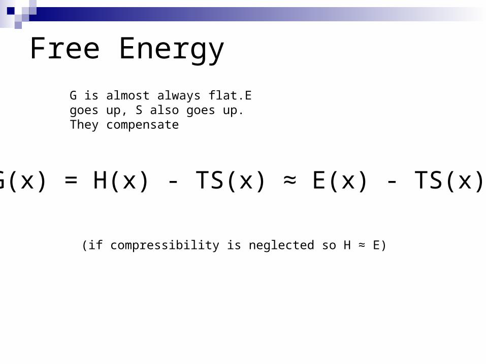
Free Energy
(if compressibility is neglected so H ≈ E)
G(x) = H(x) - TS(x) ≈ E(x) - TS(x)
G is almost always flat.E goes up, S also goes up. They compensate
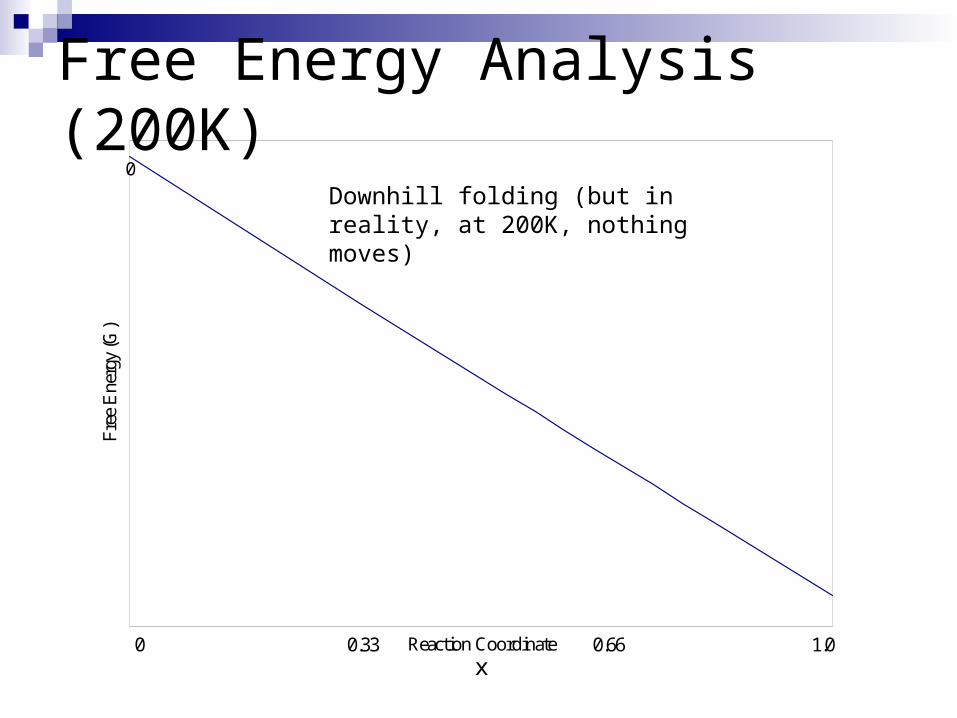
Free Energy Analysis (200K)0
Reaction Coordinate
Free
Ene
rgy
(G)
1.00.660.330x
Downhill folding (but in reality, at 200K, nothing moves)
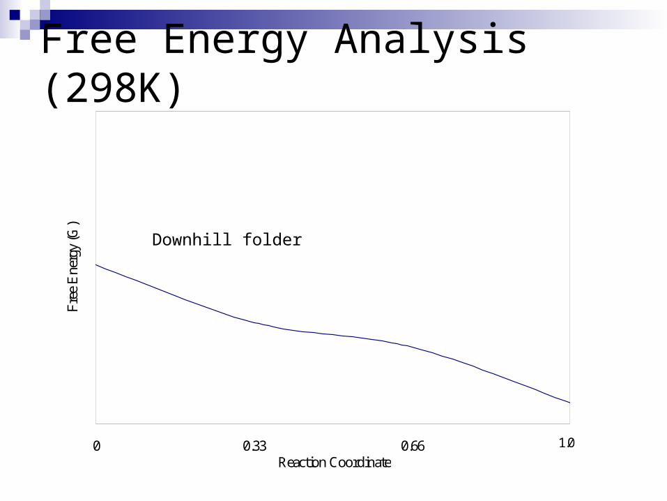
Free Energy Analysis (298K)
0 0.33 0.66 0.99Reaction Coordinate
Free
Ene
rgy
(G)
1.0
Downhill folder

Free Energy Analysis (360 K)
0 0.33 0.66 0.99Reaction Coordinate
Free
Ene
rgy
(G)
1.0
Two state folder
Unfolded state—has some structure
This is likely the equilibrium of 50:50 where they are interconverting and equally stable.
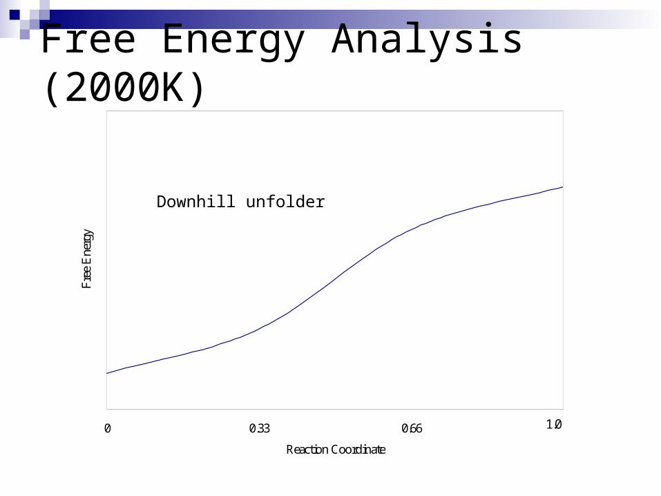
Free Energy Analysis (2000K)
0 0.33 0.66 0.99
Reaction Coordinate
Free
Ene
rgy
1.0
Downhill unfolder
aa
0-1 1
Free
ene
rgy
x
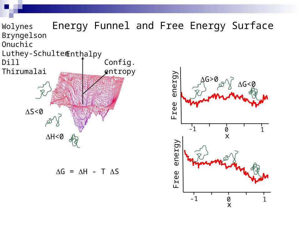
EnthalpyConfig.entropy
S<0
G>0
H<0
G<0
Wolynes BryngelsonOnuchicLuthey-SchultenDillThirumalai
0-1 1
Free
ene
rgy
x
Energy Funnel and Free Energy Surface
G = H - T S
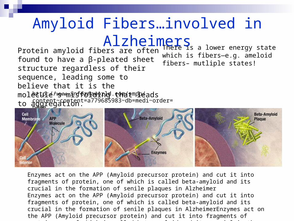
There is a lower energy state which is fibers—e.g. ameloid fibers– mutliple states!
Amyloid Fibers…involved in Alzheimers
Protein amyloid fibers are often found to have a β-pleated sheet structure regardless of their sequence, leading some to believe that it is the molecule's misfolding that leads to aggregation.
http://www.informaworld.com/smpp/content~content=a779685983~db=medi~order=page
Enzymes act on the APP (Amyloid precursor protein) and cut it into fragments of protein, one of which is called beta-amyloid and its crucial in the formation of senile plaques in AlzheimerEnzymes act on the APP (Amyloid precursor protein) and cut it into fragments of protein, one of which is called beta-amyloid and its crucial in the formation of senile plaques in AlzheimerEnzymes act on the APP (Amyloid precursor protein) and cut it into fragments of protein, one of which is called beta-amyloid and its crucial in the formation of senile plaques in Alzheimer
Proteins can fold.
Don’t need chaperones.
ΔG is always about zero. Therefore can fold fast.
Kinetics – fast cause not huge barriers
Summary of Protein Folding
Class evaluation
1. What was the most interesting thing you learned in class today?
2. What are you confused about?
3. Related to today’s subject, what would you like to know more about?
4. Any helpful comments.
Answer, and turn in at the end of class.


