Short Hypocotyl in White Light1 Interacts with ElongatedHypocotyl5 (HY5) and Constitutive Photomorphogenic1(COP1) and Promotes COP1-Mediated Degradation ofHY5 during Arabidopsis Seedling Development1[OPEN]
Anjil Kumar Srivastava, Dhirodatta Senapati, Archana Srivastava, Moumita Chakraborty,Sreeramaiah N. Gangappa, and Sudip Chattopadhyay*
Department of Biotechnology, National Institute of Technology, Durgapur 713209, India
Arabidopsis (Arabidopsis thaliana) Short Hypocotyl in White Light1 (SHW1) encodes a Ser-Arg-Asp-rich protein that acts as anegative regulator of photomorphogenesis. SHW1 and Constitutive Photomorphogenic1 (COP1) genetically interact in anadditive manner to suppress photomorphogenesis. Elongated Hypocotyl5 (HY5) is a photomorphogenesis promoting a basicleucine zipper transcription factor that is degraded by COP1 ubiquitin ligase in the darkness. Here, we report the functionalinterrelation of SHW1 with COP1 and HY5 in Arabidopsis seedling development. The in vitro and in vivo molecular interactionstudies show that SHW1 physically interacts with both COP1 and HY5. The genetic studies reveal that SHW1 and HY5 work inan antagonistic manner to regulate photomorphogenic growth. Additional mutation of SHW1 in hy5 mutant background is ableto suppress the gravitropic root growth defect of hy5 mutants. This study further reveals that the altered abscisic acidresponsiveness of hy5 mutants is modulated by additional loss of SHW1 function. Furthermore, this study shows that SHW1promotes COP1-mediated degradation of HY5 through enhanced ubiquitylation in the darkness. Collectively, this studyhighlights a mechanistic view on coordinated regulation of SHW1, COP1, and HY5 in Arabidopsis seedling development.
Plants have evolved with developmental plasticityto respond to environmental changes. Light acts asone of the most influential environmental factors forplant growth and development. Arabidopsis (Arabi-dopsis thaliana) seedlings follow two distinct devel-opmental patterns in the presence and absence oflight. The dark-grown seedlings have long hypocotylwith apical hooks and small and closed cotyledons.The light-grown seedlings, however, have short hypo-cotyl with open and expanded cotyledons (Nagataniet al., 1993; Whitelam et al., 1993; Neff et al., 2000; Chenet al., 2004; Bae and Choi, 2008). Downstream to pho-toreceptors, several positive and negative regulatorshave been identified that are intimately involved in
Arabidopsis seedling development (Jiao et al., 2007;Chen and Chory, 2011).
Constitutive Photomorphogenic1 (COP1) is a re-pressor of photomorphogenesis in the darkness (Weiand Deng, 1999; Jiao et al., 2007; Lau and Deng, 2012).COP1 acts as an E3 ubiquitin ligase and targets photo-morphogenesis promoting factors, such as ElongatedHypocotyl5 (HY5), Elongated Hypocotyl5 Homolog(HYH), Long After Far-Red Light1 (LAF1), Long Hy-pocotyl in Far-Red1 (HFR1), Blue Insensitive Trait1,and LIGHT-REGULATED ZINC FINGER PROTEIN1,for degradation in the dark (Osterlund et al., 2000;Holm et al., 2002; Saijo et al., 2003; Seo et al., 2003; Yanget al., 2005a, 2005b; Chang et al., 2011). However, COP1has been shown to be required for the optimum accu-mulation of G-box Binding Factor1 (GBF1)/Z-boxBinding Factor2 (ZBF2) protein in light (Mallappa et al.,2006, 2008). ACOP1 suppressor, CSU1, has recently beenshown to play a major role in maintaining the COP1 ho-meostasis in dark (Xu et al., 2014a). A group of SPAproteins (Suppressor of PhytochromeA1 [SPA1]–SPA4),which functions redundantly to suppress photomorpho-genesis, has been shown to physically interact with COP1and enhance its function (Saijo et al., 2003; Laubinger et al.,2004; Zhu et al., 2008). It has been shown that spaquadruple-mutant seedlings with defects in all four SPAgenes display constitutive photomorphogenesis in thedark. However, such morphological defects are not ob-served in any of the single mutants in the darkness(Laubinger et al., 2004). Also, a group of phytochrome-
1 This work was supported by the Department of Science and Tech-nology (Fast Track Young Scientist Grant to A.K.S.), the National Insti-tute of Technology-Durgapur (fellowship to D.S.), the University GrantsCommission, Government of India (fellowships to A.S. and M.C.), andJ.C. Bose (National Fellowship Grant SR/S2/JCB–85/2010 to S.C.).
* Address correspondence to [email protected] author responsible for distribution of materials integral to the
findings presented in this article in accordance with the policy de-scribed in the Instructions for Authors (www.plantphysiol.org) is:Sudip Chattopadhyay ([email protected]).
A.K.S., D.S., and S.C. designed the research; A.K.S., D.S., A.S., M.C.,and S.N.G. carried out the experiments; A.K.S., D.S., and S.C.analyzed the data and wrote the article.
[OPEN] Articles can be viewed without a subscription.www.plantphysiol.org/cgi/doi/10.1104/pp.15.01184
2922 Plant Physiology�, December 2015, Vol. 169, pp. 2922–2934, www.plantphysiol.org � 2015 American Society of Plant Biologists. All Rights Reserved. www.plantphysiol.orgon May 8, 2018 - Published by Downloaded from
interacting factors (PIFs; PIF1, PIF3, PIF4, PIF5, and PIF7)that functions redundantly in the dark to suppress pho-tomorphogenesis has been reported (de Lucas et al., 2008;Leivar et al., 2008; Leivar and Quail, 2011). Recent studieshave shown that PIF1 promotes the E3 ligase activity ofCOP1 in the dark (Xu et al., 2014b).Transcriptional regulatory networks play an impor-
tant role in light signaling pathways through the coor-dinated activation and repression of downstream genes(Ma et al., 2001; Tepperman et al., 2001; Jiao et al., 2007).HY5 is a positive regulator of light signaling pathwaysthat acts at various wavelengths of light. HY5 has beengenetically defined as a positive regulator of photomor-phogenesis based on its partially etiolated phenotype inlight-grownmutant seedlings (Ang and Deng, 1994).HY5encodes a basic leucine zipper (bZIP) protein that canphysically interact with COP1 (Ang et al., 1998; Osterlundet al., 2000). DNA-protein interaction studies have re-vealed that HY5 specifically interacts with the G box andis required for the proper activation of G box-containingpromoters in light (Chattopadhyay et al., 1998; Yadavet al., 2002). It has recently been reported that bZIP proteinGBF1/ZBF2 physically interacts with HY5 and its otherbZIP partner HYH in blue light (BL)-mediated seedlingdevelopment (Mallappa et al., 2006, 2008; Singh et al.,2012; Ram et al., 2014). HY5 is shown to cross talk withmultiple hormonal signaling pathways, including auxin,cytokinin, GA3, and abscisic acid (ABA; Jiao et al., 2007;Vandenbussche et al., 2007; Alabadí et al., 2008; Chenet al., 2008). Recent chromatin immunoprecipitation-on-chip studies have shown that HY5 binds to the pro-moters of a large number of regulatory genes in Arabi-dopsis (Lee et al., 2007; Zhang et al., 2011). Recent studieshave shown that HY5 can directly bind to its own pro-moter in associationwithCalmodulin7 to promote its ownexpression (Abbas et al., 2014).Short Hypocotyl in White Light1 (SHW1), a Ser-Arg-
Asp-rich protein, is constitutively localized in the nucleus,and its expression is developmentally regulated. SHW1acts as a negative regulator of light-mediated inhibition ofhypocotyl elongation (Bhatia et al., 2008). The shw1 mu-tants also display shorter hypocotyl in the darkness. It hasbeen shown that SHW1 acts nonredundantly with COP1to control hypocotyl elongation in the darkness andthereby, is functionally interrelated to COP1 in photo-morphogenesis (Bhatia et al., 2008). In this study, we haveinvestigated the biochemical interactions of SHW1 withHY5 and COP1 through in vitro and in vivo studies. Wehave further analyzed the in vivo functional interactionsofSHW1withHY5 andCOP1 throughmutational studies.Our data strongly support that SHW1 is an associatedfactor of COP1 that helps in the degradation of HY5 in thedarkness.
RESULTS
SHW1 Physically Interacts with COP1
The genetic interaction studies between COP1 andSHW1 had earlier revealed that shw1 and cop1 acted in
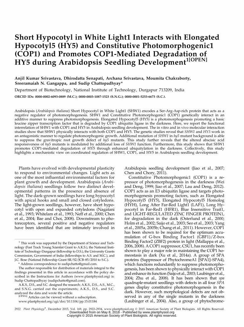
an additive manner to repress photomorphogenicgrowth in the dark (Bhatia et al., 2008). However, en-hanced inhibition in hypocotyl elongation of shw1mutants in white light (WL) requires functional COP1protein (Bhatia et al., 2008). Therefore, we ask whetherSHW1 physically interacts with COP1. To examine thephysical interactions between SHW1 and COP1, invitro pull-down assays were carried out using gluta-thione S-transferase (GST)-SHW1 and COP1-His fusionproteins. In these experiments, GST-HY5 and GBF1-Hiswere used as positive and negative controls, respec-tively (Ang et al., 1998; Singh et al., 2012). GST-SHW1and GST-HY5 proteins were separately passed throughcolumns containing Ni-NTA Agarose (Qiagen) beadsbound to GBF1-His or COP1-His protein. As shownin Figure 1A, SHW1 was retained in COP1 column,and the level of retention of SHW1 was comparablewith the amount of HY5 retained in COP1 column.SHW1 was hardly detectable, if at all, in GBF1-Hiscolumn (Fig. 1, A and B; Supplemental Fig. S1). Takentogether, these results indicate that SHW1 physicallyinteracts with COP1.
To further examine the observed physical interactionof SHW1 and COP1, yeast (Saccharomyces cerevisiae) two-hybrid protein-protein interaction assays were carriedout. In this study, we have used full-length and trun-cated versions of COP1, such as WD40-repeat or zinc-finger and coiled-coil domain of COP1. The growth ofcotransformed yeast cells on two-dimensional (deficientin Trp and Leu) and four-dimensional (deficient inTrp, Leu, His, and adenine) synthetically defined plateswas monitored. As shown in Figure 1, C and D, the full-length SHW1 was able to interact with full-lengthCOP1. Comparison of b-galactosidase activity inSHW1 and COP1 interaction revealed that theb-galactosidase activity was about 5-fold increasedthan the background and comparablewith the increasedactivity in COP1 and HY5 interaction (approximately6-fold) used as control (Fig. 1E). However, neither ofthe truncated versions of COP1 showed increasedactivity compared with background level (Fig. 1E).These results suggest that SHW1 physically interactswith COP1 in yeast cells. These results further indicatethat full-length COP1 is required for the interactionwith SHW1.
To further substantiate the physical interactions be-tween SHW1 and COP1, bimolecular fluorescencecomplementation (BiFC) experiments were carried out.For these experiments, SHW1 full-length coding se-quence was fused to the C terminus of YFP in pUC-SPYCE vector (SHW1-cYFP), and COP1 full-lengthcoding sequence was fused to the N terminus of YFPin pUC-SPYNE vector (COP1-nYFP; Sethi et al.,2014). Whereas empty vectors did not produce anyYFP fluorescence (Fig. 1F), interaction of SHW1 andCOP1 produced strong YFP fluorescence in the nu-cleus with the characteristic speckles known to beformed by COP1 (Fig. 1F). Taken together, these re-sults show that SHW1 physically interacts with COP1in vivo.
Figure 1. SHW1 physically interacts with COP1. A, In vitro binding of SHW1 and COP1: 2mg of COP1-6His or GBF1-6His (negativecontrol) was bound toNi-NTA beads, washed, and incubatedwithGST-SHW1orGST-HY5 (positive control). Beadswerewashed andfractionated in12%(w/v) SDS-PAGE.Theblotwasprobedwith anti-GSTantibodies. B,Quantificationof thedata (byBio-Radmulti-imager):retention ofGST-SHW1andGST-HY5 byCOP1-6His or GBF1-6His is shown in the graph. Error bars indicate SEM of three replicate
SHW1 and HY5 Genetically Interact to RegulateHypocotyl Growth
Because SHW1 physically interacts with COP1 andbecause earlier studies have shown that shw1 cop1double mutants display enhanced photomorphogenicgrowth in the darkness (Bhatia et al., 2008), we askwhether, similar to COP1, SHW1 also genetically in-teracts with HY5, one of the targets of COP1 in thedarkness (Osterlund et al., 2000). The hy5 mutants ex-hibit elongated hypocotyl, whereas shw1 mutants dis-play shorter hypocotyl in WL (Ang et al., 1998; Bhatiaet al., 2008). We generated shw1 hy5 double mutantsand examined the seedling growth in the dark and atvarious wavelengths of light. In the darkness, the shw1mutants displayed shorter hypocotyl with partiallyopened apical hooks, consistent with the previous ob-servation (Bhatia et al., 2008). The shw1 hy5 doublemutants showed hypocotyl length and hook anglesimilar to shw1 singlemutants in the darkness (Fig. 2, A,F, and K).The shw1 mutants display shorter hypocotyl in WL
but not in BL, red light (RL), and far-red (FR) light.When we examined the hypocotyl growth in WL, 6-d-oldshw1 hy5 double mutants displayed shorter hypocotylthan hy5 single mutants inWL, suggesting that they seemto work antagonistically to regulate the hypocotyl growthin WL (Fig. 2, B and G). Furthermore, although shw1mutants did not display any alteration in the hypocotyllength in BL, RL, and FR light (Fig. 2, C–E andH–J; Bhatiaet al., 2008) conditions, the hypocotyl length of shw1 hy5doublemutants was significantly reduced comparedwithhy5 single mutants in BL and RL (Fig. 2, C, D, H, and I).These results suggest that functional SHW1 is required forthe optimum hypocotyl phenotype of hy5 mutants in BLandRL conditions.However, the hypocotyl length of shw1hy5 double mutant was similar to hy5 in FR light (Fig. 2, Eand J), indicating that additional mutation of SHW1 in hy5mutant does not affect the phenotype of hy5 in FR light.
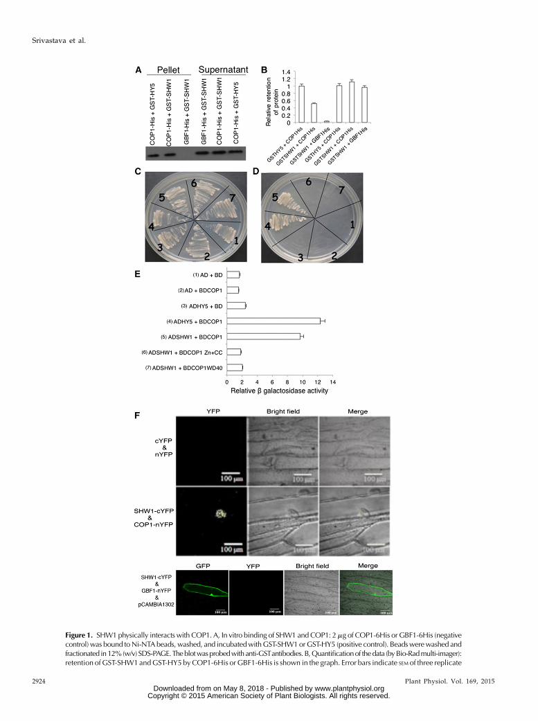
The shw1 and hy5 mutants display altered rootgrowth (Oyama et al., 1997; Ang et al., 1998; Bhatiaet al., 2008). To determine whether SHW1 and HY5 canmodulate each other’s function on root growth, weexamined the root growth of shw1 hy5 double mutantscompared with single mutants. The shw1 mutants de-veloped shorter roots than the wild type, and the rootlength of shw1 hy5 double mutants was found to besimilar to shw1 (Fig. 3, A and B). Because hy5 mutants
do not display any altered root length, these resultsindicate that SHW1works independently of HY5 in theregulation of root length.
Previous studies have shown that one of the obviousphenotypes of hy5 mutants is the direction of rootgrowth. It was nearly horizontal rather than down-ward, indicating alteration of the gravitropic response.The angles of lateral roots of hy5 mutants were largerthan the wild type (Oyama et al., 1997). As shown inFigure 3, A and C, the roots of shw1 hy5 double-mutantplants did not exhibit any defect in gravitropic re-sponses. These results suggest that additional mutationof SHW1 in hy5 mutant background is able to suppressthe gravitropic root growth defect of hy5 mutants.
HY5 and SHW1 Additively Work in ABA Responsiveness
Recent studies have revealed that light signalingpathways cross talk with multiple hormone signalingpathways. For example, HY5 acts as a point of crosstalk in light and ABA signaling pathways. It hasbeen shown that mutations in HY5 caused Arabidopsisplants to be less sensitive to ABA (Chen et al., 2008).To determine further genetic relationship betweenSHW1 and HY5, we ask whether mutations in SHW1can modulate the altered ABA responsiveness of hy5mutants. Freshly harvested seeds of wild-type andsingle- or double-mutant plants were plated on Mura-shige and Skoog medium (MS) plates without or withABA. The rate of germinationwas found to be similar inthe wild type and various mutants in untreated seeds(Supplemental Fig. S2A). Whereas 1 mM ABA reducedthe rate of germination of wild-type seeds (34%), theeffect was significantly suppressed in hy5 background(70%). The shw1 mutant seeds showed less sensitivityto ABA-mediated inhibition of seed germinationcompared with the wild type (48%). As shown inSupplemental Figure S2, the rate of seed germinationwas found to be significantly higher in shw1 hy5 doublemutants than respective single mutants (82%). Theseresults indicate that HY5 and SHW1 work in an addi-tive manner in response to ABA-mediated inhibition ofseed germination.
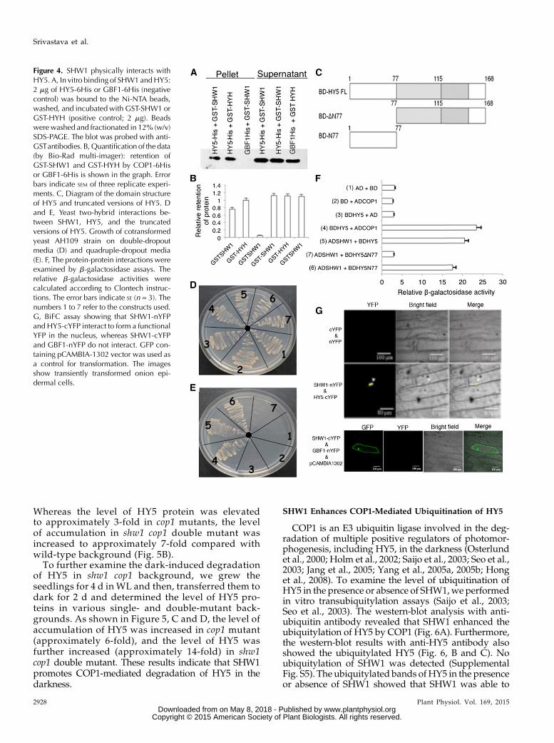
SHW1 Physically Interacts with HY5
The genetic interaction between SHW1 and HY5prompted us to investigate the possible physical inter-actions between SHW1 and HY5. To examine that,in vitro pull-down assays were performed usingGST-SHW1 and HY5-His fusion proteins. In these
Figure 1. (Continued.)experiments. C to E, Yeast two-hybrid interactions between SHW1, COP1, and the truncated versions of COP1. Growth of cotrans-formedyeast AH109 strain on double-dropout media (C) and quadruple-dropoutmedia (D). The protein-protein interactionswereexamined by b-galactosidase assays. The relative b-galactosidase activities were calculated according to Clontech instructions (E). Theerror bars indicate SE (n = 3). The numbers 1 to 7 refer to the constructs used. F, BiFC assay showing that COP1-nYFPand SHW1-cYFPinteract to form a functional YFP in the nucleus, whereas SHW1-cYFP and GBF1-nYFP do not interact. GFP containing pCAMBIA-1302 vector was used as a control for transformation. The images show transiently transformed onion (Allium cepa) epidermal cells.
experiments, we usedGST-HYHas positive control andGBF1-His as negative control (Holm et al., 2002; Singhet al., 2012). GST-SHW1 and GST-HYH proteins wereseparately passed through columns containingNi-NTAbeads bound to GBF1-His or HY5-His protein. The levelof SHW1 retained by HY5 was similar to the level ofretention of HYH by HY5, and practically no retentionof SHW1 protein was detected in GBF1 column (Fig. 4,A and B; Supplemental Fig. S3).
To further substantiate the observed physical inter-action of SHW1 and HY5, yeast two-hybrid assayswere performed. In these experiments, we used full-length and truncated versions of HY5 proteins (Fig. 4C).The growth of cotransformed yeast cells on two-dimensional (deficient in Trp and Leu) and four-dimensional (deficient in Trp, Leu, His, and adenine)synthetically defined media plates was monitored. Asshown in Figure 4, D and E, the full-length HY5 was
Figure 2. HY5 genetically interacts with SHW1. In A to E, wild-type (WT; Col-0), shw1, hy5, and shw1 hy5 seedlings are shown.A to E, Visible phenotypes of 6-d-old seedlings grown in constant dark (D), WL (30 mmol m22 s21), BL (30 mmol m22 s21), RL(30 mmolm22 s21), and FR light (30 mmol m22 s21) are shown as indicated. F to J, Quantification of hypocotyl length of wild-type,shw1, hy5, and shw1 hy5 seedlings grown under various wavelengths of light. K, Hook angle of 6-d-old constant dark-grownseedling of wild-type and various mutant lines. Results presented are obtained from five biological repeats, each having at least30 seedlings. Error bars represent SE (Student’s t test). *, P , 0.05; **, P , 0.01; ***, P , 0.001.
able to interact with full-length SHW1. Furthermore,the truncated version of HY5, HY5N77, was also able tointeract with SHW1. The comparison of b-galactosidaseactivity in SHW1 with HY5 and HY5N77 interactionsrevealed that the b-galactosidase activity was about 6-to 7-fold increased than the background and similar tothe increased activity in COP1 and HY5 interaction,used as control (Fig. 4F). These results suggest thatSHW1 is able to interact with the full length and the N-terminal domain of HY5 protein.To further substantiate thephysical interactions between
SHW1 and HY5, BiFC experiments were carried out. Forthese experiments, SHW1 full-length coding sequencewasfused to the N terminus of YFP in pUC-SPYNE vector(SHW1-nYFP), and HY5 full-length coding sequence wasfused to the C terminus of YFP in pUC-SPYCE vector(HY5-cYFP; Singh et al., 2012). The empty vectors or acombination of SHW1-cYFP and GBF1-nYFP did notproduce any YFP fluorescence; however, interaction ofSHW1 and HY5 produced strong YFP fluorescence in thenucleus (Fig. 4G). Taken together, these results show thatSHW1 physically interacts with HY5.
SHW1 Promotes COP1-Mediated Degradation of HY5 inthe Darkness
It has earlier been shown that the extent of abundanceof HY5 protein directly correlates with the level of
photomorphogenesis (Osterlund et al., 2000). Subse-quently, it was shown that COP1-SPA complexes in-teract with various photomorphogenesis-promotingfactors, such as HYH, HFR1, and LAF1, and degradethese photomorphogenesis-promoting factors throughthe ubiquitin/26S proteasome pathway in the dark(Saijo et al., 2003; Seo et al., 2003; Jang et al., 2005; Yanget al., 2005a, 2005b; Lau and Deng, 2012). Earlier studieshave shown that additional mutation of SHW1 in cop1mutants enhances the photomorphogenic phenotype ofcop1 mutants in the darkness (Bhatia et al., 2008). Thisstudy further shows that SHW1 physically interactswith both COP1 and HY5 (Figs. 1 and 4). Therefore, weask whether enhanced photomorphogenesis in shw1cop1 background in the dark is caused by an increasedabundance of HY5.
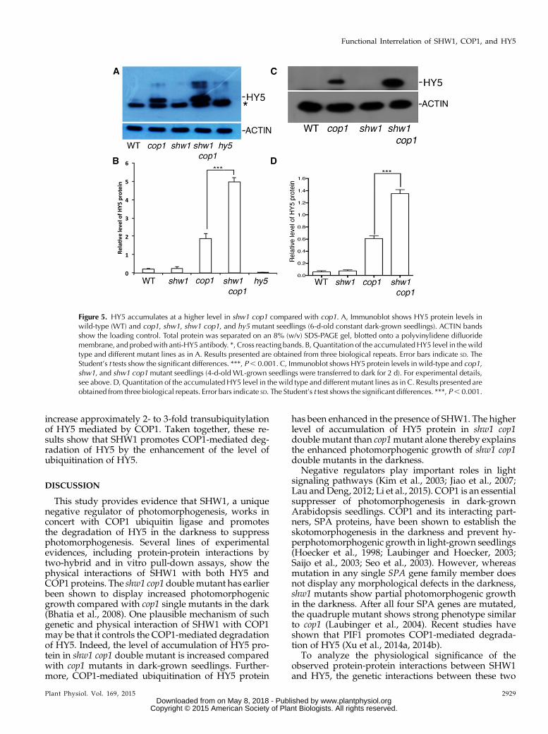
To determine that, we first examined the level of HY5protein in shw1 and shw1 cop1 double-mutant back-grounds in constant dark-grown seedlings. WhereasHY5 protein was barely detectable in wild-typebackground, it was accumulated in cop1 mutantbackground as expected (Fig. 5A; Osterlund et al.,2000). Similar to the wild type, HY5 protein was notdetectable in shw1 mutants (Fig. 5A). However, al-though the transcript level of HY5 remains the samein shw1 and shw1 cop1 backgrounds (SupplementalFig. S4), the level of accumulation of HY5 proteinwas drastically enhanced in shw1 cop1 double mu-tants compared with cop1 single mutants (Fig. 5A).
Figure 3. SHW1andHY5act antagonisticallyto regulate gravitropic root growth. A, The rootgrowthof16-d-oldWL-grown(30mmolm22 s21)wild type (WT) and shw1, hy5, and shw1 hy5mutants. B, Quantification of the root lengthof 16-d-old wild type and various mutantsgrown in WL (30 mmol m22 s21). C,Quantification of gravitropic root move-ment of the wild type and various mutantsgrown in WL (30 mmol m22 s21). Resultspresented are obtained from three bio-logical repeats, each having at least 20plants. Error bars represent SE (Student’s t test).***, P , 0.001.
Whereas the level of HY5 protein was elevatedto approximately 3-fold in cop1 mutants, the levelof accumulation in shw1 cop1 double mutant wasincreased to approximately 7-fold compared withwild-type background (Fig. 5B).
To further examine the dark-induced degradationof HY5 in shw1 cop1 background, we grew theseedlings for 4 d in WL and then, transferred them todark for 2 d and determined the level of HY5 pro-teins in various single- and double-mutant back-grounds. As shown in Figure 5, C and D, the level ofaccumulation of HY5 was increased in cop1 mutant(approximately 6-fold), and the level of HY5 wasfurther increased (approximately 14-fold) in shw1cop1 double mutant. These results indicate that SHW1promotes COP1-mediated degradation of HY5 in thedarkness.
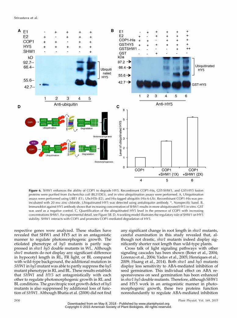
SHW1 Enhances COP1-Mediated Ubiquitination of HY5
COP1 is an E3 ubiquitin ligase involved in the deg-radation of multiple positive regulators of photomor-phogenesis, including HY5, in the darkness (Osterlundet al., 2000; Holm et al., 2002; Saijo et al., 2003; Seo et al.,2003; Jang et al., 2005; Yang et al., 2005a, 2005b; Honget al., 2008). To examine the level of ubiquitination ofHY5 in the presence or absence of SHW1,we performedin vitro transubiquitylation assays (Saijo et al., 2003;Seo et al., 2003). The western-blot analysis with anti-ubiquitin antibody revealed that SHW1 enhanced theubiquitylation of HY5 by COP1 (Fig. 6A). Furthermore,the western-blot results with anti-HY5 antibody alsoshowed the ubiquitylated HY5 (Fig. 6, B and C). Noubiquitylation of SHW1 was detected (SupplementalFig. S5). The ubiquitylated bands ofHY5 in the presenceor absence of SHW1 showed that SHW1 was able to
Figure 4. SHW1 physically interacts withHY5. A, In vitro binding of SHW1andHY5:2 mg of HY5-6His or GBF1-6His (negativecontrol) was bound to the Ni-NTA beads,washed, and incubated with GST-SHW1 orGST-HYH (positive control; 2 mg). Beadswere washed and fractionated in 12% (w/v)SDS-PAGE. The blot was probed with anti-GSTantibodies. B,Quantification of the data(by Bio-Rad multi-imager): retention ofGST-SHW1 and GST-HYH by COP1-6Hisor GBF1-6His is shown in the graph. Errorbars indicate SEM of three replicate experi-ments. C, Diagram of the domain structureof HY5 and truncated versions of HY5. Dand E, Yeast two-hybrid interactions be-tween SHW1, HY5, and the truncatedversions of HY5. Growth of cotransformedyeast AH109 strain on double-dropoutmedia (D) and quadruple-dropout media(E). F, The protein-protein interactions wereexamined by b-galactosidase assays. Therelative b-galactosidase activities werecalculated according to Clontech instruc-tions. The error bars indicate SE (n = 3). Thenumbers 1 to 7 refer to the constructs used.G, BiFC assay showing that SHW1-nYFPand HY5-cYFP interact to form a functionalYFP in the nucleus, whereas SHW1-cYFPand GBF1-nYFP do not interact. GFP con-taining pCAMBIA-1302 vector was used asa control for transformation. The imagesshow transiently transformed onion epi-dermal cells.
increase approximately 2- to 3-fold transubiquitylationof HY5 mediated by COP1. Taken together, these re-sults show that SHW1 promotes COP1-mediated deg-radation of HY5 by the enhancement of the level ofubiquitination of HY5.
DISCUSSION
This study provides evidence that SHW1, a uniquenegative regulator of photomorphogenesis, works inconcert with COP1 ubiquitin ligase and promotesthe degradation of HY5 in the darkness to suppressphotomorphogenesis. Several lines of experimentalevidences, including protein-protein interactions bytwo-hybrid and in vitro pull-down assays, show thephysical interactions of SHW1 with both HY5 andCOP1 proteins. The shw1 cop1 doublemutant has earlierbeen shown to display increased photomorphogenicgrowth compared with cop1 single mutants in the dark(Bhatia et al., 2008). One plausible mechanism of suchgenetic and physical interaction of SHW1 with COP1may be that it controls the COP1-mediated degradationof HY5. Indeed, the level of accumulation of HY5 pro-tein in shw1 cop1 double mutant is increased comparedwith cop1 mutants in dark-grown seedlings. Further-more, COP1-mediated ubiquitination of HY5 protein
has been enhanced in the presence of SHW1. The higherlevel of accumulation of HY5 protein in shw1 cop1double mutant than cop1mutant alone thereby explainsthe enhanced photomorphogenic growth of shw1 cop1double mutants in the darkness.
Negative regulators play important roles in lightsignaling pathways (Kim et al., 2003; Jiao et al., 2007;Lau andDeng, 2012; Li et al., 2015). COP1 is an essentialsuppresser of photomorphogenesis in dark-grownArabidopsis seedlings. COP1 and its interacting part-ners, SPA proteins, have been shown to establish theskotomorphogenesis in the darkness and prevent hy-perphotomorphogenic growth in light-grown seedlings(Hoecker et al., 1998; Laubinger and Hoecker, 2003;Saijo et al., 2003; Seo et al., 2003). However, whereasmutation in any single SPA gene family member doesnot display any morphological defects in the darkness,shw1 mutants show partial photomorphogenic growthin the darkness. After all four SPA genes are mutated,the quadruple mutant shows strong phenotype similarto cop1 (Laubinger et al., 2004). Recent studies haveshown that PIF1 promotes COP1-mediated degrada-tion of HY5 (Xu et al., 2014a, 2014b).
To analyze the physiological significance of theobserved protein-protein interactions between SHW1and HY5, the genetic interactions between these two
Figure 5. HY5 accumulates at a higher level in shw1 cop1 compared with cop1. A, Immunoblot shows HY5 protein levels inwild-type (WT) and cop1, shw1, shw1 cop1, and hy5 mutant seedlings (6-d-old constant dark-grown seedlings). ACTIN bandsshow the loading control. Total protein was separated on an 8% (w/v) SDS-PAGE gel, blotted onto a polyvinylidene difluoridemembrane, and probedwith anti-HY5 antibody. *, Cross reacting bands. B,Quantitation of the accumulatedHY5 level in thewildtype and different mutant lines as in A. Results presented are obtained from three biological repeats. Error bars indicate SD. TheStudent’s t tests show the significant differences. ***, P, 0.001. C, Immunoblot shows HY5 protein levels in wild-type and cop1,shw1, and shw1 cop1mutant seedlings (4-d-old WL-grown seedlings were transferred to dark for 2 d). For experimental details,see above. D, Quantitation of the accumulatedHY5 level in the wild type and different mutant lines as in C. Results presented areobtained from three biological repeats. Error bars indicate SD. The Student’s t test shows the significant differences. ***, P, 0.001.
respective genes were analyzed. These studies haverevealed that SHW1 and HY5 act in an antagonisticmanner to regulate photomorphogenic growth. Theetiolated phenotype of hy5 mutants is partly sup-pressed in shw1 hy5 double mutants in WL. Althoughshw1 mutants do not display any significant differencein hypocotyl length in RL, FR light, or BL comparedwith wild-type background, the additional mutation inSHW1 in hy5mutantwas able to partly suppress the hy5mutant phenotype in RL and BL. These results establishthat SHW1 and HY5 act antagonistically with eachother to regulate photomorphogenic growth in RL andBL conditions. The gravitropic root growth defect of hy5mutants is also suppressed by additional loss of func-tion of SHW1. Although Bhatia et al. (2008) did not find
any significant change in root length in shw1 mutants,careful examination in this study revealed that, al-though not drastic, shw1 mutants indeed display sig-nificantly shorter root length than wild-type plants.
Cross talk of light signaling pathways with othersignaling cascades has been shown (Boter et al., 2004;Lorenzo et al., 2004; Yadav et al., 2005; Henriques et al.,2009; Huang et al., 2014). Both shw1 and hy5 mutantsdisplay less sensitivity to ABA-mediated inhibition ofseed germination. This individual effect on ABA re-sponsiveness on seed germination has been enhancedin shw1 hy5 doublemutants. Therefore, although SHW1and HY5 work in an antagonistic manner in photo-morphogenic growth, these two proteins functionnonredundantly to regulate ABA-mediated inhibition
Figure 6. SHW1 enhances the ability of COP1 to degrade HY5. Recombinant COP1-His, GST-SHW1, and GST-HY5 fusionproteins were purified from Escherichia coli (BL21DE3), and in vitro ubiquitination assays were performed. A, Ubiquitinationassays were performed using UBE1 (E1), UbcH5b (E2), and His-tagged ubiquitin (His-6-Ub). Recombinant COP1-His was pre-incubated with 20 mM zinc chloride. Ubiquitinated HY5 was detected using antiubiquitin antibody. *, Nonspecific band. B,Immunoblot against HY5 antibody shows that increasing concentration of SHW1 results in more ubiquitinated HY5 in vitro. GSTwas used as a negative control. C, Quantification of the ubiquitinated HY5 level in the presence of COP1 with increasingconcentrations SHW1. For experimental detail, see Figure 5B. D, Aworkingmodel illustrates the regulatory role of SHW1onHY5stability. SHW1 interacts with COP1 and promotes COP1-mediated degradation of HY5.
of seed germination. Several reports have earlier shownthe differential regulation of a gene in two differentsignaling pathways (Anderson et al., 2004; Kazan andManners, 2008). MYC2 works as a negative regulator oflight signaling pathways (Yadav et al., 2005; Gangappaet al., 2010; Sethi et al., 2014). However, it works as bothpositive and negative regulators of jasmonic acid sig-naling pathways (Dombrecht et al., 2007; Kazan andManners 2008, 2013). Furthermore, differential geneticinteraction ofHY5 and GBF1 has been shown to work ina light intensity-dependent manner (Singh et al., 2012).Recent studies have revealed the cross talk between lightand temperature signaling pathways (Delker et al., 2014;Johansson et al., 2014; Toledo-Ortiz et al., 2014). It wouldbe interesting to investigate the possible involvement ofSHW1 and HY5 in temperature signaling pathways.Furthermore, it would also be interesting to determinethe possible involvement of SHW1, as an associatedfactor of COP1, in COP1-mediated proteasomal degra-dation of other photomorphogenesis-promoting factors,such as HYH, HFR1, and LAF1, in future studies.
MATERIALS AND METHODS
Plant Materials, Growth Conditions, and Generation ofDouble Mutants
The growth conditions and light sources are described in Singh et al. (2012).For the generation of the hy5 shw1 double mutant, homozygous hy5 mutantplants (hy5-215) in Columbia-0 (Col-0) background were crossed with shw1(shw1-1) in Col-0 background. In the F2 generation, seedlings were grown inWL (30 mmol m22 s21) for the identification of hy5 mutant phenotype, andseedlingswere selected and transferred to soil. To determine the genotype at theSHW1 locus, approximately 40 seedlings from each line were tested by genomicPCR (Bhatia et al., 2008). For this, individual plants were examined by PCRusing the left border-specific primer. F3 progeny that are homozygous for shw1and hy5 mutant plants were further examined by reverse transcription (RT)-PCR and considered as hy5 shw1 double mutants. The cop1 mutant used in thisstudy is cop1-6 allele (Bhatia et al., 2008).
Root Growth
Seeds were plated on one-half-strength MS (1% [w/v] Suc and 1% [w/v]agar) on vertical square plates and stratified at 4°C in dark conditions for 4 d toinduce uniform germination. The plates were placed vertically in racks, and theseedlings were grown under constant WL conditions (40 mmol m22 s21). Thenumbers of lateral roots of the wild type and single and double mutants werecounted from 10 to 15 d after germination and tabulated. The root angle wasmeasured by using ImageJ 1.x software (NIH).
Effect of ABA on Seed Germination
The seeds of wild-type, shw1, hy5, and shw1 hy5 mutant plants were platedonto MS plates with or without ABA (Sigma-Aldrich). The plates were kept incold and dark for 4 d for stratification and then transferred to constant WL, andthe seeds were monitored for germination.
Cloning of Constructs
For making the recombinant constructs in each case, the desired fragmentwas amplified by PCR using a specific pair of forward and reverse primers withrestriction sites at the 59 end of the primers (as indicated in the primers list).Then, the desired fragment and the vectors were digested with the same pair ofenzymes. The digested sample was purified from the agarose gel using the gelextraction kit (Qiagen). The different DNA fragments used in this study were
ligated into respective vectors with T4 DNA ligase using different molar ratios(insert:vector, 3:1 to 5:1) by incubating overnight at 16°C or 22°C (as per thecompany’s recommendation for T4 DNA ligase enzyme). Ligated DNA frag-ments were transformed into Escherichia coli DH5a-competent cells. ColonyPCR was performed to confirm the transformed cells with recombinant plas-mid. The clones were confirmed by restriction digestion of plasmids and ad-ditional sequencing.
For in vitro protein-protein interaction studies, DNA fragments encodingfull-length SHW1orHY5were cloned intopGEX-4T2vector using the respectiveprimers atBamHI and EcoRI restriction sites to get GST fusion protein. TheDNAfragments encoding full-length HY5 and COP1 were cloned into pET-20b (+)vector using the respective primers at NdeI-ClaI and EcoRI-PstI restriction sites,respectively, to obtain 63 His tag at the C terminus of the protein. For yeast(Saccharomyces cerevisiae) two-hybrid protein-protein interaction assay, full-length SHW1 was cloned into the pGADT7 vector using the respectiveprimers at EcoRI and BamHI restriction sites to produce translational fusionproteins with the activation domain. To generate full-length COP1 constructs,COP1-FL (for full length), COP1-Zn+CC (for coiled coil), and COP1-WD40were cloned into pGBKT7 vector using the respective primers (SupplementalTable S1) at EcoRI, PstI, and BamHI restriction sites to produce translationalfusion with the binding domain. The HY5-FL, HY5-N77 (for N-terminal 77 aminoacids), and HY5DN77 have been cloned into the pGBKT7 vector using the re-spective primers (Supplemental Table S1) at EcoRI and BamHI restriction sites toproduce translational fusion proteins with the binding domain. For BiFC experi-ments, full-length CDS (coding sequence) of SHW1was cloned in the vectors pUC-SPYNEandpUC-SPYCE (Walter et al., 2004) using the respective primers atBamHIand XhoI sites to obtain SHW1-YFPN-ter and SHW1-YFPC-ter fusion proteins,respectively. To obtain COP1-YFPC-ter and COP1-YFPN-ter fusion proteins, full-length CDS of COP1 was cloned in pUC-SPYCE and pUC-SPYNE vectors, re-spectively, using the respective primers at AscI and XhoI restriction sites.
Pull-Down Assay
In vitro protein-protein interaction studies were carried out as described inAbbas et al. (2014) with slight modifications. DNA fragments encoding full-length SHW1 or HY5 were cloned into pGEX-4T2 vector, obtaining transla-tional fusion constructs with the GST protein. GST-SHW1 andGST-HY5 proteinswere overexpressed and purified from E. coli by Glutathione Sepharose 4BBeads (GE). The DNA fragments encoding full-length COP1 or HY5 werecloned into pET-20b (+) vector with 63His tag at the C terminus of the protein.COP1-His protein was overexpressed and purified from E. coli cells by Ni-NTAAgarose Beads (Qiagen). For in vitro binding experiments, HY5-His and COP1-His (2.0mg each) proteins were bound toHis column by incubatingwith in vitropull-down buffer for 2 h at 4°C. Excess unbound protein was washed off,and GST-HYH, GST-SHW1, and GST proteins were added in equimolar ratioand incubated in 500 mL of in vitro pull-down buffer (50 mM Tris-Cl, pH 7.5,100 mM NaCl, 0.2% [v/v] glycerol, 1 mM EDTA, 0.1% [v/v] Nonidet P-40, 1 mM
phenylmethylsulfonyl fluoride, and full-strength protease inhibitors cocktail;Sigma) at 4°C. The Ni-NTA beads were collected by brief centrifugation (su-pernatantwas collected separately and saved for further analysis) andwashed threetimeswith 1mLof in vitropull-downbuffer. Pelletwas resuspended in full-strengthSDS loading buffer, boiled for 5 min, and analyzed by SDS-PAGE for proteinbinding. Both pellet and supernatant (2%) were probed with anti-GST antibodies.
Total Protein Extraction
The seedlings (100mg)were frozen in liquid nitrogen andground in 300mL ofgrinding buffer (400 mM Suc, 50 mM Tris-Cl, pH 7.5, 10% [v/v] glycerol, and2.5mMEDTA), andphenylmethylsulfonylfluoride (1mM stock)was added (0.5mLfor every 100mL of grinding buffer). The protein extract was transferred to freshmicrocentrifuge tube and centrifuged at 5,000 rpm for 5 min to pellet down thedebris. The supernatant was transferred to a fresh tube, and an aliquot of 5 mLwas taken out in a separate tube for the estimation of protein by Bradford assay.To the rest of the protein extract, appropriate volume of 43 sample buffer (200 mM
Tris-Cl, pH 6.8, 400 mM dithiothreitol, 4% [w/v] SDS, 0.025% [w/v] BromophenolBlue, and 20% [v/v] glycerol) was added and boiled for 5 min before loading onSDS-PAGE.
Yeast Two-Hybrid Assay
Yeast two-hybrid assays were performed using the Matchmaker GAL4-Based Two-Hybrid System as recommended (Clontech Laboratories, Inc.). To
investigate the protein-protein interaction, full-length SHW1was cloned into thepGADT7vector (ClontechLaboratories, Inc.)withEcoRI-BamHIrestriction sites toproduce translational fusion proteins with the activation domain. To generatefull-length COP1 constructs, COP1-FL, COP1-Zn+CC, and COP1-WD40 werecloned into pGBKT7 vector (Clontech Laboratories, Inc.) with EcoRI-PstI re-striction site to produce translational fusion with the binding domain. The HY5-FL, HY5-N77, and HY5ΔN77 have been individually cloned into the pGBKT7vector (Clontech Laboratories, Inc.) withEcoRI-BamHI restriction sites to producetranslational fusion proteins with the binding domain. To assess protein-protein interactions, the corresponding plasmidswere cotransformed into yeaststrain AH109 according to the protocol given by Clontech Laboratories, Inc.Successfully transformed colonies were identified on quadruple-dropout medialacking Trp, Leu, His, and adenine. Expression of GAL4 Activation Domainfused-SHW1 fusion protein was examined by Hemagglutinin antibodies, andGAL4 Binding Domain fused (BD)-COP1, BD-HY5, and their truncated ver-sions were examined by c-Myc antibodies (Supplemental Fig. S6). The protein-protein interactions were also examined by b-galactosidase assays using chlo-rophenol red-b-D-galactopyranoside as a substrate. The relative b-galactosidaseactivities were calculated according to Clontech Laboratories, Inc. instructions.
BiFC Assay
Coding sequences corresponding to full-length SHW1, COP1, andHY5wereamplified using respective primers (Supplemental Table S1) cloned under thecontrol of the 35S promoter and fused to the N- and/or C-terminal part of YFP(pUC-SPYNE/pUC-SPYCE; Walter et al., 2004). The desired constructs weremixed in equal proportions (5 mg each) and cobombarded into onion (Alliumcepa) epidermal cells as described in Abbas et al., 2014. DNA particle bom-bardment was performed using the helium-driven particle accelerator (PDS-1000) following the manufacturer’s instructions (Bio-Rad). The bombardedonion peels were kept in the dark for approximately 20 h at 22°C to allow theexpression of the transfected DNA and reconstruction of the functional YFP,and then, they were mounted onto glass slide and observed under a confocallaser-scanning microscope (Leica-TCS-SP-2) with a visible aquistic opticaltunable filter standard filter set.
RT-PCR Analysis
For RT-PCR, RNA from dark-adapted 6-d-old seedlings was extracted usingthe RNeasy Plant Mini Kit (Qiagen). One microgram of total RNA was con-verted into complementary DNA by using Thermo Scientific RevertAid HMinus First-Strand cDNASynthesis Kit followed by PCR by using gene-specificand ACTIN primers. Primer sequences are given in Supplemental Table S1. Theintensity of each band was quantified by the Gel Doc EZ Imager (BIO-RAD),and the ratio of the HY5 versus ACTIN2 band was determined and plotted.
In Vitro Ubiquitylation Assays
Invitroubiquitinationassayswereperformedasdescribedpreviously (Seoet al.,2003) with minor modifications. Ubiquitination reaction mixtures (30 mL) con-tained 30 ng ofUBE1 (E1; Boston Biochem), 30 ng ofUbcH5b (E2; Boston Biochem),10 mg of His-tagged ubiquitin (Ub; Boston Biochem), 500 ng of COP1-His (pre-viously incubated with 20 mM zinc chloride), 400 ng of GST-HY5, and 200 ng ofGST-SHW1 in a reaction buffer containing 50 mM Tris, pH 7.5, 5 mM MgCl2, 2 mM
ATP, and 0.5 mM dithiothreitol. GST was used as a negative control. After 2 h ofincubation at 30°C, the reactions were stopped by adding 53 sample buffer. Thereaction mixtures (30 mL) were then separated onto 8% (w/v) SDS-PAGE gels.Ubiquitinated HY5 was detected using anti-HY5 (antibody of HY5 was raisedagainst thepeptide [IKEGIESDEEIRRVPC]ofHY5byBangaloreGeneI) andanti-GSTantibody (Sigma) and anti-ubiquitin antibody. The intensity of the GST-HY5 bandsdetected by anti-HY5 antibody from three independent blots was quantified usingImageJ software. Briefly, protein levels in western-blot analyses were quantified byGelDocEZ Imager (BIO-RAD)using ImageLab software (version 4.0). Bandspresentin all of the lanes are selected automatically. The band that corresponds to ACTIN inthe wild type is taken as relative. By taking this band as standard, the machine au-tomatically quantifies the value of relative intensities of other bands. Conventionally,the machine takes the relative intensity of ACTIN band of the wild type as 1.
Western-Blot Analysis
Western blot was performed using the Super Signal West Pico Chemilumi-nescent Substrate Kit (Pierce) following the instructions as described in the user’s
manual providedby themanufacturer. The sampleswere then runon SDS-PAGEgel and transferred to a polyvinylidene difluoride membrane (GE) at 130 mA for1 h in transfer buffer (5.82 g of Tris, 2.93 g of Gly, and 20% [v/v] methanol in 1 L)in a Genei Dual Transfer Unit Apparatus (Banglore Genei). The membrane wasstained with Ponceau-S to confirm the protein transfer and washed with sterilemilli-Q water. The membrane was then incubated for 1 h in 25 mL of blockingbuffer (5% [w/v] nonfat dry milk [Himedia] in phosphate-buffered saline [PBS]and 0.05% [v/v] Tween 20) at room temperature on a dancing rocker. Theblocking reagent was removed, and the affinity-purified primary antibody wasdiluted (1:500 to 1:10,000) in 10 mL of PBS with 0.05% (v/v) Tween 20 and in-cubated for 2 h with shaking at room temperature. The membrane was thenwashed with 15 mL of wash buffer (PBS and 0.05% [v/v] Tween 20) threetimes for 5 min each. The secondary antibody conjugated with horseradishperoxidise diluted 1:10,000 in 10 mL of blocking buffer with 0.05% (v/v)Tween 20 was added and incubated for 1 h with shaking at room temper-ature. The membrane was washed with 15 mL of wash buffer three times atroom temperature. The working solution of substrate was prepared bymixing peroxide solution:luminol/enhancer solution (1:1), and the blotwas incubated in that working solution for 5 min in dark. The blot was thenremoved from the working solution, covered with plastic wrap in cassette,and exposed to x-ray film for different times.
Primers Used in Various Experiments
The primers used in this study are summarized in Supplemental Table S1.
Supplemental Data
The following supplemental materials are available.
Supplemental Figure S1. Physical interaction between SHW1 and COP1 asshown by in vitro binding assay.
Supplemental Figure S2. ABA-mediated responsiveness of shw1 hy5double mutants.
Supplemental Figure S3. Physical interaction between SHW1 and HY5 asshown by in vitro binding assay.
Supplemental Figure S4. Transcript levels of HY5.
Supplemental Figure S5. SHW1 enhances the ubiquitylation activity ofCOP1 but is not itself ubiquitinated by COP1.
Supplemental Figure S6. The expressed proteins in yeast cells.
Supplemental Table S1. Primers used in this study.
ACKNOWLEDGMENTS
We thank Xing-Wang Deng for providing the cop1 mutant seeds and JayPrakash Maurya for critically reading and commenting on this article.
Received July 31, 2015; accepted October 13, 2015; published October 16, 2015.
LITERATURE CITED
Abbas N, Maurya JP, Senapati D, Gangappa SN, Chattopadhyay S (2014)Arabidopsis CAM7 and HY5 physically interact and directly bind to theHY5 promoter to regulate its expression and thereby promote photo-morphogenesis. Plant Cell 26: 1036–1052
Alabadí D, Gallego-Bartolomé J, Orlando L, García-Cárcel L, Rubio V,Martínez C, Frigerio M, Iglesias-Pedraz JM, Espinosa A, Deng XW,et al (2008) Gibberellins modulate light signaling pathways to preventArabidopsis seedling de-etiolation in darkness. Plant J 53: 324–335
Anderson JP, Badruzsaufari E, Schenk PM, Manners JM, Desmond OJ,Ehlert C, Maclean DJ, Ebert PR, Kazan K (2004) Antagonistic interac-tion between abscisic acid and jasmonate-ethylene signaling pathwaysmodulates defense gene expression and disease resistance in Arabidopsis.Plant Cell 16: 3460–3479
Ang LH, Chattopadhyay S, Wei N, Oyama T, Okada K, Batschauer A,Deng XW (1998) Molecular interaction between COP1 and HY5 definesa regulatory switch for light control of Arabidopsis development. MolCell 1: 213–222
Ang LH, Deng XW (1994) Regulatory hierarchy of photomorphogenic loci:allele-specific and light-dependent interaction between the HY5 andCOP1 loci. Plant Cell 6: 613–628
Bae G, Choi G (2008) Decoding of light signals by plant phytochromes andtheir interacting proteins. Annu Rev Plant Biol 59: 281–311
Bhatia S, Gangappa SN, Kushwaha R, Kundu S, Chattopadhyay S (2008)SHORT HYPOCOTYL IN WHITE LIGHT1, a serine-arginine-aspartate-rich protein in Arabidopsis, acts as a negative regulator of photomor-phogenic growth. Plant Physiol 147: 169–178
Boter M, Ruíz-Rivero O, Abdeen A, Prat S (2004) Conserved MYC tran-scription factors play a key role in jasmonate signaling both in tomatoand Arabidopsis. Genes Dev 18: 1577–1591
Chang CS, Maloof JN, Wu SH (2011) COP1-mediated degradation ofBBX22/LZF1 optimizes seedling development in Arabidopsis. PlantPhysiol 156: 228–239
Chattopadhyay S, Ang LH, Puente P, Deng XW, Wei N (1998) ArabidopsisbZIP protein HY5 directly interacts with light-responsive promoters inmediating light control of gene expression. Plant Cell 10: 673–683
Chen H, Zhang J, Neff MM, Hong SW, Zhang H, Deng XW, Xiong L(2008) Integration of light and abscisic acid signaling during seed ger-mination and early seedling development. Proc Natl Acad Sci USA 105:4495–4500
Chen M, Chory J (2011) Phytochrome signaling mechanisms and the con-trol of plant development. Trends Cell Biol 21: 664–671
Chen M, Chory J, Fankhauser C (2004) Light signal transduction in higherplants. Annu Rev Genet 38: 87–117
de Lucas M, Davière JM, Rodríguez-Falcón M, Pontin M, Iglesias-PedrazJM, Lorrain S, Fankhauser C, Blázquez MA, Titarenko E, Prat S (2008)A molecular framework for light and gibberellin control of cell elonga-tion. Nature 451: 480–484
Delker C, Sonntag L, James GV, Janitza P, Ibañez C, Ziermann H,Peterson T, Denk K, Mull S, Ziegler J, et al (2014) The DET1-COP1-HY5pathway constitutes a multipurpose signaling module regulating plantphotomorphogenesis and thermomorphogenesis. Cell Reports 9: 1983–1989
Dombrecht B, Xue GP, Sprague SJ, Kirkegaard JA, Ross JJ, Reid JB, FittGP, Sewelam N, Schenk PM, Manners JM, et al (2007) MYC2 differ-entially modulates diverse jasmonate-dependent functions in Arabi-dopsis. Plant Cell 19: 2225–2245
Gangappa SN, Prasad VB, Chattopadhyay S (2010) Functional intercon-nection of MYC2 and SPA1 in the photomorphogenic seedling devel-opment of Arabidopsis. Plant Physiol 154: 1210–1219
Henriques R, Jang IC, Chua NH (2009) Regulated proteolysis in light-related signaling pathways. Curr Opin Plant Biol 12: 49–56
Hoecker U, Xu Y, Quail PH (1998) SPA1: a new genetic locus involved inphytochrome A-specific signal transduction. Plant Cell 10: 19–33
Holm M, Ma LG, Qu LJ, Deng XW (2002) Two interacting bZIP proteinsare direct targets of COP1-mediated control of light-dependent geneexpression in Arabidopsis. Genes Dev 16: 1247–1259
Hong SH, Kim HJ, Ryu JS, Choi H, Jeong S, Shin J, Choi G, Nam HG(2008) CRY1 inhibits COP1-mediated degradation of BIT1, a MYBtranscription factor, to activate blue light-dependent gene expression inArabidopsis. Plant J 55: 361–371
Huang X, Ouyang X, Deng XW (2014) Beyond repression of photomor-phogenesis: role switching of COP/DET/FUS in light signaling. CurrOpin Plant Biol 21: 96–103
Jang IC, Yang JY, Seo HS, Chua NH (2005) HFR1 is targeted by COP1 E3ligase for post-translational proteolysis during phytochrome A signal-ing. Genes Dev 19: 593–602
Jiao Y, Lau OS, Deng XW (2007) Light-regulated transcriptional networksin higher plants. Nat Rev Genet 8: 217–230
Johansson H, Jones HJ, Foreman J, Hemsted JR, Stewart K, Grima R,Halliday KJ (2014) Arabidopsis cell expansion is controlled by a pho-tothermal switch. Nat Commun 5: 4848
Kazan K, Manners JM (2008) Jasmonate signaling: toward an integratedview. Plant Physiol 146: 1459–1468
Kazan K, Manners JM (2013) MYC2: the master in action. Mol Plant 6: 686–703
Kim J, Yi H, Choi G, Shin B, Song PS, Choi G (2003) Functional charac-terization of phytochrome interacting factor 3 in phytochrome-mediatedlight signal transduction. Plant Cell 15: 2399–2407
Lau OS, Deng XW (2012) The photomorphogenic repressors COP1 andDET1: 20 years later. Trends Plant Sci 17: 584–593
Laubinger S, Fittinghoff K, Hoecker U (2004) The SPA quartet: a family ofWD-repeat proteins with a central role in suppression of photomor-phogenesis in Arabidopsis. Plant Cell 16: 2293–2306
Laubinger S, Hoecker U (2003) The SPA1-like proteins SPA3 and SPA4repress photomorphogenesis in the light. Plant J 35: 373–385
Lee J, He K, Stolc V, Lee H, Figueroa P, Gao Y, Tongprasit W, Zhao H, LeeI, Deng XW (2007) Analysis of transcription factor HY5 genomic bind-ing sites revealed its hierarchical role in light regulation of development.Plant Cell 19: 731–749
Leivar P, Monte E, Oka Y, Liu T, Carle C, Castillon A, Huq E, Quail PH(2008) Multiple phytochrome-interacting bHLH transcription factorsrepress premature seedling photomorphogenesis in darkness. Curr Biol18: 1815–1823
Leivar P, Quail PH (2011) PIFs: pivotal components in a cellular signalinghub. Trends Plant Sci 16: 19–28
Li K, Gao Z, He H, Terzaghi W, Fan LM, Deng XW, Chen H (2015)Arabidopsis DET1 represses photomorphogenesis in part by negativelyregulating DELLA protein abundance in darkness. Mol Plant 8:622–630
Lorenzo O, Chico JM, Sánchez-Serrano JJ, Solano R (2004) JASMONATE-INSENSITIVE1 encodes a MYC transcription factor essential to dis-criminate between different jasmonate-regulated defense responses inArabidopsis. Plant Cell 16: 1938–1950
Ma L, Li J, Qu L, Hager J, Chen Z, Zhao H, Deng XW (2001) Light controlof Arabidopsis development entails coordinated regulation of genomeexpression and cellular pathways. Plant Cell 13: 2589–2607
Mallappa C, Singh A, Ram H, Chattopadhyay S (2008) GBF1, a tran-scription factor of blue light signaling in Arabidopsis, is degraded in thedark by a proteasome-mediated pathway independent of COP1 andSPA1. J Biol Chem 283: 35772–35782
Mallappa C, Yadav V, Negi P, Chattopadhyay S (2006) A basic leucinezipper transcription factor, G-box-binding factor 1, regulates blue light-mediated photomorphogenic growth in Arabidopsis. J Biol Chem 281:22190–22199
Nagatani A, Reed JW, Chory J (1993) Isolation and initial characterizationof Arabidopsis mutants that are deficient in phytochrome A. PlantPhysiol 102: 269–277
Neff MM, Fankhauser C, Chory J (2000) Light: an indicator of time andplace. Genes Dev 14: 257–271
Osterlund MT, Hardtke CS, Wei N, Deng XW (2000) Targeted destabili-zation of HY5 during light-regulated development of Arabidopsis. Na-ture 405: 462–466
Oyama T, Shimura Y, Okada K (1997) The Arabidopsis HY5 gene encodesa bZIP protein that regulates stimulus-induced development of root andhypocotyl. Genes Dev 11: 2983–2995
Ram H, Priya P, Jain M, Chattopadhyay S (2014) Genome-wide DNAbinding of GBF1 is modulated by its heterodimerizing protein partners,HY5 and HYH. Mol Plant 7: 448–451
Saijo Y, Sullivan JA, Wang H, Yang J, Shen Y, Rubio V, Ma L, Hoecker U,Deng XW (2003) The COP1-SPA1 interaction defines a critical step inphytochrome A-mediated regulation of HY5 activity. Genes Dev 17:2642–2647
Seo HS, Yang JY, Ishikawa M, Bolle C, Ballesteros ML, Chua NH (2003)LAF1 ubiquitination by COP1 controls photomorphogenesis and isstimulated by SPA1. Nature 423: 995–999
Sethi V, Raghuram B, Sinha AK, Chattopadhyay S (2014) A mitogen-activated protein kinase cascade module, MKK3-MPK6 and MYC2, isinvolved in blue light-mediated seedling development in Arabidopsis.Plant Cell 26: 3343–3357
Singh A, Ram H, Abbas N, Chattopadhyay S (2012) Molecular interactionsof GBF1 with HY5 and HYH proteins during light-mediated seedlingdevelopment in Arabiopsis thaliana. J Biol Chem 287: 25996–26009
Tepperman JM, Zhu T, Chang HS, Wang X, Quail PH (2001) Multipletranscription-factor genes are early targets of phytochrome A signaling.Proc Natl Acad Sci USA 98: 9437–9442
Toledo-Ortiz G, Johansson H, Lee KP, Bou-Torrent J, Stewart K, Steel G,Rodríguez-Concepción M, Halliday KJ (2014) The HY5-PIF regulatorymodule coordinates light and temperature control of photosyntheticgene transcription. PLoS Genet 10: e1004416
Vandenbussche F, Habricot Y, Condiff AS, Maldiney R, Straeten DVD,Ahmad M (2007) HY5 is a point of convergence between cryptochromeand cytokinin signalling pathways in Arabidopsis thaliana. Plant J 49:428–441
Walter M, Chaban C, Schütze K, Batistic O, Weckermann K, Näke C,Blazevic D, Grefen C, Schumacher K, Oecking C, et al (2004) Visual-ization of protein interactions in living plant cells using bimolecularfluorescence complementation. Plant J 40: 428–438
Wei N, Deng XW (1999) Making sense of the COP9 signalosome. A regu-latory protein complex conserved from Arabidopsis to human. TrendsGenet 15: 98–103
Whitelam GC, Johnson E, Peng J, Carol P, Anderson ML, Cowl JS,Harberd NP (1993) Phytochrome A null mutants of Arabidopsis display awild-type phenotype in white light. Plant Cell 5: 757–768
Xu D, Lin F, Jiang Y, Huang X, Li J, Ling J, Hettiarachchi C, Tellgren-RothC, Holm M, Deng XW (2014a) The RING-finger E3 ubiquitin ligaseCOP1 SUPPRESSOR1 negatively regulates COP1 abundance in main-taining COP1 homeostasis in dark-grown Arabidopsis seedlings. PlantCell 26: 1981–1991
Xu X, Paik I, Zhu L, Bu Q, Huang X, Deng XW, Huq E (2014b)PHYTOCHROME INTERACTING FACTOR1 enhances the E3 ligaseactivity of CONSTITUTIVE PHOTOMORPHOGENIC1 to synergis-tically repress photomorphogenesis in Arabidopsis. Plant Cell 26:1992–2006
Yadav V, Kundu S, Chattopadhyay D, Negi P, Wei N, Deng XW, Chat-topadhyay S (2002) Light regulated modulation of Z-box containing
promoters by photoreceptors and downstream regulatory components,COP1 and HY5, in Arabidopsis. Plant J 31: 741–753
Yadav V, Mallappa C, Gangappa SN, Bhatia S, Chattopadhyay S (2005) Abasic helix-loop-helix transcription factor in Arabidopsis, MYC2, acts as arepressor of blue light-mediated photomorphogenic growth. Plant Cell17: 1953–1966
Yang J, Lin R, Hoecker U, Liu B, Xu L, Wang H (2005a) Repression of lightsignaling by Arabidopsis SPA1 involves post-translational regulation ofHFR1 protein accumulation. Plant J 43: 131–141
Yang J, Lin R, Sullivan J, Hoecker U, Liu B, Xu L, Deng XW, Wang H(2005b) Light regulates COP1-mediated degradation of HFR1, a tran-scription factor essential for light signaling in Arabidopsis. Plant Cell 17:804–821
Zhang H, He H, Wang X, Wang X, Yang X, Li L, Deng XW (2011) Genome-wide mapping of the HY5-mediated gene networks in Arabidopsis thatinvolve both transcriptional and post-transcriptional regulation. Plant J65: 346–358
Zhu D, Maier A, Lee JH, Laubinger S, Saijo Y, Wang H, Qu LJ, HoeckerU, Deng XW (2008) Biochemical characterization of Arabidopsis com-plexes containing CONSTITUTIVELY PHOTOMORPHOGENIC1 andSUPPRESSOR OF PHYA proteins in light control of plant development.Plant Cell 20: 2307–2323


![The greedy triangulation can be computed from the Delaunay ...algorithm is known [20]. In the latter case, it achieves a non-trivial approximation factor2. p n/[11,13] and, with a](https://static.documents.pub/doc/80x56/60045a287bb42a666835a4c6/the-greedy-triangulation-can-be-computed-from-the-delaunay-algorithm-is-known.jpg)


