Short-range order and near-field effectson optical scattering and structural
coloration
S. F. Liew,1,∗ J. Forster,2 H. Noh,1 C. F. Schreck,3 V. Saranathan,4
X. Lu,5 L. Yang,6 Richard O. Prum,4 C. S. O’Hern,2,3
E. R. Dufresne,2,3,7,8 and H. Cao1,3
1Department of Applied Physics, Yale University, New Haven, Connecticut 06511, USA2Department of Mechanical Engineering and Materials Science, Yale University,
New Haven, Connecticut 06511, USA3Department of Physics, Yale University, New Haven, Connecticut 06511, USA
4Department of Ecology and Evolutionary Biology, Peabody Museum of Natural History,Yale University, New Haven, Connecticut 06511, USA
5Department of Condensed Matter Physics and Materials Science,Brookhaven National Laboratory, Upton, New York 11973, USA
6National Synchrotron Light Source, Brookhaven National Laboratory,Upton, New York 11973, USA
7Department of Chemical Engineering, Yale University, New Haven, Connecticut 06511, USA8Department of Cell Biology, Yale University, New Haven, Connecticut 06511, USA
Abstract: We have investigated wavelength-dependent light scatteringin biomimetic structures with short-range order. Coherent backscatteringexperiments are performed to measure the transport mean free path over awide wavelength range. Overall scattering strength is reduced significantlydue to short-range order and near-field effects. Our analysis explains whysingle scattering of light is dominant over multiple scattering in similarbiological structures and is responsible for color generation.
References and links1. P. Vukusic and J. R. Sambles, “Photonic structures in biology,” Nature 424, 852–855 (2003).2. S. Kinoshita, S. Yoshioka, and J. Miyazaki, “Physics of structural colors,” Rep. Prog. Phys. 71, 076401 (2008).3. R. O. Prum, R. H. Torres, S. Williamson, and J. Dyck, “Coherent light scattering by blue bird feather barbs,”
Nature 396, 28–29 (1998).4. E. R. Dufresne, H. Noh, V. Saranathan, S. G. J. Mochrie, H. Cao, and R. O. Prum, “Self-assembly of amorphous
biophotonic nanostructures by phase separation,” Soft Matter 5, 1792–1795 (2009).5. B. Q. Dong, X. H. Liu, T. R. Zhan, L. P. Jiang, H. W. Yin, F. Liu, and J. Zi, “Structural coloration and photonic
pseudogap in natural random close-packing photonic structures,” Opt. Express 18, 14430–14438 (2010).6. H. Noh, S. F. Liew, V. Saranathan, R. O. Prum, S. G. J. Mochrie, E. R. Dufresne, and H. Cao, “How non-iridescent
colors are generated by quasi-ordered structures of bird feathers,” Adv. Mater. 22, 2871–2880 (2010).7. J. D. Forster, H. Noh, S. F. Liew, V. Saranathan, C. F. Schreck, L. Yang, J.-G. Park, R. O. Prum, S. G. J. Mochrie,
C. S. O’Hern, H. Cao, and E. R. Dufresne, “Biomimetic isotropic nanostructures for structural coloration,” Adv.Mater. 22, 2939–2944 (2010).
8. M. H. Ur-Rashid, A. B. Imran, T. Seki, M. Ishii, H. Nakamura, and Y. Takeoka, “Angle-independent structuralcolor in colloidal amorphous arrays,” Chemphyschem 11, 579–583 (2010).
9. Y. Takeoka, M. Honda, T. Seki, M. Ishii, and H. Nakamura, “Structural colored liquid membrane without angledependence,” ACS Appl. Mater. Interfaces 1, 982–986 (2009).
#142190 - $15.00 USD Received 7 Feb 2011; accepted 28 Mar 2011; published 14 Apr 2011(C) 2011 OSA 25 April 2011 / Vol. 19, No. 9 / OPTICS EXPRESS 8208
10. K. Ueno, A. Inaba, Y. Sano, M. Kondoh, and M. Watanabe, “A soft glassy colloidal array in ionic liquid, which ex-hibits homogenous, non-brilliant and angle-independent structural colors,” Chem. Commun. 3603–3605 (2009).
11. I. Lee, D. Kim, J. Kal, H. Baek, D. Kwak, D. Go, E. Kim, C. Kang, J. Chung, Y. Jang, S. Ji, J. Joo, andY. Kang, “Quasi-amorphous colloidal structures for electrically tunable full-color photonic pixels with angle-independency,” Adv. Mater. 22, 4973–4977 (2010).
12. H. Noh, S. F. Liew, V. Saranathan, R. O. Prum, S. G. J. Mochrie, E. R. Dufresne, and H. Cao, “Double scatteringof light from biophotonic nanostructures with short-range order,” Opt. Express 18, 11942–11948 (2010).
13. H. Noh, S. F. Liew, V. Saranathan, R. O. Prum, S. G. J. Mochrie, E. R. Dufresne, and H. Cao, “Contributionof double scattering to structural coloration in quasi-ordered nanostructures of bird feathers,” Phys. Rev. E 81,051923 (2010).
14. S. Torquato, T. M. Truskett, and P. G. Debenedetti, ”Is random close packing of spheres well defined?,“Phys.Rev. Lett. 84, 2064–2067 (2000).
15. J. G. Berryman, “Random close packing of hard spheres and disks,” Phys. Rev. A 27, 1053–1061 (1983).16. E. Akkermans and G. Montambaux, Mesoscopic Physics of Electrons and Photons (Cambridge University Press,
2007).17. C. F. Schreck and C. S. O’Hern, “Computational methods to study jammed systems,” in Experimental and Com-
putational Techniques in Soft Condensed Matter Physics, ed. by J. S. Olafsen, (Cambridge University Press,2010).
18. Y. Chonde and I. M. Krieger, “Emulsion polymerization of styrene with ionic comonomer in the presence ofmethanol,” J. Appl. Polymer Sci. 26, 1819–1827 (1981).
19. S. Fraden and G. Maret, “Multiple light scattering from concentrated, interacting suspensions,” Phys. Rev. Lett.65, 512–515 (1990).
20. L. Blum and G. J. Stell, “Scattering function for polydisperse fluids of hard or permeable spheres,” J. Chem.Phys. 71, 42–46 (1979).
21. W. L. Griffith, R. Triolo, and A. L. Compere, “Analytical scattering function of a polydisperse Percus-Yevickfluid with Schulz -(Γ-) distributed diameters,” Phys. Rev. A 35, 2200–2206 (1987).
22. Y. Huang, Z. Sun, and E. M. Sevick-Muraca, “Assessment of electrostatic interactions in dense colloidal suspen-sions with multiply scattered light,” Langmuir 18, 2048–2053 (2002).
23. H. C. van de Hulst, Light Scattering by Small Particles (Dover, 1981).24. C. S. O’Hern, L. E. Silbert, A. J. Liu, and S. R. Nagel, “Jamming at zero temperature and zero applied stress: the
epitome of disorder,” Phys. Rev. E 68, 011306 (2003).25. G.-J. Gao, J. Blawzdziewicz, and C. S. O’Hern, “Frequency distribution of mechanically stable disk packings,”
Phys. Rev. E 74, 061304 (2006).26. L. F. Rojas-Ochoa, J. M. Mendez-Alcaraz, J. J. Saenz, P. Schurtenberger, and F. Scheffold, “Photonic properties
of strongly correlated colloidal liquids,” Phys. Rev. Lett. 93, 073903 (2004).27. R. W. Hart and R. A. Farrell, “Light scattering in the cornea,” J. Opt. Soc. Am. 59, 766–773 (1969).28. X. T. Peng and A. D. Dinsmore, “Light propagation in strongly scattering, random colloidal films: the role of the
packing geometry,” Phys. Rev. Lett. 99, 143902 (2007).29. L. E. McNeil and R. H. French, “Multiple scattering from rutile TiO2 particles,” Acta Mater. 48, 4571–4576
(2000).30. E. V. Petrova, V.P. Tishkovets, and K. Jockers, “Interaction of particles in the near field and opposition effects in
regolith-like surfaces,” Solar Syst. Res. 43, 100–115 (2009).31. A. F. Koenderink, M. Megens, G. van Soest, W. L. Vos and A. Lagendijk, “Enhanced backscattering from pho-
tonic crystals,” Phys. Lett. A 268, 104–111 (2000).32. J. Huang, N. Eradat, M. E. Raikh, and Z. V. Vardeny, “Anomalous coherent backscattering of light from optical
photonic crystals,” Phys. Rev. Lett. 86, 4815–4818 (2001).33. A. F. Koenderink, A. Lagendijk, and W. L. Vos, “Optical extinction due to intrinsic structural variations of
photonic crystals,” Phys. Rev. B 72, 153102 (2005).34. P. D. Garcıa, R. Sapienza, L. S. Froufe-Perez, and C. Lopez, “Strong dispersive effects in the light-scattering
mean free path in photonic gaps,” Phys. Rev. B 79, 241109 (2009).
1. Introduction
Structural color, which has been widely employed in nature, originates from light scattering bynanostructures with spatial variation in the refractive index on the scale of optical wavelengths.The most studied examples are periodic structures that produce iridescent color via Bragg scat-tering [1, 2]. In recent years, there is a growing interest in the previously unappreciated classof quasi-ordered structures that can generate non-iridescent color [3–7]. Such structures haveonly short-range order and are isotropic, making color invariant with viewing angle in natu-ral lighting conditions. Biomimetic samples have been fabricated by self-assembly of colloidal
#142190 - $15.00 USD Received 7 Feb 2011; accepted 28 Mar 2011; published 14 Apr 2011(C) 2011 OSA 25 April 2011 / Vol. 19, No. 9 / OPTICS EXPRESS 8209
particles [7, 8], and have potential applications in wide-angle color displays [9–11].We recently studied the mechanism of coloration of quasi-ordered nanostructures in feather
barbs of many bird species [4]. Single scattering of light [6], with contributions from dou-ble scattering [12,13], is shown to determine the color. The angular dispersion and polarizationcharacteristic of the major/secondary peak in the scattering spectra agree well to the predictionsof single/double scattering. Local structural correlation leads to strong backward scattering oflight within a narrow frequency range, which is selected by the characteristic length scale of thestructure. A puzzle left from our previous study is why only low-order scattering events are ob-served from those structures even though simple estimations of scattering length would predictstrong multiple scattering. As an example, Fig. 1(a) shows part of a cross-sectional transmissionelectron micrograph (TEM) of the feather barb of Cotinga maynana (a blue green bird). Thenanostructured layer underneath the cortex is about 10 μm thick, and consists of random close-packed [14, 15] spherical air cavities in a β -keratin matrix. The transport mean free path lt oflight in this structure, assuming independent scattering approximation, is significantly smallerthan the thickness of the nanostructured layer. Thus, one might expect that multiple scattering todominate over single scattering and remove wavelength dependence in the reflection spectrum.However, such structures create vivid colors in reflection as shown in Ref. [4]. The dominantpeak in the optical scattering spectrum coincides with the X-ray scattering peak, confirming theformer is from single scattering of light [6].
To resolve this puzzle, we have directly measured the transport mean free path lt inbiomimetic samples, which are made of random close-packed dielectric spheres in air[Fig. 1(b)]. The reason we use biomimetic samples instead of biological samples is that thenanostructured layers in the latter [Fig. 1(a)] are too thin for the coherent backscattering (CBS)experiment which we perform to extract lt [16]. We find an order of magnitude differencebetween the measured value of lt and the estimated one over a broad frequency range. Our the-oretical analysis reveals that short-range order and near-field effects reduce the overall (angle-integrated) scattering strength and increase lt dramatically. Thus, in the biological sampleswhich have similar scattering strength as the biomimetic samples, the transport mean free pathis comparable to the thickness of nanostructured layer. Consequently, single scattering is muchstronger than multiple scattering, and dominates coloration.
Fig. 1. (a) Transmission electron micrograph (TEM) showing the amorphous photonicstructure (right part) in a feather barb of Cotinga maynana that produces blue color. Uni-form spherical air cavities (white) are closely packed in β -keratin (grey). The structure isisotropic and has only short-range order. (b) Scanning electron micrograph (SEM) of ourbiomimetic sample made of random close-packed polystyrene spheres of two sizes. Insetis a photo image of the entire sample.
#142190 - $15.00 USD Received 7 Feb 2011; accepted 28 Mar 2011; published 14 Apr 2011(C) 2011 OSA 25 April 2011 / Vol. 19, No. 9 / OPTICS EXPRESS 8210
2. Measurement of transport mean free path
To avoid polycrystalline structures that can be easily formed with monodisperse spheres, wefabricate the biomimetic samples by self-assembly of bi-disperse spheres of polystyrene [17].First, the monodisperse spheres are synthesized using a surfactant-free polymerization tech-nique [7, 18]. The sphere size can be varied by changing the methanol concentration. Then,equal volumes of monodisperse suspensions with polydispersity of 2% are mixed to make a bi-disperse suspension. An approximate 0.5 mL droplet of this bi-disperse suspension is pipettedinto a 5 cm diameter petri dish containing 5 mL of Fluorinert FC-70. The suspension droplet isalmost completely surrounded by Fluorinert, allowing slow evaporation of water. After all thewater has evaporated from the suspension, the sample is removed from Fluorinert, and placedon a Kimwipe to allow any residual Fluorinert to drain from the sample. The final sample hasa dome shape [inset of Fig. 1(b)]. The maximum thickness at the center is about 1 mm, muchlarger than the thickness of nanostructured layers in bird feather barbs [Fig. 1(a)].
Figure 1(b) is a scanning electron micrograph (SEM) taken from the interior surface of acracked sample. It shows the polystyrene spheres are random close-packed. This structure isthe inverse of that with air spheres in the bird feather [Fig. 1(a)]. It also resembles the color-producing structures made of dielectric spheres in some species of beetles [5]. We have per-formed small-angle X-ray scattering (SAXS) measurement on the sample for quantitative struc-tural characterization. The SAXS data yield a diffused ring pattern as shown in the inset ofFig. 2(a), indicating the structure is isotropic. Azimuthal-averaged SAXS intensity (black solidcurve) in Fig. 2(a) reveals that our structure has a dominant spatial frequency qo = 0.03 nm−1.The corresponding spatial periodicity is a = 2π/qo = 210 nm. The form factor of monodispersespheres causes regular oscillation of SAXS intensity at large q value, where the structure factordiminishes. Since our sample has bi-disperse spheres, there are two oscillations with slightlydifferent periods. Their beating can be clearly seen in Fig. 2(b) (black solid curve). By fittingthe oscillation and beating of the SAXS intensity with the analytical expression of form factors[red dashed line in Fig. 2(b)], the diameters of two spheres are found to be 223 nm and 265 nmrespectively.
To characterize the scattering properties of our biomimetic samples, we have performed co-herent backscattering (CBS) experiment to obtain the transport mean free path lt as a functionof wavelength λ . A supercontinuum light source is used to cover a broad range of λ from 520nm to 700 nm. Beyond this range, several lasers with operation wavelengths of 406 nm, 445nm, and 473 nm are used to probe scattering at shorter λ . Supercontinuum light is generatedin a photonic crystal fiber by femtosecond pulses from a mode-locked Ti:Sapphire laser (pulsewidth ∼ 200 fs, repetition rate 76 MHz). The output beam is dispersed by a diffraction grating,and a slit picks light at certain wavelength (with a bandwidth of 5 nm). The filtered light iscollimated and incident on the sample after passing through a linear polarizer. The illuminationspot on the sample surface is about 2 mm in diameter. The sample is tilted in such a way thatthe surface reflection of the incident beam deviates from the backscattering direction.
The scattered light with polarization parallel to the incident one is detected by a photomul-tiplier tube. An optical chopper and a lock-in amplifier are used to enhance the signal to noiseratio. The scattered light intensity Is is measured as a function of angle θB from the backscatte-ring direction. The sample is rotated during the measurement to smear out the speckle pattern.Figure 3(a) shows the measured Is(θB) at λ = 473 nm, 580 nm and 660 nm. As λ increases,the CBS cone becomes narrower, indicating lt is longer. The measured Is(θB) is fitted by theanalytical expression of CBS intensity [16], taking into account the finite angular resolution δθof the experimental apparatus. By replacing the scattering sample with a highly-reflective mir-ror, we determine δθ � 0.6 mrad. Fitting parameters for Is(θB) are the enhancement factor, theconstant background from single scattering, and the transport mean free path lt . By repeating
#142190 - $15.00 USD Received 7 Feb 2011; accepted 28 Mar 2011; published 14 Apr 2011(C) 2011 OSA 25 April 2011 / Vol. 19, No. 9 / OPTICS EXPRESS 8211
Fig. 2. Small Angle X-ray Scattering (SAXS) measurement of the biomimetic sample.(a) SAXS pattern (inset) showing an isotropic ring pattern. The azimuthal-averaged SAXSintensity (black solid line in main panel) reveals the existence of a dominant spatial fre-quency in the structure. The red-dashed curve is obtained from the power Fourier spectrumof a computer-simulated structure shown in Fig. 4(a). (b) Log-linear plot of SAXS intensity(black solid line) at high q value featuring the oscillation and beating, caused by the formfactors of bi-disperse spheres. Red dashed curve is from the calculation with form factors oftwo spheres with diameters 265 nm and 223 nm. The red dashed curve is shifted verticallyfor better comparison.
the CBS measurement at many wavelengths, we obtain lt as a function of λ [black squares inFig. 3(b)]. The fitting error for all data points are below 5%.
3. Theoretical analysis
To interpret the experimental data, we estimate lt = (ρσt)−1 from the transport cross section
σt [19],
σt =πk2
∫ π
0B(θ)sinθ(1− cosθ)dθ , (1)
where
B(θ) = xF11(θ)S11(θ)+(1− x)F22(θ)S22(θ)+2√
x(1− x)F12(θ)S12(θ). (2)
The partial structure factors for the bi-disperse system are
S11(q) =1√
N1N1〈∑n,m
eiq�(r(1)n −r(1)m )〉−√N1N1δ (q), (3)
S22(q) =1√
N2N2〈∑n,m
eiq�(r(2)n −r(2)m )〉−√N2N2δ (q), (4)
S12(q) =1√
N1N2〈∑n,m
eiq�(r(1)n −r(2)m )〉−√N1N2δ (q). (5)
The binary form factors are F11 = fs,1 f ∗s,1 + fp,1 f ∗p,1, F22 = fs,2 f ∗s,2 + fp,2 f ∗p,2, and F12 =Re[ fs,1 f ∗s,2 + fp,1 f ∗p,2] [20–22]. Particles with diameter 265 nm are labeled as 1, and 223 nm as2. N1 and N2 denote the numbers of larger and smaller particles respectively. x = N1/(N1 +N2)is the fraction of larger particles in the mixture, which is 0.4 in our sample. fs and fp are the
#142190 - $15.00 USD Received 7 Feb 2011; accepted 28 Mar 2011; published 14 Apr 2011(C) 2011 OSA 25 April 2011 / Vol. 19, No. 9 / OPTICS EXPRESS 8212
scattering amplitudes of two orthogonal polarizations from a spherical particle, which can becalculated by Mie scattering theory [23]. k = 2πne f f /λo is the scattering wave vector, wherene f f is the effective refractive index of the scattering medium. q = 2k sin(θ/2) is the spatialfrequency, where θ is the scattering angle ranging from 0° in the forward direction to 180° inthe backward direction. The particle density is ρ = φ/[xv1+(1−x)v2], where v1 and v2 are thevolumes of particles with diameter 265 nm and 223 nm respectively.
Fig. 3. (a) Coherent backscattering (CBS) measurement (a) CBS intensity Is vs. scatteringangle θB, measured at λ = 660 nm (red triangle), 580 nm (orange circle) and 473 nm(blue square). θB = 0 in the backscattering direction. Black solid lines represent the fittedcurves. (b) Measured (black square) and estimated (lines) transport mean free path lt vs.wavelength λ . Green dash-dots curve represents lt estimated without short-range order andnear-field effects, blue dashed line is with short-range order but no near-field effects, andred solid curve is with both.
We start with a simple estimation of lt in our sample with two assumptions. First, we assumeindependent scattering of light by individual particles. Secondly, we ignore the short-rangeorder by assuming the particles are randomly located without any correlation, namely, S11 = 1,S22 = 1, and S12 = 0. The dielectric spheres have the refractive index of n= 1.58, and the fillingfraction of φ = 64% [24]. The form factors are calculated from optical scattering of individualdielectric spheres in air. The computed value of lt is plotted by the green dash-dotted line inFig. 3(b).
Next, we take into account local correlation of particle position in the random close-packedstructure by including the structure factors of the bi-disperse system in the estimation of lt .However, it is very difficult, if not impossible, to accurately extract all partial structure fac-tors S11, S22, and S12 from the SAXS data. Alternatively, they are obtained from a computer-simulated random close-packed system of 1000 bi-disperse spheres of diameters equal to theexperimental values [Fig. 4(a)] using the methods described in Ref. [25]. The filling fraction is64% [24]. The partial structure factors are calculated from the center positions of all spheres andplotted in Fig. 4(b). Power Fourier spectrum, computed by Fourier transform of this structure,matches well the azimuthal-averaged SAXS intensity in Fig. 2(a). This agreement confirms thatour sample has a filling fraction of 64%, because with identical sphere sizes and number ratio xof bi-disperse spheres, the dominant spatial frequency would coincide only if the filling fractionis the same. The estimated lt is plotted by the blue dashed line in Fig. 3(b). Its value is increasedfrom the previous estimation as a result of short-range order. This result is a little surprising, asstructural correlation is often thought to enhance light scattering, at least, at certain wavelength.Short-range structural order introduces the phase correlation of light scattered by adjacent par-ticles, leading to constructive interference in certain direction and destructive interference in
#142190 - $15.00 USD Received 7 Feb 2011; accepted 28 Mar 2011; published 14 Apr 2011(C) 2011 OSA 25 April 2011 / Vol. 19, No. 9 / OPTICS EXPRESS 8213
Fig. 4. (a) Computer-simulated structure of random close-packed spheres of diameters 265nm (blue) and 223 nm (yellow). (b) Partial structure factors computed for the structure in(a). Blue solid curve is S11, green dashed curve S22, and red dash-dots curve S12.
other directions. To be more concrete, let us consider light with an incident wave vector ki
being scattered to a wave vector ko. The scattering is elastic, |ki| = |ko| = k. The differencebetween ki and ko is provided by the spatial vector q of the structure, ko−ki = q. For example,in the backward scattering direction, ko = −ki, thus 2k = q. The dominant spatial frequencyqo of the random close-packed system causes the strongest backward scattering at k = qo/2.Consequently, lt exhibits a shallow “dip” at the corresponding λ ≈ 550 nm [blue dashed linein Fig. 3(b)] [26]. More generally, for any k > qo/2, short-range structural order introduces aphase correlation of light scattered by adjacent particles. They constructively interfere in spe-cific directions, enhancing the scattered light intensity. However, in all other directions theyinterfere destructively, suppressing light scattering. The suppression of scattering intensity oc-curs in many more directions than the enhancement. Hence, the total (angle-integrated) scat-tering strength reduces and lt increases. It has been shown in Ref. [27] that short-range orderreduces scattering and is responsible for the transparency of cornea to visible light. However,in Ref. [27], the characteristic length scale of the nanostructure is about an order of magnitudesmaller than the optical wavelength, while in our case they are comparable.
In the above two estimations of lt , we calculate the form factors by assuming the particlesare situated in a background of air. However, in a random close-packed system, particles are incontact with each other, and the scattering cross section of a particle is affected by the presenceof nearby particles [28]. The near-field coupling of the adjacent particles modifies the formfactor of an “average” particle. Such near-field effects have been reported in the study of whitepigmentation using TiO2 particles [29] and it is called optical crowding. Scattering of solarelectromagnetic radiation by dust particles in the atmosphere or on the surface of celestialbodies are also affected by the near-field effects [30]. To take into account the near-field effectsin our random close-packed sample, the form factors of particles are computed by assumingeach particle is effectively surrounded by a homogeneous dielectric background of refractiveindex nb. The value of nb is obtained by averaging the actual refractive index surrounding aparticle with a weighting factor from an exponentially-decaying evanescent field. Namely,
nb =
∫ ∞0 n(r)e−αr/λor2dr∫ ∞
0 e−αr/λor2dr(6)
#142190 - $15.00 USD Received 7 Feb 2011; accepted 28 Mar 2011; published 14 Apr 2011(C) 2011 OSA 25 April 2011 / Vol. 19, No. 9 / OPTICS EXPRESS 8214
where r is the distance from a particle’s surface, n(r) is the ensemble-averaged refractive indexbased on on the sphere packings described above, and α is a fitting parameter that we expectto depend on the refractive indices of the particles and the surrounding medium as well asthe local particle packing geometry. We can calculate lt for any value of α using the modifiedform factors with background refractive index nb from Eq. (6). The least square fitting be-tween the measured and estimated values of lt for all probed wavelengths gives α = 14.1±3.2.The relative standard error is ≈ 9%. With larger/smaller α value, the magnitude of lt de-creases/increases, but the shape of the curve remain constant. The final estimated lt whichincludes the short-range order and near-field effects is plotted by red solid line in Fig. 3(b), andin good agreement with the experimental values at all measured wavelengths. This result indi-cates that short-range order and near-field effects reduce the scattering strength by one order ofmagnitude in random close-packed structures.
4. Discussion and conclusion
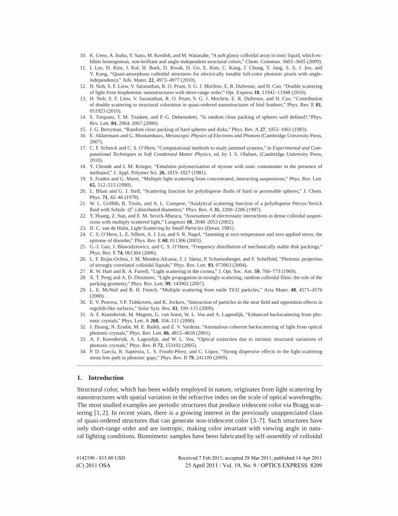
Although the transport mean free path has been well studied in disordered photonic crystalswith long-range order [31–34], there have been only a few studies on amorphous photonicstructures with short-range order. It has been shown in colloidal liquids, local order induces alocal minimum of the transport mean free path [26]. Since the particles are not closely packed,the near-field effect is negligible. In Ref. [28], lt is shown to increase at high filling fraction of arandom close-packed random film of monodisperse dielectric spheres. This result is explainedby accounting for evanescent wave coupling of contacting spheres. The modification of near-field scattering environment is included in the form factors of isolated scatterers in an effectivedielectric background. The background refractive index is obtained after setting a couplinglength that scales linearly with λ . Following this method, we calculate nb as a function ofwavelength and plot (with blue dashed line) in Fig. 5(a). Its value displays a sharp rise atλ ∼ 200 nm. Within the wavelength range of our study, its value is nearly invariant with λ .The lt calculated with this background index is roughly 3 times larger than the measured valuesfor our samples (not shown). For comparison, we plot (with black solid line) in Fig. 5(a) thevalue of nb obtained from Eq. (6) with α = 14.1. It has a much more gradual increase with λ .The background index nb is notably lower within the wavelength range of our measurement.The good agreement between the estimated lt with this nb and the measured values over abroad wavelength range clearly shows that our proposed model works better to incorporate thenear-field effects in a random close-packed scattering environment. The asymptotic behaviorof nb can be understood as follows. At longer wavelength, the near-field of a particle wouldextend farther away, coupling to more particles and probe the global environment. Thus thebackground refractive index approaches that of a homogenized medium which is nh = 1.39 inour case. In the short-wavelength limit, the particle would only be able to sense its immediateenvironment and nb approaches the refractive index of air. As an example, we plot in Fig. 5(b)the scattering efficiency Qsca of a dielectric sphere in different backgrounds. Qsca is defined asthe ratio of the total scattering cross section over the geometrical cross section of a sphere. Thesphere diameter is 244 nm and the refractive index is 1.58. Qsca calculated with background nb
from Eq. (6) approaches that of scattering in air at short wavelength, and that in homogenizedmedium of nh at long wavelength.
Assuming the same α value, we have estimated lt ≈ 5μm at λ = 540 nm the center wave-length of the major reflection peak for the nanostructures of bird feather barb as shown inFig. 1(a). Thus, lt is comparable to the total thickness of the scattering nanostructures, andsingle scattering is dominant over multiple scattering to produce color. However, the nanos-tructures of bird feather barb (air cavities embedded in β -keratin) are inverse of the biomimeticstructures (dielectric spheres in air), and the value of α is likely to be different. We expect the α
#142190 - $15.00 USD Received 7 Feb 2011; accepted 28 Mar 2011; published 14 Apr 2011(C) 2011 OSA 25 April 2011 / Vol. 19, No. 9 / OPTICS EXPRESS 8215
Fig. 5. (a) Near-field effects on form factors can be included in an effective backgroundrefractive index nb, whose value is calculated from Eq. (6). It approaches the refractiveindex of air at short wavelength, and that of a homogenized medium at long-wavelength.The wavelength range of our CBS measurement is highlighted with color. For comparison,the value of nb obtained from Ref. [28] is plotted with blue dashed line. (b) Calculatedscattering efficiency Qsca of a dielectric sphere in different backgrounds. The sphere hasa diameter 244 nm and a refractive index of 1.58. Qsca = σsca/σgeo is the ratio of thescattering cross section σsca to the geometical cross section σgeo. The refractive index ofthe background is equal to nb from Eq. (6) (black solid line), nh = 1.39 of the homogenizedmedium (blue dashed line), and that of air 1 (red dash-dotted line).
value determined in this paper is not universal, since it depends on the properties of the scattererand its surrounding. It is sensitive to the parameters such as ratio of refractive index of scattererwith its background, size of scatterer and maybe the packing geometry. Hence, further studyof light scattering by systematic tuning of the above mentioned parameters has the potential offully characterizing α , and providing physical insight to dependent scattering.
In summary, we have measured the transport mean free path lt with coherent backscatteringin amorphous photonic structures over a broad wavelength range. Such structures are madeof random close-packed dielectric spheres of two sizes, and have only short-range order. Themeasured lt is significantly larger than the estimated value based on the assumptions of inde-pendent scattering and the absence of structural order. With particles in close contact with oneanother, we must consider the phase correlation of scattered light and local scattering environ-ment. Short-range order accounts for the interference of light scattered from particles locatedin close proximity. Near-field effects originate from the evanescent wave coupling of adja-cent particles and leads to reduced refractive index contrast between particles and surrounding.Both increase the transport mean free path. Since many color-producing biological nanostruc-tures consist of random close-packed dielectric or air spheres, we expect both effects exist.They increase lt and make it comparable to the total size of the nanostructure. Consequently,single scattering becomes dominant over multiple scattering, and is responsible for structuralcoloration.
Acknowledgments
We thank Professor Simon Mochrie for stimulating discussions. This work is supported withseed funding from the Yale NSFMRSEC (DMR-0520495) and NSF grants to HC (PHY-0957680), ERD (CAREER CBET-0547294) and CSOH (DMS-0835742). Use of the NationalSynchrotron Light Source, Brookhaven National Laboratory, was supported by the U.S. Depart-
#142190 - $15.00 USD Received 7 Feb 2011; accepted 28 Mar 2011; published 14 Apr 2011(C) 2011 OSA 25 April 2011 / Vol. 19, No. 9 / OPTICS EXPRESS 8216
ment of Energy, Office of Science, Office of Basic Energy Sciences, under Contract No. DE-AC02-98CH10886. SAXS data were collected with the help of Drs. Alec Sandy and SureshNarayanan at beamline 8-ID-I of the Advanced Photon Source, Argonne National Labs, andsupported by the US Department of Energy, Office of Science, Office of Basic Energy Sci-ences, under Contract DE-AC02-06CH11357.
#142190 - $15.00 USD Received 7 Feb 2011; accepted 28 Mar 2011; published 14 Apr 2011(C) 2011 OSA 25 April 2011 / Vol. 19, No. 9 / OPTICS EXPRESS 8217


![PHOTONICS REVIEWS · 2012-11-09 · ability of NPs to squeeze light into the nanoscale provides near-field optical microscopy with unprecedented resolu-tion. [15–17] Arrays of](https://static.documents.pub/doc/80x56/5e39bf3a1bea677f7d2ca1c7/photonics-reviews-2012-11-09-ability-of-nps-to-squeeze-light-into-the-nanoscale.jpg)


