j ourna l homepage: www.e lsev ie r .com/ locate / jpedsurg
Should the ovary always be conserved in torsion? A tertiary care institute experience
Sandesh V. Parelkar ⁎, Dinesh Mundada, Beejal V. Sanghvi, Prashant B. Joshi, Sanjay N. Oak,Satish P. Kapadnis, Shishira Shetty, Hemangi Athawale, Pooja MultaniDepartment of Pediatric Surgery, King Edward Memorial Hospital, Parel, Mumbai, India
a b s t r a c ta r t i c l e i n f o
⁎ Corresponding author at: Department of Pediatric SuHospital, E. Borges road, Parel, Mumbai, India. Pin: 40(Mobile).
Aim: The aim of this study was to analyze our experience in conserving ovarian tissue in cases of ovariantorsion, irrespective of grade of necrosis at exploration.Materials and methods: All children with a diagnosis of ovarian torsion admitted to our hospital from January2009 to January 2013 were included. Patients with underlying ovarian pathology were excluded.Results: There were 13 torsions in 12 children (one bilateral). All underwent detorsion with or withoutevacuation of hematoma. Follow-up ultrasonography (USG) with color Doppler was done for all 13 ovaries,which showed an ovary with good vascularity and follicular development in 12 ovaries (92%). In 76% (10 of13) of cases, intraoperatively, the ovary was judged to be moderately to severely ischemic/necrotic. Yet,
follow-up sonograms showed the ovary with follicular development in all cases except one (7%). There wereno major complications in our series.Conclusion: Simple detorsion, instead of traditionally advocated oophorectomy, was not accompanied by anincrease in morbidity. On follow-up, almost all patients studied had functioning ovarian tissue despite thegrave ischemia observed intraoperatively. Detorsion should be the procedure of choice for all cases of simpleovarian torsion in children.
Torsion of ovary is a surgical emergency in prepubertal girls. Itaccounts for up to 2.7% of all cases with acute abdominal pain inchildren [1]. Clinical presentation of torsion of ovary masquerades asany acute abdomen surgical or otherwise. One must approach allprepubertal girls with an acute abdomen with a high degree ofsuspicion of torsion of ovary. The earlier recommended treatment ofovarian torsion was oophorectomy [2,3]. Recent reports, describeovarian conservation with untwisting of the ischemic adnexa as a safeand successful procedure [4–6]. The arguments in favor of oophorec-tomy are: risk of missing an underlying malignancy, thromboembo-lism after detorsion and a belief that a grossly black hemorrhagicadnexa is irreversibly damaged [2–4]. Our primary aimwas to analyzewhether irreversible ovarian damage was inevitable or not withobjective follow-up data following conservative management ofovarian torsion.
1. Materials and methods
All children with diagnosis of ovarian torsion admitted to ourhospital from January 2009 to January 2013 were included. A plainradiograph of the abdomen and pelvis, and abdominal ultrasonogra-phy (USG) with Color Doppler were obtained. After USG, the patient
rgery, King Edward Memorial0012. Tel.: +91 9869039091
lkar).
l rights reserved.
was taken to the operating theater (OR) on an emergency basis andtumor markers were obtained in all patients as a protocol. Laboratorywork was done according to an institutional protocol. Patients withunderlying ovarian malignancy or cyst were excluded (i.e, a case oftorsed gonadoblastoma was excluded). Laparoscopy was the initialmode of management followed by laparotomy if required. Dataregarding patients’ demographics, duration of symptoms, site of pain,atypical symptoms, investigations, complications, and length of staywere obtained. Also, time taken from presentation to hospital tosurgery was tabulated. To reduce subjective operator bias, surgerywas performed by designated staff members.
In addition, torsion of ovary was graded as follows:
Grade 1: Slightly discolored, normal size ovary, which promptlyreverted to normal color after detorsion.
Grade 2: Dark red to brown, mildly enlarged ovary, which becamehyperemic with multiple pin-point petechiae after detorsion.
Grade 3: Brown to black, grossly enlarged ovary with hematomawith slight improvement in color, small pin-point oozing afterdetorsion and hematoma evacuation.
Grade 4: Completely black, grossly enlarged ovary with hematomaand no improvement in color after detorsion and hematomaevacuation.
All ovaries underwent either simple detorsion or detorsion withevacuation of hematoma. None of the ovaries or contralateral ovaries
466 S.V. Parelkar et al. / Journal of Pediatric Surgery 49 (2014) 465–468
were fixed. On follow up at 3 months, all girls were evaluated withUSG and color Doppler. Data were analyzed.
Fig. 2. Right ovarian torsion (grade 3) presented as right sided strangulated inguinalhernia.
2. Results
There were 13 torsions in 12 children with age ranging from 6 to12 years (mean = 8.84). Pain (87%), nausea (56%), and vomiting(56%) were the most common presenting symptoms. Of these, onepresented with constipation and mild abdominal pain and anotherwith left flank pain. A palpable mass on presentationwas noted in onegirl. The time from onset of symptoms to hospital ranged from 14 to54 hours (mean = 31 hours). The time lapse from presentation tohospital until surgery was 4–30 hours (mean = 10 hours).
Diagnostic USG with color Doppler was done in all patients, whichshowed ovarian torsion with decreased vascularity in all and ovarianenlargement with hemorrhagic collection in 10 ovaries (70%). Thediameter of ovary measured by USG averaged 5.1 cm (range 3.9–12 cm). In one patient with constipation and mild abdominal pain,initial USGmissed the torsion. Review USG for persistent pain showedtorsion. The torsion was right-sided in 46% and left sided in 54%.Laparoscopy was performed in 84% of patients; 28% cases requiredconversion to an open procedure, laparotomy was primarily done in asingle patient and another presented as an incarcerated right inguinalhernia and required open herniotomy.
Using the aforementioned grading system for ovarian ischemia,the following results were obtained: 3 of 13 (24%) ovaries weregraded as grade 2 ischemic (Fig. 1) and, 5 of 13 (38%) as grade 3(Fig. 2). Grade 4 ischemia was present in 5 (38%) ovaries (Fig. 3A).Three patients underwent simple detorsion while 10 (76%) requireddetorsion with evacuation of hematoma and unroofing of the cavity(Fig. 3B). Median postoperative stay was 72 hours (range 48–120 hours). One patient had postoperative ileus with low-gradefever, and one patient had ileus and wound infection (both were openlaparotomies). Six patients had low-grade fever for a day or twopostoperatively. All were managed conservatively.
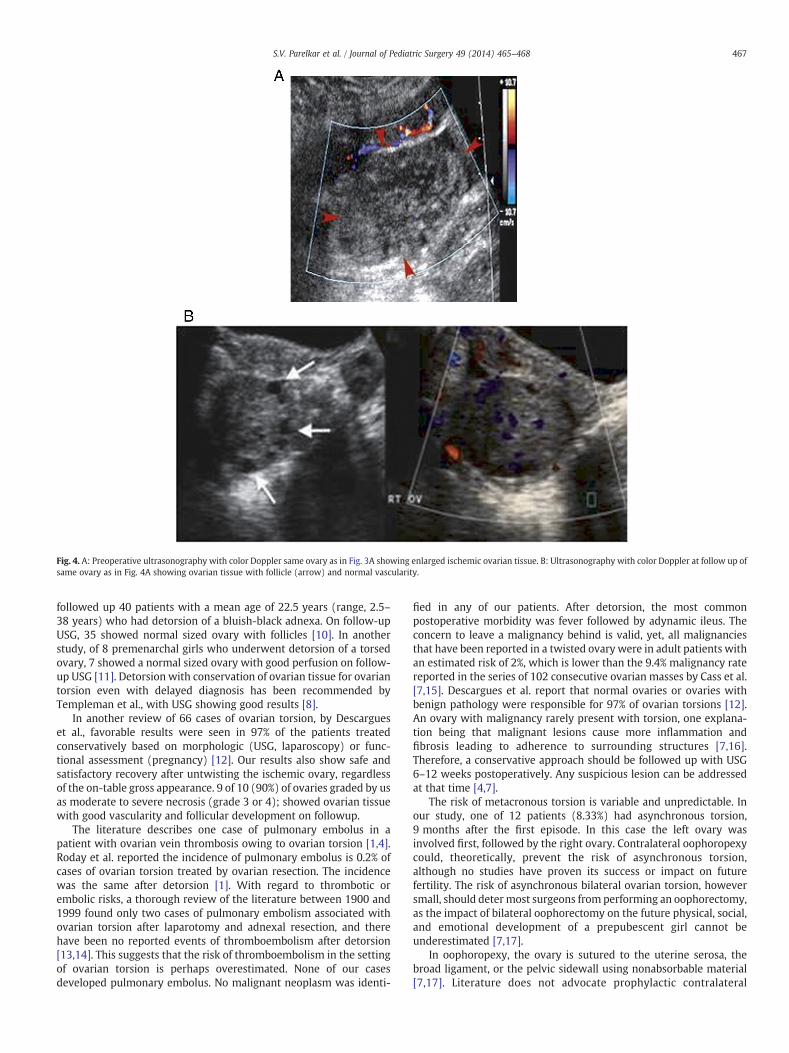
Follow-up was available in all 12 patients (13 ovaries). All thepatients were asymptomatic at 3 month follow-up. Repeated USGwith color Doppler showed ovarian tissue with good vascularity andfollicular development in 12 ovaries (92%) (Fig. 4A and B). However,in the child with bilateral asynchronous occurrence, left ovarian tissuewas not identified on follow up USG. As noted almost 76% ovarieswere initially labeled as having moderate to severe necrosis (grade 3or 4), but of these 10 ovaries, 9 ovaries had ovarian tissue with good
Fig. 1. Left ovarian torsion with Grade 2 ischemia.
vascularity and with follicular development on follow up USG withcolor Doppler.
3. Discussion
Torsion of ovary is a surgical emergency. The usual presentation islower abdominal pain, which may be indistinguishable from acuteappendicitis when the pain is located in the right lower quadrant [7].Nausea and vomiting, mild fever, and leucocytosis are associatedfeatures. The reported incidence of ovarian torsion is 2.7% in thegeneral population. As differential diagnosis includes appendicitis,gastroenteritis and renal colic, diagnosis is frequently delayed. USGwith color Doppler remains the most useful investigation.
Traditionally, the management of ovarian torsion was excision ofthe twisted ovary for fear of (1) embolic phenomenon on detorsion,(2) leaving a malignancy behind, and more importantly, (3) the beliefthat grossly black hemorrhagic ovary was irreversibly damaged [7–9].
Recent studies, however, advocate conservative management bydetorsion as the treatment of choice in prepubertal girls. Oelsner et al.
Fig. 3. A: Right ovarian torsion with Grade 4 ischemia. B: Same ovary as in Fig. 4A afterevacuation of hematoma.
Fig. 4. A: Preoperative ultrasonography with color Doppler same ovary as in Fig. 3A showing enlarged ischemic ovarian tissue. B: Ultrasonography with color Doppler at follow up ofsame ovary as in Fig. 4A showing ovarian tissue with follicle (arrow) and normal vascularity.
467S.V. Parelkar et al. / Journal of Pediatric Surgery 49 (2014) 465–468
followed up 40 patients with a mean age of 22.5 years (range, 2.5–38 years) who had detorsion of a bluish-black adnexa. On follow-upUSG, 35 showed normal sized ovary with follicles [10]. In anotherstudy, of 8 premenarchal girls who underwent detorsion of a torsedovary, 7 showed a normal sized ovary with good perfusion on follow-up USG [11]. Detorsion with conservation of ovarian tissue for ovariantorsion even with delayed diagnosis has been recommended byTempleman et al., with USG showing good results [8].
In another review of 66 cases of ovarian torsion, by Descargueset al., favorable results were seen in 97% of the patients treatedconservatively based on morphologic (USG, laparoscopy) or func-tional assessment (pregnancy) [12]. Our results also show safe andsatisfactory recovery after untwisting the ischemic ovary, regardlessof the on-table gross appearance. 9 of 10 (90%) of ovaries graded by usas moderate to severe necrosis (grade 3 or 4); showed ovarian tissuewith good vascularity and follicular development on followup.
The literature describes one case of pulmonary embolus in apatient with ovarian vein thrombosis owing to ovarian torsion [1,4].Roday et al. reported the incidence of pulmonary embolus is 0.2% ofcases of ovarian torsion treated by ovarian resection. The incidencewas the same after detorsion [1]. With regard to thrombotic orembolic risks, a thorough review of the literature between 1900 and1999 found only two cases of pulmonary embolism associated withovarian torsion after laparotomy and adnexal resection, and therehave been no reported events of thromboembolism after detorsion[13,14]. This suggests that the risk of thromboembolism in the settingof ovarian torsion is perhaps overestimated. None of our casesdeveloped pulmonary embolus. No malignant neoplasm was identi-
fied in any of our patients. After detorsion, the most commonpostoperative morbidity was fever followed by adynamic ileus. Theconcern to leave a malignancy behind is valid, yet, all malignanciesthat have been reported in a twisted ovary were in adult patients withan estimated risk of 2%, which is lower than the 9.4% malignancy ratereported in the series of 102 consecutive ovarian masses by Cass et al.[7,15]. Descargues et al. report that normal ovaries or ovaries withbenign pathology were responsible for 97% of ovarian torsions [12].An ovary with malignancy rarely present with torsion, one explana-tion being that malignant lesions cause more inflammation andfibrosis leading to adherence to surrounding structures [7,16].Therefore, a conservative approach should be followed up with USG6–12 weeks postoperatively. Any suspicious lesion can be addressedat that time [4,7].
The risk of metacronous torsion is variable and unpredictable. Inour study, one of 12 patients (8.33%) had asynchronous torsion,9 months after the first episode. In this case the left ovary wasinvolved first, followed by the right ovary. Contralateral oophoropexycould, theoretically, prevent the risk of asynchronous torsion,although no studies have proven its success or impact on futurefertility. The risk of asynchronous bilateral ovarian torsion, howeversmall, should deter most surgeons from performing an oophorectomy,as the impact of bilateral oophorectomy on the future physical, social,and emotional development of a prepubescent girl cannot beunderestimated [7,17].
In oophoropexy, the ovary is sutured to the uterine serosa, thebroad ligament, or the pelvic sidewall using nonabsorbable material[7,17]. Literature does not advocate prophylactic contralateral
468 S.V. Parelkar et al. / Journal of Pediatric Surgery 49 (2014) 465–468
oophoropexy, and questions regarding its success and effects onfuture fertility remain unanswered. There is increasing evidence ofsuccessful oophoropexy in patients receiving pelvic irradiation fortreatment of malignancy for shielding the ovary and not forpreventing torsion.
A high index of suspicion and early diagnosis affords bettersalvage of the ischemic ovaries. Hence, ovarian torsion should beconsidered in every girl presenting with lower abdominal pain. USGremains the most useful investigation, and loss of blood flow onDoppler is diagnostic. Ovarian tissue conservation is stronglyrecommended [1,4,7,15]. Gross appearance of ovary and intra-operative grading of ischemia by the surgeon are not reliableindicators of ovarian viability. Ovarian malignancy occurs rarely andshould not delay the surgical procedure in a girl with ovariantorsion. Repeat USG can be done postoperatively and an appropriateintervention performed if required [4,7]. Thus, we conclude andadvocate maximal conservation and salvage of ovarian tissue for allcases of ovarian torsion in prepubertal girls, irrespective of theintraoperative bleak appearance of the ovary. Thus, potentialinfertility and negative impact on physical, social, and emotionalimpact on prepubescent girls can be avoided.
Detorsion with maximal conservation of ovarian tissue should bethe procedure of choice for all cases of simple ovarian torsion inchildren, irrespective of the perceived degree of ischemia.
Acknowledgment
We would like to acknowledge and thank Dr. Renuka S. Parelkar,who helped us in writing and modifying this article.
References
[1] Roday A, Jackish C, Klockenbusch W, et al. The conservative management ofadnexal torsion—A case report and review of the literature. Eur J Obstet GynecolReprod Biol 2002;101:83–862.
[2] Mordehai J, Mares AJ, Barki Y, et al. Torsion of uterine adnexa in neonates andchildren, A report of 20 cases. J Pediatr Surg 1991;26:1195–9.
[3] Chen M, Chen CD, Yang YS. Torsion of the previously normal uterine adnexa.Evaluation of the correlation between the pathological changes and the clinicalcharacteristics. Acta Obstet Gynecol Scand 2001;80:58–61.
[4] Aziz C, Davis V, Allen L, et al. Ovarian torsion in children: is oophorectomynecessary? J Pediatr Surg 2004;39(5):750–3.
[5] Dolgin SE, Lublin M, Shlasko E. Maximizing ovarian salvage when treatingidiopathic adnexal torsion. J Pediatr Surg 2000;35:624–6.
[6] Meynol F, Steyaert H, Valla JS. Adnexal torsion in children earlier diagnosis andtreatment by laparoscopy. Arch Pediatr 1997;4:416–9.
[8] Templeman C, Hertweck P, Fallat M. The clinical course of unresected ovariantorsion. J Pediatr Surg 2000;35:1385–7.
[9] Hibbard LT. Adnexal torsion. Am J Obstet Gynecol 1985;152:456–61.[10] Oelsner G, Bider G, Goldenberg M, et al. Long term follow-up of the twisted
ischemic adnexa managed by detorsion. Fertil Steril 1993;60:976–97918.[11] PanskyM, Abargil A, Dreazen E, et al. Conservative management of adnexal torsion
in premenarchal girls. J Am Assoc Gynecol Laparosc 2000;7:121–4.[12] Descargues G, Tinlot-Mauger F, Gravier A, et al. Adnexal torsion, a report on forty-
five cases. Eur J Obstet Gynecol Reprod Biol 2001;98:91–6.[13] Tsafrir Ziv, Azem Foad, Hasson Joseph, et al. Risk factors, symptoms and treatment
of ovarian torsion in children: The twelve-year experience of one centre. J MinimInvasive Gynecol 2012;19:29–33.
[14] McGovern PG, Noah R, Koenigsburg R, et al. Adnexal torsion and pulmonaryembolism: case report and review of literature. ObstetGynecol Surv 1999;54:601–8.
[15] Cass D, Hawkins E, Brandt M, et al. Surgery for ovarian masses in infants, childrenand adolescents: 102 consecutive patients treated in a 15 year period. J PediatrSurg 2001;36:693–9.
[16] Kokoska ER, Keller MS, Weber TR. Acute ovarian torsion in children. Am J Surg2000;180:462–5.
[17] Eckler K, LauferM, Perlman SE. Conservativemanagement of bilateral asynchronousadnexal torsion with necrosis in prepubescent girl. J Pediatr Surg 2000;35:1248–51.