1 Shoulder retractor strengthening exercise to minimize rhomboid muscle activity and subacromial impingement Jeremy Fennell, PT, MD; * Chetan P. Phadke, BPhT, PhD; *†‡ George Mochizuki, PhD; †§ Farooq Ismail, MD; *† Chris Boulias, MD, PhD *† From the: *Spasticity Research Program, West Park Healthcare Centre, Toronto; †Faculty of Medicine, University of Toronto; ‡Faculty of Health, York University, Toronto; §Sunnybrook Research Institute, Toronto. Correspondence to: Chetan P. Phadke, 82 Buttonwood Ave., Toronto, ON M6M 2J5; [email protected]. Acknowledgements: We would like to thank all our participants. The West Park Foundation provided salary support for Dr. Phadke. ABSTRACT Purpose: To investigate the best position for shoulder retractor strengthening exercise to maximize middle trapezius activity and minimize rhomboid major activity. Though both are scapular retractors, rhomboids also act as downward rotators of the scapula, which can worsen subacromial impingement. Methods: Twelve healthy participants (age 30 [SD 6] years) with no history of shoulder pain were recruited for this study, which used fine wire electromyography to examine maximal muscle activation of the middle trapezius and rhomboid major muscle fibres during in different positions: shoulder in 90° abduction position with elbow completely extended and (a) shoulder internal rotation, (b) shoulder neutral rotation, and (c) shoulder external rotation,
Transcript
1
Shoulder retractor strengthening exercise to minimize rhomboid muscle activity and
subacromial impingement
Jeremy Fennell, PT, MD;* Chetan P. Phadke, BPhT, PhD;
*†‡ George Mochizuki, PhD;
†§ Farooq
Ismail, MD;*†
Chris Boulias, MD, PhD*†
From the:
*Spasticity Research Program, West Park Healthcare Centre, Toronto;
†Faculty of Medicine, University of Toronto;
‡Faculty of Health, York University, Toronto;
§Sunnybrook Research Institute, Toronto.
Correspondence to: Chetan P. Phadke, 82 Buttonwood Ave., Toronto, ON M6M 2J5;
(3) shoulder external rotation (SER), used to isolate middle
trapezius;18
and (4) rowing position (shoulder neutral rotation with 90° elbow flexion ), reported
to activate middle trapezius.19
Lying prone with the shoulder on the edge of the bed, participants
were asked to retract their shoulder and resist as hard as possible for 3 seconds as the examiner
provided maximal downward force in the direction of horizontal adduction just proximal to the
elbow joint. The examiner provided standardized verbal encouragement in all trials to assist in
maximal force output. Participants performed three trials of maximal voluntary contraction
(MVC) in each position.
Data Analysis
EMG data were sampled at 10 KHz, band-pass filtered (10-450 Hz), and full-wave rectified
using EMGworks 4.0 (Delsys Inc., Boston, MA). We calculated the root mean squared (RMS)
for the middle 1 second of the MVC EMG data for both muscles. Data from RMS of the middle
trapezius were divided by the RMS of the rhomboids to calculate the muscle activation ratio. The
muscle activation ratio from condition 1 (SNR) was considered a control condition, since the
shoulder was in neutral rotation, and the rest of the conditions (2–4) were normalized to MVC in
neutral rotation. We chose to use non-parametric statistics because of the small sample size. To
assess which of four scapular retraction exercises preferentially activated the middle trapezius as
opposed to the rhomboids, we performed Wilcoxon signed rank tests using SPSS 16.0 (IBM
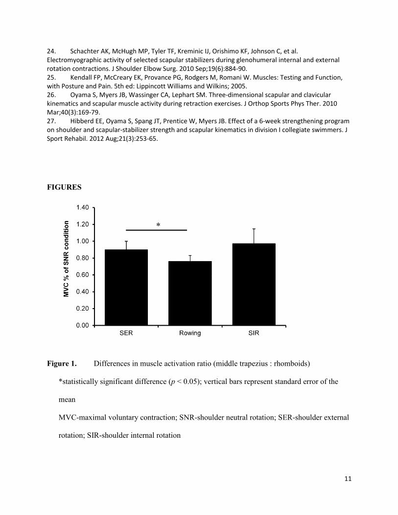
Corp., New York), with the threshold for significance set at 0.05. Figure 1 was prepared using
the Microsoft Excel add-on XL Toolbox (Daniel Kraus, Würzburg, Germany).
6
RESULTS
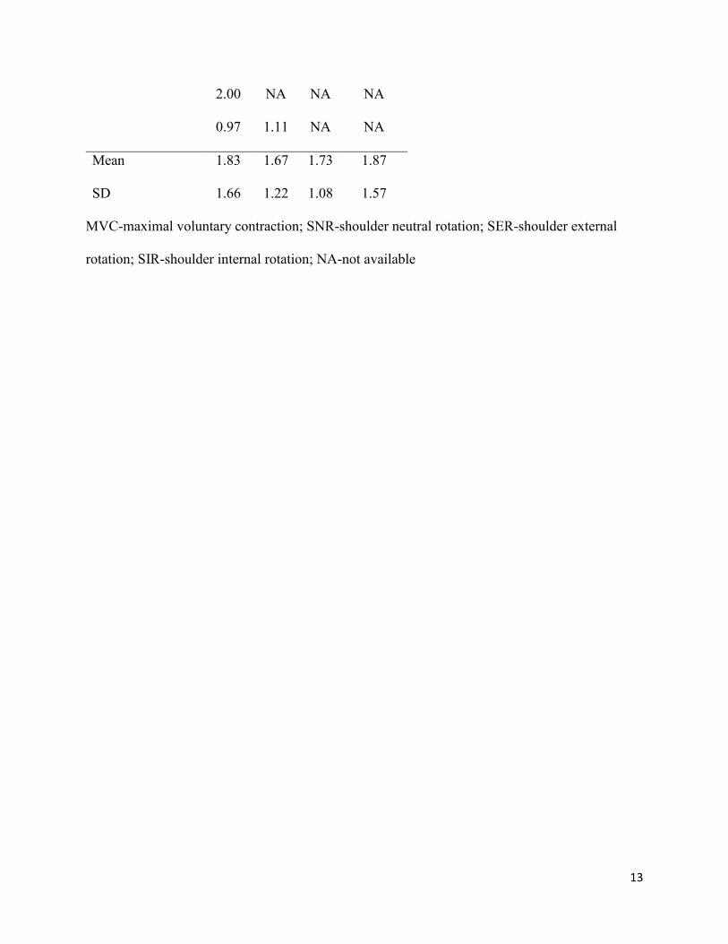
We were not able to collect complete data for three participants, and EMG data for a fourth
participant were not of good quality; therefore, we report average data from eight healthy
participants (see Table 1 for raw values). We found that the middle trapezius : rhomboid ratio
was significantly lower (22% lower) in rowing position than in SER (p = 0.031; see Figure 1).
The rhomboid was more active than middle trapezius in the rowing position (elbow flexed) than
in the SER (elbow extended) position. All four exercise positions produced coactivation of
trapezius and rhomboids. There was no significant difference between SNR and SER (p = 0.46)
or between SNR and rowing conditions (p = 0.31).
DISCUSSION
Strengthening exercises are an important component of rehabilitation for people with
impingement syndrome and can reduce pain, improve function, and prevent surgery.20-23
Knowledge of the types of exercise that address muscle weakness without deteriorating the
subacromial space or exacerbating the condition is important. Strengthening of scapular
retractors is an important aspect of a shoulder strengthening exercise regime for impingement
syndrome. Our results suggest that shoulder retraction with the shoulder either in internal or
external rotation does not preferentially activate middle trapezius or rhomboids. In the rowing
condition, however, rhomboid activity increased by 10% while middle trapezius activity
decreased by 15%, thus significantly decreasing the middle trapezius : rhomboid ratio in the
rowing condition relative to the SER condition. The decrease in middle trapezius in rowing
relative to SER can be attributed to the known hyperactivity of the middle trapezius during the
SER movement relative to the rowing activity.24
Our results reflect the differences in muscle
7
activation patterns between the two chief scapular retractors during different types of shoulder
exercises.
We found no difference in muscle activation ratio (middle trapezius : rhomboids)
between SIR and SER, which supports previous findings.18
Although muscle testing principles
described in standard textbooks25
suggest preferential activation of the middle trapezius in SER
position, our data do not support this finding. Similarly, previous studies have shown no
statistically significant difference between muscle activation patterns in a variety of testing
positions recommended for isolating other scapular muscles.18
Because differences in muscle
activation patterns are difficult to perceive clinically (manually or visually),18
clinicians must
rely on EMG findings to help them make objective assessments of muscle strength and plan
muscle strengthening regimes. Scapular retractor muscle activation increases with increasing
angle of retraction, but muscle activation also varies even when scapular retraction angle remains
unchanged while the shoulder is placed in different positions.26
Our data using fine-wire EMG
on deep scapular muscles (inaccessible by surface EMG) provide clinical insights on best
positions to strengthen scapular retractors in impingement syndrome, but these results need to be
replicated in people with impingement syndrome.
No objective information or scientifically tested clinical guidelines are available to guide
clinicians in understanding the impact of various forms of shoulder retraction exercises on
muscle activation patterns. A recent study reported that two types of shoulder retraction exercises
performed with scapular upward and downward rotation were not effective in strengthening
retractors or improving scapular kinematics,27
but we lack evidence on potentially beneficial
exercises. Our study examined the differences in muscle activation patterns between two major
scapular retractors. Our findings suggest that exercise performed in rowing position (90°
8
shoulder abduction and 90° elbow flexion position) increases rhomboid activation and decreases
middle trapezius activation. The testing position used for strengthening the posterior deltoid
muscle (shoulder abduction 90°, slight extension, and internal rotation with resistance in antero-
medial direction) is also reported to result in highest rhomboid muscle activation.18
For people
with impingement syndrome, therefore, it may be best to avoid the rowing position and the
posterior deltoid testing position for shoulder retractor strengthening exercise, as these positions
are likely to increase rhomboid activity and cause further impingement on the subacromial space.
Limitations
Our study has several limitations. First, rhomboids are deep muscles, and thus inaccessible to
surface EMG techniques; however, fine-wire sensors are small and do not capture EMG signals
from the entire muscle. Second, we tested a small sample of healthy control participants, which
may have biased the results due to the lower power of the study. Third, we did not record
scapular retraction kinematics such as position and degree of retraction, which may have not
been consistent across conditions. Finally, we did not measure muscle activation in other
shoulder muscles; it is important to study muscle activity in the rest of the shoulder muscles, as
compensatory muscle activation can occur after shoulder impingement.
CONCLUSIONS
Our data suggest that the optimal exercise for shoulder retractors is in shoulder abduction and
external rotation position (with elbow extended), providing maximal trapezius and minimal
rhomboid activation. These findings need to be confirmed in people with shoulder impingement.
We recommend that future studies test this hypothesis as well as the impact of a rehabilitation
9
programme incorporating shoulder retractor exercise in the optimal position of shoulder
abduction and external rotation (with elbow extension).
KEY MESSAGES
What is already known on this topic
The middle trapezius and rhomboid muscles are responsible for retraction of the scapula,
but the middle fibres of the trapezius function as pure scapular retractors, whereas the rhomboids
act both to retract the scapula and to rotate it to depress the glenoid fossa. This inferior rotation
of the scapula leads to a decrease in the subacromial space which may contribute to the
development or persistence of impingement symptoms. Middle trapezius muscle activation is
decreased and rhomboid muscle activation is increased in subjects with shoulder impingement
syndrome.
What this study adds
Our fine-wire EMG data suggests that scapular retractor isometric contraction exercise
performed in 90° shoulder abduction with external rotation and elbow extended position can
produce higher middle trapezius and lower rhomboid muscle activation. Rowing position (90°
shoulder abduction, neutral shoulder rotation and elbow flexed 90°) may not be the best position
for scapular retractor strengthening in patients with impingement syndrome because it
preferentially activates rhomboid more than middle trapezius.
REFERENCES
1. van der Heijden GJ. Shoulder disorders: a state-of-the-art review. Baillieres Best Pract Res Clin Rheumatol. 1999 Jun;13(2):287-309. 2. Bot SD, van der Waal JM, Terwee CB, van der Windt DA, Schellevis FG, Bouter LM, et al. Incidence and prevalence of complaints of the neck and upper extremity in general practice. Ann Rheum Dis. 2005 Jan;64(1):118-23. 3. Rockwood C, Matsen F, Arntz C. The Shoulder. Philadelphia: WB Saunders Company; 1990.
10
4. van der Windt DA, Koes BW, de Jong BA, Bouter LM. Shoulder disorders in general practice: incidence, patient characteristics, and management. Ann Rheum Dis. 1995 Dec;54(12):959-64. 5. Neer CS. Anterior acromioplasty for the chronic impingement syndrome in the shoulder: a preliminary report. J Bone Joint Surg Am. 1972 Jan;54(1):41-50. 6. Papadonikolakis A, McKenna M, Warme W, Martin BI, Matsen FA. Published evidence relevant to the diagnosis of impingement syndrome of the shoulder. J Bone Joint Surg Am. 2011 Oct;93(19):1827-32. 7. Vitale MA, Arons RR, Hurwitz S, Ahmad CS, Levine WN. The rising incidence of acromioplasty. J Bone Joint Surg Am. 2010 Aug;92(9):1842-50. 8. Coghlan JA, Buchbinder R, Green S, Johnston RV, Bell SN. Surgery for rotator cuff disease. Cochrane Database Syst Rev. 2008 (1):CD005619. 9. Solem-Bertoft E, Thuomas KA, Westerberg CE. The influence of scapular retraction and protraction on the width of the subacromial space. An MRI study. Clin Orthop Relat Res. 1993 Nov(296):99-103. 10. Lewis JS, Wright C, Green A. Subacromial impingement syndrome: the effect of changing posture on shoulder range of movement. J Orthop Sports Phys Ther. 2005 Feb;35(2):72-87. 11. Chester R, Smith TO, Hooper L, Dixon J. The impact of subacromial impingement syndrome on muscle activity patterns of the shoulder complex: a systematic review of electromyographic studies. BMC Musculoskelet Disord. 2010;11:45. 12. Cools AM, Declercq GA, Cambier DC, Mahieu NN, Witvrouw EE. Trapezius activity and intramuscular balance during isokinetic exercise in overhead athletes with impingement symptoms. Scand J Med Sci Sports. 2007 Feb;17(1):25-33. 13. Kibler WB, Sciascia A. Current concepts: scapular dyskinesis. Br J Sports Med. 2010 Apr;44(5):300-5. 14. Reinold MM, Escamilla RF, Wilk KE. Current concepts in the scientific and clinical rationale behind exercises for glenohumeral and scapulothoracic musculature. The Journal of Orthopaedic and Sports Physical Therapy. 2009 Feb;39(2):105-17. 15. Moore K, Dalley A, Agur A. Clinically oriented anatomy. 6th ed: Lippincott Williams & Wilkins; 2010. 16. Fu FH, Harner CD, Klein AH. Shoulder impingement syndrome. A critical review. Clin Orthop Relat Res. 1991 Aug(269):162-73. 17. Urbaniak GC, Plous S. [15 May 2015]; Available from: www.randomizer.org. 18. Smith J, Padgett DJ, Kaufman KR, Harrington SP, An KN, Irby SE. Rhomboid muscle electromyography activity during 3 different manual muscle tests. Arch Phys Med Rehabil. 2004 Jun;85(6):987-92. 19. Moseley JB, Jobe FW, Pink M, Perry J, Tibone J. EMG analysis of the scapular muscles during a shoulder rehabilitation program. Am J Sports Med. 1992 1992 Mar-Apr;20(2):128-34. 20. Lewis JS. A specific exercise program for patients with subacromial impingement syndrome can improve function and reduce the need for surgery. J Physiother. 2012;58(2):127. 21. Holmgren T, Björnsson Hallgren H, Öberg B, Adolfsson L, Johansson K. Effect of specific exercise strategy on need for surgery in patients with subacromial impingement syndrome: randomised controlled study. Bmj. 2012;344:e787. 22. Litchfield R. Progressive strengthening exercises for subacromial impingement syndrome. Clin J Sport Med. 2013 Jan;23(1):86-7. 23. Conaghan PG. Steroid injection and regular shoulder-specific exercises reduce the need for surgery in subacromial impingement syndrome. Evid Based Med. 2013 Feb;18(1):e3.
24. Schachter AK, McHugh MP, Tyler TF, Kreminic IJ, Orishimo KF, Johnson C, et al. Electromyographic activity of selected scapular stabilizers during glenohumeral internal and external rotation contractions. J Shoulder Elbow Surg. 2010 Sep;19(6):884-90. 25. Kendall FP, McCreary EK, Provance PG, Rodgers M, Romani W. Muscles: Testing and Function, with Posture and Pain. 5th ed: Lippincott Williams and Wilkins; 2005. 26. Oyama S, Myers JB, Wassinger CA, Lephart SM. Three-dimensional scapular and clavicular kinematics and scapular muscle activity during retraction exercises. J Orthop Sports Phys Ther. 2010 Mar;40(3):169-79. 27. Hibberd EE, Oyama S, Spang JT, Prentice W, Myers JB. Effect of a 6-week strengthening program on shoulder and scapular-stabilizer strength and scapular kinematics in division I collegiate swimmers. J Sport Rehabil. 2012 Aug;21(3):253-65.
FIGURES
Figure 1. Differences in muscle activation ratio (middle trapezius : rhomboids)
*statistically significant difference (p < 0.05); vertical bars represent standard error of the