18
Cytological Changes Of Oral Mucosa Following Lateral Cephalometry & Panoramic radiograph Presented by: Shweta Srivastava JR-1 Dept. of Oral Medicine and Radiology KGMU, Lucknow
| Date post: | 22-Jan-2018 |
| Category: |
Health & Medicine |
| Upload: | shweta-sharma |
| View: | 288 times |
| Download: | 0 times |
Cytological Changes Of Oral MucosaFollowing
Lateral Cephalometry & Panoramic radiograph
Presented by:
Shweta Srivastava
JR-1
Dept. of Oral Medicine and
Radiology
KGMU, Lucknow
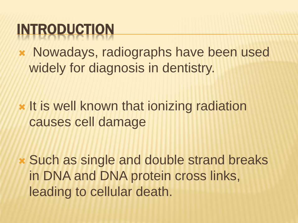
INTRODUCTION
Nowadays, radiographs have been used
widely for diagnosis in dentistry.
It is well known that ionizing radiation
causes cell damage
Such as single and double strand breaks
in DNA and DNA protein cross links,
leading to cellular death.
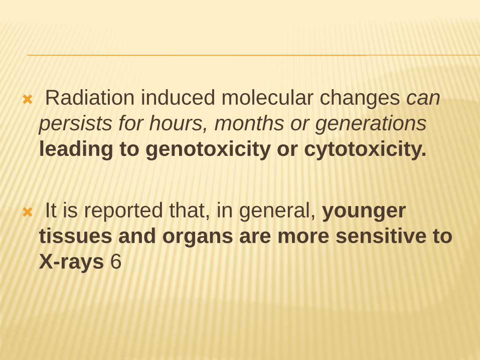
Radiation induced molecular changes can
persists for hours, months or generations
leading to genotoxicity or cytotoxicity.
It is reported that, in general, younger
tissues and organs are more sensitive to
X-rays 6
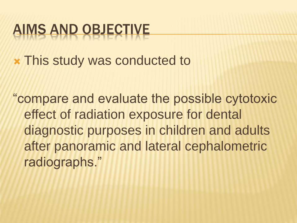
AIMS AND OBJECTIVE
This study was conducted to
“compare and evaluate the possible cytotoxic
effect of radiation exposure for dental
diagnostic purposes in children and adults
after panoramic and lateral cephalometric
radiographs.”
MATERIALS AND METHOD
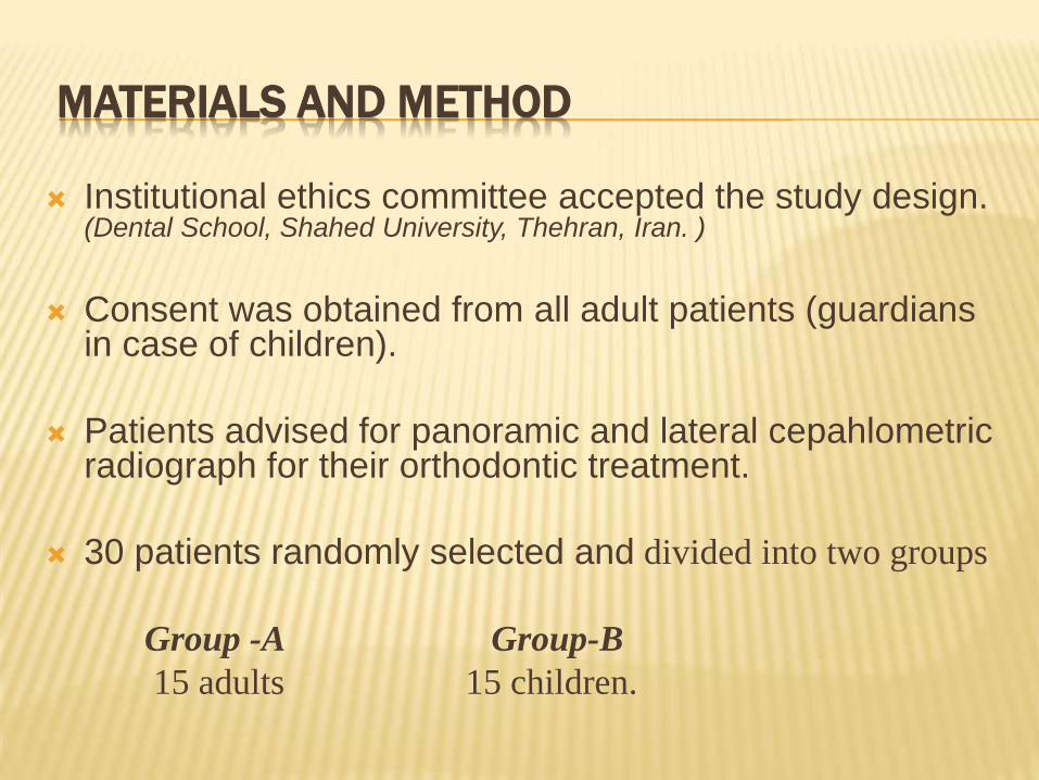
Institutional ethics committee accepted the study design. (Dental School, Shahed University, Thehran, Iran. )
Consent was obtained from all adult patients (guardians in case of children).
Patients advised for panoramic and lateral cepahlometricradiograph for their orthodontic treatment.
30 patients randomly selected and divided into two groups
Group -A Group-B
15 adults 15 children.
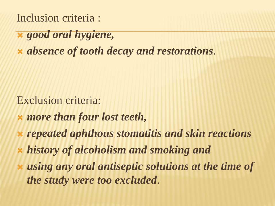
Inclusion criteria :
good oral hygiene,
absence of tooth decay and restorations.
Exclusion criteria:
more than four lost teeth,
repeated aphthous stomatitis and skin reactions
history of alcoholism and smoking and
using any oral antiseptic solutions at the time of
the study were too excluded.
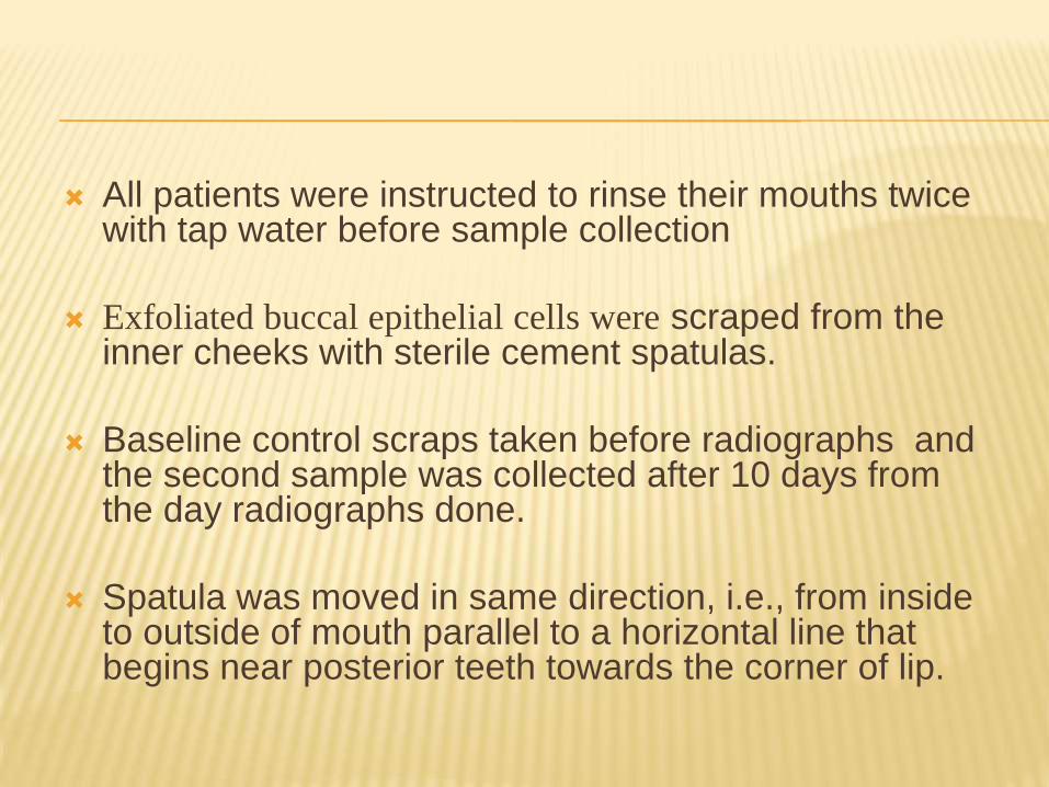
All patients were instructed to rinse their mouths twice with tap water before sample collection
Exfoliated buccal epithelial cells were scraped from the inner cheeks with sterile cement spatulas.
Baseline control scraps taken before radiographs and the second sample was collected after 10 days from the day radiographs done.
Spatula was moved in same direction, i.e., from inside to outside of mouth parallel to a horizontal line that begins near posterior teeth towards the corner of lip.
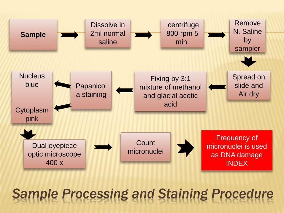
Sample Processing and Staining Procedure
Sample
Dissolve in
2ml normal
saline
Fixing by 3:1
mixture of methanol
and glacial acetic
acid
Spread on
slide and
Air dry
Remove
N. Saline
by
sampler
centrifuge
800 rpm 5
min.
Count
micronucleiDual eyepiece
optic microscope
400 x
Nucleus
blue
Cytoplasm
pink
Papanicol
a staining
Frequency of
micronuclei is used
as DNA damage
INDEX
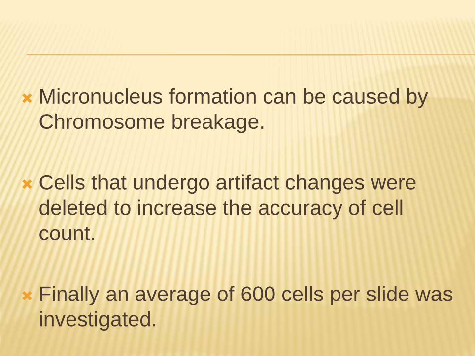
Micronucleus formation can be caused by
Chromosome breakage.
Cells that undergo artifact changes were
deleted to increase the accuracy of cell
count.
Finally an average of 600 cells per slide was
investigated.
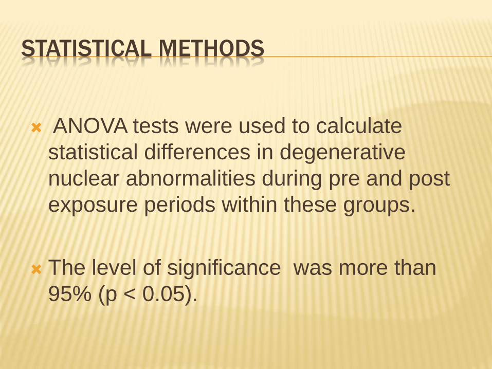
STATISTICAL METHODS
ANOVA tests were used to calculate
statistical differences in degenerative
nuclear abnormalities during pre and post
exposure periods within these groups.
The level of significance was more than
95% (p < 0.05).
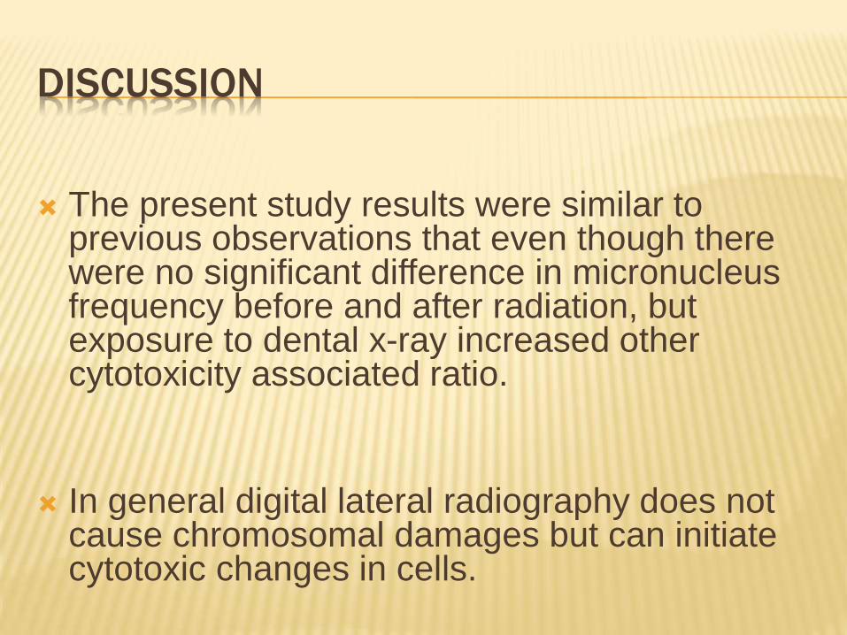
DISCUSSION
The present study results were similar to previous observations that even though there were no significant difference in micronucleus frequency before and after radiation, but exposure to dental x-ray increased other cytotoxicity associated ratio.
In general digital lateral radiography does not cause chromosomal damages but can initiate cytotoxic changes in cells.
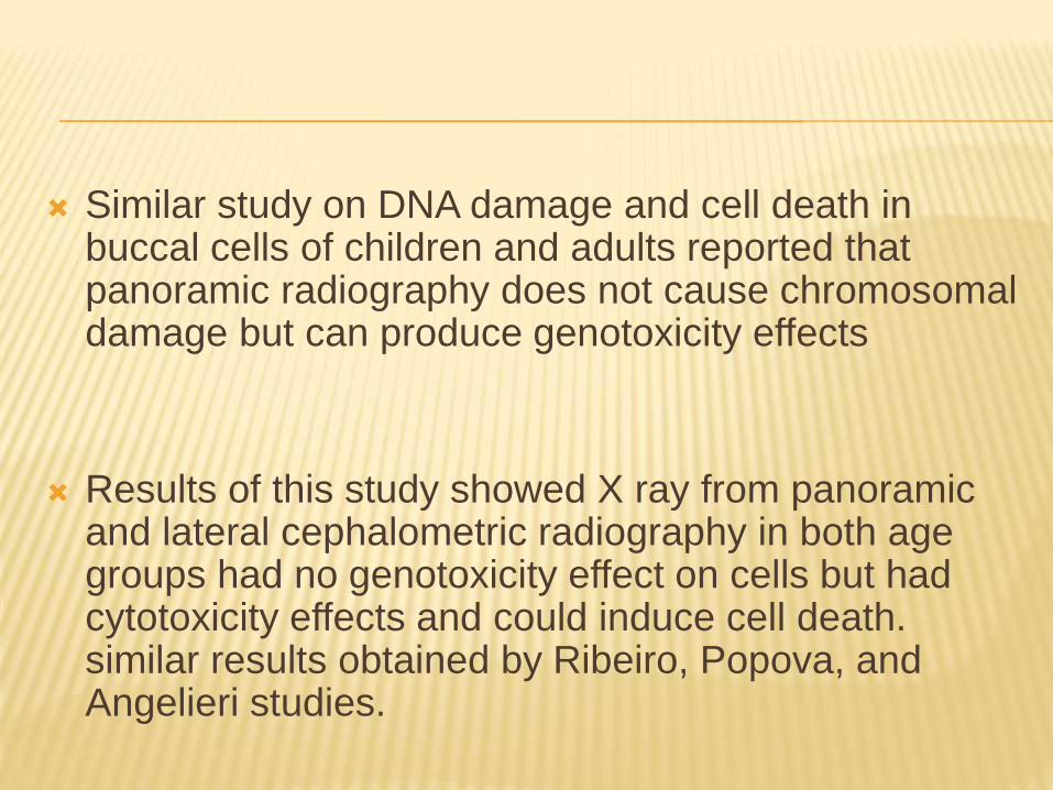
Similar study on DNA damage and cell death in buccal cells of children and adults reported that panoramic radiography does not cause chromosomal damage but can produce genotoxicity effects
Results of this study showed X ray from panoramic and lateral cephalometric radiography in both age groups had no genotoxicity effect on cells but had cytotoxicity effects and could induce cell death. similar results obtained by Ribeiro, Popova, and Angelieri studies.
In present study confounding factors such as smoking excluded because only the radiation role be compared in two age groups.
Angelieri investigated cytogenic effects of panoramic radiography in buccal mucosa and marginal surface of tongue in smoking and non- smoking adults.
Cerqueira investigated genotoxicity effects of panoramic radiography in gingival epithelial cells and showed X ray increased genotoxicity in these cells that caused chromosomal damages
Micronucleus index reflects genomic instability.
Diagnosis of micronucleus increased frequency in a population shows increased risk for cancer
The damage because micronuclei formation happens in epithelial basal cells where mitosis is happens.
Cells carring exposure induced genotoxic factors reaches at surface in one to three weeks.
That is why, in this study cells were collected 10 days after x-ray exposure.
Necrotic cells (karyolysis, karyorexy, and
pyknosis) were evaluated as an index for cell
death (cytotoxicity).
Results of this study showed
“amounts of absorbed dose in dental radiation
cannot cause genotoxicity changes but since
repeating use of cytotoxic factors can result
to chronic cell damages and degenerative
changes and finally to neoplastic changes.
CONCLUSION
In conclusion, dental radiographs should be
used only when absolutely necessary and
every effort should be made to keep the dose
to all individuals as low as possible.
REFERENCES Ribeiro DA. Cytogenetic biomonitoring in oral mucosa cells following dental X-ray. Dentomaxillofac Radiol. 2012;41(3):181-4.
Ribeiro DA, Sannomiya EK, Pozzi R, Miranda SR, Angelieri F. Cellular death but not genetic damage in oral mucosa cells after exposure to digital lateral radiography. Clinical oral investigations. 2011;15(3):357-60.
Angelieri F, de Oliveira GR, Sannomiya EK, Ribeiro DA. DNA damage and cellular death in oral mucosa cells of children who have undergone panoramic dental radiography. Pediatric radiology. 2007;37(6):561-5.
Suk WA, Murray K, Avakian MD. Environmental hazards to children’s health in the modern world. Mutation Research/Reviews in Mutation Research. 2003;544(2):235-42.
Robbins SL, Kumar V, Abbas AK, Aster JC. Robbins basic pathology: Elsevier Health Sciences; 2012.
McDonald RE, Avery DR, Dean JA. Dentistry for the Child and Adolescent: CV Mosby; 1969.
Wang C-C. Radiation therapy for head and neck neoplasms. Chicago, IL (USA): Year Book Medical Publishers; 1990.
Carlin V, Artioli AJ, Matsumoto MA, Borgo E, Oshima CTF, Ribeiro DA. Biomonitoring of DNA damage and cytotoxicity in individuals exposed to cone beam computed tomography. 2014.
Popova L, Kishkilova D, Hadjidekova VB, Hristova RP, Atanasova P, Hadjidekova VV, et al. Micronucleus test in buccal epithelium cells from patients subjected to panoramic radiography. Dentomaxillofacial Radiology 2007;36:168-71.
Sari-Minodier I, Orsiere T, Bellon L, Pompili J, Sapin C, Botta A. Cytogenetic monitoring of industrial radiographers using the micronucleus assay. Mutation Research/Genetic Toxicology and Environmental Mutagenesis. 2002;521(1):37-46.
White SC, Pharoah MJ. Oral radiology: principles and interpretation: Elsevier Health Sciences; 2013.
Xu G-L, Bestor TH, Bourc’his D, Hsieh C-L, Tommerup N, Bugge M, et al. Chromosome instability and immunodeficiency syndrome caused by mutations in a DNA methyltransferase gene. Nature. 1999;402(6758):187-91.
Ribeiro DA, De Oliveira G, De Castro GM, Angelieri F. Cytogenetic biomonitoring in patients exposed to dental X-rays: comparison between adults and children. Dentomaxillofacial Radiology. 2008;37(7):404-7.
Ribeiro DA, Angelieri F. Cytogenetic biomonitoring of oral mucosa cells from adults exposed to dental X-rays. Radiation medicine. 2008;26(6):325-30.
Angelieri F, Moleirinho TdCG, Carlin V, Oshima CTF, Ribeiro DA. Biomonitoring of oral epithelial cells in smokers and non-smokers submitted to panoramic X-ray: comparison between buccal mucosa and lateral border of the tongue. Clinical oral investigations. 2010;14(6):669-74.
Cerqueira EMM, Meireles JRC, Lopes MA, Junqueira VC, Gomes-Filho IS, Trindade S, et al. Genotoxic effects of X-rays on keratinized mucosa cells during panoramic dental radiography. Dentomaxillofacial Radiology. 2008;37(7):398-403.
Baciuchka-Palmaro M, Orsiere T, Duffaud F, Sari-Minodier I, Pompili J, Bellon L, et al. Acentromeric micronuclei are increased in peripheral blood lymphocytes of untreated cancer patients. Mutation Research/Genetic Toxicology and Environmental Mutagenesis. 2002;520(1):189-98.
Stick HF, Rosin MP. Quantitating the synergistic effect of smoking and alcohol consumption with the micronucleus test on human buccal mucosa cells. International Journal of Cancer. 1983;31(3):305-8.
King RJB, Robins MW. Cancer biology. 3rd ed. London: Pearson Education; 2006.
Madhavan R, Kumaraswamy M, Kailasam S, Kumar SM. Genetic Damage in Exfoliated Cells from Oral Mucosa of Individuals Exposed to X-rays after Panoramic Radiograph: A Cross-sectional Study. Journal of Indian Academy of Oral Medicine and Radiology. 2012;24(2):102-5.

Thank you


