Siderite dissolution in the presence of chromate Yuanzhi Tang, Scot T. Martin ⇑ School of Engineering and Applied Sciences and Department of Earth and Planetary Sciences, Harvard University, Cambridge, MA 02138, USA Received 30 July 2010; accepted in revised form 7 June 2011; available online 23 June 2011 Abstract Siderite (FeCO 3 ) is an important reduced phase iron mineral and end product of bacteria anaerobic respiration. This study addresses its dissolution behavior in the presence of the oxidant chromate, which is a common environmental contaminant. Macroscopic dissolution experiments combined with microscopic observations by atomic force microscopy show that at pH < 4.5 the dissolution rate with chromate is slower than that in control solution without chromate. Isolated deep dissolu- tion pits and clustered shallow pits occur simultaneously with surface precipitation. The implication is that the surface pre- cipitate inhibits further dissolution. For 5 < pH < 9.5, the slowest dissolution and the fastest precipitation rates are observed, both at edge steps and on terraces. For pH > 10, the dissolution rate in the presence of chromate exceeds that of the control, plausibly due to electron transfer facilitated by Fe 3þ ðOHÞ 4 . Dissolution and re-precipitation of round hillocks are observed. X-ray photoelectron spectroscopy indicates the presence of Cr(III) as well as reaction products in a hydroxide form. Based on the redox reaction mechanism, macroscopic dissolution behavior, and previous studies on the reaction products of Fe(II) with Cr(VI), we propose the formation of a low solubility nano-sized Cr(III)–Fe(III)-hydroxide as the surface precip- itate. Results from this study provide a basis for understanding and quantifying the interactions between reduced-iron min- erals and aqueous-phase oxidants. Ó 2011 Elsevier Ltd. All rights reserved. 1. INTRODUCTION Iron-containing minerals are key regulators in controlling the redox reactions and the energy flow between inorganic (e.g., redox sensitive metals) and organic components (e.g., microbes or molecular shuttles) in nature (Lovley, 1991; Nealson and Saffarini, 1994; Wielinga et al., 2001; Hansel et al., 2003). Among various reduced-phase iron minerals, siderite (FeCO 3 ) has wide occurrence in various environmen- tal settings, such as marine deposits and terrestrial sedimen- tary rocks. Although the pE–pH range experienced by siderite in these natural settings is limited, siderite nonethe- less plays an important role in the biogeochemical cycle of iron. Siderite is an important end product of bacterial anaer- obic respiration in nature (Coleman et al., 1993) and in lab- oratory studies (Zachara et al., 1998; Liu et al., 2001). In aquatic environments, the seasonal change of pH and dissolved oxygen (La Force et al., 2002; Smedley and Kinniburgh, 2002) can cause the dissolution of siderite and the subsequent release of Fe(II), which can be converted to Fe(III) under oxidizing conditions and subsequently precip- itate as iron (oxy)hydroxide minerals at intermediate to alka- line pH range. These iron (oxy)hydroxides generally have limited solubility and high surface area. They can be utilized by bacteria anaerobic respiration and thereby reduced to Fe(II). The hydroxides are also active scavengers of heavy metal and radionuclide contaminants through mechanisms of surface adsorption, precipitation, or incorporation, there- by affecting the fate and transport of these contaminants (Ahmed et al., 2004). Many studies have addressed the formation, dissolution, and oxidation of siderite at the field scale (Stel, 2009) as well as its bulk precipitation and dissolution rate in low-tempera- ture environments (Braun, 1991; Greenberg and Tomson, 1992; Jensen et al., 2002; Silva et al., 2002). The dissolution rate of siderite has also been studied at higher temperatures and pressures, as related to CO 2 sequestration and deep rock injection (Benezeth et al., 2009; Golubev et al., 2009; 0016-7037/$ - see front matter Ó 2011 Elsevier Ltd. All rights reserved. doi:10.1016/j.gca.2011.06.024 ⇑ Corresponding author. E-mail address: [email protected](S.T. Martin). www.elsevier.com/locate/gca Available online at www.sciencedirect.com Geochimica et Cosmochimica Acta 75 (2011) 4951–4962
Transcript
Available online at www.sciencedirect.com
www.elsevier.com/locate/gca
Geochimica et Cosmochimica Acta 75 (2011) 4951–4962
Siderite dissolution in the presence of chromate
Yuanzhi Tang, Scot T. Martin ⇑
School of Engineering and Applied Sciences and Department of Earth and Planetary Sciences, Harvard University, Cambridge, MA 02138, USA
Received 30 July 2010; accepted in revised form 7 June 2011; available online 23 June 2011
Abstract
Siderite (FeCO3) is an important reduced phase iron mineral and end product of bacteria anaerobic respiration. This studyaddresses its dissolution behavior in the presence of the oxidant chromate, which is a common environmental contaminant.Macroscopic dissolution experiments combined with microscopic observations by atomic force microscopy show that atpH < 4.5 the dissolution rate with chromate is slower than that in control solution without chromate. Isolated deep dissolu-tion pits and clustered shallow pits occur simultaneously with surface precipitation. The implication is that the surface pre-cipitate inhibits further dissolution. For 5 < pH < 9.5, the slowest dissolution and the fastest precipitation rates are observed,both at edge steps and on terraces. For pH > 10, the dissolution rate in the presence of chromate exceeds that of the control,plausibly due to electron transfer facilitated by Fe3þðOHÞ4
� ��. Dissolution and re-precipitation of round hillocks are
observed. X-ray photoelectron spectroscopy indicates the presence of Cr(III) as well as reaction products in a hydroxide form.Based on the redox reaction mechanism, macroscopic dissolution behavior, and previous studies on the reaction products ofFe(II) with Cr(VI), we propose the formation of a low solubility nano-sized Cr(III)–Fe(III)-hydroxide as the surface precip-itate. Results from this study provide a basis for understanding and quantifying the interactions between reduced-iron min-erals and aqueous-phase oxidants.� 2011 Elsevier Ltd. All rights reserved.
1. INTRODUCTION
Iron-containing minerals are key regulators in controllingthe redox reactions and the energy flow between inorganic(e.g., redox sensitive metals) and organic components (e.g.,microbes or molecular shuttles) in nature (Lovley, 1991;Nealson and Saffarini, 1994; Wielinga et al., 2001; Hanselet al., 2003). Among various reduced-phase iron minerals,siderite (FeCO3) has wide occurrence in various environmen-tal settings, such as marine deposits and terrestrial sedimen-tary rocks. Although the pE–pH range experienced bysiderite in these natural settings is limited, siderite nonethe-less plays an important role in the biogeochemical cycle ofiron. Siderite is an important end product of bacterial anaer-obic respiration in nature (Coleman et al., 1993) and in lab-oratory studies (Zachara et al., 1998; Liu et al., 2001). Inaquatic environments, the seasonal change of pH and
0016-7037/$ - see front matter � 2011 Elsevier Ltd. All rights reserved.
dissolved oxygen (La Force et al., 2002; Smedley andKinniburgh, 2002) can cause the dissolution of siderite andthe subsequent release of Fe(II), which can be converted toFe(III) under oxidizing conditions and subsequently precip-itate as iron (oxy)hydroxide minerals at intermediate to alka-line pH range. These iron (oxy)hydroxides generally havelimited solubility and high surface area. They can be utilizedby bacteria anaerobic respiration and thereby reduced toFe(II). The hydroxides are also active scavengers of heavymetal and radionuclide contaminants through mechanismsof surface adsorption, precipitation, or incorporation, there-by affecting the fate and transport of these contaminants(Ahmed et al., 2004).
Many studies have addressed the formation, dissolution,and oxidation of siderite at the field scale (Stel, 2009) as wellas its bulk precipitation and dissolution rate in low-tempera-ture environments (Braun, 1991; Greenberg and Tomson,1992; Jensen et al., 2002; Silva et al., 2002). The dissolutionrate of siderite has also been studied at higher temperaturesand pressures, as related to CO2 sequestration and deeprock injection (Benezeth et al., 2009; Golubev et al., 2009;
4952 Y. Tang, S.T. Martin / Geochimica et Cosmochimica Acta 75 (2011) 4951–4962
Testemale et al., 2009). Relatively few studies, however, haveaddressed the surface-controlled dissolution behavior of sid-erite under various environmental conditions (Van Cappellenet al., 1993; Pokrovsky and Schott, 2002; Duckworth andMartin, 2004a, b; Duckworth et al., 2004; Testemaleet al., 2009). Duckworth and Martin (2004a, b) comparedsiderite dissolution behavior under anoxic and oxic condi-tions and found that the anoxic dissolution rate of sideriteunder various pH conditions followed parallel proton- andwater hydrolysis-promoted dissolution pathways. In thepresence of molecular oxygen, the dissolution rate betweenpH of 6.0 and 10.3 decreased below the limit of detection,as explained by the surface precipitation of ferrihydrite.For pH > 10.3, the oxic dissolution rate was greater thanunder the corresponding anoxic conditions, as was ex-plained by rapid electron transfer between dissolved O2
or ½Fe3þðOHÞ4�� and surficial >FeII hydroxyl groups.
These results were a first step in providing a basis forunderstating siderite surface reactivity and redox reactionsfor the complex yet realistic variability of environmentalconditions.
In this study, we examined the role of Cr(VI) in the sur-face-controlled dissolution behavior of siderite. We choseCr(VI) because it is a strong oxidant and a significantanthropogenic metal contaminant (Palmer and Wittbrodt,1991). Several studies have examined the use of sideritefor Cr(VI) remediation through mechanisms of Cr(VI)reduction to Cr(III) by Fe(II), sorption of Cr(VI) andCr(III) ions to the surface of siderite, and the precipitationof low solubility Cr(III)–Fe(III)-(oxy)hydroxides (Bloweset al., 1997; Erdem et al., 2004). These studies primarily fo-cused on the remediation of Cr(VI) and did not address thedissolution behavior of siderite, which is the mechanisticrequirement for understanding the redox and precipitationprocesses. Herein we use atomic force microscopy (AFM)equipped with a flow-through setup to measure simulta-neously macroscopic and microscopic dissolution. Chemi-cal and structural characteristics of the surface reactionproducts are examined ex situ using X-ray photoelectronspectroscopy (XPS).
2. EXPERIMENTAL
2.1. Mineral specimen and surface preparation
A siderite specimen (sample # 134827, Ivigtut, Green-land) was obtained from the Harvard University Mineral-ogical Museum. Yellowish to greenish translucent crystalsof rhombohedral shapes were selected by visual inspec-tion. Some of the selected crystals were finely groundfor X-ray diffraction analysis on a Scintag XDS2000 dif-fractometer, which confirmed the siderite structure(PDF# 831764) and the absence of detectable impurityphases. The nominal detection limit for a well-crystallizedmaterial was 3%. Composition analysis of the same sider-ite specimen using proton-induced X-ray emission spec-troscopy (PIXE) was previously reported as 95.35% Fepurity, with 4.47% Mn and trace amounts (<0.04%) ofother species including Zn, Ca, and Sr (Duckworth andMartin, 2004b).
Before each set of dissolution experiments, a fresh sider-ite surface was prepared by cleaving a crystal along the
10�14� �
plane of perfect cleavage. The cleaved crystalwas then attached to a steel puck using warmed dentalmodeling wax (Cavex, Holland), with the fresh surface fac-ing up. The wax covered the edge of the sample so that onlythe top surface was exposed to the dissolution liquor. Anoptical image of the surface was recorded using a digitalcamera and the geometric surface area was calculated usingAdobe Photoshop.
2.2. Preparation of dissolution liquor
The solutions used in this study were prepared with ACSgrade reagents and deionized water (18 MO cm). K2CrO4
and NaNO3 (Alfa Aesar) were dissolved to prepare a disso-lution liquor of 1 mM K2CrO4 in 10 mM NaNO3 back-ground electrolyte, hereafter referred to as the Crsolution. Two sets of control solutions were prepared:deionized water and 10 mM NaNO3 solution, referred toas control-water and control-NaNO3, respectively. Beforethe start of each dissolution experiment, 200 mL of eachof the three solutions was deoxygenated by purging over-night with water-saturated argon gas at a rate of 1 L min�1.The pH values were adjusted between 1 and 11.5 using HCl(VWR) and NaOH (Acros) solutions. The prepared disso-lution liquors were immediately drawn into 60 mL syringesand capped until use.
2.3. Speciation calculation
Aqueous speciation and saturation ratios with respect tosolid phases at experimental conditions were calculatedusing the program PHREEQC Interactive (Parkhurst andAppelo, 1999). Saturation ratios of solid phases were quan-tified by the saturation index (SI), defined as SI = log(IAP/Keq), where IAP is the ion activity product and Keq is thesolubility product.
2.4. In situ atomic force microscope (AFM) flow-through
experiments
A Nanoscope IIIa Multimode SPM (Digital Instru-ments) equipped with a fluid cell was used for in situAFM image acquisition. In a typical experiment, the sider-ite surface that was cleaved and mounted on the steel puckwas placed on the piezoelectric scanner, and a drop of deox-ygenated deionized water was immediately placed on thesample surface, thereby obviating surface oxidation by O2
from the air as well as the formation of bubbles. The fluidcell was immediately placed over the sample, and the con-trol solution or Cr solution was introduced in continuousflow using a syringe pump through the cell at a rate of0.2–0.7 mL min�1. In a typical dissolution experiment withCr solution, the surface was first cleaned using deoxygen-ated water to eliminate possible contaminants or debrisremaining after cleavage. This process generally took2–4 h, after which the Cr solution was introduced. Effluentfrom the fluid cell was monitored using a vertical-flow pHelectrode (Cole Parmer) and was collected periodically into
Siderite dissolution in the presence of chromate 4953
test tubes by a fraction collector (Teledyne Isco). The col-lected solution was immediately acidified using 5 lL of con-centrated nitric acid and analyzed off-line for total ironconcentration [Fe]T (nM) using an atomic absorptionspectrometer equipped with a graphite furnace (AAS-GF;Perkin Elmer).
The siderite dissolution rate R (mol m�2 s�1) of eachexperiment was calculated as:
R ¼ ½Fe�TQ=A ð1Þ
for a flow rate Q (L s�1) and a geometric surface area A
(m2) (Shiraki et al., 2000; Duckworth and Martin, 2003,2004b). Dissolution rates obtained at different flow rateswere self-consistent (variation <3.5%), suggesting that theoverall dissolution was surface-controlled.
AFM images of the surface were collected in situ incontact mode using oxide-sharpened Si3N4 nanoprobes(Digital Instruments). Images were collected for areas of1–100 lm2 at scan rates of 1–3 Hz. Off-line image analysisfollowed standard procedures, including plane fitting,flattening, and height analysis using the programs WSxMv4.0 Develop 12.6 (Horcas et al., 2007) and Igor Pro 6.1(WaveMetrics).
surface Cr solution introducedpreparation
Fig. 1. Representative time series of total Fe concentration [Fe]T inthe effluent (left axis) for step changes in pH (right axis). [Fe]T andpH are shown in open and closed circles, respectively.
XPS data were collected on siderite surfaces that werereacted with Cr solutions at pH values of 2.8, 6.9, and11.2 for varied time. After reaction, surfaces were gentlywashed with 1 mL deoxygenated deionized water at theend of AFM experiments to remove any solution residue,dried under a nitrogen gas, and analyzed ex situ by XPS.Several reference compounds were also analyzed, includinga freshly cleaved and unreacted FeCO3 surface, a-Fe2O3
(hematite), a-FeOOH (goethite), Cr2O3 (Acros Organics),and K2CrO4 (Alfa Aesar). Hematite (sample #138797, Bra-zil) was obtained from the Harvard University Mineralog-ical Museum. Goethite was synthesized following themethods of Schwertmann and Cornell (2000) and its struc-ture confirmed by X-ray diffraction.
Chromium-reacted siderite crystals on the AFM puckswere fixed to the XPS sample stage using carbon tape. Analuminum foil, cut with a small opening to expose onlythe mineral surface, was placed around the crystal to in-crease conductivity. Unreacted siderite and hematite crys-tals were mounted directly on the sample stage withcarbon tape, and a conductive path was made betweenthe sample and stage with carbon paint. Powdered sampleswere pressed onto a carbon tape fixed to a stainless steeldisc, and the exposed surface was pressed flat using a glassslide.
XPS analysis was conducted using an ESCA SSX-100X-ray photoelectron spectrometer. The spectrometer wasequipped with a monochromatized Al Ka X-ray source.The base pressure in the analytical chamber was 10�9 Torr.Spectra were collected at Fe 2s, Cr 2s, O 1s, and C 1s ener-gies using a 600 lm spot size and a fixed pass-energy of50 eV. Binding energies were corrected using the C 1s
adventitious carbon peak (284.6 eV). Data processing usedthe program CasaXPS v2315.
3. RESULTS AND DISCUSSION
3.1. Macroscopic dissolution behavior
Fig. 1 shows representative time course profiles of pH andtotal iron concentration for the dissolution experiments. ThepH value reaches steady state, both after the injection of thecontrol solution and after switching to the Cr solution. Afteran initial transient at the beginning of each injection, total Feconcentration also obtains steady state. Initial transientshave previously been observed for siderite and rhodochrosite(Duckworth and Martin, 2004a, b). They are possibly ex-plained by the dissolution of high-energy sites created duringcleavage or alternatively by the initial formation of deep pitsat dislocations (Duckworth and Martin, 2003, 2004a).
Fig. 2A shows the dissolution rate of siderite for the twosets of controls in which no oxidant is present. The resultsof each follow similar trends. The dissolution rate is�10�8.6 mol m�2 s�1 for pH > 6 and gradually increasesfor more acidic pH, reaching �10�6 mol m�2 s�1 atpH = 2. The dissolution kinetics of divalent metal carbon-ates have been attributed to two parallel reactions occur-ring at the mineral–water interface: protonation ofmineral surface at acidic pH and hydration of surface metalsites at neutral to alkaline pH (Plummer et al., 1978; Pok-rovsky and Schott, 2002; Duckworth and Martin, 2004a,b; Testemale et al., 2009). For our experimental conditions,the calculated saturation index with respect to siderite is be-low 0, indicating that the system is far from equilibrium. Inthis case, the overall dissolution rate optimized to our dataset can be expressed by a mechanistic model as:
Rcontrol ¼ 10�5:34½Hþ�0:61 þ 10�8:61 ð2Þ
This model is shown as the solid line in Fig. 2A and B.Table 1 shows that the model parameters are in good agree-ment with those of previous studies conducted at similarexperimental conditions.
-9
-8
-7
-6L
og(r
ate)
(m
ol m
-2 s
-1)
Log
(rat
e) (
mol
m-2
s-1
)
(A) controls
(B) experiment
pH
Fig. 2. Macroscopic dissolution rate of siderite as a function of pHfor (A) controls of deionized water or 10 mM NaNO3 solutionsand (B) the experiment of 1 mM Cr(VI) as oxidant in 10 mMNaNO3. The solid line is the mechanistic model calculated for thedissolution rate in control water (cf. Eq. (2)). The uncertainty barsthat extend to the abscissa indicate lower limits based on theanalytical detection limit of the experiments.
4954 Y. Tang, S.T. Martin / Geochimica et Cosmochimica Acta 75 (2011) 4951–4962
Fig. 2B shows the pH-dependent dissolution rate in thepresence of the oxidant Cr(VI). For acidic pH (pH < 4.5),RCr is consistently lower than Rcontrol. For example, at
Table 1Summary of parameters for the mechanistic model used to describe prot
Log k(H+)(mol m�2 s�1)*
n* Log k(H2O)(mol m�2 s�1)*
Siderite source E
pH
�4.42 0.75 Powder 0–
�4.60 0.75 �8.65 Single crystal 1.
�8.9 Powder 5–
�3.75 0.75 Polycrystalline 2–�2.20 0.96
0.94�1.42 0.90
�4.35 1 Single crystal(Fe0.87Mg0.12Ca0.01)CO3
1.
�3.40 1 1.�2.77 1 1.
�5.34 0.61 �8.61 Single crystal
(a) Dresel et al. (1989) as cited by Duckworth and Martin (2004b).(b) Duckworth and Martin (2004b).(c) Pokrovsky and Schott (2002).(d) Golubev et al. (2009).(e) Testemale et al. (2009).
* Rate constants and order of reaction as defined in mechanistic dissolutDuckworth and Martin, 2004a, b.)
pH = 2 RCr is �10�7.5 mol m�2 s�1 and Rcontrol is�10�6 mol m�2 s�1. For intermediate pH (5 < pH < 9.5),[Fe]T from the effluent is below the detection limit of GF-AAS. A lower limit of RCr is therefore established. Foralkaline pH (pH > 10), RCr is significantly higher than thecorresponding Rcontrol. Specifically, at pH = 11 RCr is�10�7.9 mol m�2 s�1 compared to RCr of �10�8.6 molm�2 s�1.
The results using Cr(VI) as an oxidant can be comparedto those reported by Duckworth and Martin (2004b) fordissolution with dissolved oxygen as an oxidant. In thepresence of oxygen, the siderite dissolution rate atpH < 5.5 was the same as in anoxic conditions. This resultdiffers from our study using Cr(VI) as an oxidant, showingsmaller dissolution rates compared to the control solution.At pH values between 6 and 10.3, the dissolution rate ofDuckworth and Martin (2004b) was below detection limit,in agreement with our results. For pH > 10.3, Duckworthand Martin (2004b) also observed rapid dissolution,approaching 10�7.5 mol m�2 s�1, again similar to our obser-vations (�10�7.9 mol m�2 s�1) using Cr(VI) as the oxidant.
3.2. Microscopic observations
In the control solutions, the images collected during dis-solution show widespread surface etching, edge step retreat-ing, and isolated deep dissolution pits. Under circumneutralto alkaline pH conditions, pits typically distribute alonglines. Most pits have the shape of inverted rhombohedralpyramids, reflecting the symmetry of siderite. Some areflat-bottomed, truncated pyramids (Fig. S1). The formationof these deep isolated pits can arise from the presence ofsurface reactive sites such as dislocations (Macinnis andBrantley, 1992; Duckworth and Martin, 2004a). Underacidic conditions, isolated dissolution pits also form but
on- and water-hydrolysis-promoted siderite dissolution.
xperimental conditions Ref.
Temp. (�C) Solution PCO2 (bar)
6 (a)
5–12 25 (b)
8 25 0.01 M NaCl (c)
4 25 0.1 M NaCl Up to 50 (d)6080100
1–1.6 50 0.1 M HCl 300 (e)
1–2.5 751–4 100
25 0.01 M NaNO3 This study
ion model R ¼ kðHþÞ½Hþ�n þ kðH2OÞ (Pokrovsky and Schott, 2002;
Siderite dissolution in the presence of chromate 4955
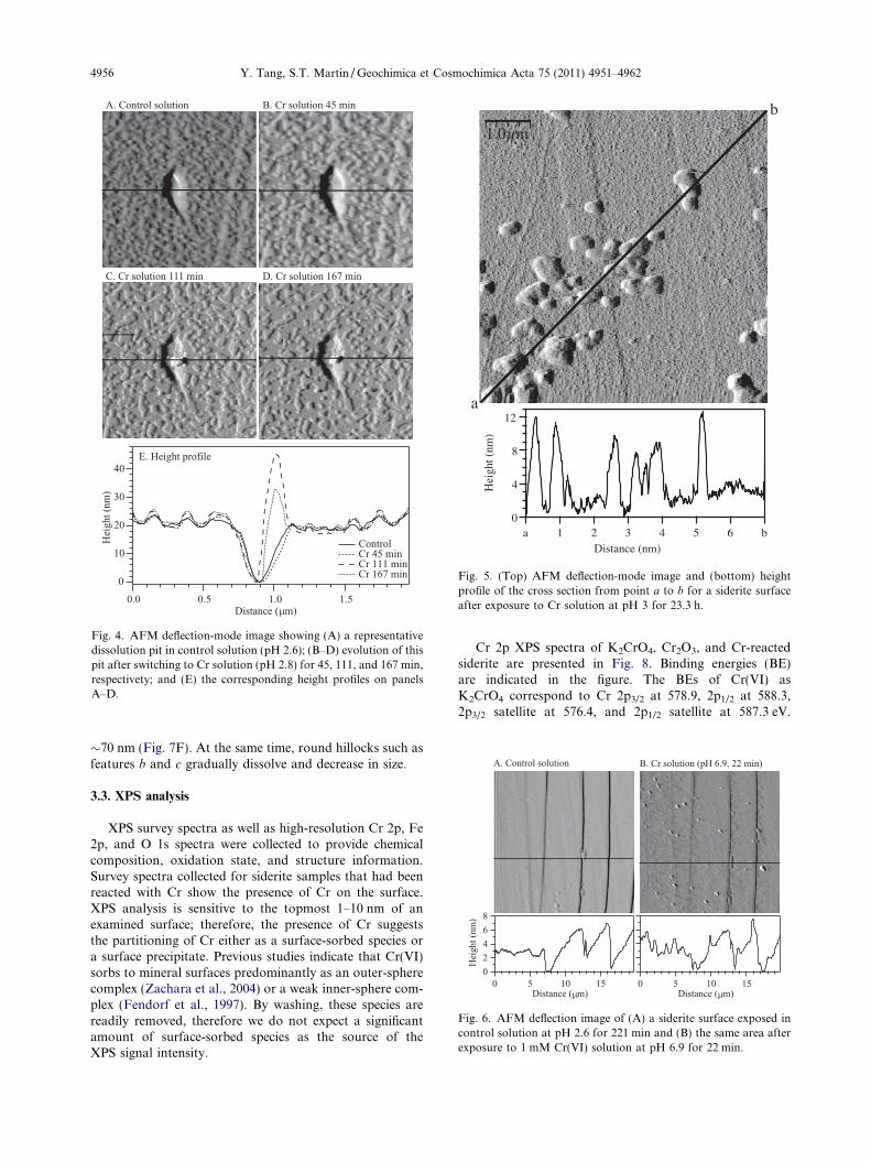
in this case generally show an elongated feature (Fig. S1A).Clustered small shallow dissolution pits are also present atfine scale. Previous studies have attributed the formation ofthese shallow pits to the presence of clustered point defects(Macinnis and Brantley, 1992; Duckworth and Martin,2004a). These observations are consistent with the previ-ously reported pit morphologies of siderite under anoxicconditions (Duckworth and Martin, 2004a). In the presenceof Cr(VI), no significant modification to the pit morpholo-gies was observed. However, a new feature of simultaneousprecipitation was observed, which was more pronouncedfor acidic and circumneutral conditions. In the followingparagraphs we show representative images collected foracidic (Figs. 3–5), circumneutral (Fig. 6), and alkaline(Fig. 7) conditions.
The time series of images in Fig. 3 shows the develop-ment of clustered shallow pits as well as a precipitating sur-face overlayer in the presence of Cr solution at pH = 3.Fig. 3A–C shows that the pits on the left and lower sideof the images (i.e., area outside of the black ellipse) coalesceinto larger rhombohedral shaped pits. The histograms re-veal that the pit depth gradually increases from 0.4 to3.2 nm during this process. At the same time, small pitsare filled by surface precipitates. After 60 min (image notshown), all pits disappear, primarily by filling.
In addition to widespread shallow pits at fine scale,development of isolated deep pits is also observed. Fig. 4shows the evolution of a representative isolated deep pitafter switching from the control solution (pH = 2.6) tothe Cr solution (pH = 2.8). The height profiles in Fig. 4Eindicate that the pit has an unchanged depth of �20 nmafter switching to Cr solution for 45 min but widens tothe right side. After 111 min, the pit also widens to the left,
A. Cr solution 336 min B. + 5
segamIthgie
Hsega
mInoitcelfe
D
1200
800
400
04
His
togr
ams 1500
1000
500
0
ycneuqerF
1.61.20.80.4Height (nm) Heigh
0.37 (+0.03)_ nm 2.4
750nm
Fig. 3. AFM micrographs illustrating dissolution and precipitation oversolution at pH 3. The black ellipse in each image highlights areas that aevident in the series of images. The arrow serves as a point of reference
with a hillock-shaped precipitate filling the right side. Thisprecipitate is �200 nm in diameter and �12 nm higher thanthe surface. After 167 min, no further widening of the pit isobserved. The precipitate grows to �26 nm above the sur-face. In these images, widespread surface etching aroundthe pit is also evident. With extended reaction time, the sur-face precipitate gradually builds up. Fig. 5 shows the deflec-tion-mode image and height profile of a siderite surfaceafter exposure to a Cr solution of pH = 3 for 23.3 h.Rounded hillock precipitates are apparent across the sur-face, with no preferential distribution to terrace or steps.The vertical heights of these precipitates are �10 nm, withlateral diameters varying between 200 and 1000 nm.
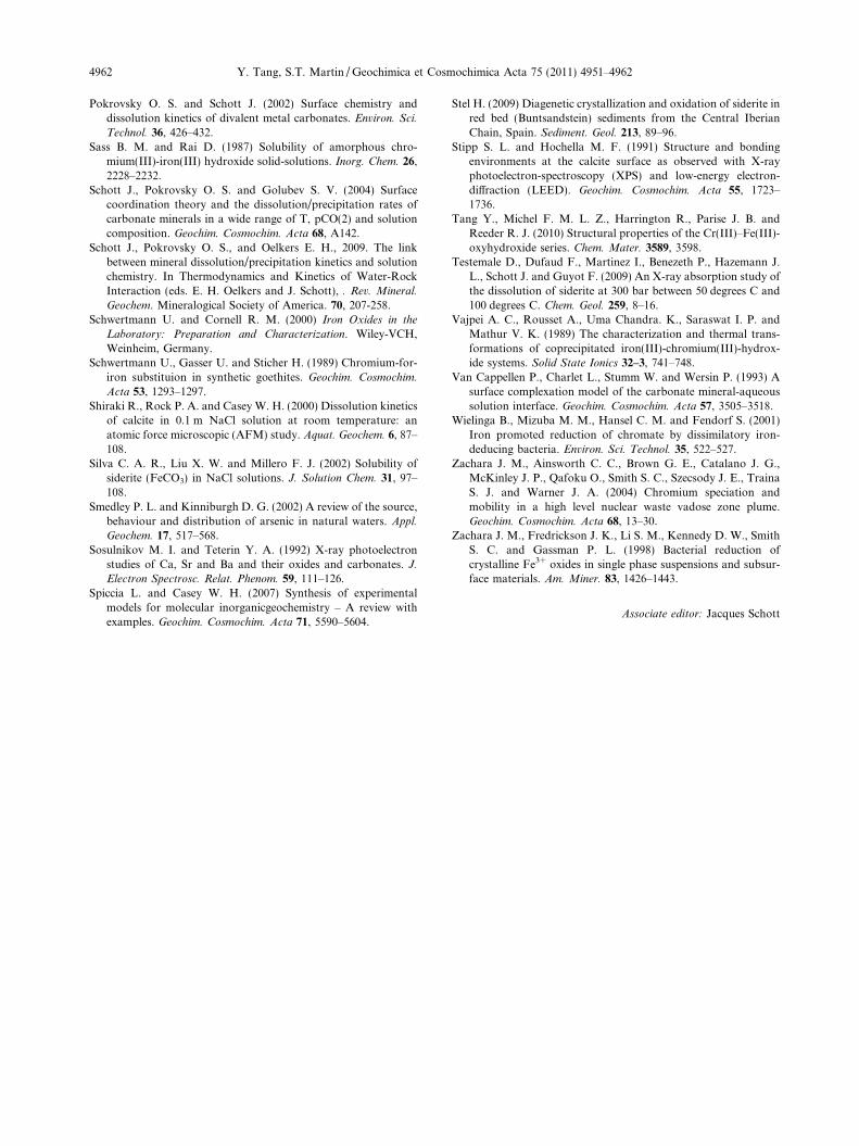
Fig. 6 shows a set of representative images collected atcircumneutral pH in the Cr solution. After the inlet isswitched from the control to the Cr solution, the surfaceis rapidly covered by a film of precipitates. The height pro-file in Fig. 6B shows that the height of these precipitates isfrom 0 to 2 nm. Some of the larger precipitates grow asrounded hillocks, with sizes ranging from 2 to 3 nm. Theseprecipitates, especially the less developed small ones, areloosely attached at the initial stage and can be removedby scanning the AFM tip at large contact force. Again,no preferential locations are observed for these precipitates;they appear both at edge steps and terraces on the surface.
At alkaline pH, surface morphology is dominated bydissolution, although simultaneous precipitation and reor-ganization of surface features are also sometimes observed.Fig. 7 shows the evolution of a siderite surface after expo-sure to Cr solution of pH = 11.5 for up to �4 h. Surfacefeature a gradually dissolves and reorganizes from an irreg-ular shape to a rounded shape. After 222 min, precipitatesform on top of it, increasing the height from �40 to
C. + 11 minmin
128
1200
800
400
0108642
t (nm) Height (nm)
3.23 (+0.05) nm_3 (+0.18)_ nm
time on the surface of siderite when exposed to the Cr(VI) oxidantre evident of precipitation. Outside the ellipse, dissolution is morereferred to in the text.
ig. 4. AFM deflection-mode image showing (A) a representativeissolution pit in control solution (pH 2.6); (B–D) evolution of thisit after switching to Cr solution (pH 2.8) for 45, 111, and 167 min,espectivety; and (E) the corresponding height profiles on panels–D.
1.0µm
a
b
12
8
4
0
Hei
ght (
nm)
b654321aDistance (nm)
Fig. 5. (Top) AFM deflection-mode image and (bottom) heightprofile of the cross section from point a to b for a siderite surfaceafter exposure to Cr solution at pH 3 for 23.3 h.
4956 Y. Tang, S.T. Martin / Geochimica et Cosmochimica Acta 75 (2011) 4951–4962
FdprA
Fig. 6. AFM deflection image of (A) a siderite surface exposed incontrol solution at pH 2.6 for 221 min and (B) the same area afterexposure to 1 mM Cr(VI) solution at pH 6.9 for 22 min.
�70 nm (Fig. 7F). At the same time, round hillocks such asfeatures b and c gradually dissolve and decrease in size.
3.3. XPS analysis
XPS survey spectra as well as high-resolution Cr 2p, Fe2p, and O 1s spectra were collected to provide chemicalcomposition, oxidation state, and structure information.Survey spectra collected for siderite samples that had beenreacted with Cr show the presence of Cr on the surface.XPS analysis is sensitive to the topmost 1–10 nm of anexamined surface; therefore, the presence of Cr suggeststhe partitioning of Cr either as a surface-sorbed species ora surface precipitate. Previous studies indicate that Cr(VI)sorbs to mineral surfaces predominantly as an outer-spherecomplex (Zachara et al., 2004) or a weak inner-sphere com-plex (Fendorf et al., 1997). By washing, these species arereadily removed, therefore we do not expect a significantamount of surface-sorbed species as the source of theXPS signal intensity.
Cr 2p XPS spectra of K2CrO4, Cr2O3, and Cr-reactedsiderite are presented in Fig. 8. Binding energies (BE)are indicated in the figure. The BEs of Cr(VI) asK2CrO4 correspond to Cr 2p3/2 at 578.9, 2p1/2 at 588.3,2p3/2 satellite at 576.4, and 2p1/2 satellite at 587.3 eV.
A. Control solution B. Cr solution, 22 min
C. Cr solution, 122 min D. Cr solution, 166 min
E. Cr solution, 222 min
500nm
a
b
c
80
60
40
20
0
)mn(thgie
H
2.01.51.00.50.0Distance ( m)
(i) (ii) (iii)
(i)
(ii)
(iii)F. Height profiles
Fig. 7. AFM deflection images showing the evolution of siderite in (A) control solution (pH 2.4) and (B–E) after switching to Cr solution ofpH 11.5 for 22, 122, 166, and 222 min, respectively. Panel (F) shows the height profiles of surface feature a across panels A, C, and E.
Siderite dissolution in the presence of chromate 4957
These values are in agreement with previously reportedvalues of Cr 2p3/2 at 579.3 and 2p1/2 at 588.5 eV, with aseparation of 9.1–9.2 eV (Stipp and Hochella, 1991; Sos-ulnikov and Teterin, 1992; Aronniemi et al., 2005).Cr2O3 lacks the two satellite peaks and has BEs of 2p3/2
at 576.4 and 2p1/2 at 586.3 eV, significantly lower thanthose of Cr(VI) and in agreement with previous studies(Aronniemi et al., 2005). The two Cr-reacted samples re-acted at pH < 10 for 20 h have BEs at 576.8–576.9 eVand 586.3–596.8 eV for 2p3/2 and 2p1/2 peaks, respectively,similar to those of Cr(III) and significantly lower thanthose of Cr(VI), indicating that the Cr is retained on the
siderite surface as Cr(III). Additional samples that are re-acted at similar pH but shorter time (<4 h) also show peakpositions that are indicative of the presence of Cr(III). CrXPS spectra were also collected on five samples reactedwith Cr(VI) at pH > 10. Two of the samples exposed toCr(VI) to less than 4 hr show Cr 2p peaks, although noisy,with positions similar to that of Cr(III) (data not shown).Interestingly, no Cr signals were observed on the threecrystals that are reacted for extended time (20 h). An at-tempt to resolve the Fe oxidation state from Fe 2p spectrawas not successful due to a large interference from theunderlying FeCO3 substrate.
(A) K2CrO4
(B) Cr2O3
595 590 585 580 575 570
(D) pH 6.8
(C) pH 3.2
Inte
nsity
(a.
u.)
Binding Energy (eV)
a'=576.4
586.8
578.9
588.3
586.3576.4
576.8586.3
576.9
a
a
a
a
b
b
b
b
b'=587.3
Fig. 8. High-resolution Cr 2p XPS spectra of (A) K2CrO4, (B)Cr2O3, and (C–D) siderite samples that were reacted with Cr(VI)solution at pH 2.8 and 6.9, respectively. Raw and fitted data areshown in gray and black lines, respectively. The Cr 2p3/2, 2p3/2
satellite, 2p1/2, and 2p1/2 satellite peaks are labeled as a, a0, b,and b0, respectively. Binding energies (eV) determined from peakpositions are labeled next to each peak.
(A) FeCO3
(B) hematite
(D) pH 2.8
(E) pH 6.9
538 536 534 532 530 528 526 524
(F) pH 11.2
(C) goethite
Inte
nsity
(a.
u.)
Binding Energy (eV)
531.6 eV
529.7 eV
529.6 eV 531.0 eV
530.9 eV
530.9 eV532.6 eV
532.5 eV
531.8 eV
531.2 eV
c
a
b a
b’
c b
bc
c
Fig. 9. High-resolution O 1s XPS spectra of (A) unreacted siderite,(B) hematite, (C) goethite, and (D)–(F) Cr-reacted siderite sampleat pH 2.8, 6.9, and 11.2, respectively. Raw and fitted data areshown in open circles and black lines, respectively. The O 1s peakas in O2�, OH�, adventitious water/OH�, and CO2�
3 are labeled asa, b, b0, and c, respectively. Binding energies (eV) determined frompeak positions are labeled next to each peak.
4958 Y. Tang, S.T. Martin / Geochimica et Cosmochimica Acta 75 (2011) 4951–4962
O 1s XPS spectra were collected for known referencecompounds to determine characteristic peaks for oxygenspeciated as O2�, OH�, and CO2�
3 . The O 1s spectra andBEs are shown in Fig. 9. The spectrum collected for unre-acted FeCO3 has a major peak centered at 531.6 eV, similarto that reported for oxygen in CO2�
3 , e.g., BaCO3 at531.2 eV, CaCO3 at 531.6–531.9 eV, MnCO3 at 532.2 eV,and FeCO3 at 532.3 eV (Stipp and Hochella, 1991; Sosulni-kov and Teterin, 1992; Duckworth and Martin, 2004b). TheFWHM of this peak (2.7 eV) is similar to the values ofFeCO3 reported by Duckworth and Martin (2004b), butslightly higher than reported values of other carbonate min-erals, e.g., �2.0 eV for BaCO3 and CaCO3 (Sosulnikov andTeterin, 1992). However, fitting the peak with multiplecomponents did not improve the result. It is possible thatthe sample surface is slightly oxidized during the shortexposure to atmosphere before putting into the XPS samplechamber. Hematite has a major peak centered at 529.7 eV,consistent with previously reported values of 529.8–530.2 eV for oxygen in O2� (Junta Rosso and Hochella,1996; Duckworth and Martin, 2004b; Spiccia and Casey,2007). A minor shoulder at 531.2 eV is also present, likelydue to the contribution from adventitious dissociatedH2O (Junta Rosso and Hochella, 1996). Goethite shows abroad peak with FWHM of 3.0 eV, which can be deconvo-luted into two peaks: an O2� peak at 529.6 eV and an OH�
peak at 531.0 eV. The BEs and separation values of thesetwo peaks (1.4 eV) agree with literature values for O2�
(529.8–530.2 eV) and OH� (531.0–531.9 eV), with a separa-tion value of 1.1–1.9 eV (Duckworth and Martin, 2004b;Spiccia and Casey, 2007).
O 1s spectra were also recorded for the Cr-reacted sider-ite surfaces. The peaks for samples reacted at pH of 2.8 and6.9 are broad (FWHM both at 3.1) and asymmetric. Theycan be deconvoluted into two peaks: (1) a peak at530.9 eV that corresponds to the oxygen in OH�, suggest-ing that the surface precipitate is a hydroxide; and (2) apeak at 532.5–532.6 eV that we assign as CO2�
3 peak. TheCO2�
3 peak arises from the contribution of the underlyingFeCO3 substrate. Position of this peak is slightly higherthan that observed value for the unreacted siderite reference(531.6 eV). For the siderite sample reacted at pH = 11.2,the spectra show a dominant peak centered at 531.8 eV,which is assigned to oxygen in CO2�
3 .
Fe2+
HCrO4/CrO42-
CrxFe1-x(OH)3(s)H+/H2O
HCO3-
Fe3+
Cr3+
H2O/OH-
FeCO3
CrO42-
H2O
CO32-
[Fe3+(OH)4]-
[Cr3+(OH)4]-
FeCO3
[Fe2+(OH)3]-
>FeIIOH />Fe0[Fe3+(OH)4]-
Fe2+
HCrO4-
CrxFe1-x(OH)3(s)H+
HCO3-
Fe3+
Cr3+
H2O
FeCO3
(A) pH < 4.5
(B) 4.5 < pH < 10
(C) pH > 10
[Fe]T
[Fe]T
IIO-
Fig. 10. Illustration of the proposed reaction mechanisms for theCr-initiated oxidation of the siderite surface, including surfaceprecipitation, for three different pH regimes.
Siderite dissolution in the presence of chromate 4959
3.4. Reaction products in the presence of Cr(VI)
Duckworth and Martin (2004b) showed that in the pres-ence of dissolved oxygen, Fe(II) released from siderite disso-lution is oxidized to Fe(III) and precipitates as Fe hydroxide(ferrihydrite) for the pH range of 6–10.3. For pH <5, ferrihy-drite precipitation is not thermodynamically favorable, andthe macroscopic dissolution rate of Duckworth and Martin(2004b) is therefore similar to anoxic conditions. This resultis much different from our results obtained using Cr(VI) as anoxidant. The macroscopic dissolution rate in Cr solution isalmost an order of magnitude lower than that in the controlsolution. As a check, our control conditions are similar tothose of Duckworth and Martin (2004b). We also observeprecipitation for acidic pH (<5). The combination of thisobservation with that of a reduced dissolution rate indicatesthat the surface precipitates formed in the presence of Cr(VI)are different from the ferrihydrite formed under oxygen con-ditions. In particular, this new precipitate is much less solubleunder acidic pH conditions.
From our XPS analysis, although we are not able to di-rectly resolve Fe(III) species from Fe 2p spectra due to largeinterferences from the underlying FeCO3 substrate, the pres-ence of the Cr(III) species strongly points to a redox reactionbetween Fe(II) and Cr(VI) that results in Cr(III) and Fe(III)species. Combining these results with the O 1s observationshowing the presence of a hydroxide species on the surfaceleads to the conclusion that the surface precipitate is either(1) a mixed Cr(III)–Fe(III)-hydroxide phase or (2) a discretesingle solid assemblage of mixed Fe(III)-hydroxide andCr(III)-hydroxide phases.
Many studies have shown that the reduction of Cr(VI) byFe(II) and S2� results in insoluble Cr1�xFexðOHÞ3 � nH2Ophase (Sass and Rai, 1987; Patterson et al., 1997). Eary andRai (1988) and Palmer and Wittbrodt (1991) report the rapidredox reaction:
The resulting Cr(III) and Fe(III) can hydrolyze, com-bine, and precipitate as a solid solution in slightly acidicto alkaline conditions:
xCrðIIIÞðaqÞ þ ð1� xÞFeðIIIÞðaqÞ þ ð3þ nÞH2O
! CrxFe1�xðOHÞ3ðsÞ � nH2Oþ 3Hþ ð4Þ
for which the Fe:Cr ratio is consistently measured as approx-imately 3.0 (i.e., x = 0.25), as one would expect from the stoi-chiometry of reaction 3. Solubility and structure studies havealso suggested that the Cr(III)–Fe(III)-hydroxide phase isstable during the fast precipitation of Cr(III) and Fe(III)aqueous ions (Sass and Rai, 1987; Vajpei et al., 1989; Amo-nette and Rai, 1990; Tang et al., 2010). We are not awareof any study reporting separate Cr(III)- and Fe(III)-hydrox-ide phases during the redox reaction between Cr(VI) andFe(II) or the co-precipitation of Cr(III) and Fe(III). There-fore, we assign the precipitates as nanocrystalline particleswith a likely composition of Cr0.25Fe0.75(OH)3�nH2O. Inagreement with the size dimensions, Tang et al. (2010) showedthat the CrxFe1�x(OH)3(s)�nH2O series has particles sizes of 1–2 nm. Although Schwertmann et al. (1989) and Amonette andRai (1990) report the transformation of the initial nanocrystal-
line Cr–Fe-hydroxide phase into crystalline goethite with ex-tended aging time (100 days) at high pH or temperature, forour experimental conditions at room temperature and with areaction time shorter than 24 h we do not expect and did not ob-serve such structural transformation.
3.5. Reaction mechanisms in the presence of Cr(VI)
A schematic presentation of the proposed reactionmechanisms for the Cr-initiated oxidation of the sideritesurface, including surface precipitation, is shown inFig. 10 for three different pH regimes. The mechanism in-cludes the following aspects (A–C):
(A) The dissolution of siderite via parallel protonationand water hydrolysis reactions (cf. Section 3.1):
(B) A redox reaction between Cr(VI) and Fe(II). The spe-ciation calculation of the Cr(VI) influent (Cr(VI) = 1 mM,NaNO3 = 0.01 M, and PO2 = 0 atm) indicates that belowpH of 5 HCrO�4 dominates (>95%) with less than 5%Cr2O2�
7 . For increasing pH, the relative percentage of
4960 Y. Tang, S.T. Martin / Geochimica et Cosmochimica Acta 75 (2011) 4951–4962
HCrO�4 decreases and CrO2�4 becomes more important.
HCrO�4 and CrO2�4 have equal abundance at pH = 6.5.
CrO2�4 dominates the Cr(VI) speciation for pH > 8. Previous
studies show that the reduction of Cr(VI) by Fe(II)-bearingminerals such as biotite and Fe(II)-containing natural hema-tite only occurs in solutions in which dissolved Fe(II) exists(i.e., homogeneous reaction) instead of occurring directlyat the mineral surface (i.e., heterogeneous reaction) (Earyand Rai, 1989). The rate of Cr(VI) reduction and Fe(II) oxi-dation thus depends on the rate of mineral dissolution.Therefore, depending on the pH, the redox reaction betweenFe(II) and Cr(VI) can be written as:
3Fe2þ þHCrO�4 þ 7Hþ ! 3Fe3þðaqÞ þ Cr3þ
ðaqÞ þ 4H2O ð7Þ3Fe2þ þ CrO2�
4 þ 8Hþ ! 3Fe3þðaqÞ þ Cr3þ
ðaqÞ þ 4H2O ð8Þ
(C) The precipitation of Cr(III)–Fe(III)-hydroxide phase(cf. Section 3.4):
Cr3þðaqÞ þ 3Fe3þ
ðaqÞ þ 12H2O! 4Cr0:25Fe0:75ðOHÞ3ðsÞ þ 12Hþ
ð9ÞCr3þðaqÞ þ 3Fe3þ
ðaqÞ þ 12OH� ! 4cr0:25Fe0:75ðOHÞ3ðsÞ ð10Þ
The macroscopic, microscopic, and spectroscopic obser-vations are the net result of the above competing reactions,and are explained as followed.
For pH < 4.5 (Fig. 10A), proton-promoted siderite dis-solution dominates. Precipitation occurs simultaneouslywith the redox reaction, resulting in a reduced net dissolu-tion rate as compared to control observations. At circum-neutral pH values of 4.5–10 (Fig. 10B), the solubility ofCr–Fe-hydroxide precipitates is at a minimum (Sass andRai, 1987). Rapid and widespread surface precipitation atboth terraces and edge steps inhibits the further dissolution.
For pH > 10 (Fig. 10C), the dissolution rate is unusuallyhigh as compared to the control condition. As discussed inSection 3.1, the classic model for carbonate minerals disso-lution involves parallel protonation and water hydrolysisreactions, which also implies pH-independent dissolutionrates for pH > 6 (Plummer et al., 1978). Recent surfacecomplexation modeling inputs have suggested the consider-ation of surface speciation, especially when consideringexperimental carbonate mineral dissolution rates as a func-tion of
PCO2 (Schott et al., 2009). The increased dissolu-
tion rate of calcite and dolomite in CO2-free and stronglyalkaline solution (pH > 12) can be explained by the forma-tion of the negatively charged >MeO� species at the ex-pense of neutral >MeOH0 species (Pokrovsky and Schott,2001; Schott et al., 2004, 2009). Interestingly, the redox-sen-sitive mineral siderite shows the classic pH-independent dis-solution behavior in anoxic conditions both in this studyand in Duckworth and Martin (2004b) yet has elevated dis-solution rate at pH > 10 in the presence of oxidants, i.e.,chromate in this study and oxygen in Duckworth andMartin (2004b).
Duckworth and Martin (2004b) proposed two possiblemechanisms for the elevated dissolution rate in the presenceof oxygen: (1) the rapid electron transfer between aqueous½Fe3þðOHÞ4�
� and surfacial >FeIIOHðsÞ groups or (2) the di-rect attack of surface >FeII groups by aqueous oxygen. Forour study of siderite dissolution in the presence of chromate,
an explanation of the elevated dissolution rate in alkalineconditions requires an examination both of the solutionand of the surface speciation. Speciation calculation using40 nM Cr(III), Fe(III) or Fe(II) shows that the predominantCr(III), Fe(II), and Fe(III) species for pH > 10 are½Cr3þðOHÞ4�
�, ½Fe2þðOHÞ3��, and ½Fe3þðOHÞ4�
�, respec-tively. The observation that rapid dissolution occurs forpH > 10 only in the presence of oxidant (i.e., chromate in thisstudy or oxygen in Duckworth and Martin (2004b)) indicatesthat the oxidation of Fe(II) to Fe(III) and the involvement ofthese hydroxylated species play important roles. In our caseof Cr(VI) as the oxidant, because the redox reaction betweenCr(VI) and Fe(II) species only occurs in solution rather thanat the mineral surfaces (Eary and Rai, 1989), we thereforeconsider the second mechanism unlikely in our system. Basedon the above information on both solution and surface spe-ciation, we propose that the fast dissolution rate is due to theelectron transfer between solution ½Fe3þðOHÞ4�
� and surfa-cial >FeIIOH0 or >FeIIO� groups. In this mechanism,the dissolved ½Fe2þðOHÞ3�
� is oxidized by CrO2�4 to
½Fe3þðOHÞ4��, which then scavenges one electron from the
surfacial >FeIIOH or >FeIIO� groups, causing continueddissolution of siderite. In tandem, the ½Fe3þðOHÞ4�
� speciesis reduced to ½Fe2þðOHÞ3�
�, which is re-cycled for furtherreduction of CrO4
2�.
4. CONCLUSIONS AND IMPLICATIONS
The present study combines macroscopic, microscopic,and spectroscopic observations of the siderite surface inthe presence of chromate. For all investigated pH condi-tions, simultaneous dissolution and precipitation occur.Surface precipitation caused by the introduction of Cr(VI)solution appear as round hillocks, which grow into largersizes after extended reaction time. In addition, regardlessof experimental pH, these hillock precipitates are locatedboth at terraces and edge steps with no preferential distri-bution. These observations are strikingly different fromthose on siderite or rhodochrosite (MnCO3) surface in thepresence of dissolved oxygen as oxidant (Duckworth andMartin, 2004b). The oxidation of siderite by dissolved oxy-gen under alkaline condition causes the precipitation of hill-ocks with 10–20 nm height by 100–200 nm diameter, whichpreferentially form at step sites. In contrast, rhodochrositesurface oxidation by dissolved oxygen results in a continu-ous growth of layer at neutral pH, as well as precipitationof a patchy layer of hillocks 20–25 nm high by 1–2 lm diameter (Duckworth and Martin, 2004b).
In the presence of Cr(VI) as an oxidant, reactions occurat the siderite surface and involve parallel proton- andwater hydrolysis-promoted dissolution, oxidation of dis-solved Fe(II) by Cr(VI), and surface precipitation. Thesereactions occur simultaneously and have different relativeimportance for varied pH. The dissolution rate forpH < 4.5 is slower than that in the control solution. Iso-lated deep dissolution pits and clustered shallow pits occursimultaneously with surface precipitation. For5 < pH < 9.5, the dissolution rate is the slowest, and rapidprecipitation is observed at both edge steps and terraces.For pH > 10, the dissolution rate is faster than that in the
Siderite dissolution in the presence of chromate 4961
control solution and can be explained by the electron trans-fer facilitated by ½Fe3þðOHÞ4�
�. Dissolution and re-precipi-tation of round hillocks are observed. The surfaceprecipitates are identified as low-solubility nano-sizedCr(III)–Fe(III)-hydroxide. These results provide importantinsights for the more general topic of the interactions be-tween reduced iron in minerals and aqueous-phase inor-ganic or organic oxidants.
ACKNOWLEDGEMENTS
The authors acknowledge Christopher Letini (SEAS) for pro-viding a synthetic hematite sample and David Lange (CNS) forassistance with XPS data collection. The Center for Nanoscale Sys-tems (CNS) is a member of the National Nanotechnology Infra-structure Network (NNIN), which is supported by the NationalScience Foundation under NSF award no. ECS-0335765. Com-ments from the editor and two anonymous reviewers greatly im-proved the manuscript.
APPENDIX A. SUPPLEMENTARY DATA
Supplementary data associated with this article can befound, in the online version, at doi:10.1016/j.gca.2011.06.024.
REFERENCES
Ahmed K. M., Bhattacharya P., Hasan M. A., Akhter S. H., AlamS. M. M., Bhuyian M. A. H., Imam M. B., Khan A. A. andSracek O. (2004) Arsenic enrichment in groundwater of thealluvial aquifers in Bangladesh: an overview. Appl. Geochem.
19, 181–200.
Amonette J. E. and Rai D. (1990) Identification of noncrystalline(Fe, Cr)(OH)3 by infrared-spectroscopy. Clays Clay Miner. 38,
129–136.
Aronniemi M., Sainio J. and Lahtinen J. (2005) Chemical statequantification of iron and chromium oxides using XPS: theeffect of the background subtraction method. Surf. Sci. 578,
108–123.
Benezeth P., Dandurand J. L. and Harrichoury J. C. (2009)Solubility product of siderite (FeCO3) as a function oftemperature (25–250 degrees C). Chem. Geol. 265, 3–12.
Blowes D. W., Ptacek C. J. and Jambor J. L. (1997) In-situremediation of Cr(VI)-contaminated groundwater using per-meable reactive walls: laboratory studies. Environ. Sci. Technol.
31, 3348–3357.
Braun R. D. (1991) Solubility of iron(III) carbonate at tempera-tures between 30 and 80-degrees. Talanta 38, 205–211.
Coleman M. L., Hedrick D. B., Lovley D. R., White D. C. and PyeK. (1993) Reduction of Fe(III) in sediments by sulfate-reducingbacteria. Nature 361, 436–438.
Duckworth O. W., Cygan R. T. and Martin S. T. (2004) Linear freeenergy relationships between dissolution rates and molecularmodeling energies of rhombohedral carbonates. Langmuir 20,
2938–2946.
Duckworth O. W. and Martin S. T. (2003) Connections betweensurface complexation and geometric models of mineral disso-lution investigated for rhodochrosite. Geochim. Cosmochim.
Acta 67, 1787–1801.
Duckworth O. W. and Martin S. T. (2004a) Dissolution rates andpit morphologies of rhombohedral carbonate minerals. Am.
Miner. 89, 554–563.
Duckworth O. W. and Martin S. T. (2004b) Role of molecularoxygen in the dissolution of siderite and rhodochrosite.Geochim. Cosmochim. Acta 68, 607–621.
Eary L. E. and Rai D. (1988) Chromate removal from aqueouswastes by reduction with ferrous ion. Environ. Sci. Technol. 22,
972–977.
Eary L. E. and Rai D. (1989) Kinetics of chromate reduction byferrous-ions derived from hematite and biotite at 25-degrees-C.Am. J. Sci. 289, 180–213.
Erdem M., Gur F. and Tumen F. (2004) Cr(VI) reduction inaqueous solutions by siderite. J. Hazard. Mater. 113, 219–224.
Fendorf S., Eick M. J., Grossl P. and Sparks D. L. (1997) Arsenateand chromate retention mechanisms on goethite.1. Surfacestructure. Environ. Sci. Technol. 31, 315–320.
Golubev S. V., Benezeth P., Schott J., Dandurand J. L. andCastillo A. (2009) Siderite dissolution kinetics in acidic aqueoussolutions from 25 to 100 degrees C and 0 to 50 atm pCO2.Chem. Geol. 265, 13–19.
Greenberg J. and Tomson M. (1992) Precipitation and dissolutionkinetics and equlibria of aqueous ferrous carbonate vs temper-ature. Appl. Geochem. 7, 185–190.
Hansel C. M., Wielinga B. W. and Fendorf S. R. (2003) Structuraland compositional evolution of Cr/Fe solids after indirectchromate reduction by dissimilatory iron-reducing bacteria.Geochim. Cosmochim. Acta 67, 401–412.
Horcas I., Fernandez R., Gomez-Rodriguez J. M., Colchero J.,Gomez-Herrero J., and Baro A. M. (2007) WSXM: a softwarefor scanning probe microscopy and a tool for nanotechnology.Rev. Sci. Instrum. 78.
Jensen D. L., Boddum J. K., Tjell J. C. and Christensen T. H.(2002) The solubility of rhodochrosite (MnCO3) and siderite(FeCO3) in anaerobic aquatic environments. Appl. Geochem.
17, 503–511.
Junta Rosso J. L. and Hochella M. F. (1996) The chemistry ofhematite {001} surfaces. Geochim. Cosmochim. Acta 60, 305–
314.
La Force M. J., Hansel C. M. and Fendorf S. (2002) Seasonaltransformations of manganese in a palustrine emergent wet-land. Soil Sci. Soc. Am. J. 66, 1377–1389.
Liu C. X., Kota S., Zachara J. M., Fredrickson J. K. and BrinkmanC. K. (2001) Kinetic analysis of the bacterial reduction ofgoethite. Environ. Sci. Technol. 35, 2482–2490.
Lovley D. R. (1991) Dissimilatory Fe(III) and Mn(IV) reduction.Microbiol. Rev. 55, 259–287.
Macinnis I. N. and Brantley S. L. (1992) The role of dislocationsand surface-morphology in calcite dissolution. Geochim. Cos-
mochim. Acta 56, 1113–1126.
Nealson K. H. and Saffarini D. (1994) Iron and manganese inanaerobic respiration–environmental significance, physiology,and regulation. Annu. Rev. Microbiol. 48, 311–343.
Palmer C. D. and Wittbrodt P. R. (1991) Processes affecting theremediation of chromium-contaminated sites. Environ. Health
Perspect. 92, 25–40.
Parkhurst D. L. and Appelo C. A. J., 1999. User’s Guide toPHREEQC (Version 2) – A computer Program for Speciation,Batch-reaction, One-dimensional Transport, and Inverse Geochem-ical Calculations. US Geol. Surv. Water Res. Inv. Rept. 99-4259.
Patterson R. R., Fendorf S. and Fendorf M. (1997) Reduction ofhexavalent chromium by amorphous iron sulfide. Environ. Sci.
Technol. 31, 2039–2044.
Plummer L. N., Wigley T. M. L. and Parkhurst D. L. (1978) Kinetcsof calcite dissolution in CO2-water systems at 5-degree-C to 60-degree-C and 0.0 to 1.0 atm CO2. Am. J. Sci. 278, 179–216.
Pokrovsky O. S. and Schott J. (2001) Kinetics and mechanism ofdolomite dissolution in neutral to alkaline solutions revisited.Am. J. Sci. 301, 597–626.
4962 Y. Tang, S.T. Martin / Geochimica et Cosmochimica Acta 75 (2011) 4951–4962
Pokrovsky O. S. and Schott J. (2002) Surface chemistry anddissolution kinetics of divalent metal carbonates. Environ. Sci.
Technol. 36, 426–432.
Sass B. M. and Rai D. (1987) Solubility of amorphous chro-mium(III)-iron(III) hydroxide solid-solutions. Inorg. Chem. 26,
2228–2232.
Schott J., Pokrovsky O. S. and Golubev S. V. (2004) Surfacecoordination theory and the dissolution/precipitation rates ofcarbonate minerals in a wide range of T, pCO(2) and solutioncomposition. Geochim. Cosmochim. Acta 68, A142.
Schott J., Pokrovsky O. S., and Oelkers E. H., 2009. The linkbetween mineral dissolution/precipitation kinetics and solutionchemistry. In Thermodynamics and Kinetics of Water-RockInteraction (eds. E. H. Oelkers and J. Schott), . Rev. Mineral.
Geochem. Mineralogical Society of America. 70, 207-258.Schwertmann U. and Cornell R. M. (2000) Iron Oxides in the
Laboratory: Preparation and Characterization. Wiley-VCH,Weinheim, Germany.
Schwertmann U., Gasser U. and Sticher H. (1989) Chromium-for-iron substituion in synthetic goethites. Geochim. Cosmochim.
Acta 53, 1293–1297.
Shiraki R., Rock P. A. and Casey W. H. (2000) Dissolution kineticsof calcite in 0.1 m NaCl solution at room temperature: anatomic force microscopic (AFM) study. Aquat. Geochem. 6, 87–
108.
Silva C. A. R., Liu X. W. and Millero F. J. (2002) Solubility ofsiderite (FeCO3) in NaCl solutions. J. Solution Chem. 31, 97–
108.
Smedley P. L. and Kinniburgh D. G. (2002) A review of the source,behaviour and distribution of arsenic in natural waters. Appl.
Geochem. 17, 517–568.
Sosulnikov M. I. and Teterin Y. A. (1992) X-ray photoelectronstudies of Ca, Sr and Ba and their oxides and carbonates. J.
Electron Spectrosc. Relat. Phenom. 59, 111–126.
Spiccia L. and Casey W. H. (2007) Synthesis of experimentalmodels for molecular inorganicgeochemistry – A review withexamples. Geochim. Cosmochim. Acta 71, 5590–5604.
Stel H. (2009) Diagenetic crystallization and oxidation of siderite inred bed (Buntsandstein) sediments from the Central IberianChain, Spain. Sediment. Geol. 213, 89–96.
Stipp S. L. and Hochella M. F. (1991) Structure and bondingenvironments at the calcite surface as observed with X-rayphotoelectron-spectroscopy (XPS) and low-energy electron-diffraction (LEED). Geochim. Cosmochim. Acta 55, 1723–
1736.
Tang Y., Michel F. M. L. Z., Harrington R., Parise J. B. andReeder R. J. (2010) Structural properties of the Cr(III)–Fe(III)-oxyhydroxide series. Chem. Mater. 3589, 3598.
Testemale D., Dufaud F., Martinez I., Benezeth P., Hazemann J.L., Schott J. and Guyot F. (2009) An X-ray absorption study ofthe dissolution of siderite at 300 bar between 50 degrees C and100 degrees C. Chem. Geol. 259, 8–16.
Vajpei A. C., Rousset A., Uma Chandra. K., Saraswat I. P. andMathur V. K. (1989) The characterization and thermal trans-formations of coprecipitated iron(III)-chromium(III)-hydrox-ide systems. Solid State Ionics 32–3, 741–748.
Van Cappellen P., Charlet L., Stumm W. and Wersin P. (1993) Asurface complexation model of the carbonate mineral-aqueoussolution interface. Geochim. Cosmochim. Acta 57, 3505–3518.
Wielinga B., Mizuba M. M., Hansel C. M. and Fendorf S. (2001)Iron promoted reduction of chromate by dissimilatory iron-deducing bacteria. Environ. Sci. Technol. 35, 522–527.
Zachara J. M., Ainsworth C. C., Brown G. E., Catalano J. G.,McKinley J. P., Qafoku O., Smith S. C., Szecsody J. E., TrainaS. J. and Warner J. A. (2004) Chromium speciation andmobility in a high level nuclear waste vadose zone plume.Geochim. Cosmochim. Acta 68, 13–30.
Zachara J. M., Fredrickson J. K., Li S. M., Kennedy D. W., SmithS. C. and Gassman P. L. (1998) Bacterial reduction ofcrystalline Fe3+ oxides in single phase suspensions and subsur-face materials. Am. Miner. 83, 1426–1443.