Sign-conserving amacrine neurons in the fly’sexternal plexiform layer
JOHN K. DOUGLASS and NICHOLAS J. STRAUSFELDArizona Research Laboratories, Division of Neurobiology, University of Arizona, Tucson
(Received February 3, 2004; Accepted February 16, 2005!
Abstract
Amacrine cells in the external plexiform layer of the fly’s lamina have been intracellulary recorded and dye-filledfor the first time. The recordings demonstrate that like the lamina’s short photoreceptors R1–R6, type 1 laminaamacrine neurons exhibit nonspiking, “sign-conserving” sustained depolarizations in response to illumination. Thiscontrasts with the sign-inverting responses that typify first-order retinotopic relay neurons: monopolar cells L1–L5and the T1 efferent neuron. The contrast frequency tuning of amacrine neurons is similar to that of photoreceptorsand large lamina monopolar cells. Initial observations indicate that lamina amacrine receptive fields are alsophotoreceptor-like, suggesting either that their inputs originate from a small number of neighboring visual samplingunits ~VSUs!, or that locally generated potentials decay rapidly with displacement. Lamina amacrines also respondto motion, and in one recording these responses were selective for the orientation of moving edges. This functionalorganization corresponds to the anatomy of amacrine cells, in which postsynaptic inputs from several neighboringphotoreceptor endings are linked by a network of very thin distal processes. In this way, each VSU can receiveconvergent inputs from a surround of amacrine processes. This arrangement is well suited for relaying responses tolocal intensity fluctuations from neighboring VSUs to a central VSU where amacrines are known to be presynapticto the dendrites of the T1 efferent. The T1 terminal converges at a deeper level with that of the L2 monopolar cellrelaying from the same optic cartridge. Thus, the localized spatial responses and receptor-like temporal responseproperties of amacrines are consistent with possible roles in lateral inhibition, motion processing, or orientationprocessing.
In the fly visual system, as in those of other insects and crustaceans~Strausfeld & Nässel, 1980!, the most peripheral amacrine cellsoriginate from cell bodies beneath the lamina, the first level ofphotoreceptor outputs. Each amacrine cell body sends a stoutneurite distally through the lamina’s external plexiform layer as faras the surface of this synaptic neuropil, where the neurite dividesto provide numerous tangential processes that extend some dis-tance across the lamina. These process provide two types ofspecializations: swellings at the plexiform layer’s surface andvaricose specializations, called a processes ~Boschek, 1971! thatextend down through the plexiform layer. Each a process abuts,and is postsynaptic to, the outer surface of a photoreceptor ending~Campos-Ortega & Strausfeld, 1973!.
Receptor terminals in the lamina are arranged in groups of six,each group forming a hollow column ~Braitenberg, 1967!. Eachsextet derives from six rhabdomeres distributed in the retina
among six ommatidia. These rhabdomeres sample the sameregion of visual space and are referred to as a visual sampling unit~VSU; Franceschini, 1975!. Each column—also called an opticcartridge—is associated with at least six a processes. These arelinked by their tangential fibers to other a processes that reside inneighboring or more distant optic cartridges. Each optic cartridgeis equipped with six distinctive types of efferent neurons ~Straus-feld, 1970!. Three of these, called the large monopolar cells~LMCs! L1, L2, and L3, are postsynaptic to all six receptorendings of their cartridge and thus sample one VSU. However, afourth efferent neuron, called the T1 basket cell, is not onlypostsynaptic to all six receptor endings but is also postsynaptic toall the a processes at its optic cartridge ~Campos-Ortega & Straus-feld, 1973!. Thus, because amacrine processes are linked directlyor indirectly to other amacrines at surrounding optic cartridges,each T1 dendritic tree could receive inputs from a surround ofnearby optic cartridges. Amacrine neurons thus occupy a uniqueanatomical position, which is in some respects analogous to that ofhorizontal cells in the mammalian outer plexiform layer but, as hasbeen proposed from immunocytological studies ~Sinakevitch &Strausfeld, 2004!, might be functionally comparable to certaintypes of amacrine cells of the mammalian inner plexiform layer, atthe level of bipolar endings.
Address correspondence and reprint requests to: John K. Douglass,Arizona Research Laboratories, Division of Neurobiology, 611 Gould-Simpson Building, University of Arizona, Tucson, AZ 85721, USA. E-mail:[email protected]
Because they occupy a peripheral location in visual pathwaysand because their synaptic connections provide a system of lateralconnections, lamina amacrines are expected to play a fundamentalrole in visual processing ~Strausfeld & Campos-Ortega, 1977;Shaw, 1984; Douglass & Strausfeld, 2003a!. For example, theycould support wide-field neural adaptation to ambient lightingconditions, lateral inhibition, or, as computational and anatomicalmodels of the lamina suggest, motion detection ~Higgins et al.,2004; Douglass & Strausfeld, 2003a; Sinakevitch & Strausfeld,2004!. Here, we describe the first intracellular recordings to beclearly identified with lamina amacrines. These recordings revealresponse properties that are consistent with a spatially localizedrole in lateral inhibition and0or motion computation.
Materials and methods
Flies used for intracellular recordings ~Phaenicia sericata! wereraised and maintained in the laboratory under a 12-h light012-hdark cycle at 258C, as described previously ~Douglass & Straus-feld, 1998, 2003b!. Ultrastructural and Golgi observations derivefrom P. sericata and Musca domestica, also reared under theseconditions. We find no difference between the anatomical featuresof the lamina in these two species. The methods of intracellularrecording, computerized visual stimulus presentation, and histo-logical processing were similar to those described previously~Douglass & Strausfeld, 1996, 1998, 2003b!, with new features asdescribed below.
Electrophysiology
Flies were immobilized with a low-melting point mixture ofbeeswax and violin rosin and fixed to a pedestal in front of a 300 �218 mm computer-controlled CRT display that was used to presentvisual stimuli. Room temperature was approximately 208C. Thefly’s eyes were positioned 18 cm from the center of the display, orin some cases the distance was set at 12.5 cm to provide brighterstimuli. Thus, at the fly’s head, the stimulus display was “full-field” at either 80 deg wide � 62 deg high ~at 18 cm!, or 100 degwide � 82 deg high ~at 12.5 cm!. The fly and pedestal wereattached to a vibration isolation table ~TMC 78 Series, Peabody,MA! under a Faraday cage, with the fly’s head aligned either for abinocular frontal view, or rotated approximately 45 deg for alateral, monocular view of the monitor. The fly viewed the monitorthrough a rectangular opening cut in one side of the Faraday cage,and the opening was covered with a grounded indium-titaniumoxide-coated glass plate ~Thin Film Devices, Anaheim, CA! toshield the preparation from high-frequency CRT noise. The fly’sthorax and abdomen were fixed at 45 deg from horizontal, bringingthe head of the fly to a horizontal position. The cuticle at the backof the head was removed along with the first few rows of the mostdorsal ommatidia of one or the other eye, providing a clear view ofthe lamina and first optic chiasma. Exposed portions of the brainwere flooded with insect saline ~after O’Shea & Adams, 1981;buffered with 5.0 mM TES ~Sigma, St. Louis, MO! and adjusted topH 7.2!, and air sacs located dorsally and posterior to the brainwere carefully removed using forceps and cactus spines.
Single neurons were impaled in the right lamina or the outerchiasma, using sharp borosilicate or quartz pipettes that werefabricated with a laser puller ~P2000, Sutter Instruments, Novato,CA! and backfilled either with 4% Lucifer yellow CH ~lithiumsalt, Molecular Probes, Eugene, OR! and 0.1–0.2M LiCl, or with10% Neurobiotin ~Vector Laboratories, Burlingame, CA! and 1 M
KCl. The pipette was connected to the headstage of an intracellularamplifier ~Axoprobe 1A, Axon Instruments, Foster City, CA! via achlorided silver electrode, and a silver wire immersed in insectsaline behind the brain served as the ground electrode. In situpipette resistances were from ca. 120–260 MV. The headstage wasattached to a micromanipulator ~Leica, Bannockburn, IL!, themanual fine advance of which was augmented by a piezoelectricdrive ~IW-800, Burleigh Instruments, Fishers, NY! that was con-figured to advance the pipette in rapid, 0.5-µm steps. The amplifier“buzz” and the piezoelectric drive were used to assist in penetra-tions. Recordings showed initial membrane potentials from ca.�50 mV to �30 mV, and were continued from 2–20 min prior toiontophoretic injection of Lucifer yellow ~�1 to 3nA! or Neuro-biotin ~�1 to 3nA!, using constant current or 1-Hz current pulses.
Intracellular voltages were amplified and digitized at 20 kHzfor storage on a PC hard disk ~Power 1401 with Spike 2 version 5software, Cambridge Electronic Design ~CED!, Cambridge, En-gland!. Additional analog0digital ~A0D! channels recorded a voicerecord of events during each experiment, as well as stimulustiming signals provided by two photodiodes ~PIN 10 DP, UnitedDetector Technologies, Hawthorne, CA! that received, respec-tively, small-field and wide-field views of the CRT display ~Dou-glass & Strausfeld, 2003b!.
Visual stimuli
During intracellular recordings, visual stimuli were presented usingVisionworks Neurosequence v3.0 software, a VGA adapter, and acustom monochrome monitor equipped with ultrashort persistencegreen “P46” phosphors and operated at a vertical refresh rate of200 Hz ~Vision Research Graphics, Durham, NH!. Stimulus ir-radiance spectra and absolute irradiances at the position of the fly’shead ~integrated from 300–700 nm! were measured with a fixed-grating spectrometer ~model S2000 with “Ooirrad2” software,Ocean Optics, Dunedin, FL!, fitted with a 100-µm-diameter fiberoptic cable designed to transmit ultraviolet ~UV! and visiblewavelengths. The input end of the fiber optic was fitted with acosine collector, and the spectrometer was operated with suffi-ciently long integration times to encompass multiple vertical re-traces of the stimulus monitor. The spectrometer is calibrated inthis configuration to an NIST-traceable standard tungsten lamp~Ocean Optics LS-1-CAL!. The monitor’s irradiance spectrumshows a single broad peak region from 510–590 nm, with a fullhalf-bandwidth of 110 nm. With the display set for maximumbrightness, the absolute irradiance of a 10-mm-diameter circlemeasured at a distance of 125 mm from the CRT was approxi-mately 4 � 1010 q{cm�2{s�1.
Visual stimuli included full-field square-wave flicker, full-fieldsquare-wave grating motion, and horizontal or vertical motion ofsingle bright bars. All stimuli were defined with constant pixeldimensions, and ~if applicable! a constant onscreen speed and themaximum possible brightness per pixel. Thus, for a given neuron,the apparent dimensions and speeds of stimuli were partly afunction of the viewing distance and the location of the receptivefield. Except where noted in the Results for bar motion stimula-tion, these considerations do not affect the conclusions regardingthe basic response properties of lamina amacrines.
The full-field flicker stimulus was designed to test basic On0Off responses as well as temporal response properties, and wascomprised of a series of logarithmically spaced flicker frequenciesranging from 1 Hz to 100 Hz and back to 1 Hz. The grating motionwas presented in eight directions to test for sensitivity to motion
346 J.K. Douglass and N.J. Strausfeld
orientation or direction, at speeds and spatial frequencies that areknown to produce robust responses in lamina neurons and else-where in the dipteran visual system ~e.g. Laughlin, 1981; Heng-stenberg, 1982; Douglass & Strausfeld, 1996!. Each single verticalor horizontal bar extended the full height or width of the CRTdisplay, and was moved from one edge of the display to the other~from left-to-right or top-to-bottom! and back again. Single barmotion provides a rapid means of evaluating the location andspatial extent of a cell’s receptive field, while simultaneouslytesting for sensitivity to the direction of edge motion. Spatialreceptive-field properties were also evaluated by presenting briefflashes of stationary circles, which appeared one at a time in apseudorandom sequence of 48 overlapping locations such that notwo successive positions were adjacent ~see Fig. 4A!.
Histology and anatomical reconstructions
Following intracellular recording and staining, the brain was re-moved from the head capsule, fixed in phosphate-buffered 4%formaldehyde, and rinsed in Millonig’s phosphate buffered saline~PBS!. For neurobiotin-stained preparations, the brain was perme-abilized by dehydration in an alcohol series to propylene oxide,rehydration, treatment with 0.1% Triton-X-PBS, and incubation in1:5000 avidin-Texas Red conjugate ~Molecular Probes! in PBS.All brains were then dehydrated again, embedded in Spurr’splastic, sectioned on a sliding microtome, and viewed with aconfocal epifluorescence microscope ~LSM 5 Pascal, Carl Zeiss,Thornwood, NY!. Fluorescent profiles of intracellularly stainedneurons were identified according to characteristic morphologicalfeatures that are well known from previous anatomical studies ofthe dipteran optic lobe ~Strausfeld, 1976, 1989; Strausfeld &Nässel, 1980!.
Golgi impregnations
Opened heads were processed using a previously described com-bined Golgi Colonnier-Golgi rapid method ~Strausfeld & Li, 1999!.Observations on lamina amacrines take into account impregnatedcells at locations that include the frontal, lateral, ventral, and dorsalfields of view.
Electron microscopy
The compound eye was sliced open and then it and its optic lobe,as far as the level of the medulla, were removed from the headunder fixative comprising 2.5% EM grade glutaraldehyde and 2%EM grade formaldehyde ~Electron Microscopy Sciences, Ft. Wash-ington, PA! carried in 0.1 M Millonig’s PBS, pH. 7.2. After 12 hfixation at room temperature, tissue was washed in buffer, im-mersed in 0.1% osmium tetroxide ~in water! for 12 h at roomtemperature, then washed, dehydrated, and embedded in Durcupanplastic via propylene oxide. Ten-micron sections were cut on asliding microtome, mounted between gelatin sheets in Durcupan,and examined under a compound microscope. Those parallel to theouter surface of the plexiform layer were removed, reembedded,polymerized, and then serial-sectioned with a Reichardt ultramicro-tome to obtain silver to gray sections. Sections were mounted oncopper grids, stained with lead citrate and uranyl acetate, andexamined with a JEOL 2000 electron microscope. Negatives weredigitally scanned, then converted to positive images using AdobePhotoshop.
Data analysis
Electrophysiological data were analyzed with Spike 2 ~version 5,CED!, Excel 2000 ~Microsoft! and Origin 7.5 ~OriginLab, North-ampton, MA!. Raw intracellular records were low-pass filtered~Spike 2 finite impulse response filter! above 100 Hz to attenuatehigh-frequency noise ~�3 dB point � 167 Hz!. Data analysisscripts written in Spike2 were used to measure response param-eters from voltage traces by using manually placed markers.Flicker response amplitudes and latencies were measured from theprestimulus “On” or “Off” baseline membrane voltage to themaximum response level. The latency was defined as the intersec-tion of the prestimulus baseline with a line defined by the pointswhere the intracellular voltage reached 20% and 80% of themaximum initial upward or downward deflection. Stimulus On andOff times were obtained from the photodiode record of eachvertical CRT scan within a small circular region at the center of thestimulus monitor. The flicker latency measurements therefore in-corporate a6 2.5 ms correction according to the vertical distanceof a cell’s receptive field from the center of the display.
Results
Amacrine morphology
Salient anatomical features of lamina amacrine cells are illustratedin Fig. 1. The large, ellipsoid-shaped cell bodies of type 1 laminaamacrines ~Fig. 1G! lie 10–20 µm proximal to the lamina amongaxons forming the outer part of the first optic chiasma, and are theonly neurons originating from this level. Cell bodies of laminamonopolar cells lie distal to the plexiform layer. The cell bodies ofT1 efferent neurons, as well as those of centrifugal tangential cells,lie above the medulla and are displaced to the side of the inner partof the first optic chiasma ~Strausfeld, 1976!.
The arborizations of type 1 lamina amacrines are distinctive,reflecting the known synaptic relationships of their a processeswith photoreceptor terminals and T1 efferent neurons ~Campos-Ortega & Strausfeld, 1973!. The plexus of tangential processes thatextends over the outer surface of the plexiform layer is less welldescribed. Where the tangential processes of several amacrinesconverge and intermingle, above and to the side of each opticcartridge ~Figs. 1A & 1B!, they give rise to varicose swellings.Electron microscopy of these tangential processes suggests thatamacrines are presynaptic and postsynaptic to each other at thesenodes, and can be postsynaptic to receptor endings as well at thisdistal level ~Figs. 1C–1E!. The tangential processes provide asystem of looped fibers that first extend down through the plexi-form layer and then recurve to provide a tuberous specialization,called the a process ~Fig. 1F!, which ascends up the outside of thecartridge alongside a receptor ending. Each a process gives rise toa comb of spines to one side. Previous electron-microscopicalstudies of muscid and calliphorid flies ~Boschek, 1971; Campos-Ortega & Strausfeld, 1973! have identified these tubers as beingpostsynaptic to receptor endings, as they also are in Drosophila~Meinertzhagen & O’Neil, 1991!. Immunocytology reveals type 1amacrines to be immunoreactive to sera raised against the neuro-transmitter, glutamate ~Sinakevitch & Strausfeld, 2004!. The aprocesses also have been shown to be presynaptic to the dendritesof T1 efferent neurons ~Campos-Ortega & Strausfeld, 1973!, thedendrites of which correspond to elements that are revealed byantibodies against a NMDAR1 receptor protein ~Sinakevitch &Strausfeld, 2004!.
Lamina amacrine neurons in fly vision 347
Amacrine cells extend across several cartridges. The smallestdomain observed was eight cartridges in close proximity, where asingle amacrine provided two a processes to three adjacent car-tridges and one a process to a fourth cartridge. Other amacrineshave been observed with wider spreads. However, each cartridge isequipped with six a processes, and a regular network of tangentialamacrine processes covers the surface of the plexiform layer,providing a symmetrical arrangement of synaptic nodes. Thus,despite the individual variation in the size of their domains,amacrines together provide an isotropic organization across theentire lamina. Presynaptic and postsynaptic connections within this
plexus allow each a process at one cartridge to be connected to thea processes that supply surrounding cartridges. For example, evenamacrines with quite small domains can be diffusely arranged yetstill contribute six a processes to several cartridges ~Fig. 1F!.
As will become clear below, the visual responses of laminaamacrines and photoreceptors are very similar, at least in theirbasic features. As a result, in preparations that include both ama-crine and photoreceptor staining, it can be difficult to identify thesource of the recording. To avoid mistaken identification of arecording, the following criteria were used for identifying a laminaamacrine recording: ~1! basic responses that are “sign-conserving”
Fig. 1. Anatomical characteristics of lamina amacrine cells, revealed by Golgi staining and electron microscopy. ~A! Golgiimpregnation showing a top–down view of the superficial plexus of amacrine tangential processes with regularly spaced nodescomprising varicose specializations ~one node shown boxed!. ~B! Golgi impregnation showing converging processes from severalamacrines providing apposed varicose specializations at one node ~boxed!. ~C–E! Low-power electron micrographs showing amacrinetangential process ~arrowed am in C!, amacrine swellings distal in the lamina ~am in D!, and convergence of amacrine processes, asin boxed areas, denoted by boutons equipped with synaptic specializations ~arrowed in D & E!. Receptor profiles are labeled R.~F! Lamina amacrines can have various sizes, spreading through between 6 and 12 optic cartridges. In this schematic, the minimalspacing necessary to provide one optic cartridge with its six amacrine processes ~indicated by arrowed bracket 2–6! is reconstructedby overlapping five identical amacrine cells ~2–6!, the shapes of which correspond to the single element ~1! shown to the left, whichsupplies one or two a processes to each of five optic cartridges ~under bracket 1!. ~G! Golgi-impregnated amacrine including cell bodyand cell body fiber. Scale for A � 10 µm; scale for B � 5 µm; 1 µm scale in C also applies to D, E. Scales for F,G � 10 µm.
348 J.K. Douglass and N.J. Strausfeld
~as in photoreceptors!, ~2! the absence of any stained photoreceptorthat could account for these responses, and ~3! a retinotopiclocation of the stained amacrine material that is consistent with thephysiologically determined receptive-field location. Among sev-eral type 1 lamina amacrines that have been intracellularly stainedto date, three preparations satisfy these criteria. All of the data weattribute to amacrines are derived from these three preparations.Similarly, all data attributed to photoreceptors are from recordingsthat were clearly identified with one or more stained photoreceptorterminals in the lamina and thus belong to R1–R6 receptors.
Fig. 2 shows examples of intracellularly stained fluorescentprofiles belonging to the three type 1 lamina amacrines mentionedabove ~panels numbered i–iii!, as well as a fourth amacrine thatwas co-stained with several photoreceptors and thus did not satisfythe above criteria ~panel iv!. The boxed profiles are clearly iden-tifiable as portions of lamina amacrines, as they correspond to thecharacteristic morphological features that have been boxed in theaccompanying drawing of a complete lamina amacrine cell. Thesefeatures include a superficial plexus that connects varicose spe-cializations ~Fig. 2, see boxes labeled “A”!, a cell body locatedbeneath the lamina, with the cell body fiber extending to the outersurface of the lamina ~Fig. 2 “B” boxes!, relatively thick columnarfibers that are located in the spaces between unstained photorecep-tor terminals and which may be seen with or without clear evi-
dence of dendritic specializations ~Figs. 2C and 2D boxes,respectively!, and columnar a loops in the inner lamina ~Fig. 2Eboxes!. In the Lucifer yellow-stained preparation ~Fig. 2, panel i!,the dye appears to have extended beyond the boundary expectedfrom a Golgi impregnation of a single lamina amacrine cell. Dyemigration into the processes of one or more adjacent amacrines isnot implausible, as some distal profiles within the amacrine-to-amacrine tangential network appear to possess small gap junction-like zones ~N.J. Strausfeld, personal observation!, which couldpermit migration of Lucifer yellow to other amacrines.
Physiological properties of amacrine neurons
We now turn to the physiological recordings from lamina ama-crines i–iii, and compare their properties with those of otherlamina neurons. Previous intracellular investigations have docu-mented the basic electrophysiological properties of most of the 20distinguished types of lamina neurons. These include the eightphotoreceptor types R1–R8, five lamina monopolar cell types, atype of centrifugal tangential neuron to the lamina known as LamTan 1, the small-field centrifugal cell C2, and the T1 neuron ~seeDiscussion for details!. Here, we focus on comparing laminaamacrine responses with those of photoreceptors, because these
Fig. 2. Characteristic structures of four intracellularly stained lamina amacrine neurons denoted i–iv in the text. Type 1 laminaamacrines are clearly identifiable according to characteristic morphological features ~boxed in blue and white! that have previouslybeen identified from Golgi-stained material ~red boxes A–E!. All figures are oriented with the outer lamina up and the first opticchiasma down. Scale bar � 20 µm for i, ii, iv, and center diagram; 10 µm for iii.
Lamina amacrine neurons in fly vision 349
two types share certain basic properties which distinguish themfrom all other known lamina neurons.
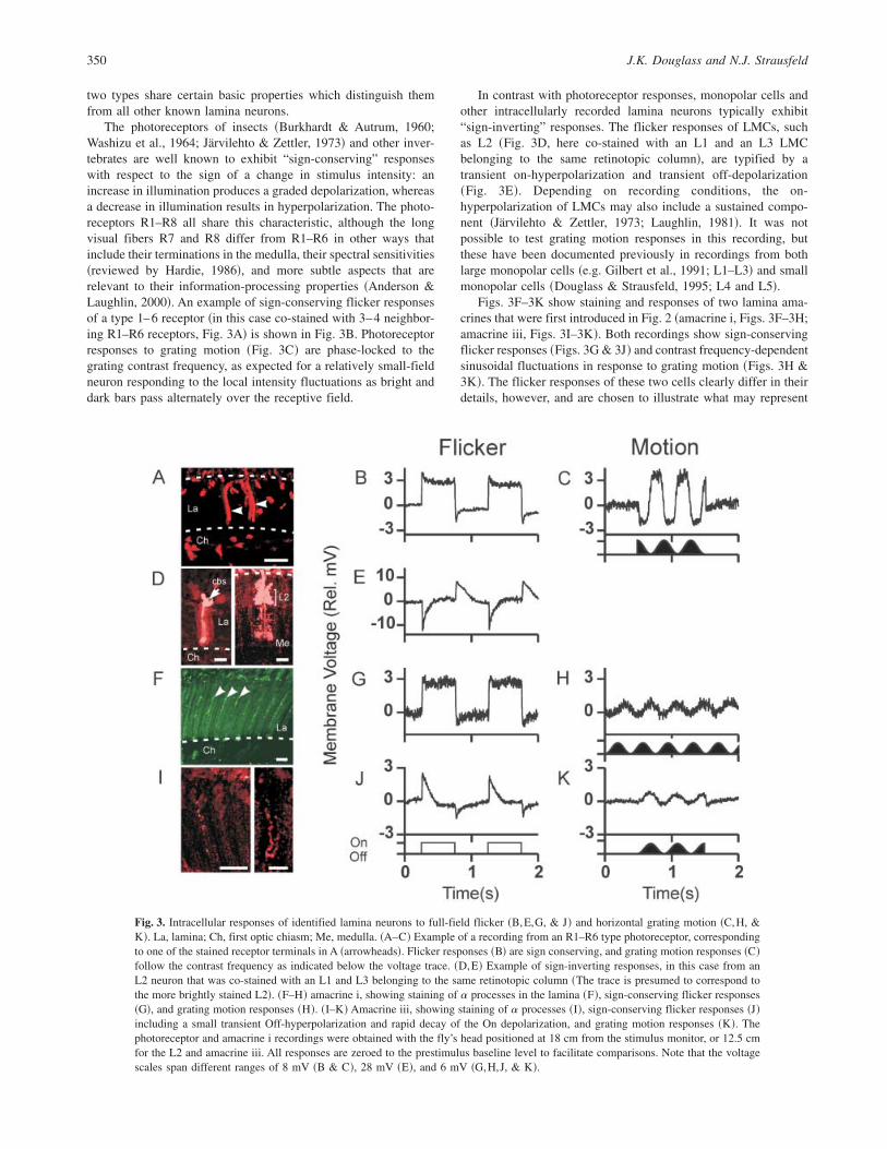
The photoreceptors of insects ~Burkhardt & Autrum, 1960;Washizu et al., 1964; Järvilehto & Zettler, 1973! and other inver-tebrates are well known to exhibit “sign-conserving” responseswith respect to the sign of a change in stimulus intensity: anincrease in illumination produces a graded depolarization, whereasa decrease in illumination results in hyperpolarization. The photo-receptors R1–R8 all share this characteristic, although the longvisual fibers R7 and R8 differ from R1–R6 in other ways thatinclude their terminations in the medulla, their spectral sensitivities~reviewed by Hardie, 1986!, and more subtle aspects that arerelevant to their information-processing properties ~Anderson &Laughlin, 2000!. An example of sign-conserving flicker responsesof a type 1–6 receptor ~in this case co-stained with 3–4 neighbor-ing R1–R6 receptors, Fig. 3A! is shown in Fig. 3B. Photoreceptorresponses to grating motion ~Fig. 3C! are phase-locked to thegrating contrast frequency, as expected for a relatively small-fieldneuron responding to the local intensity fluctuations as bright anddark bars pass alternately over the receptive field.
In contrast with photoreceptor responses, monopolar cells andother intracellularly recorded lamina neurons typically exhibit“sign-inverting” responses. The flicker responses of LMCs, suchas L2 ~Fig. 3D, here co-stained with an L1 and an L3 LMCbelonging to the same retinotopic column!, are typified by atransient on-hyperpolarization and transient off-depolarization~Fig. 3E!. Depending on recording conditions, the on-hyperpolarization of LMCs may also include a sustained compo-nent ~Järvilehto & Zettler, 1973; Laughlin, 1981!. It was notpossible to test grating motion responses in this recording, butthese have been documented previously in recordings from bothlarge monopolar cells ~e.g. Gilbert et al., 1991; L1–L3! and smallmonopolar cells ~Douglass & Strausfeld, 1995; L4 and L5!.
Figs. 3F–3K show staining and responses of two lamina ama-crines that were first introduced in Fig. 2 ~amacrine i, Figs. 3F–3H;amacrine iii, Figs. 3I–3K!. Both recordings show sign-conservingflicker responses ~Figs. 3G & 3J! and contrast frequency-dependentsinusoidal fluctuations in response to grating motion ~Figs. 3H &3K!. The flicker responses of these two cells clearly differ in theirdetails, however, and are chosen to illustrate what may represent
Fig. 3. Intracellular responses of identified lamina neurons to full-field flicker ~B,E,G, & J! and horizontal grating motion ~C,H, &K!. La, lamina; Ch, first optic chiasm; Me, medulla. ~A–C! Example of a recording from an R1–R6 type photoreceptor, correspondingto one of the stained receptor terminals in A ~arrowheads!. Flicker responses ~B! are sign conserving, and grating motion responses ~C!follow the contrast frequency as indicated below the voltage trace. ~D,E! Example of sign-inverting responses, in this case from anL2 neuron that was co-stained with an L1 and L3 belonging to the same retinotopic column ~The trace is presumed to correspond tothe more brightly stained L2!. ~F–H! amacrine i, showing staining of a processes in the lamina ~F!, sign-conserving flicker responses~G!, and grating motion responses ~H!. ~I–K! Amacrine iii, showing staining of a processes ~I!, sign-conserving flicker responses ~J!including a small transient Off-hyperpolarization and rapid decay of the On depolarization, and grating motion responses ~K!. Thephotoreceptor and amacrine i recordings were obtained with the fly’s head positioned at 18 cm from the stimulus monitor, or 12.5 cmfor the L2 and amacrine iii. All responses are zeroed to the prestimulus baseline level to facilitate comparisons. Note that the voltagescales span different ranges of 8 mV ~B & C!, 28 mV ~E!, and 6 mV ~G,H,J, & K!.
350 J.K. Douglass and N.J. Strausfeld
two extremes in the possible range of lamina amacrine responsesto abrupt changes in stimulus intensity. Amacrine i ~Fig. 3G!showed a sustained “On” depolarization and little or no “Off”hyperpolarization beyond the baseline. In amacrine iii ~Fig. 3J!,the On-depolarizations initially lacked a strong sustained compo-nent, instead showing a gradual decay toward the baseline voltagelevel, followed by a transient Off-hyperpolarization. Later in therecording from amacrine iii ~data not shown!, its depolarizingactivity became gradually more “sustained,” yet the hyperpolariz-ing Off transients persisted. In conclusion, the responses of laminaamacrines to wide-field flicker are fundamentally sign-conserving,as in photoreceptors. This basic characteristic has also been con-firmed in lamina amacrine ii, which was not stimulated withflicker, but nonetheless exhibited robust sign-conserving responsesto single bar motion ~see Fig. 5B below!.
Figs. 3C, 3H, and 3K compare the responses of the photorecep-tor and amacrines i and iii to movement of a wide-field verticalgrating. Although these amacrine motion responses are weak com-pared to the flicker responses, this appears to reflect the fact thatgrating motion was presented late in these two recordings, whenoverall response amplitudes were diminishing. Nevertheless, themere presence of voltage fluctuations at the grating contrast fre-quency provides an initial clue about the spatial receptive-fieldproperties of lamina amacrines. In general, if the wavelength of ahigh-contrast grating is far less than the diameter of a neuron’sprincipal receptive field, then grating motions should produce littleor no fluctuation in membrane voltage. If the grating wavelengthis much greater than the receptive-field diameter, one can expect tosee relatively sudden depolarizations and repolarizations, withintervening periods when the voltage remains relatively stable. Thepresent recordings suggest that in these two amacrines, as in thephotoreceptor, the approximate limits of the principal receptive-field region were roughly comparable to the grating stripe width~half of the grating wavelength!. The apparent stripe width de-pends on the viewing distance and the receptive-field location onthe stimulus screen, which was possible to estimate in somerecordings according to the responses to single bar motion ~seebelow!. For the receptor in Figs. 3A–3C, the apparent stripe widthwas approximately 9 deg. For amacrine i, the receptive-fieldlocation is unknown, but the grating dimensions at the center of thedisplay predict an upper limit of 10 deg for the apparent stripewidth. For amacrine iii ~from a fly that was positioned closer to thedisplay!, the apparent stripe width was approximately 14 deg. Thus,without embarking upon a more detailed analysis, the grating mo-tion responses in Fig. 3 suggest that lamina amacrines and photo-receptors have very comparable spatial receptive-field properties.
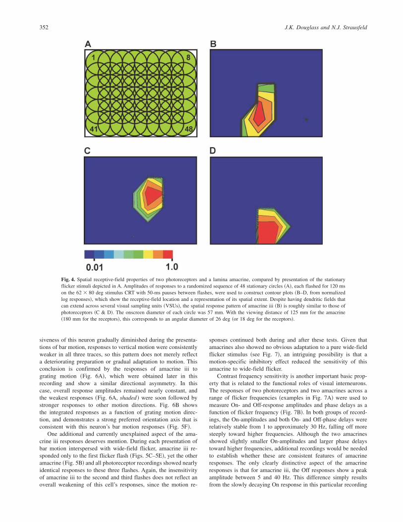
Two additional measures of spatial receptive-field propertiesare provided by responses to flashing stationary circular spots andto bar motion. Fig. 4A illustrates an 8 � 6 array of overlappingcircles which were presented in a pseudorandom sequence of 48individual “flicker” flashes ~for spot dimensions, see Fig. 4 cap-tion!. This stimulus has been used during several recordings fromphotoreceptors, and despite the large size of the flashed spotscompared to full-width half-maximum acceptance angles of lessthan 2 deg ~Burton et al., 2001!, it is possible for several of thespots to elicit a photoreceptor response due to their mutual overlapand the more extensive receptive field beyond the region ofgreatest sensitivity ~Smakman et al., 1984!. The stationary flashstimulus was also presented during the recording from amacrineiii. The contour plots in Fig. 4 compare its responses ~Fig. 4B! withthose of two identified photoreceptors ~Figs. 4C & 4D!. Like thegrating motion responses above, these results are suggestive of
similar spatial receptive fields in lamina amacrines and photorecep-tors. Due to the large size of the flash stimuli, however, these datastill only offer a low-resolution glimpse of actual receptive fields.Some experiments have also tested responses to spots as small as6 deg in diameter ~the smallest size that produces a reliableresponse due to the limited brightness of the CRT display!, but todate, none of these recordings has subsequently been identifiedwith an amacrine cell.
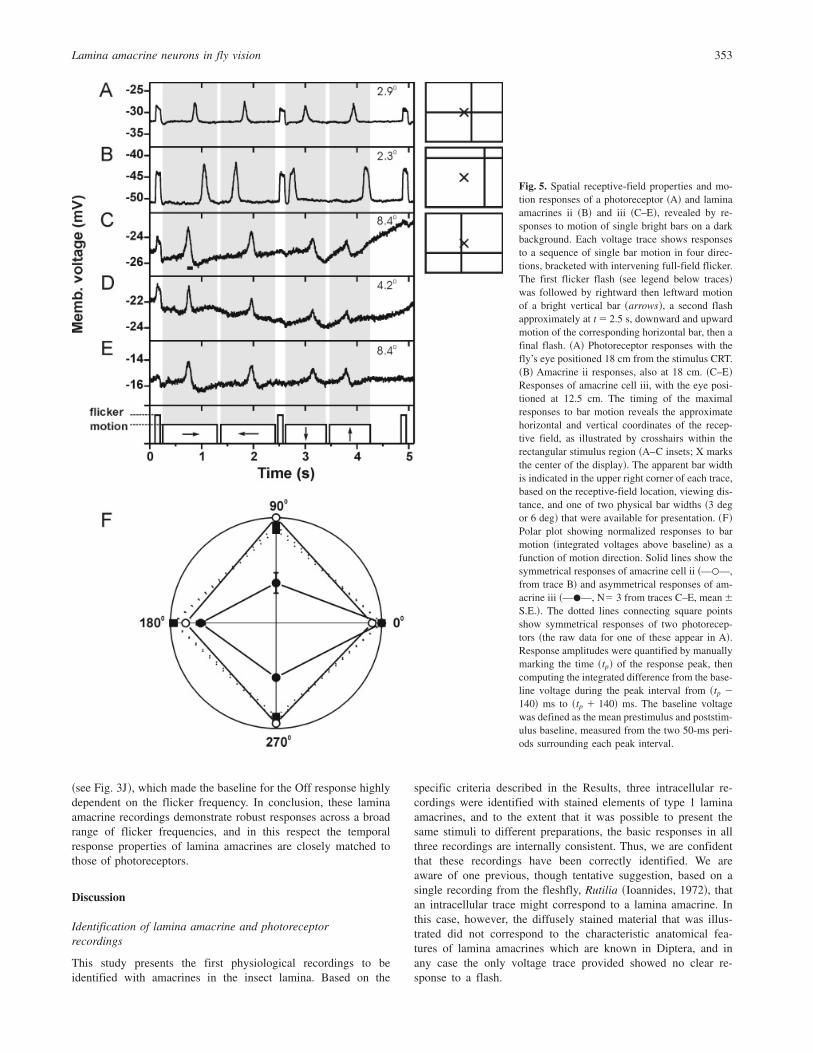
The most detailed information to date on the receptive-fieldproperties of amacrines has come from two recordings of re-sponses to single moving bars. The voltage traces in Fig. 5compare photoreceptor ~Fig. 5A!, amacrine ii ~Fig. 5B!, andamacrine iii ~Figs. 5C–5E! responses to a sequence of bar motionsin four directions; these were interspersed with full-field On andOff flicker stimuli. Ignoring the flicker responses for the moment,three distinct aspects of the bar motion responses reveal crucialfeatures of the recorded neurons’ response characteristics. First, thetiming of the maximal responses to bar motion discloses theapproximate horizontal and vertical coordinates of the receptivefield. These are illustrated in the insets to Figs. 5A–5C, andindicate that the receptive-field center of the photoreceptor ~Fig. 5A!viewed a point approximately 8 deg from the center of the CRT,whereas those of amacrine ii and iii ~Figs. 5B–5E! were some30 deg and 10 deg from the center, respectively.
A second useful aspect of the responses to bar motion is theirdurations; these provide information concerning the horizontal andvertical extent of the recorded neuron’s receptive-field as a func-tion of the width of a moving bar. The apparent width of a bar,however, depends not only on its fixed physical dimensions, butalso on the distance from the fly’s head to the CRT and the positionof each neuron’s receptive-field center. Thus, for the two physicalbar widths used in these experiments, these parameters were usedto compute the apparent bar widths shown in the upper rightcorner of each voltage trace. In general, the photoreceptor ~A! andamacrine ~B–E! response durations were similar, consistent withthe above lines of evidence that these two types of lamina neuronshare comparable spatial characteristics. Other recordings fromphotoreceptors ~not shown! have also shown similar responsedurations. A closer look at Fig. 5, however, suggests there may besome interesting differences among the receptive-field propertiesof these neurons. First, comparing the two amacrine recordings,the durations of responses in amacrine ii ~Fig. 5B! were about thesame as those of amacrine iii ~Figs. 5C–5E!, despite the substan-tially wider apparent bar widths seen by amacrine iii. If the twocells had identical receptive-field properties, one would expectamacrine ii to display the shortest response durations. Second,amacrine ii’s responses ~Fig. 5B! lasted as long or longer thanthose of the photoreceptor ~Fig. 5A!, despite the wider apparentbar width for the photoreceptor. These observations suggest that,on an individual basis, there may be receptive-field differencesboth among amacrines, and between amacrines and photoreceptors.
A third informative feature of the bar motion responses is theiramplitudes as a function of motion direction. Whereas amacrine iiand the two photoreceptors ~Figs. 5A and 5B! showed nearlysymmetrical responses to the four directions of bar motion, thethree repeated traces from amacrine iii consistently showed thelargest responses to horizontal motion. These patterns are quanti-fied in Fig. 5F, which represents all of the data in Figs. 5A–5E aswell as an additional dataset from a second R1–R6 photoreceptor.The polar plots ~Fig. 5F! confirm that the responses of amacrine iiiwere asymmetrical, with stronger responses to horizontal than tovertical motion. Although the resting potential and overall respon-
Lamina amacrine neurons in fly vision 351
siveness of this neuron gradually diminished during the presenta-tions of bar motion, responses to vertical motion were consistentlyweaker in all three traces, so this pattern does not merely reflecta deteriorating preparation or gradual adaptation to motion. Thisconclusion is confirmed by the responses of amacrine iii tograting motion ~Fig. 6A!, which were obtained later in thisrecording and show a similar directional asymmetry. In thiscase, overall response amplitudes remained nearly constant, andthe weakest responses ~Fig. 6A, shaded ! were soon followed bystronger responses to other motion directions. Fig. 6B showsthe integrated responses as a function of grating motion direc-tion, and demonstrates a strong preferred orientation axis that isconsistent with this neuron’s bar motion responses ~Fig. 5F!.
One additional and currently unexplained aspect of the ama-crine iii responses deserves mention. During each presentation ofbar motion interspersed with wide-field flicker, amacrine iii re-sponded only to the first flicker flash ~Figs. 5C–5E!, yet the otheramacrine ~Fig. 5B! and all photoreceptor recordings showed nearlyidentical responses to these three flashes. Again, the insensitivityof amacrine iii to the second and third flashes does not reflect anoverall weakening of this cell’s responses, since the motion re-
sponses continued both during and after these tests. Given thatamacrines also showed no obvious adaptation to a pure wide-fieldflicker stimulus ~see Fig. 7!, an intriguing possibility is that amotion-specific inhibitory effect reduced the sensitivity of thisamacrine to wide-field flicker.
Contrast frequency sensitivity is another important basic prop-erty that is related to the functional roles of visual interneurons.The responses of two photoreceptors and two amacrines across arange of flicker frequencies ~examples in Fig. 7A! were used tomeasure On- and Off-response amplitudes and phase delays as afunction of flicker frequency ~Fig. 7B!. In both groups of record-ings, the On-amplitudes and both On- and Off-phase delays wererelatively stable from 1 to approximately 30 Hz, falling off moresteeply toward higher frequencies. Although the two amacrinesshowed slightly smaller On-amplitudes and larger phase delaystoward higher frequencies, additional recordings would be neededto establish whether these are consistent features of amacrineresponses. The only clearly distinctive aspect of the amacrineresponses is that for amacrine iii, the Off responses show a peakamplitude between 5 and 40 Hz. This difference simply resultsfrom the slowly decaying On response in this particular recording
Fig. 4. Spatial receptive-field properties of two photoreceptors and a lamina amacrine, compared by presentation of the stationaryflicker stimuli depicted in A. Amplitudes of responses to a randomized sequence of 48 stationary circles ~A!, each flashed for 120 mson the 62 � 80 deg stimulus CRT with 50-ms pauses between flashes, were used to construct contour plots ~B–D, from normalizedlog responses!, which show the receptive-field location and a representation of its spatial extent. Despite having dendritic fields thatcan extend across several visual sampling units ~VSUs!, the spatial response pattern of amacrine iii ~B! is roughly similar to those ofphotoreceptors ~C & D!. The onscreen diameter of each circle was 57 mm. With the viewing distance of 125 mm for the amacrine~180 mm for the receptors!, this corresponds to an angular diameter of 26 deg ~or 18 deg for the receptors!.
352 J.K. Douglass and N.J. Strausfeld
~see Fig. 3J!, which made the baseline for the Off response highlydependent on the flicker frequency. In conclusion, these laminaamacrine recordings demonstrate robust responses across a broadrange of flicker frequencies, and in this respect the temporalresponse properties of lamina amacrines are closely matched tothose of photoreceptors.
Discussion
Identification of lamina amacrine and photoreceptorrecordings
This study presents the first physiological recordings to beidentified with amacrines in the insect lamina. Based on the
specific criteria described in the Results, three intracellular re-cordings were identified with stained elements of type 1 laminaamacrines, and to the extent that it was possible to present thesame stimuli to different preparations, the basic responses in allthree recordings are internally consistent. Thus, we are confidentthat these recordings have been correctly identified. We areaware of one previous, though tentative suggestion, based on asingle recording from the fleshfly, Rutilia ~Ioannides, 1972!, thatan intracellular trace might correspond to a lamina amacrine. Inthis case, however, the diffusely stained material that was illus-trated did not correspond to the characteristic anatomical fea-tures of lamina amacrines which are known in Diptera, and inany case the only voltage trace provided showed no clear re-sponse to a flash.
Fig. 5. Spatial receptive-field properties and mo-tion responses of a photoreceptor ~A! and laminaamacrines ii ~B! and iii ~C–E!, revealed by re-sponses to motion of single bright bars on a darkbackground. Each voltage trace shows responsesto a sequence of single bar motion in four direc-tions, bracketed with intervening full-field flicker.The first flicker flash ~see legend below traces!was followed by rightward then leftward motionof a bright vertical bar ~arrows!, a second flashapproximately at t � 2.5 s, downward and upwardmotion of the corresponding horizontal bar, then afinal flash. ~A! Photoreceptor responses with thefly’s eye positioned 18 cm from the stimulus CRT.~B! Amacrine ii responses, also at 18 cm. ~C–E!Responses of amacrine cell iii, with the eye posi-tioned at 12.5 cm. The timing of the maximalresponses to bar motion reveals the approximatehorizontal and vertical coordinates of the recep-tive field, as illustrated by crosshairs within therectangular stimulus region ~A–C insets; X marksthe center of the display!. The apparent bar widthis indicated in the upper right corner of each trace,based on the receptive-field location, viewing dis-tance, and one of two physical bar widths ~3 degor 6 deg! that were available for presentation. ~F!Polar plot showing normalized responses to barmotion ~integrated voltages above baseline! as afunction of motion direction. Solid lines show thesymmetrical responses of amacrine cell ii ~—�—,from trace B! and asymmetrical responses of am-acrine iii ~—�—, N� 3 from traces C–E, mean6S.E.!. The dotted lines connecting square pointsshow symmetrical responses of two photorecep-tors ~the raw data for one of these appear in A!.Response amplitudes were quantified by manuallymarking the time ~tp! of the response peak, thencomputing the integrated difference from the base-line voltage during the peak interval from ~tp �140! ms to ~tp � 140! ms. The baseline voltagewas defined as the mean prestimulus and poststim-ulus baseline, measured from the two 50-ms peri-ods surrounding each peak interval.
Lamina amacrine neurons in fly vision 353
Though the intracellularly stained profiles in Fig. 2 clearlybelong to lamina amacrines, it is unknown why it has not yet beenpossible to visualize an entire stained amacrine. Lamina amacrineshave also been known to stain incompletely with silver techniques~Shaw, 1981; N.J. Strausfeld, personal observation!. When a lam-ina amacrine neuron has been injected with dye, it is possible thatin the very thin processes that connect different portions of thecell, the dye concentrations are too low to be distinguished frombackground fluorescence. An unusual feature of the dipteran brainis that illumination with violet to blue wavelengths producesstrong background autofluorescence that obscures Lucifer yellowstained material ~Fig 2, panel i!. This problem is exacerbated in theperipheral optic lobe by the diffusion of ommochrome ocularaccessory pigments into neighboring tissues during fixation, and issomewhat alleviated by removing the retina during dissection. Asan alternative to Lucifer yellow, we have had good success atinjecting lamina cells with neurobiotin and subsequently visualiz-ing them with avidin-Texas Red ~Fig. 2, panels ii–iv!. Althoughour current procedure results in nonspecific fluorescence of glialcells and their cell bodies, as well as fainter staining of a subset ofvisual interneurons, the neurobiotin-injected material fluorescesparticularly brightly, and can be clearly distinguished from thebackground when viewed with the naked eye.
Maximal peak-to-peak responses from amacrines and R1–R6photoreceptors ~from ca. 4–8 mV! were observed in response tofull-field flicker or single bar motion at irradiances of approxi-mately 4 � 1010 q{cm�2{s�1 ~see Materials and methods!. Previ-ous investigators have reported obtaining similar responseamplitudes from R1–R6 receptors at dimmer flash intensities ~4–7mV responses at 1 to 2 � 109 q{cm�2{s�1; Scholes & Reichardt,1969; Laughlin & Hardie, 1978!. The discrepancy between theseirradiance values appears to be due to differences in stimulusconditions. Whereas these previous studies employed fully dark-adapted flies and stimulus wavelengths that were very close to the490 nm lmax of R1–R6 receptors ~Hardie, 1979!, the flies in the
present investigation were partly light adapted, and the spectraldistribution of our CRT stimulus lies predominately above the lmax
of R1–R6 receptors. As lamina amacrines receive their inputsexclusively from R1–R6 receptors, they are expected to share thesame spectral sensitivity. Thus, though the present results representonly one stimulus intensity, they suggest that unlike LMCs ~Laugh-lin & Hardie, 1978!, the absolute sensitivity of lamina amacrines issimilar to that of R1–R6 receptors.
Since the basic visual response properies of lamina amacrinesand photoreceptors are so similar, it is possible that some previ-ously published recordings that were thought to have come fromphotoreceptors may actually represent lamina amacrines. At present,however, this seems unlikely for both methodological and anatom-ical reasons. In experiments that target insect photoreceptors, thetypical procedure ~cf. Hardie, 1979; Anderson & Laughlin, 2000!has been to aim the pipette tip toward a preselected retinal regionvia a small cut at the surface of the compound eye. With thistechnique, one expects to impale those portions of photoreceptorsthat reside entirely within the retina ~namely, cell bodies or cyto-plasmic regions adjacent to the rhabdomeres!, and the pipette tipmay never reach the lamina. On the other hand, if the pipette tipdoes reach the lamina, or if the lamina is specifically targeted ~e.g.Scholes, 1969; Zettler & Weiler, 1976!, it still seems much morelikely that a large R1-R6 photoreceptor terminal would be impaledthan a thin amacrine process.
Sign-conserving and sign-inverting responsesof lamina neurons
Other than receptors, all of the eight types of lamina neurons thathave previously been identified by intracellular recordings and dyefills exhibit sign-inverting responses. The large monopolar cells~LMCs! L1–L3 ~Järvilehto & Zettler, 1973; Gilbert et al., 1991;Anderson & Laughlin, 2000!, the small monopolar cells ~SMCs!,
Fig. 6. Orientation- and weakly direction-selective responses of lamina amacrine iii to grating motion. ~A! three successivepresentations of wide-field grating motion in a sequence of eight directions. The raw data were high-pass fast Fourier-filtered to removeslow fluctuations below 1 Hz, then zeroed to a common baseline. Shaded bars indicate the two directions that produced the weakestresponses. ~B! Polar plot derived from the traces in A, showing normalized mean responses ~N � 3, mean 6 S.E.! as a function ofmotion direction. Each motion direction was presented for 1.0 s, preceded and followed by 1.0 s of a blank screen having the samemean luminance as the gratings. Response amplitudes were defined as the integrated rectified voltage ~a sum of both positive andnegative departures from the baseline! for 1100 ms from the start of each motion interval. The extra 100 ms allowed for recovery ofthe membrane voltage to baseline after cessation of motion. The preferred axis of orientation is at 159 deg ~arrowed dotted line!.
354 J.K. Douglass and N.J. Strausfeld
L4 and L5 ~Douglass & Strausfeld, 1995!, the “basket” T1 efferentneuron ~Järvilehto & Zettler, 1973; Douglass & Strausfeld, 1995!,the centrifugal neuron C2, and the type 1 lamina tangential cell~Lam Tan1; Strausfeld, 1970; Douglass & Strausfeld, 1995! allexhibit hyperpolarizing “On” responses and depolarizing “Off”responses. In contrast, the present results show that type 1 laminaamacrine cells have sign-conserving responses which are verymuch like those of photoreceptors.
Among all anatomically identified cell types that have pro-cesses within the lamina, only three have yet to be identifiedfollowing an intracellular recording: the type 2 lamina tangentialcell ~Lam Tan2!, the centrifugal neuron C3, and the type 2 laminaamacrine ~Am2!. Could any of the recordings we have attributed totype 1 lamina amacrines ~Am1! have arisen from Lam Tan 2, C3,or Am 2 neurons? This is unlikely, because it would be difficult tomistake even partial stainings of these neurons with an Am1 cell.Lam Tan 2 processes, like those of Am 2 ~Strausfeld & Nässel,1980!, reside only at the outer surface of the plexiform layer andshould be easily distinguished from staining of the a processes oftype 1 amacrines. Moreover, C3 and Lam Tan 2 have their prin-cipal dendritic fields in the inner and outer medulla, respectively,where they are likely to receive inputs from higher-order retino-topic neurons and thus should have significantly longer responselatencies than we have seen here.
Implications of sign-conserving responses of laminaamacrine neurons
The demonstration that lamina amacrines have sign-conservingresponses has interesting implications for the nature of neurotrans-mission from photoreceptors to amacrines. It is well establishedthat histamine is the neurotransmitter used by the photoreceptorsof various insects and other arthropods at synapses to LMCs~Nässel et al., 1988; Nässel, 1999; Stuart, 1999!, and that theinhibitory postsynaptic responses of LMCs to histamine are di-rectly gated by chloride channels ~Hardie, 1989; Geng et al.,2002!. In contrast, our data imply that neurotransmission fromphotoreceptors to lamina amacrines is excitatory. This suggeststhree basic hypotheses regarding the nature of photoreceptor-amacrine synapses: they could function via a metabotropic hista-mine receptor related to those of vertebrates, they could be electrical,or they could use an additional, nonhistamine neurotransmittersuch as acetylcholine. The evidence for metabotropic histaminereceptors in insects is controversial, with one molecular studysuggesting there is no arthropod homologue to vertebrate metab-otropic histamine receptors ~Roeder, 2003!. Although these obser-vations do not exclude the possibility that such receptors may haveevolved independently in vertebrates and invertebrates ~Roeder,2003!, the rapid flicker responses of lamina amacrines are incon-
Fig. 7. Responses of lamina amacrines and photorecep-tors as a function of contrast frequency. ~A! Rawresponse of a photoreceptor ~upper trace! and ama-crine i ~middle trace! to the full-field flicker rampstimulus ~bottom trace!. ~B! On- and Off-responseamplitudes ~upper plots! and phase delays ~lower plots!of two lamina amacrines ~solid lines! and photorecep-tors ~dashed lines!, measured from responses such asthose shown in A. The amplitudes in B are normalizedto individual response maxima.
Lamina amacrine neurons in fly vision 355
sistent with the slow temporal dynamics of conventional metabo-tropic receptors. This leaves the possibility of electrical synapsesor a second type of transmitter. But whereas anatomical studieshave described only chemical synapses between receptors andamacrines ~Boschek, 1971; Strausfeld & Campos-Ortega, 1977!,three studies provide evidence for the presence of acetylcholine ininsect photoreceptors. In honey bee, histochemical tests havedemonstrated acetylcholinesterase in the photoreceptors ~Kral &Schneider, 1981!. This enzyme has also been identified in devel-oping photoreceptors of mid-pupal Drosophila ~Wolfgang & Forte,1989!. Although the presence of the enzyme could underlie bio-chemical pathways unrelated to synaptic transmission, it is con-sistent with the possibility that fly photoreceptors employ a dualfast transmitter system. A more recent finding, that rhabdomericphotoreceptors of the Drosophila imago eyelet stain both forhistamine and with an antibody against Drosophila choline ace-tyltransferase ~Yasuyama & Meinertzhagen, 1999!, is of specialinterest in this regard. If this antibody has not clearly revealed theenzyme in other photoreceptors, this may only be a matter of itsrelative concentration.
As for neurons that receive inputs from lamina amacrines, theirresponses may be either sign reversing ~e.g. T1, Järvilehto &Zettler, 1973; Douglass & Strausfeld, 1995! or sign conserving~other type 1 lamina amacrines!. This suggests that amacrineoutputs also may involve more than one neurotransmitter or trans-mitter pathway. Currently, evidence that lamina amacrines areglutamate immunoreactive ~Sinakevitch & Strausfeld, 2004! sug-gests that glutamate alone could serve both excitatory and inhib-itory roles, as has been suggested for the outputs from mammalianphotoreceptors to On- and Off-bipolar cells ~Bloomfield & Dacheux,2001!. The situation in flies is complicated, however, by the factthat T1 also receives some inputs from photoreceptors ~Campos-Ortega & Strausfeld, 1973!.
Functional properties and possible functional roles oflamina amacrine neurons
Until now, the observation that amacrine dendritic fields canextend across several VSUs, coupled with the presence of connec-tions among neighboring amacrines, has led many workers ~in-
cluding ourselves! to expect their receptive fields to encompassmore than a single VSU. In a notable exception to this point ofview, Shaw ~1981! debated whether lamina amacrines might ex-hibit some degree of signal decrement that could result in func-tional isolation of individual a processes. Here, we have shownthat amacrine responses to grating motion, stationary spots, andbar motion are all similar to those of photoreceptors. Given thelarger dendritic fields of amacrines, these results suggest thatlamina amacrines have fairly short space constants, meaning thatpassive conduction of local voltage fluctuations may decrementquite rapidly and not be strongly propagated to neighboring car-tridges. This contrasts with vertebrate horizontal cells, whichoccupy an analogous position with respect to photoreceptor inputsbut have receptive fields which are considerably wider than theirdendritic fields ~e.g. Tomita, 1965; Naka & Rushton, 1967; Lamb,1976; Quian & Ripps, 1992!. Nevertheless, the receptive-fieldinformation provided by the current results is constrained by therelatively low stimulus intensities provided by a CRT display. Infuture experiments, we plan to employ much brighter point-sourcestimuli which are more comparable to natural irradiances and mayreveal more extensive spatial receptive fields.
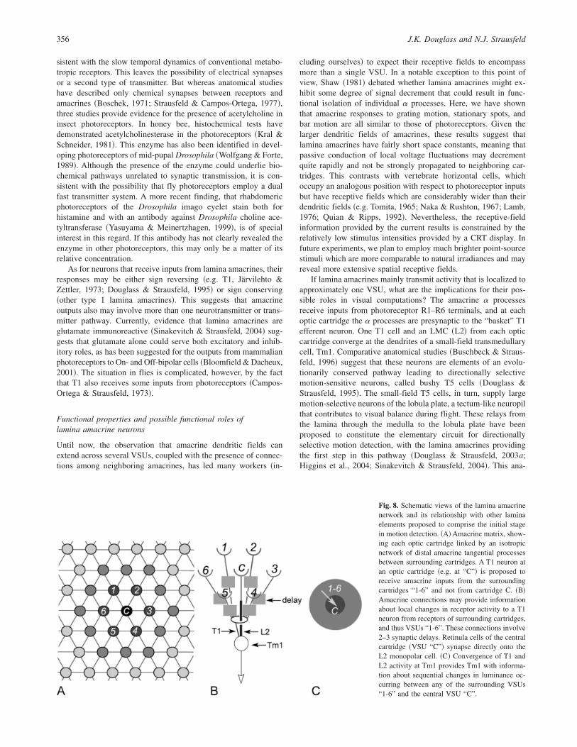
If lamina amacrines mainly transmit activity that is localized toapproximately one VSU, what are the implications for their pos-sible roles in visual computations? The amacrine a processesreceive inputs from photoreceptor R1–R6 terminals, and at eachoptic cartridge the a processes are presynaptic to the “basket” T1efferent neuron. One T1 cell and an LMC ~L2! from each opticcartridge converge at the dendrites of a small-field transmedullarycell, Tm1. Comparative anatomical studies ~Buschbeck & Straus-feld, 1996! suggest that these neurons are elements of an evolu-tionarily conserved pathway leading to directionally selectivemotion-sensitive neurons, called bushy T5 cells ~Douglass &Strausfeld, 1995!. The small-field T5 cells, in turn, supply largemotion-selective neurons of the lobula plate, a tectum-like neuropilthat contributes to visual balance during flight. These relays fromthe lamina through the medulla to the lobula plate have beenproposed to constitute the elementary circuit for directionallyselective motion detection, with the lamina amacrines providingthe first step in this pathway ~Douglass & Strausfeld, 2003a;Higgins et al., 2004; Sinakevitch & Strausfeld, 2004!. This ana-
Fig. 8. Schematic views of the lamina amacrinenetwork and its relationship with other laminaelements proposed to comprise the initial stagein motion detection. ~A!Amacrine matrix, show-ing each optic cartridge linked by an isotropicnetwork of distal amacrine tangential processesbetween surrounding cartridges. A T1 neuron atan optic cartridge ~e.g. at “C”! is proposed toreceive amacrine inputs from the surroundingcartridges “1-6” and not from cartridge C. ~B!Amacrine connections may provide informationabout local changes in receptor activity to a T1neuron from receptors of surrounding cartridges,and thus VSUs “1-6”. These connections involve2–3 synaptic delays. Retinula cells of the centralcartridge ~VSU “C”! synapse directly onto theL2 monopolar cell. ~C! Convergence of T1 andL2 activity at Tm1 provides Tm1 with informa-tion about sequential changes in luminance oc-curring between any of the surrounding VSUs“1-6” and the central VSU “C”.
356 J.K. Douglass and N.J. Strausfeld
tomically and physiologically inspired circuit involves multiplelevels, thus departing from the traditional Reichard-Hassensteinmodel, which supposes that both the presence of motion and itsdirection are detected in a single step. The central hypothesis~Fig. 8! is that amacrine processes converging from the immedi-ately surrounding optic cartridges drive the T1 pathway of acentral optic cartridge, while the photoreceptors supplying thiscentral cartridge drive its L2 monopolar cell. The present evidencefor spatially localized activity in lamina amacrines appears to beconsistent with the proposal that amacrine activity from neighbor-ing optic cartridges converges to a central T1 cell. This does notexclude the possibility that inputs from amacrines to T1, L4, or L5may also produce lateral inhibitory effects, for which there is someevidence in T1 based on evidence its receptive field is narrowerthan those of receptors ~Järvilehto & Zettler, 1973!.
In summary, the present recordings reveal several basic visualresponse properties of lamina amacrines which are relevant totheir possible functional role~s!. These properties include fast,photoreceptor-like responses to flicker, and spatial properties thatare very localized and thus suggestive of passive conductionproperties. Although our contrast frequency data were obtained byusing a square-wave flicker stimulus, they agree qualitatively withpreviously published data from dipteran photoreceptors and LMCsinvolving either sinusoidally varied ~Järvilehto & Zettler, 1973;Coombe et al., 1989! or pseudorandomly modulated ~Juusola et al.,1994; Anderson & Laughlin, 2000! flicker stimulation. If laminaamacrines were to play a neuromodulatory role related to long-term aspects of light and dark adaptation and0or circadian rhythms,they would not necessarily need fast temporal properties, andmight be expected to have larger receptive fields. Although theproperties demonstrated here are consistent with a variety ofcomputational roles including lateral inhibition, light0dark adap-tation, motion processing, or edge processing, the only directphysiological evidence so far comes from the demonstration oforientation-selective motion responses, suggesting the involve-ment of lamina amacrines in processing the orientations of movingedges.
Acknowledgments
We would like to thank Drs. Konrad E. Zinsmaier and Charles M. Higginsfor discussions, and two anonymous reviewers for their suggestions. Thisresearch was supported by NIH R01-RR08868.
References
Anderson, J.C. & Laughlin, S.B. ~2000!. Photoreceptor performanceand the co-ordination of achromatic and chromatic inputs to the flyvisual system. Vision Research 40, 13–31.
Bloomfield, S.A. & Dacheux, R.F. ~2001!. Rod vision: Pathways andprocessing in the mammalian retina. Progress in Retinal and EyeResearch 20, 351–384.
Boschek, C.B. ~1971!. On the fine structure of the peripheral retina and thelamina of the fly, Musca domestica. Zeitschrift für Zellforschung undMikroskopische Anatomie 110, 369–409.
Braitenberg, V. ~1967!. Patterns of projection in the visual system of thefly. I. Retina-lamina projections. Experimental Brain Research 3,271–298.
Burkhardt, D. & Autrum, H. ~1960!. Die Belichtungspotentiale einzel-ner Sehzellen von Calliphora erythrocephala Meig. Zeitschrift fürNaturforschung 15b, 612–616.
Burton, B.G., Tatler, B.W. & Laughlin, S.B. ~2001!. Variations inphotoreceptor response dynamics across the fly retina. Journal ofNeurophysiology 86, 950–960.
circuits in flies: Small-field retinotopic elements responding to motionare evolutionarily conserved across taxa. Journal of Neuroscience 16,4563–4578.
Campos-Ortega, J.A. & Strausfeld, N.J. ~1973!. Synaptic connectionsof intrinsic cells and basket arborizations in the external plexiformlayer of the fly’s eye. Brain Research 59, 119–136.
Coombe, P., Srinivasan, M.V. & Guy, R.G. ~1989!. Are the large mono-polar cells of the insect lamina on the optomotor pathway? Journal ofComparative Physiology A 166, 23–35.
Douglass, J.K. & Strausfeld, N.J. ~1995!. Visual motion detectioncircuits in flies: Peripheral motion computation by identified small-field retinotopic neurons. Journal of Neuroscience 15, 5596–5611.
Douglass, J.K. & Strausfeld, N.J. ~1996!. Visual motion-detectioncircuits in flies: Parallel direction- and non-direction-sensitive path-ways between the medulla and lobula plate. Journal of Neuroscience16, 4551–4562.
Douglass, J.K. & Strausfeld, N.J. ~1998!. Functionally and anatomi-cally segregated visual pathways in the lobula complex of a calliphoridfly. Journal of Comparative Neurology 396, 84–104.
Douglass, J.K. & Strausfeld, N.J. ~2003a!. Anatomical organization ofretinotopic motion-sensitive pathways in the optic lobes of flies. Mi-croscopy Research and Technique 62, 132–150.
Douglass, J.K. & Strausfeld, N.J. ~2003b!. Retinotopic pathways pro-viding motion-selective information to the lobula from peripheralelementary motion detecting circuits. Journal of Comparative Neurol-ogy 456, 326–344.
Franceschini, N. ~1975!. Sampling of the visual environment by thecompound eye of the fly: Fundamentals and applications. In Photo-receptor Optics, ed. Snyder, A.W. & Menzel, R., pp. 8–125. Berlin,Heidelberg, New York: Springer.
Geng, C., Leung, H.-T,, Skingsley, D.R., Iovchev, M.I., Yin, Z., Se-menov, E.P,, Burg, M.G., Hardie, R.C. & Pak, W.L. ~2002!. Thetarget of Drosophila photoreceptor synaptic transmission is a histamine-gated chloride channel encoded by ort ~hclA!. Journal of BiologicalChemistry 277, 42113–42120.
Gilbert, C., Penisten, D.K. & DeVoe, R.D. ~1991!. Discrimination ofvisual motion from flicker by identified neurons in the medulla of thefleshfly Sarcophaga bullata. Journal of Comparative Physiology A168, 653–673.
Hardie, R.C. ~1979!. Electrophysiological analysis of fly retina. I. Com-parative properties of R1-R6 and R7 and 8. Journal of ComparativePhysiology A 129, 19–33.
Hardie, R.C. ~1986!. The photoreceptor array of the dipteran retina. Trendsin Neuroscience 9, 419–423.
Hardie, R.C. ~1989!. A histamine-activated chloride channel involved inneurotransmission at a photoreceptor synapse. Nature ~London! 339,704–706.
Hengstenberg, R. ~1982!. Common visual response properties of giantvertical cells in the lobula plate of the blowfly Calliphora. Journal ofComparative Physiology 149, 179–193.
Higgins, C.M., Douglass, J.K. & Strausfeld, N.J. ~2004!. The compu-tational basis of an identified neuronal circuit for elementary motiondetection in dipterous insects. Visual Neuroscience 21, 567–586.
Ioannides, A.C. ~1972! Light adaptation and signal transmission in thecompound eye of the giant water bug Lethocerus (Belastomatidae:Hemiptera). Ph.D. Thesis, Canberra National University, Australia.
Järvilehto, M. & Zettler, F. ~1973!. Electrophysiological-histologicalstudies on some functional properties of visual cells and second-orderneurons of an insect retina. Zeitschrift für Zellforschung und Mikrosko-pische Anatomie 136, 291–306.
Juusola, M., Kouvalainen, E., Järvilehto, M. & Weckström, M.~1994!. Contrast gain, signal-to-noise ratio, and linearity in light-adapted blowfly photoreceptors. Journal of General Physiology 104,593–621.
Kral, K. & Schneider, L. ~1981!. Fine structural localisation of acetyl-cholinesterase activity in the compound eye of the honeybee ~Apismellifica L.!. Cell and Tissue Research 221, 351–359.
Lamb, T.D. ~1976!. Spatial properties of horizontal cell responses in theturtle retina. Journal of Physiology 263, 239–255.
Laughlin, S.B. ~1981!. Neural principles in the peripheral visual systemsof invertebrates. In Handbook of Sensory Physiology, VII/6B, ed.Autrum, H., pp. 133–280. Berlin: Springer.
Laughlin, S.B. & Hardie, R.C. ~1978!. Common strategies for lightadaptation in the peripheral visual systems of fly and dragonfly. Jour-nal of Comparative Physiology 128, 319–340.
Lamina amacrine neurons in fly vision 357
Meinertzhagen, I.A. & O’Neil, S.D. ~1991!. Synaptic organization ofcolumnar elements in the lamina of the wild-type in Drosophila mel-anogaster. Journal of Comparative Neurology 305~2!, 232–263.
Naka, K.I. & Rushton, W.A.H. ~1967!. The generation and spread ofs-potentials in fish ~cyprinidae!. Journal of Physiology 192, 437–461.
Nässel, D.R. ~1999!. Histamine in the brain of insects: A review. Micros-copy Research and Technique 44, 121–136.
Nässel, D.R., Holmqvist, M.H., Hardie, R.C., Hakanson, R. & Sund-ler, F. ~1988!. Histamine-like immunoreactivity in photoreceptors ofthe compound eyes and ocelli of the flies Calliphora erythrocephalaand Musca domestica. Cell and Tissue Research 253, 639–646.
O’Shea, M. & Adams, M.E. ~1981!. Pentapeptide ~proctolin! associatedwith an identified neuron. Science 21, 567–569.
Quian, H. & Ripps, H. ~1992!. Receptive-field properties of rod-drivenhorizontal cells in the skate retina. Journal of General Physiology 100,457–478.
Roeder, T. ~2003!. Metabotropic histamine receptors—nothing for inver-tebrates? European Journal of Pharmacology 466, 85–90.
Scholes, J. ~1969!. The electrical responses of the retinal receptors and thelamina in the visual system of the fly Musca. Kybernetik 6, 149–162.
Scholes, J. & Reichardt, W. ~1969!. The quantal content of optomotorstimuli and the electrical responses of receptors in the compound eye ofthe fly Musca. Kybernetik 6, 74–79.
Shaw, S.R. ~1981!. Anatomy and physiology of identified non-spikingcells in the photoreceptor-lamina complex of the compound eye ofinsects, especially Diptera. In Neurones Without Impulses: Their Sig-nificance for Vertebrate and Invertebrate Nervous Systems, eds. Rob-erts, A. & Bush, B.M.H., pp. 61–116. Cambridge: CambridgeUniversity Press.
Shaw, S.R. ~1984!. Early visual processing in insects. Journal of Experi-mental Biology 112, 225–251.
Sinakevitch, I. & Strausfeld, N.J. ~2004!. Chemical neuroanatomy ofthe fly’s movement detection pathway. Journal of Comparative Neu-rology 468, 6–23.
Smakman, J.G.J., van Hateren, J.H. & Stavenga, D.G. ~1984!. Angularsensitivity of blowfly photoreceptors: Intracellular measurements andwave-optical predictions. Journal of Comparative Physiology A 155,239–247.
Strausfeld, N.J. ~1970!. Golgi studies on insects. Part II. The optic lobes
of Diptera. Philosophical Transactions of the Royal Society B ~London!258, 135–223.
Strausfeld, N.J. ~1976!. Mosaic organizations, layers, and visual path-ways in the insect brain. In Neural Principles in Vision, ed. Zettler,F. & Weiler, R., pp. 245–279. Berlin, Heidelberg, New York: Springer.
Strausfeld, N.J. ~1989!. Beneath the compound eye: Neuroanatomicalanalysis and physiological correlates in the study of insect vision. InFacets of Vision, ed. Stavenga, D.G. & Hardie, R.C., pp. 317–359.Heidelberg: Springer.
Strausfeld, N.J. & Campos-Ortega, J.A. ~1977!. Vision in insects:Pathways possibly underlying neural adaptation and lateral inhibition.Science 195, 894–897.
Strausfeld, N.J. & Li, Y. ~1999!. Organization of olfactory and multi-modal afferent neurons supplying the calyx and pedunculus of thecockroach mushroom bodies. Journal of Comparative Neurology 409,603–625.
Strausfeld, N.J. & Nässel, D.R. ~1980!. Neuroarchitecture of brainregions that subserve the compound eyes of Crustacea and Insects. InHandbook of sensory physiology, VII/6B, ed. Autrum, H., pp. 1–133.Heidelberg: Springer.
Stuart, A.E. ~1999!. From fruit flies to barnacles, histamine is theneurotransmitter of arthropod photoreceptors. Neuron 22, 431–433.
Tomita, T. ~1965!. Electrophysiological study of the mechanisms subserv-ing color coding in the fish retina. Cold Spring Harbor Symposia onQuantitative Biology 30, 559–566.
Washizu, Y., Burkhardt, D. & Streck, P. ~1964!. Visual field of singleretinula cells and interommatidial inclination in the compound eye ofthe blowfly Calliphora erythrocephala. Zeitschrift für VergleichendePhysiologie 48, 413–428.
Wolfgang, W.J. & Forte, M.A. ~1989!. Expression of acetylcholinester-ase during visual system development in Drosophila. DevelopmentalBiology 131, 321–330.
Yasuyama, K. & Meinertzhagen, I.A. ~1999!. Extraretinal photorecep-tors at the compound eye’s posterior margin in Drosophila melanogas-ter. Journal of Comparative Neurology 412, 193–202.
Zettler, F. & Weiler, R. ~1976!. Neuronal processing in the first opticneuropile of the compound eye of the fly. In Neural Principles inVision, ed. Zettler, F. & Weiler, R., pp. 227–237. New York:Springer.