10
Silhouette Sign
| Date post: | 16-Dec-2015 |
| Category: |
Documents |
| Upload: | hugo-berks |
| View: | 215 times |
| Download: | 1 times |

Silhouette Sign

Frontal X-ray Signs of Lobar Consolidation
• RUL – loss of upper right mediastinal border
• RML – loss of right heart border• RLL – loss of right hemidiaphram• LUL – loss of upper left mediastinal
border• LINGULA – loss of left heart border• LLL – loss of left hemidiaphram

Frontal X-ray Signs of Lobar Consolidation
• SILHOUETTE SIGN – for localizing lesion from frontal chest film. Intra-thoracic air space occupying lesions touching a soft tissue structure will obliterate the air soft tissue interface
• Examples include:– Atelectasis– Pneumonia– Neoplasm

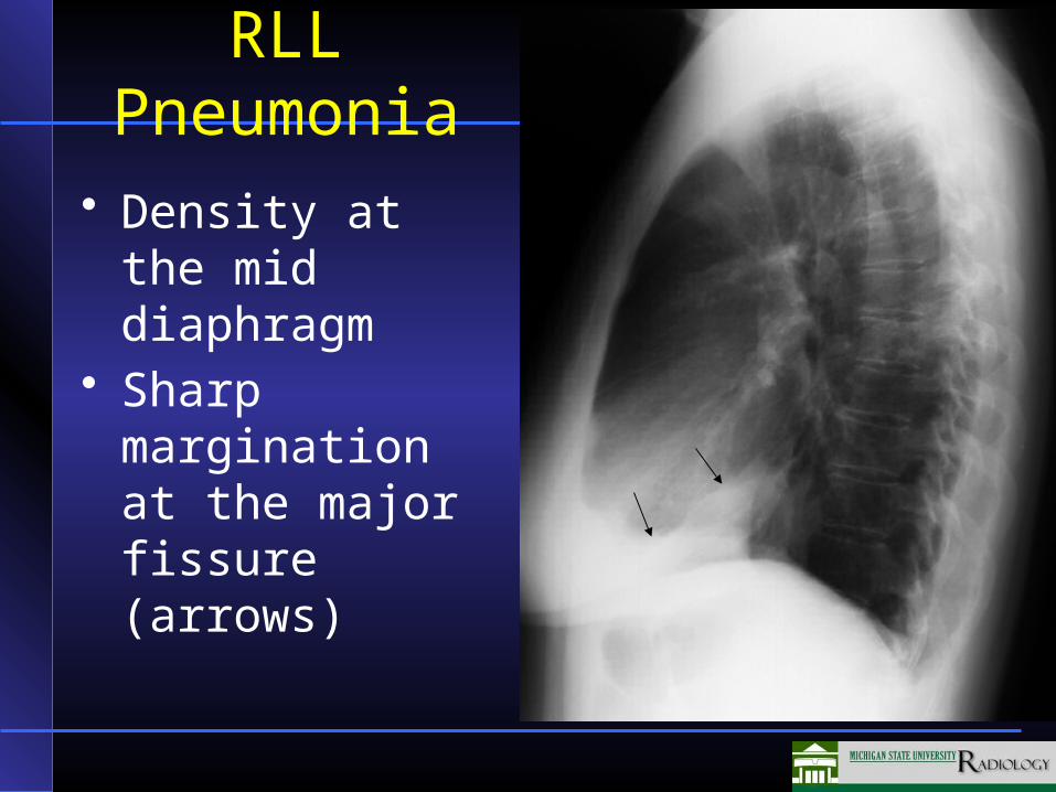
RLL Pneumonia
• Density at the right lateral diaphragm
• Obliteration of lateral diaphragm border
RLL Pneumonia
• Density at the mid diaphragm
• Sharp margination at the major fissure (arrows)

Right Middle Lobe Pneumonia

LUL Lingular
Pneumonia
• Obliterated left cardiac border

LULLingular
Pneumonia Lateral
• Consolidation anterior to the major fissure
• Compare to PA exam

LLL Pneumonia
• Air space disease left lower lobe
• Density behind heart
• Obliteration of left diaphragm at edge of heart
• Left heart border preserved

LLL Pneumonia• Note
obliteration of the posterior portion of the left diaphragm (arrows)
• Right diaphragm clearly seen


