Silicon, silica and its surface patterning/activation with alkoxy- and amino-silanes for nanomedical applications
Silicon & silica � Silicon
Silicon is the second most abundant element on earth (26.3%), preceded only by oxygen (48.9%). The two elements have a great affinity for each other and, thus, are almost always found combined in the form of silicates [1]. Silicates are derived from the anhydride silicon dioxide or sil-ica (SiO
2), which is the main constituent of the
earth’s crust (60.6%). The term silica denotes silicon dioxide in all of its forms – crystalline, amorphous, soluble and combined.
Compared with its congener carbon, the chemistry of silicon is characterized by its signif icantly larger van der Waals radius and relatively low electronegativity (rw
Si/C
= 210/170 pm; ENSi/C
= 1.74/2.50, respec-tively) [2,3]. This has consequent implications for the polarity of bonds formed between sili-con and other elements, where coordination is generally tetravalent with silicon usually
positively charged. The element also shows a very poor tendency to form π-bonds, which are generally unstable and highly reactive. In addition, the electronegativity of silicon is lower than that of hydrogen (EN
H = 2.20), causing a
reversal of bond polarization when comparing C-H and Si-H in methane (CH
4) and silane
(SiH4). Such compounds therefore exhibit dif-
ferent reactivities (e.g., hydrogen evolution on reaction with an acid H+ with SiH
4). For more
electronegative elements (X), compounds pos-sessing Si-X or Si-X-Si in many cases exhibit, in contradiction to the rule, higher bind-ing energies and shorter bond lengths (e.g., C-O/Si-O = 358/444 kJ mol-1; C-F/Si-F = 489/595 kJ mol-1) [2]. This provides the driv-ing force in many of the reactions of silanes with oxygen and fluoride nucleophiles. Another consequence of this is the so-called ‘β-effect’ stabilization of a positive charge on a carbon atom β to silicon [4].
Silica and silicates are widely used in nanomedicine with applications as diverse as medical device coatings to replacement materials in tissue engineering. Although much is known about silica and its synthesis, relatively few biomedical scientists fully appreciate the link that exists between its formulation and its resultant structure and function. This article attempts to provide insight into relevant issues in that context, as well as highlighting their importance in the material’s eventual surface patterning/activation with alkoxy- and organo-silanes. The use of aminosilanes in that context is discussed at some length to permit an understanding of the specific variables that are important in the reproducible and robust aminoactivation of surfaces using such molecules. Recent investigative work is cited to underline the fact that although aminosilanization is a historically accepted mechanism for surface activation, there is still much to be explained about how and why the process works in the way it does. In the last section of this article, there is a detailed discussion of two classical approaches for the use of aminosilanized materials in the covalent immobilization of bioligands, amino-aldehyde and amino-carboxyl coupling. In the former case, the use of the homobifunctional coupler glutaraldehyde is explored, and in the latter, carbodiimides. Although these chemistries have long been employed in bioconjugations, it is apparent that there are still variables to be explored in the processes (as witnessed by continuing investigations into the chemistries concerned). Aspects regarding optimization, standardization and reproducibility of the fabrication of amino functionalized surfaces are discussed in detail and illustrated with practical examples to aid the reader in their own studies, in terms of considerations to be taken into account when producing such materials. Finally, the article attempts to remind readers that although the chemistry and materials involved are ‘old hat’, there is still much to be learnt about the methods involved. The article also reminds readers that although many highly specific and costly conjugation chemistries now exist for bioligands, there still remains a place for these relatively simple and cost-effective approaches in bioligand conjugate fabrication.
Dag Rother1,2, Tapas Sen1,3, Daniel East1 & Ian James Bruce†1
1Nanobiotechnology Research Group, School of Biosciences, University of Kent, Canterbury, Kent, CT2 7NJ, UK 2Bundesanstalt für Arbeitsschutz und Arbeitsmedizin, Freidrich-Henkel-Weg 1-25 D-44149 Dortmund, Germany 3Centre for Materials Science, School of Forensic & Investigative Sciences, University of Central Lancashire, Preston, PR1 2HE, UK †Author for correspondence:Tel.: +44 122 782 4557 Fax: +44 122 782 4659 [email protected]
SPECIAL FOCUS y New Nanomaterials for Forensics & Diagnostics
� SilicaUnsurprisingly, the chemistry of silicon can be largely characterized by its affinity for oxy-gen and thereby its existence as silica, where Si is generally tetrahedrally coordinated with four oxygen atoms. Silicates can be described as derivatives of the relatively weak acid mono silic acid (H
4SiO
4), which possesses a
very strong tendency for elimination of water, causing intermolecular condensation/poly-merization leading to the formation of many different types of branched silicate networks. Silicate types range from single (or island) SiO
4
groups and pairs to finite clusters, including rings, chains, double chains and sheets, up to 3D frameworks of SiO
2 (Figure 1) [2]. The anhy-
dride silicon dioxide, especially in its crystal-line state, is relatively inert and only attacked by hydrofluoric acid or very slowly by aqueous alkalis [2]. Chemically unmodified silicates and their surfaces display several properties that make them useful materials for a variety of biomedical applications. These are that silica is biocompatible (i.e., nontoxic and does not easily undergo degradation under physio logical conditions) and that SiO
2 surfaces display a
high level of mechanical and thermal robust-ness, as well as resistance to a wide range of pHs and chemical conditions.
Silica is a common substrate used in nano-medicine as either a flat or particulate material and its synthesis is commonly reliant on ‘sol-gel’
processes [5,6]. A ‘sol’ is referred to as a stable solution of colloidal particles (i.e., dispersed particles of amorphous silica) with a size of between approximately 5 and 200 nanometers. Crosslinking of such colloidal particles by poly-condensation reactions can rigidify the sol so it then becomes a ‘gel’, which is defined as a 3D network contained within a liquid medium, which exhibits no flow in the steady state [7]. The sol-gel process is especially useful in areas such as thin-film formation, surface patterning and surface modification at the nanoscale [8], as well as synthesis of silica particles of control-led shape, size and size distribution. The goals of sol-gel processing in general are to control the nanostructure, composition homogeneity and the surface and interface properties of sili-cate materials during the earliest stages of their p roduction [9,10].
Sol-gel silicate synthesis is a relatively simple and versatile process [11]. The synthesis is simple as it is usually carried out under mild condi-tions, such as low temperature and pressure, often under ambient conditions. The synthesis is versatile as a wide range of structurally different silicates, from amorphous to ordered products, can be synthesized by one general approach. The first report of a sol-gel reaction dates back to the mid-1800s, when Ebelman observed the forma-tion of a glass-like substance at the neck of a bottle of tetraethylorthosilicate (TEOS), which had been accidentally left unsealed for several
RO
RO
RO
ORSi
RO
RO
RO
OH
OR
OR
OR
OR
OR
OR
OR
OR
Si
HO
RO
RO
RO
RO
RO
R
RORO
OHSi
Si
Si
Si
Si
Si Si
Si
Si
HO
RO
HO
OHSi
HO
HO
HO
OHSi
RO
RO
RO
O
O
O
O
O
O
O
O
Si Si
OH
OR
OR
RO
RO
RO
OSi SiH2O H2O
H2O
H2O
H2O
n
Chains
Rings
n
Figure 1. Polymerization/condensation of silica by sol-gel synthesis from a tetraalkoxysilane precursor.Adapted from [140].
www.futuremedicine.com 283future science group
Silicon, silica & its aminosilanization in nanomedicine Review
weeks under ambient conditions [5,12]. Sol-gel chemistry usually employs a tetraalkoxysilane as a precursor to silica synthesis and TEOS pos-sessing the formula Si(OC
2H
5)
4 (Figure 2) is a pro-
totypical tetraalkoxide/alkoxysilane (see later; Table 1). It consists of four ethyl groups attached to an orthosilicate ion, SiO
44- and rapidly con-
verts into silicon dioxide upon addition of water (in the presence of an acid or base). TEOS is the most commonly used precursor for the synthesis of silica by sol-gel chemistry.
The full potential of sol-gel chemistry remained underutilized until the invention of the base catalyzed synthesis of silica particles from alkoxysilanes, with sizes ranging from 10 nm to 2µm. This process is now commonly referred to as the Stöber process [13], named after one of the inventors, and represents probably the most prevalent method for the synthesis of col-loidal silica particles [14]. It is also the primary route for the synthesis of nanoparticulate silica for biomedical applications [15,16].
While a number of different other routes exist for the synthesis of silica [2], the converse to base catalysis is the use of acid and both reac-tion mechanisms are described in Figure 3 for the hydrolysis of silane alkoxy groups.
The basis of sol-gel chemistry is the poly-condensation/polymerization of monosilicic acid, leading to the formation of silicon–oxy-gen–silicon bonds (siloxane rather than silicon– silicon bonds), thus generating a silica sol, which, driven by ongoing intramolecular elimination of water, then forms a polymeric network of col-loidal silica gel. The most investigated route for silica synthesis uses alkoxide (or alkoxysilane) precursors (Si[-OR]
4; R = alkyl), which may
also include organically modified alkoxysilanes (organosilanes like R -́Si[-OR]
3, where R´ is the
organic moiety). Sol-gel chemistry also allows the combination/mixing of alkoxy- and organo-silanes (see later), the latter of which can possess a broad range of chemical functionalities, thus offering a very convenient route for the prepara-tion of functionalized hybrid silicate materials with specific chemical and biological properties.
CH3CH2O
CH3CH2O
OCH2CH3
OCH2CH3
Si
Figure 2. Tetraethylorthosilicate.
RO
RO
RO RO
RO-OR
OR
OR
OR
ORROH
ROH
OR
OR
Si Si
RO
RO
RO
ORSi
RO
RO
RO
ORSi
RO
RO
RO
ORSi
HO- HO
HO
Alkoxysilane
SiHO
δ- δ-
RO
OR
OR
OSiO
δ+ δ+
H2O
OR
OR
OR
Si
+
+
++
+H+
H+
-H+
H+
H+
H+
OH-
Base catalyzed mechanism
Acid catalyzed mechanism
Fast
O
H
H
H
H R
H
Figure 3. Base and acid catalysis of alkoxysilane polymerization. (A) Under basic conditions, a hydroxyl anion produced by the dissociation of water to its conjugate acid and base (H+ and OH-) attacks the electrophilic silicon atom. As a result, the formation of a pentacoordinate transition state with significant SN
2-type character occurs. Inversion of the silicon tetrahedron results in displacement
of OR (the alkoxy group) [5], which is subsequently protonated to the corresponding alcohol. (B) Under acidic conditions, alkoxide groups are rapidly protonated, resulting in increased electron withdrawal from the silicon atom, thereby enhancing its susceptiblity to nucleophilic attack by water, again by a SN
2-type mechanism. Inversion of the silicon tetrahedron results in the displacement of an
alcohol and formation of a silanol group.
Nanomedicine (2011) 6(2)284 future science group
Review Rother, Sen, East & Bruce
The first step in the hydrolysis of the alkoxy-silane (such as TEOS) occurs in water or an organic solvent, usually an alcohol (most com-monly ethanol) and can be either acid or base catalyzed, as mentioned previously.
1) Hydrolysis: Si(–OR)
4 + H
2O → HO–Si(–OR)
3 + R–OH
(in the presence of H+ or OH-)
Depending on the amount of water and cata-lyst present, hydrolysis may proceed to comple-tion, so that all of the alkoxy OR groups become OH groups:
Si(–OR)4 + 4 H
2O → Si(–OH)
4 + 4 R–OH
Silicic acid is the product of the reaction, which accumulates in solution and after exceeding its solubility limit (i.e., becoming super saturated)commences nucleation by condensation. The product of condensation can be silica particles, but exactly what results is consequent from the particular conditions applied in the reaction (see later). Condensation can also occur with any partially hydrated intermediate species HO–Si(–OR)
3; (HO–)
2Si(–OR)
2 or (HO–)
3Si–OR,
forming siloxane bonds, (≡Si–O–Si≡) [17].
2) Condensation: ≡Si–OH + HO–Si≡ → ≡Si–O–Si≡ + H
2O
or ≡Si–OH + RO–Si≡ → ≡Si–O–Si≡ + R–OH
Further (ongoing) condensation of additional ≡Si–OH species forms a siloxane polymer, even-tually resulting in a SiO
2 network. In the case
of a Stöber synthesis, particle diameter can be adjusted by choice of precursors and solvent(s), and control of reaction parameters, such as tem-perature, concentrations of starting material, amount and addition rate of base or acid. The condensation reaction (and hence eventual silica particle morphology) can also be influenced by the addition of surfactants or an electrolyte, allowing templating of pore structures of con-trolled size and distribution pattern, meso porous and microporous, among others (see later) [18,19]. Alkaline catalysis of the process (usually car-ried out with ammonia as a base) causes partial deprotonation of the polysilisic acid molecules, that is, leads to the presence of O- rather than OH groups at the interface and a negatively charged product. Electrostatic repulsion of the particles then inhibits further condensation and subsequent gelation, and under these conditions the process ends at the sol state, that is, produces stable suspensions of colloidal silica particles. If the process is acid catalyzed it is believed that once again stabilization of the sol is by electro-static repulsion. In this case the particles will be positively charged at their surfaces at pHs below the isoelectric point of silicia, but at pH values around -3 (the point of zero charge of silica), the colloid particle surfaces become uncharged, which leads to their aggregation and gelation (by formation of a 3D crosslinked silica network). In the case of acid catalysis, silicon oxide networks/gels are primarily built of linear or randomly branched polymers, while silica networks/gels derived from alkaline catalysis yield highly branched, discrete clusters [20].
When the sol-gel process is carried out with-out any templating system present, it yields amorphous silica. However, if a template is added to the condensing/polymerizing system, which can be aggregates of surfactants or block copolymers (and their mixtures), or particle arrays of uniform polymer spheres, it is possible to synthesize highly ordered structures of silica with a tailored macro-, meso- or micro-porous architecture (with ordered pore sizes ranging from few Å to µm) [21–24]. Figure 4 represents this process in brief.
In this context, the key to efficient synthesis of ordered porous materials is the spontaneous molecular or physical self-assembly of the tem-plating systems into aggregates of well-defined and stable structures [25], around which silica can condense and that allows 2D and 3D control of the structure of the product silicates [26,27]. For recent comprehensive reviews on ordered sili-cates see [28–32] and references cited therein. Such
Figure 4. Two approaches to templating porous silica (A) using a surfactant and (B) using polystyrene microspheres.
Colloidalcrystal-type templating
Surfactant-type templating
Self-assembly
Templated synthesis
A
B
www.futuremedicine.com 285future science group
Silicon, silica & its aminosilanization in nanomedicine Review
ordered, porous silicate materials have been used in applications as diverse as energy storage [33,34], heterogeneous catalysis [35,36], adsorption [37], molecular separation [38,39], supports [40], imag-ing [41], matrices for optical [42], electronic [43] and sensing devices [44,45], to drug-delivery agents [46,47] and tissue engineering [48].
Alkoxysilanes & organosilanesAlkoxysilane molecules consist of four organic groups bonded to the negatively charged oxy- bonded to the negatively charged oxy-bonded to the negatively charged oxy-gen atoms of orthosilicate ions (SiO
44-) and
in their simplest forms, possess four identical moieties, commonly methoxy (OCH
3), ethoxy
(OCH2CH
3) or propoxy (OCH
2CH
2CH
3).
When one or more of the alkoxy groups are replaced by a hydrocarbon substituent, which is directly bonded to the silicon atom by a Si–C bond, they are termed organosilanes or organi-cally modified alkoxysilanes (ORMOSILs) [49]. These hydrocarbon residues are usually based on the chemical structure X-(CH
2)
m, where the
CH2 moieties of the alkyl chain form a flexible
spacer of variable length (usually m = 3 to 17) and X represents the headgroup functionality (X = NH
2, OH, COOH, CH=CH
2, CN, epoxy
groups [e.g., -Br, -Cl, -SH]) [30,50]. Some com-mon alkoxysilane and organosilane structures with their names and applications are given in Table 1.
Table 1. Common organo-/alkoxy-silanes.
Alkoxysilane Headgroup functionality
Applications
O O
O
O
Si
Tetraethylorthosilicate
– Precursors for sol-gel synthesis of ordered or amorphous silica
O
O
O
Si
Triethoxy(octyl)silane
Alkyl Changing surface properties from hydrophilic to hydrophobic (e.g., for solid support in chromatography or silica excipients)
NH2OO
O
Si
(3-aminopropyl)triethoxysilane
NH2
O
OSi
(3-aminopropyl)diethoxymethylsilane
NH2OSi
(3-aminopropyl)ethoxydimethylsilane
OO
O
NH
NH2Si
N-[3-(trimethoxysilyl)propyl]ethylenediamine
Amino Activation of surfaces for conjugation of biomolecules/polymers
Nanomedicine (2011) 6(2)286 future science group
Review Rother, Sen, East & Bruce
Silica & its surface Silica’s surface can be regarded as representing the outer parts of the polymers of silicic acid from which it is composed (i.e., interlinked tetra hedral SiO
4). These polymers can terminate in one of
several forms of silanol groups (≡Si–OH) or in a siloxane group (≡Si–O–Si≡), with the bridging oxygen at the silica surface. The surface siloxane sites are usually considered to be hydrophobic, whereas the silanols are strong adsorption sites,
Table 1. Common organo-/alkoxy-silanes (cont.).
Alkoxysilane Headgroup functionality
Applications
O
O
OClSi
(3-chloropropyl)trimethoxysilane
Chloro Activation of surfaces for ongoing functionalization
SHOO
O
Si
(3-mercaptopropyl)trimethoxysilane
Mercapto Binding of inorganic particles (Au/Ag) or biomolecules
OCN
O
O
O
Si
(3-isocyanatopropyl)triethoxysilane
Isocyanato Crosslinking biomolecules and interlinking inorganic particles with polymer matrices
Epoxy Interlinking inorganic particles with polymer matrices (polymerization; e.g., dental materials or bone cements)
Si
OO
O
O
O
3-(trimethoxysilyl)propyl methacrylate
Methacryl Interlinking inorganic particles with polymer matrices (polymerization; e.g., dental materials or bone cements)
O
O
O
Si
Allyltrimethoxysilane
Vinyl Interlinking inorganic particles with polymer matrices; also activation of surfaces
www.futuremedicine.com 287future science group
Silicon, silica & its aminosilanization in nanomedicine Review
also capable of hydrogen bonding [51]. Therefore, the surface chemistry of silica and its reactiv-ity are determined by its surface hydroxyl and siloxane groups in the first instance by their number (surface density) and silanol type pres-ent, but also by their geometric organization [17]. Consequently, the surface properties of silicates and hybrid organosilicates can be complicated.
Silanols can be differentiated into isolated (or free), vicinal (or bridged) or geminal groups (Figure 5). The OH groups of isolated, as well as vic-inal silanols, are attached to a silicon atom, which is linked by its other three bonds into the bulk structure of a material’s surface. Vicinal silanols are in close enough proximity for intermolecular hydrogen bonding to occur, while isolated silanol groups are either surrounded by siloxane groups as direct neighbours where no hydrogen bonding is possible, or are at such a low surface density so as to preclude intermolecular hydrogen bond-ing. Geminal silanol groups consist of two OH groups attached to the same silicon atom. This conformation does not lead to intramolecular hydrogen bonding, as the bonding geometry of O–Si–O is a tetrahedral angle of approximately 109–115° [52], meaning that the OH groups point in different directions with respect to the surface normal [53,54]. Therefore, as may be predicted, geminal silanols display almost the same reac-tivity as isolated -OH groups [55]. Silica surface geometry can also greatly influence its properties, especially in the case of small p articles [56].
As mentioned previously, the surface density of free silanols on silica can depend upon many factors, such as the temperature, time or solvents used during its fabrication. Consequently, under-standing something about a silicate’s preparation is essential for understanding its chemistry [57]. Interestingly, although silica is a relatively stable and inert material, condensation of silanols to siloxanes continues at room temperature post-synthesis and therefore aging of silicates inevi-tably leads to changes in their surface chemis-try and consequently their reactivity [17]. This is largely related to a gradual decrease in the number of free silanol groups on their surfaces. In that context, the estimated number of silanol
groups on silica surfaces accessible for chemical bond formation may vary between 0 and 9.5 sila-nol groups per nm2, depending on the method of synthesis (and subsequent treatment); an average value of 4.9 OH groups per nm2 is found on untreated amporphous silica [58–61].
Silica surfaces can be modified in terms of their functionality by either chemical or physi-cal treatment, and whilst physical treatment (thermal or hydrothermal) will change the ratio of silanol to siloxane groups (and therefore the materials surface absorption/conjugation proper-ties) [62], chemical treatment allows alteration of surface functional groups, that is, its chemical characteristics (e.g., wettability, binding affini-ties and specific reactivity) [63]. In its unmodified form, colloidal silica is one of the most important and frequently used pharmaceutical excipients allowing the adjustment of a number of drug and pharmaceutical formulation properties, for exam-ple flowability and stability [64,65]. Unmodified silica (either in the form of particles or mono-liths) is also one of the most commonly used solid support materials for the production of station-ary phases used in chromatography, particularly high-performance liquid chromatography and thin-layer chromatography [66]. In fact, one of the most common biomedical applications for silica is its use as a solid support material in bio-molecular separation and purification [67,68]. In that context, ‘unmodified’ silica has proved to be an especially useful material for the purification of nucleic acids (DNA and RNA) from biologi-cal or biomedical samples in a fast, cost-efficient and simple way, requiring a minimum of effort in sample preparation [69]. The principle behind this particular type of separation is the specific interaction between silica and nucleic acids in the presence of chaotropic salts at high concentra-tion and alkali pH. Silica is weakly acidic under physiological conditions (its isoelectric point is ~pH 2); thus, its surface at physiological pH is represented mostly by Si–O- rather than Si–OH groups [70]. At this pH, in the presence of very high chaotrope salt concentrations, cations in solution can form a more or less stable layer of counter ions around the negatively charged silica,
Si
OH
Isolated silanol group
Silica
Vicinal silanol groups
Si
OH
Si
OH
Si
OH
Silica
Germinal silanol groups
SiOO
H H
Silica
Siloxane groups
SiOO
SiO
Silica
Figure 5. Different types of surface groups present on silica (A to D).
Nanomedicine (2011) 6(2)288 future science group
Review Rother, Sen, East & Bruce
which effectively turns its surface from negative to positive. Almost all biomolecules in aqueous solution bear some charge at physiological pH, however, chaotropic conditions denature most of them by disrupting their hydration shells, often resulting in their precipitation. An exception is DNA, which is remarkably stable against chao-trope denaturation and which at physiological pH, has one of the highest negative charge densi-ties of any biomolecule [71]. The phosphate groups of DNA are strongly acidic, that is, mostly depro-tonated in aqueous media at physiological pH, with an effective density of one negative charge per 0.17 nm [72,73]. Consequently, nucleic acids bind very effectively to the positively charged silica under chaotropic conditions, outcompet-ing other molecules for the silica’s surface, leaving them in solution. The DNA–silica complex is then washed in a salt solution or an ethanol/water mixture to remove less tightly bound ‘impurities’,
after which DNA recovery is achieved by elution in a low salt concentration buffer or water [74]. Figure 6 graphically represents this process.
DNA separation by silica adsorption can also be carried out with silica-coated material in automated systems and at small scales, such as microchannels in so-called ‘lab-on-a-chip’ devices or biosensors, or by hybrid nanoparticles [75,76]. The latter approach provides a versatile and very convenient route for bioseparation of DNA or RNA by the use of magnetizable solid-phase sup-port material, that is, silica shell–magnetic core nanohybrid particles [77]. This type of magnetic silica separation is based on the same principles as those described previously, except that the solid phase, instead of being packed into a column, exists as a suspension [78]. The advantages of the medium are ease of sample preparation, speed and simplicity of process in terms of user input, and the procedure is also far less susceptible to
K+K+K+
SiSi Si
Si
Si
Si
SiSiSi
Si
Si
Si-O
-O
-O
-O
-O O-O-
O-
O-
O-
O-O-
K+K+
K+
K+
K+K+
K+K+ K+
K+ K+K+ K+ K+
K+K+
K+ K+
K+
K+
K+
K+
K+
K+
K+
K+
K+K+
K+K+K+
SiSi Si
Si
Si
Si
SiSiSi
Si
Si
Si-O
-O
-O
-O
-O O-O-
O-
O-
O-
O-O-
K+K+
K+
K+
K+K+
K+K+ K+
K+ K+K+ K+ K+
K+K+
K+ K+
K+
K+
K+
K+
K+
K+
K+
K+
K+K+
K+
SiSi Si
Si
Si
Si
SiSiSi
Si
Si
Si-O
-O
-O
-O
-O O-O-
O-
O-
O-
O-O-
K+K+
K+
K+
K+K+
K+
K+KCI
K+ K+
K+ K+K+ K+ K+
K+K+
K+ K+
K+
K+
K+
K+
K+
K+
K+
K+
K+K+
SiSi Si
Si
Si
Si
SiSiSi
Si
Si
Si-O
-O
-O
-O
-O O-O-
O-
O-
O-
O-O-
Sample(nucleic acids andother biomolecules)
70% EtOHaq wash
H2O wash
Regenerate material
K+K+
- -
---
- -
---
-
---
-
-
---
-
-
- -
--
-
- -
--
Figure 6. Silica particles under physiological and chaotropic conditions used in nucleic acid purification.
www.futuremedicine.com 289future science group
Silicon, silica & its aminosilanization in nanomedicine Review
the negative effects of high sample viscosity and particulate density of the mixtures to be sepa-rated. It is also less time consuming and cheaper to use, and more convenient for automation [79,80]. There are many reports in the literature relating to the use of such materials and one example is the rapid, inexpensive and scalable method for extraction of DNA from soil samples and phy-toplankton, developed by Sebastianelli et al. [81] and Bertozzini et al. [82,83], yielding DNA of high molecular weight, which is suitable for subsequent molecular b iological manipulations.
Unmodified silica (deposited by sol-gel proc-esses or by chemical vapor deposition) can also be used to provide protective coatings with adjustable thicknesses ranging from a few nm to µm on large scales in a variety of applications, ranging from implants and prostheses [84], bone-replacement materials [40], catheters, tubing [85] and microelec-trodes [86], to surface sealing of nanoparticles such as Au, Ag, Fe
3O
4 or ZnS:CdSe quantum dots [87].
Monolithic silica, amorphous or porous, has also been widely exploited in hard tissue engineering, as both cement and bone-replacement material [40].
However, for many biomedical applications unmodified silica does not promote good inter-action with living tissues, cell adhesion, bio-molecules etc., or ease with which its surface can be chemically functionalized. In biomedicine, materials used in in vivo implantation (repair or replacement), such as cements or prostheses, must be biocompatible, but just as importantly need to be bioactive with a range of different properties, for example, able to stimulate growth of cells or be better integrated into surrounding tissue(s) [88]. Alternatively, devices such as indwelling cath-eters or artificial valves should ideally possess surfaces that are antifouling and self-cleaning to diminish the risks of thromboses, inflamma-tory reactions or fibrous encapsulation [89,90]. For such applications, surfaces with a minimum of cell or bio molecule adhesion are the materials of choice [91,92]. For in vitro biomedical applica-tions, surfaces often need to be immobilized with bioligands capable of specifically registering and measuring the concentration of specific analytes present in body fluids and tissues that are indica-tors of disease state, for example cancer or infec-tion or immunity [93–95]. Such surface properties can be tailored by surface modification/activa-tion with organosilanes. In the context of spe-cific surface patterning with alkoxy silanes, three different approaches exist for the synthesis of such materials, namely cocondensation (one-pot synthesis), grafting (postsynthesis modification) and imprint coating, and any of these routes can
be employed in the fabrication of ‘smart’ bioac-tive materials [96]. However, since the majority of such materials are composites, postsynthesis modification with organo- or alkoxy-silanes is most c ommonly employed.
The surface functionalization of materials with biomolecules (e.g., nucleic acids, or antibodies or antibody [Ab] fragments) is of particular importance for many biomedical applications in vivo and in vitro; for example, immunoassays such as ELISA, the production of nucleic acid conjugated particles in the food industry, and in health, for the detection of pathogenic and food spoilage organisms and in smart contrast agents [97–107]. In the case of either flat surfaces, such as microarrays or particles, their surface activation ideally needs to be carried out in a controlled and reproducible fashion, so as to yield materi-als with optimized performance in the desired application and ones that will always work in the same way (see later). In that context, production of optimal and reproducible surface densities and activities of conjugated ligands also depend upon the availability of appropriate assay strategies to permit accurate quantification of the specific activity of the materials produced. Typical struc-tured surfaces consist of three key components: a solid, inert, impermeable support (typically glass or silica, but also latex, plastic and metal oxides); a coating that links the inorganic substrate to an organic biomolecular species or cell (a coupler), and a biomolecular probe or array of probes, such as DNA, antibodies or antigens, to which they conjugate [108]. The coupler role is frequently pro-vided by an organosilane [109], and a coupling agent is defined as a compound that chemically bonds two dissimilar materials (usually an inor-ganic with an organic) [110]. In the context of organosilanes, aminosilanes (Table 1) are the best studied for this purpose and, consequently, one of most commonly used in the field of bioconju-gation chemistry [111,112]. This is the case even if they do not always offer what is the immediate best choice of functional group for biomolecular conjugation. Specifically, surface amino groups do not always allow for regio-specific attachment of bioligands, such as proteins, as they are unable to take advantage of a specific functional group, which may be uniquely added to, or represented in, a ligand’s structure to achieve conjugation; nor do amino groups always allow for the direct conjugation of a bioligand where the functional
Nanomedicine (2011) 6(2)290 future science group
Review Rother, Sen, East & Bruce
group to be used in the coupling process is also an amine. In this latter case, the surface amino group is often converted to an aldehyde prior to conjugation. However, surface amino groups are particularly useful in that they can be used to conjugate a wide variety of nonmodified natural and synthetic bioligands (e.g., an amino-modi-fied oligo nucleotide) directly to a surface where either an amine or carboxyl group is present. Other benefits of aminosilanes are their (rela-tively) low cost and the extensive range of litera-ture already available concerning their use, which can facilitate their application in new activation strategies and ‘problem shooting’. Finally, ami-nosilanes are useful because they are relatively easy to assay, particularly on inorganic substrates or ones not already p ossessing nitrogen.
Amino functionalities are weak bases and can drive the reaction of organosilane molecules with surface silanol groups to form siloxane bonds. In fact, unlike TEOS, (3-aminopropyl)triethoxy-silane (APTS) autocatalyzes its own hydrolysis and condensation due to the basic character of its amino group (an aqueous solution of APTS exhibits a pH of approximately 11 depending on its concentration) [113,114]. For this reason, APTS hydrolyzes (and polymerizes) spontaneously and completely within a few minutes when dissolved in water, but reactions in organic solvents pro-ceed less quickly. Consequently, APTS polymer-ization/condensation in water occurs in a largely uncontrolled fashion [115]. In addition and as a complication, the amine groups of amino silanes can also hydrogen bond with surface silanol or OH groups during condensation, leading to a decrease in the effective amine density on the surface of the materials present (the possible interactions between an amino silane and a silica surface are illustrated in Figure 7).
Introduction of amino functionality to silica surfaces is most commonly achieved with either APTS or (3-aminopropyl)diethoxymethylsi-lane (APDS) [50]. APTS possesses three poten-tial anchoring functionalities (alkoxy groups, OCH
2CH
3) and is believed to be more susceptible
to intermolecular crosslinking than APDS during condensation [116]. As a result, APTS-activated surfaces, especially when produced in aqueous media, are thought to be relatively dis ordered multilayer structures with decreased effective densities of amino functionalities available for conjugation of bioligands [117]. This unwanted intermolecular crosslinking of aminosilane mol-ecules can be reduced by decreasing the number of alkoxygroups present. Exchanging one alkoxy-group for a nonreactive methyl functionality, as in APDS surface activation, usually results in less intermolecular crosslinking and, therefore, more ordered silane surface bonding, which allows bet-ter control of the final density of surface amino groups present and therefore their availability for bonding [118,119]. In the case of (3-aminopropyl)ethoxydimethylsilane, which possesses only one alkoxy anchor group, crosslinking of the mol-ecule to form networks is impossible and Moon et al. reported that this aminosilane produced surface amine layers with the lowest efficiency in imine formation [118], as measured by conjuga-tion of 4-nitrobenzaldehyde (see later). However, results from more recent studies have tended to c ontradict that finding [117,120].
Consequently, finding the optimum reaction conditions for preparing uniform thin films of aminosilanes is crucial for their successful use as surface coupling agents for applications in nano-science and biomedicine, and remains a topic of intensive research. Examples of such work are given in [50,121].
H2N
H2N
NH2NH2
Si Si
Si
Si
O O
O
O
O OR
R
RR
H H H
R
R
R
R
R1
R’
O
Figure 7. The possible types of surface interaction between aminopropylsilane and silica. (A) Desired configuration and (B to D) undesirable configurations.
www.futuremedicine.com 291future science group
Silicon, silica & its aminosilanization in nanomedicine Review
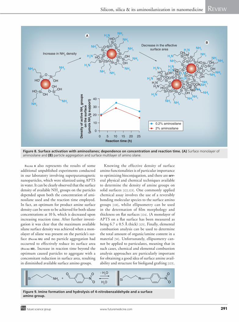
Figure 8 also represents the results of some additional unpublished experiments conducted in our laboratory involving superparamagnetic nano particles, which were silanized using APTS in water. It can be clearly observed that the surface density of available NH
2 groups on the particles
depended upon both the concentration of ami-nosilane used and the reaction time employed. In fact, an optimum for product amine surface density can be seen to be achieved for both silane concentrations at 10 h, which is decreased upon increasing reaction time. After further investi-gation it was clear that the maximum available silane surface density was achieved when a mon-olayer of silane was present on the particle’s sur-face (Figure 8a) and no particle aggregation had occurred to effectively reduce its surface area (Figure 8b). Increase in reaction time beyond the optimum caused particles to aggregate with a concomitant reduction in surface area, resulting in diminished available surface amino groups.
Knowing the effective density of surface amino functionalities is of particular importance to optimizing bioconjugation, and there are sev-sev-eral physical and chemical techniques available to determine the density of amine groups on solid surfaces [122,123]. One commonly applied chemical assay involves the use of a reversibly bonding molecular species to the surface amino groups [118], whilst ellipsometry can be used in the determation of film morphology and thickness on flat surfaces [124]. (A monolayer of APTS on a flat surface has been measured as being 6.7 ± 0.5 Å thick) [125]. Finally, elemental combustion analysis can be used to determine the total amount of organic/amine content in a material [50]. Unfortunately, ellipsometry can-not be applied to particulates, meaning that in such cases, chemical and elemental combustion analysis approaches are particularly important for obtaining a good idea of surface amine avail-ability and structure for bioligand grafting [123].
Si Si
Si
SiSi
SiSi
Si Si
Si
SiSiSi
Si
Si
Si
SiSiSiSiSiSi
Si
SiSi
SiSi
Si
Si
SiHSiSi
Si
Si
Si
Si
Si
Si SiSi
Si Si
SiSi
HO
HO
HO
HO
OH
OHOH
OH
O
O
O O O
OO
O
O
O
O
OO
OO
OO
OO
OO
O
O
O OO
OO
OO
O O
O O
O
O
OO
OO
OO
O
NH2NH2
NH2
NH2
NH2
NH2
NH2
NH2
NH2
NH2
H2N
H2N
H2N
H2NH2N
H2N
H2N
H2NH2N
H2N
H2N
NH2 NH2
NH2NH2
NH2NH2
NH2
NH2NH2
H2N
H2N
H2N
NH2NH2
Increase in NH2 densityOH
OHOH
OHHO
OH
OH
OH
OH
OH
OH
HOHO
HO
HOOHHO
HO
OH
Decrease in the effectivesurface area
NH2NH2
NH2NH2
NH2
NH2
0.2% aminosilane
2% aminosilane
Reaction time (h)
Den
sity
of
acti
ve N
H2
gro
up
so
n t
he
surf
ace
(µm
ols
NH
2/g
su
pp
ort
)
0
0
5 10
10
15 20
20
30
40
25
Figure 8. Surface activation with aminosilanes; dependence on concentration and reaction time. (A) Surface monolayer of aminosilane and (B) particle aggregation and surface multilayer of amino silane.
NH2Si + NO O
O
- H2O
H2OSi N
O
ON
Figure 9. Imine formation and hydrolysis of 4-nitrobenzaldehyde and a surface amino group.
Nanomedicine (2011) 6(2)292 future science group
Review Rother, Sen, East & Bruce
In the former context, a particularly common analytical method involves the reaction of a strongly UV absorbing aromatic aldehyde (e.g., 4-nitrobenzaldehyde [117] or 9-anthracecarboxal-dehyde [118], see Figure 9) with the surface amine groups of a sample. The aldehyde groups form reversible imine bonds with the amino groups and after washing the conjugated sample to remove unreacted aromatic aldehyde, hydroly-sis of the surface imines in a known volume of solution is carried out. Subsequently, it is pos-sible to indirectly quantify the surface density of amine by UV-vis spectroscopy of the h ydrolyzed a romatic species in solution [126].
Any surface terminated with OH (including glass, silica and metal oxides) possesses the oppor-tunity to be modified using any organo-/alkoxy-silanes and whilst aminosilanes are probably the most frequent choice for surface activation of solid substrates, there are many other organosilanes that can be used for the conjugation of bio molecules to the solid surfaces. These include ones with
functionalities, such as carboxyl (which must be applied in protected form such as an ester COOR, as the condensation/polymerization conditions would otherwise render it inactivated), sulfhydryl (or thiol, SH), isocyanato (RNCO) and epoxy groups [127–129]. Carboxyl groups can be activated to an active ester by carbodiimide coupling chem-istry, enabling them to undergo covalent bonding to biomolecules containing free amino groups via formation of a secondary amine (amide; see later). Terminal sulfhydryl/thiol groups of mercaptosi-lanes are capable of disulfide bonding to other thiol or disulfide groups in bioligands, whilst iso-cyanate groups are highly reactive to nucleophiles and enable covalent bonding of biomolecules pos-sessing either hydroxyl groups (R’OH) to form either a urethane (RNHCOOR’) or with termi-nal amino groups (R’NH
2) to form an isourea
(RNHCONHR’). Finally, highly strained cyclic ethers, such as epoxide groups enable covalent bonding of biomolecules with nucleophilic func-tional groups, such as sulf hydryl, amino and hydroxyl groups, to form thioether, amine or ether bonds.
� Conjugating bioligands to aminosilylated surfacesTwo very common approaches for grafting bio-ligands to aminosilylated surfaces involve the use of the coupler glutaraldehyde [130] or a water soluble carbodiimide (Figure 10) [131].
N N
NOO
C
Figure 10. Structures of (A) glutaraldehyde and (B) 1-ethyl-3-(3-dimethylaminopropyl) carbodiimide.
Deprotonation
Effective proton transfer from the quaternized nitrogen to the oxygen atom
Elimination of water to generate the resonancestabilized cation
The unshared pair of electrons on the amine nitrogen bond to the positively charged carbon. Nitrogen is relatively weakly nucleophilic so to push the carbonyl pi electrons ‘out of the way’ acid has been added
1. A proton donated by acid bonds to the carbonyl oxygen2. Electrons involved in double bonding (pi electrons) are used3. This leaves the carbon positively charged and susceptible to attack by nitrogen
Selectivereduction
Secondaryamine
Imine
C CO O
H+
H
H
H
HH
R
R’
R
N
+
+ CO H
HHH
R’
R
N+ C
O H
HH
R’
R R
N+ C
O HH
HH
R’N
+
R C HH
R’N
+R C HH
R’N+
R C H
R’N
R C H
R’N H
H+
+
H2O
- H+
NaCNBH3
Figure 11. Formation of secondary amines from primary amines and aldehydes.
www.futuremedicine.com 293future science group
Silicon, silica & its aminosilanization in nanomedicine Review
Both these approaches make it possible to take advantage of functional groups that are frequently present in a wide range of bio ligands or which can be added to their synthetic deriva-tives, especially oligonucloetides, namely car-boxyl and primary amino groups. They also offer ubiquitous, cheap and flexible routes for the biofunctionalization of materials and represent some of the most common approaches adopted in bioconjugation chemistry [132,133].
Glutaraldehyde is a homobifunctional cou-pling molecule (Figure 10) and can be used to convert surface amino groups to aldehydes [134]. Aldehyde functionalities react more or less spon-taneously at physiological pH with amines in an SN
2-type reaction, in which elimination of
a water molecule results in the formation of an imine bond (Schiff base; see Figure 11).
Commonly, a 0.1 to 1.0% anhydrous solution of glutaraldehyde is used in this process, in the presence of a small amount of acid. After washing the glutaraldehyde-modified surface in water, it can be used to couple to any biomolecule pos-sessing terminal amino groups. Since the imine product is sensitive to hydrolysis, the biomolecule conjugates are selectively reduced with sodium cyanoborohydride (NaCNBH
3) to the stable
secondary amine. Owing to a difference in pKa
values of terminal amino groups (~pH 7.6 to 8.0) to those present in amino acid side chains (~pH 10.0 to 10.2), using this approach it is sometimes possible to optimize the selectivity of the reac-tion for protein surface conjugation via terminal amino groups by performing the second step of
the reaction under slightly acidic conditions (at ~pH 5) [135]. In the same context, amino-modified oligonucleotides can be conjugated in a similar manner [50].
In the second type of coupling approach an aminosilylated surface can be conjugated to car-boxyl groups present in/on a ligand and a car-bodiimide is employed (Figure 12). Carbodiimide couplings can be performed in aqueous or non-aqueous, organic solvent systems, but bioconjuga-tions are ideally performed in aqueous media to prevent organic solvent denaturation of the bio-molecular ligands involved. The watersoluble car-bodiimide 1-ethyl-3-(3-dimethylaminopropyl) carbodiimide (EDC) is most often employed for this purpose [136]. Carboxyl groups do not react spontaneously with amino groups in water and their activation is necessary to make this possible. This is the role of the carbodiimide. Treatment of the carboxyl-possessing ligand with carbodiimide converts the carboxyl functionality into the cor-responding O-acylisourea intermediate, which can then react with primary amino groups to form a stable secondary amine/amide [137]. This reaction can be carried out as a one- or two-step process, and the preferred approach depends on the ligand involved (see [134] for more detail).
Carbodiimide coupling procedures are typi-cally carried out at a pH range of 3.0 to 5.0 and it is necessary to keep accurate control over the pH during the coupling reaction. This is because, on the one hand, the carbodiimide needs to be acti-vated by protonation of a nitrogen (in the case of EDC at a pK
Figure 12. Formation of amides from carboxylic acids and amines in the presence of carbodiimide.
Nanomedicine (2011) 6(2)294 future science group
Review Rother, Sen, East & Bruce
other hand the carboxyl group should remain mostly in its deprotonated form (RCOO-), that is the pH should be above the pK
a value of the
organic acid functionality. The optimum pH for the reaction is therefore the arithmetic mean of the pK
a values of EDC and the carboxyl group,
(i.e., pH = ½ {pKa(EDC) + pK
a(RCOOH)}).
EDC is relatively unstable in its protonated form and its activity decreases significantly over time at low pH. Consequently, the coupling reaction should be carried out for as short a period of time as is possible.
Simplistically it is frequently imagined that an excess of EDC is all that is required to medi-ate efficient conjugation, but in reality this is not the case and the pH issue raised above gives some insight into why. The reaction actually requires optimization and although the general protocol for EDC coupling is applicable for any
carboxyl-amine conjugation reaction, the exact relative concentration of EDC to amine and car-boxyl and pH must be optimized to maximize conjugation efficiency in each different reaction scenario. Table 2 represents some data from EDC-mediated carboxyl-amine conjugation experi-ments performed in our laboratory involving carboxyl-terminated quantum dots (QD) and anti-Cdc 8 antibodies (an Ab-targeting actin protein from yeast). The ultimate aim was to make cheap material that could be used for imag-ing the cytoskeleton of yeast cells, but which possessed the advantage that it was insensitive to photobleaching and so could be used for longer periods than classical fluorophore conjugates.
While these data are clearly complex, for ease of reference the most relevant section of the table is highlighted in blue. Columns 1 and 2 refer to the relative mole ratios of QD carboxyl group to
Table 2. Fluorescence, OD280 and quantity of IgG present in quantum dot–IgG conjugates.
data for R and FT of QD–IgG conjugates produced under various conditions. The concentration of IgG present in both R and FT determined colorimetrically is displayed in nanomoles, and is also expressed as a percentage of the total IgG added to the reaction. Optimum conditions are highlighted in green, control reactions are highlighted in gray. Ab: Antibody; EDC: 1-ethyl-3-(3-dimethylaminopropyl) carbodiimide; FT: Flow-through; QD: Quantum dot; R: Retentate.
www.futuremedicine.com 295future science group
Silicon, silica & its aminosilanization in nanomedicine Review
EDC and Ab used in the various reactions and columns 3, 4, 5 and 6 relate to the amount of fluorescence (measured by excitation at λ 468nm and emission at λ 495nm) and protein (measured spectro photometrically by OD
280) present in the
conjugate (columns 3 and 4) and nonconjugated remainder of the reaction (columns 5 and 6). Separation of the conjugate product from unre-acted reaction components was by ultrafiltration in Vivaspin500 filter units (Sartorius) and retentate relates to the conjugate (retained above the filter), while flow-through relates to the unreacted reac-tion components (flow-through). It is clear that different ratios of the three reaction components produce conjugate possessing different degrees of fluorescence and amounts of protein. Our aim was to make a product with a good degree of fluores-cence and that also possessed a significant fraction of Ab, to make it active in the imaging process. Consequently, we were looking for conjugate with high fluor and OD
280 values. At the same time,
the supply of polyclonal Ab at our disposal was limited and we therefore needed to conserve, as much as possible, the amount used in conjugation reactions. With these considerations in mind, the green row in Table 2 shows the eventual mix used for the routine production of the conjugate and imaging. Figure 13 illustrates results from the use of the conjugate in imaging yeast cell actin structure compared with classical organic fluorophore fluo-rescein isothiocyanate (FITC)–Ab conjugates and clearly demonstrates that the QD–Ab conjugate produced surpassed the performance of the clas-sical conjugate. Figure 14 shows data related to the relative resistance of the QD–Ab and FITC–Ab conjugates, described above, to photobleaching, where it is clear that the QD–Ab conjugate was much more resistant to photobleaching than the classical organic fluorophore conjugates.
Carbodiimide coupling efficiency can be improved by addition of compounds such as N-hydroxybenzotriazole to the reaction mix, which form active esters of the isoacylurea car-boxyl ligand adduct that can then react with high efficiency with primary amine groups [139].
ConclusionA place still remains for simple bioligand con-jugation chemistries in the synthesis of nano-medical materials. Notwithstanding the relative lack of specificity of the methods described, they are still commonly employed in many laborato-ries on a daily basis, worldwide, to make biocon-jugate materials. Their benefits are familiarity, cost and availability of the reagents and infor-mation required to make the processes work.
Furthermore, the methods can be made to func-tion with native biomaterials, which do not need to be chemically modified in any way. Their prin-cipal drawback is that they often lack flexibility in permitting the regio-specific association of a
Figure 13. A comparison of quantum dot–antibody conjugate and fluorescein isothiocyanate–antibody conjuate immunofluorescence. Immunofuorescence microscopy was performed against Cdc8 in wild-type Schizosaccharomyces pombe cells using a QD-labeled primary antibody (direct), and a FITC-labeled secondary antibody (indirect). The top two panels show a maximum projection of 21 sections taken at separate focal planes, using identical exposure settings. The contractile actomyosin ring is clearly visible (arrow heads). The bottom two panels show the same cells after deconvolution by imaging software. Cdc8-decorated actin filaments can be seen (fine arrows). An unknown structure can be observed in the images produced with QDs (thick arrows). FITC: Fluorescein isothiocyanate; QD: Quantum dot.
5 µm 5 µm
FITC QD
Maximum projection
Deconvolvedimage
0.30 100 200 300
Time (s)
Flu
ore
scen
t in
ten
sity
400 500 600
0.4
0.5
0.6
0.7
0.8
0.9
1
FITC–IgG
QD–IgG
Figure 14. The effects of photobleaching on quantum dot–antibody and fluorescein isothiocyanate–antibody conjugates. Fluorescent intensity of a QD–IgG conjugate and a FITC–IgG conjugate over time. Values for intensity have been normalized against the maximum for each fluorophore. FITC: Fluorescein isothiocyanate; QD: Quantum dot.
Nanomedicine (2011) 6(2)296 future science group
Review Rother, Sen, East & Bruce
bioligand with a surface. For many applications this is not a primary concern, so long as the required specific activity for a conjugate material can be produced and, consequently, there is no need for recourse to more costly and specific con-jugation chemistries. However, it must be born in mind that although these approaches have been used historically for the purposes described, there is still much that needs to be considered in any specific surface activation with silane and conju-gation of bioligand before one can be certain the processes are working efficiently. This article has attempted to highlight this fact in a holistic way from substrate synthesis to bioligand conjugation.
Future perspectiveThe future of silica and silica surface modifi-cation by alkoxy- and organo-silanes in nano-medicine looks as strong as it ever did and has not been inhibited by the development of other, more costly, more specific surface activation strat-egies for bioligand surface conjugation. Silica and, by analogy, glasses remain eminently suit-able materials for use in bulk applications such as tissue engineering in vivo, as well as in drug
formulation and in vitro diagnostic procedures, and there is still much research interest into amino silylation of such surfaces for bioconjuga-tion using classical chemistries such as carbodi-imides (e.g., [14,138,139]). Consequently, we feel that there is still much potential to be exploited in the materials and procedures described and discussed here in the field of nanomedicine.
AcknowledgementsThe authors would like to thank Dan Mulvihill (School of Biosciences, University of Kent, UK) for assistance in yeast cell imaging.
Financial & competing interests disclosureThis article includes unpublished data from the authors’ laboratory, which were produced with support from the European Commission and the Integrated Project NACBO (contract number NMP4-CT-2004-500804). The authors have no other relevant affiliations or financial involvement with any organization or entity with a financial interest in or financial conflict with the subject matter or materials discussed in the manuscript apart from those disclosed.
No writing assistance was utilized in the production of this manuscript.
Executive summary
Silica in nanomedicine � Silica is, and will continue to be, a major material used in the nanomedical field owing to its desirable chemical and physical properties,
which render it fully biocompatible. � Silica’s surface functionality and structure are closely linked to the specific approach used in its preparation. � Unmodified amorphous and ordered silicates possess surfaces whose chemistry and reactivity can be described in terms of their silanol
type, density and geometry, along with the ratio between silanol and siloxane groups present. � Silica offers a surface that is ideal for activation and chemical modification by alkoxy- and organo-silanes.
Alkoxy- & organo-silanes � Tetra alkoxysilanes are the pre-eminent precursors in silica and silicate synthesis. � Organosilanes, particularly aminosilanes, are the primary route for the surface activation of silica. � Alkoxy- and amino-silanes can also be used to surface activate other materials possessing hydroxyl groups, particularly glass, plastic,
latex and metal oxides. � Aminosilanes are still the most widely and frequently used molecules in silica surface activation. � Aminosilanes allow for common biofunctionalization protocols to be applied to a very wide range of bioligands in their surface grafting. � Aminosilanes are cheap to use and much literature exists to support their future use and application. � Simple, cheap assays exist for the characterization of amino-silylated surfaces in terms of their available amino group density for
bioconjugation and bulk amine density.
Bioconjugation to aminosilylated surfaces � Amino-activated surfaces can be coupled directly to carboxyl groups possessed by a bioligand, via the use of a carbodiimide. � Amino-activated surfaces can be coupled directly to an amino group of a bioligand by prior conversion to aldehyde, by treatment
with glutaraldehyde. � Both approaches require optimization for the specific surface and bioligand involved to be maximally efficient. � Aminosilylation remains a very viable approach for bioligand coupling to a solid surface.
Bibliography1 Rudnick RL, Gao S: Composition
of the continental crust. In: The Crust (Volume 3). Rudnick RL (Ed.). Elsevier-Pergamon, Oxford, UK, 1–64 (2003).
2 Holleman-Wieberg: Inorganic Chemistry. Wiberg N, Aylett BJ (Ed.). Academic Press, London, UK (2001).
3 Bondi A: Van der Waals volumes and radii. J. Phys. Chem. 68(3), 441–451 (1964).
4 Brook MA: Silicon in Organic, Organometallic, and Polymer Chemistry. John Wiley & Sons, NY, USA (2000).
5 Brinker CJ, Scherrer GW: Sol-Gel Science: The Physics and Chemistry of Sol-Gel Processing. Academic Press, London, UK (1990).
www.futuremedicine.com 297future science group
Silicon, silica & its aminosilanization in nanomedicine Review
297www.futuremedicine.com
6 Rao CNR: Chemical synthesis of solid inorganic materials. Mater. Sci. Eng. B 18(1), 1–21 (1993).
7 Pierre AC: Introduction to Sol-Gel Processing. Kluwer Academic Publishers, Dordrecht, The Netherlands, 2–3 (2002).
8 Dislich H: Thin films from the sol-gel process. In: Sol-Gel Technology for Thin Films, Fibers, Preforms, Electronics, and Specialty Shapes. Klein LC (Ed.). Noyes Publications, NJ, USA, 50–79 (1988).
9 Wang ZL, Liu Y, Zhang Z: Handbook of Nanophase and Nanostructured Materials (Volume 1). Kluwer Academic Publishers, NY, USA, 74 (2001).
11 Wright JD, Sommerdijk NAJM: Sol Gel Materials, Chemistry and Applications. Wright JD, Sommerdijk AJM (Eds). Taylor and Francis, London, UK, 4–5 (2001).
12 Ebelman M: Sur les combinaisons des acides borique et silicique avee les ethers. Ann. Chim. Phys. 16, 129 (1846).
13 Stöber W, Fink A, Bohn E: Controlled growth of monodisperse silica spheres in the micron size range, J. Colloid Interface Sci. 26(1), 62–69 (1968).
15 Barik TK, Sahu B, Swain V: Nanosilica – from medicine to pest control. Parasitol. Res. 103(2), 253–258 (2008).
16 Tabatabaei S, Shukohfar A, Aghababazadeh R, Mirhabibi A: Experimental study of the synthesis and characterisation of silica nanoparticles via the sol-gel method. J. Phys.: Conf. Ser. 26, 371–374 (2006).
17 Vansant EF, van der Voort P, Vrancken KC: Characterization and Chemical Modification of the Silica Surface. Elsevier-Science, Amsterdam, The Netherlands (1995).
18 Kumar CSSR, Hormes J, Leuschner C: Nanofabrication Towards Biomedical Applications: Techniques, Tools and Impact. Wiley-VCH, Weinheim, Germany (2005).
19 Ozin GA, Arsenault AC: Nanochemistry: a Chemical Approach to Nanomaterials. RCS Publishing, Dorchester, UK (2006).
20 Iler RK: The Chemistry of Silica: Solubility, Polymerization, Colloid and Surface Properties. John Wiley & Sons, NY, USA (1979).
21 Nakanishi K: Porous gels made by phase separation: recent progress and future directions. In: Sol-Gel Science and Technology: Topics in Fundamental Research and Applications (Volume 4).
Sakka S (Ed.). Kluwer Academic Publishers, Dodrecht, The Netherlands, 43–48 (2003).
22 Li Q, Retsch M, Wang J, Knoll W, Jonas U: Porous networks through colloidal templates. In: Templates in Chemistry III (Volume 287). Broekmann P, Schalley CA, Dötz KH (Eds). Springer, Heidelberg, Germany, 135–180 (2009).
24 Zhao B, Collinson MM: Well-defined hierarchical templates for multimodal porous material fabrication. Chem. Mater. 22(14), 4312–4319 (2010).
25 Whitesides GM, Boncheva M: Beyond molecules: self-assembly of mesoscopic and macroscopic components. Proc. Natl Acad. Sci. USA 99(8), 4769–4774 (2002).
26 El-Safty SA, Kiyozumi Y, Hanaoka T, Mizukami F: Cationic surfactant templates for newly developed cubic Fd3m silica mesocage structures. Mater. Lett. 62(17–18), 2950–2953 (2008).
27 Bagshaw SA, Bruce IJ: Rapid calcination of high quality mesostructured MCM-41, MSU-X, and SBA-15 silicate materials: a step towards continuous processing? Microporous Mesoporous Mater. 109, 199–209 (2008).
28 Wan Y, Zhao D: On the controllable soft-templating approach to mesoporous silicates. Chem. Rev. 107(7), 2821–2860 (2007).
29 Zou H, Wu S, Shen J: Polymer/silica nanocomposites: preparation, characterization, properties, and applications. Chem. Rev. 108(9), 3893–3957 (2008).
30 Hatton B, Landskron K, Whitnall W, Perovic D, Ozin GA: Past, present, and future of periodic mesoporous organosilicas: the PMOs. Acc. Chem. Res. 38(4), 305–312 (2005).
31 Wang Y, Price AD, Caruso F: Nanoporous colloids: building blocks for a new generation of structured materials. J. Mater. Chem. 19, 6451–6464 (2009).
33 Serrano E, Rus G, García-Martínez J: Nanotechnology for sustainable energy. Renew. Sust. Energy Rev. 13(9), 2373–2384 (2009).
34 Wang D, Kou R, Choi D et al.: Ternary self-assembly of ordered metal oxide−graphene nanocomposites for electrochemical energy storage. ACS Nano 4(3), 1587–1595 (2010).
35 Sen T, Bruce IJ, Mercer T: Fabrication of novel hierarchically ordered porous magnetic nanocomposites for bio-catalysis. Chem. Commun. 46, 6807–6809 (2010).
36 Taguchi A, Schüth F: Ordered mesoporous materials in catalysis. Micropor. Mesopor. Mater. 77(1), 1–45 (2005).
37 Walcarius A, Mercier L: Mesoporous organosilica adsorbents: nanoengineered materials for removal of organic and inorganic pollutants. J. Mater. Chem. 20, 4478–4511 (2010).
38 Sen T, Sebastianelli A, Bruce IJ: Meso-structured superparamagnetic nanospheres and nanotubes: smart materials for bioseparations. J. Am. Chem. Soc. 128(22), 7130–7131 (2006).
39 Gabashvili A, Medina DD, Gedanken A, Mastai Y: Templating mesoporous silica with chiral block copolymers and its application for enantioselective separation. J. Phys. Chem. B 111(38), 11105–11110 (2007).
40 Hertz A, Bruce IJ: Inorganic materials for bone repair or replacement applications. Nanomedicine 2(6), 899–918 (2007).
41 Kim J, Piao Y, Hyeon T: Multifunctional nanostructured materials for multimodal imaging, and simultaneous imaging and therapy. Chem. Soc. Rev. 38, 372–390 (2009).
42 Klichko Y, Liong M, Choi E et al.: Mesostructured silica for optical functionality, nanomachines, and drug delivery. J. Am. Ceram. Soc. 92(S1), S2–S10 (2009).
43 Richman EK, Kang CB, Brezesinski T, Tolbert ST: Ordered mesoporous silicon through magnesium reduction of polymer templated silica thin films. Nano Lett. 8(9), 3075–3079 (2008).
45 El-Safty SA, Prabhakaran D, Ismail AA, Matsunaga H, Mizukami F: Three-dimensional wormhole and ordered mesostructures and their applicability as optically ion-sensitive probe templates. Chem. Mater. 20(8), 2644–2654 (2008).
46 Wang Y, Yan Y, Cui J et al.: Encapsulation of water-insoluble drugs in polymer capsules prepared using mesoporous silica templates for intracellular drug delivery. Adv. Mater. 22(38), 4293–4297 (2010).
47 Wang Y, Bansal V, Zelikin AN, Caruso F: Templated synthesis of single-component polymer capsules and their application in drug delivery. Nano Lett. 8(6), 1741–1745 (2008).
Nanomedicine (2011) 6(2)298 future science group
Review Rother, Sen, East & Bruce
48 Yabuta T, Bescher EP, Mackenzie JD, Tsuru K, Hayakawa S, Osaka A: Synthesis of PDMS-based porous materials for biomedical applications. J. Sol-Gel Sci. Technol. 26, 1219–1222 (2003).
49 Pagliario M, Ciriminna R, Man MWC, Campestrini S: Better chemistry through ceramics: the physical bases of the outstanding chemistry of ORMOSIL. J. Phys. Chem. B 110, 1976–1988 (2006).
50 Bruce IJ, Sen T: Surface modification of magnetic nanoparticles with alkoxysilanes and their application in magnetic bioseparations. Langmuir 21(15), 7029–7035 (2005).
51 Yazaydin AO, Thompson RW: Molecular simulation of water adsorption in silicalite: effect of silanol groups and different cations. Microporous Mesoporous Mater. 123(1), 169–176 (2009).
52 Carteret C: Vibrational properties of silanol group: from alkylsilanol to small silica cluster.Effects of silicon substituents. Spectrochimica Acta A 64(3), 670–680 (2006).
53 Dijkstra TW, Duchateau R, van Santen RA, Meetsma A, Yap GPA: Silsesquioxane models for geminal silica surface silanol sites. A spectroscopic investigation of different types of silanols. J. Am. Chem. Soc. 124(33), 9856–9864 (2002).
54 Zhao XS, Lu GQ, Whittaker AK, Millar GJ, Zhu HY: Comprehensive study of surface chemistry of MCM-41 using 29Si CP/MAS NMR, FTIR, pyridine-TPD, and TGA. J. Phys. Chem. B 101(33), 6525–6531 (1997).
55 Sauer J, Ugliengo P, Garrone E, Saunders VR: Theoretical study of van der Waals complexes at surface sites in comparison with the experiment. Chem. Rev. 94(7), 2095–2160 (1994).
56 Kamiya H, Mitsui M, Takano H, Miyazawa S: Influence of particle diameter on surface silanol structure, hydration forces, and aggregation behavior of alkoxide-derived silica particles. J. Am. Ceram. Soc. 83(2), 287–93 (2000).
57 Alkan M, Dogan M: Silica gels, surface chemistry. In: Encyclopedia of Surface and Colloid Science (Volume 7), 2nd Revision Edition. Somasundaran P (Ed.). CRC Press, NY, USA, 5608–5620 (2006).
58 Armistead CG, Tyler AJ, Hambleton FH, Mitchell SA, Hockey JA: Surface hydroxylation of silica. J. Phys. Chem. 73(11), 3947–3953 (1969).
59 Zhuravlev LT: Concentration of hydroxyl groups on the surface of amorphous silicas. Langmuir 3(3), 316–318 (1987).
60 Zhuravlev LT: The surface chemistry of amorphous silica. Zhuravlev model. Colloids Surf. A 173(1–3), 1–38 (2000).
61 Ishikawa T, Matsuda M, Yasukawa A et al.: Surface silanol groups of mesoporous silica FSM-16. J. Chem. Soc. Faraday Trans. 92(11), 1985–1989 (1996).
62 Biliski B: The influence of thermal treatment of silica gel on surface-molecule interactions: I. Finite coverage region. J. Colloid Interface Sci. 201(2), 180–185 (1998).
63 Jal PK, Patel S, Mishra BK: Chemical modification of silica surface by immobilization of functional groups for extractive concentration of metal ions. Talanta 62(5), 1005–1028 (2004).
64 Jonat S, Hasenzahl S, Drechsler M, Albers P, Wagner KG, Schmidt PC: Investigation of compacted hydrophilic and hydrophobic colloidal silicon dioxides as glidants for pharmaceutical excipients. Powder Technol. 141(1–2), 31–43 (2004).
65 Technical Information – No. 1281: AEROSIL® Colloidal Silicon Dioxide for Pharmaceuticals. Evonik Degussa, Frankfurt, Germany (2008).
66 Snyder LR, Kirkland JJ, Dolan JW: Introduction to Modern Liquid Chromatography (3rd Edition). John Wiley & Sons, Hoboken, NJ, USA (2010).
67 Giesche H: Medical and technological application of monodispersed colloidal silica particles. In: Medical Applications of Colloids. Matijevic E (Ed.). Springer, NY, USA, 43–68 (2008).
68 Jungbauer A: Chromatographic media for bioseparation. J. Chromatogr. A 1065(1), 3–12 (2005).
69 Vogelstein B, Gillespie D: Preparative and analytical purification of DNA from agarose. Proc. Natl Acad. Sci. USA 76(2), 615–619 (1979).
73 Fogolari F, Brigo A, Molinari H: The Poisson–Boltzmann equation for biomolecular electrostatics: a tool for structural biology. J. Mol. Recogn. 15(6), 377–392 (2002).
74 Melzak KA, Sherwood CS, Turner RFB, Haynes CA: Driving forces for DNA adsorption to silica in perchlorate solutions. J. Colloid Interface Sci. 181(2), 635–644 (1996).
75 Magnani M, Galluzzi L, Bruce IJ: The use of magnetic nanoparticles in the development of new molecular detection systems. J. Nanosci. Nanotech. 6(8), 2302–2311 (2006).
76 Sen T, Bruce IJ: Mesoporous silica–magnetite nanocomposites: fabrication, characterisation and applications in biosciences. Microporous Mesoporous Mater. 120(3), 246–251 (2009).
77 Taylor JI, Hurst CD, Davies MJ, Sachsinger N, Bruce IJ: Application of magnetite and silica–magnetite composites to the isolation of genomic DNA. J. Chromatogr. A 890(1), 159–166 (2000).
78 Odenbach S: Colloidal Magnetic Fluids: Basics, Development and Application of Ferrofluids. Springer, Heidelberg, Germany (2009).
79 Bruce IJ, Taylor J, Todd M et al.: Synthesis, characterisation and application of silica-magnetite nanocomposites. J. Magn. Magn. Mater. 284, 145–160 (2004).
80 Amagliani G, Brandi G, Omiccioli E, Casiere A, Bruce IJ, Magnani M: Direct detection of Listeria monocytogenes from milk by magnetic based DNA isolation and PCR. Food Microbiol. 21(5), 597–603 (2004).
81 Sebastianelli A, Sen T, Bruce IJ: Extraction of DNA from soil using nanoparticles by magnetic bioseparation. Lett. Appl. Microbiol. 46(4), 488–491 (2008).
82 Bertozzini E, Penna A, Pierboni E, Bruce IJ, Magnani M: Development of new procedures for the isolation of phytoplankton DNA from fixed samples. J. Appl. Phycol. 17, 223–229 (2005).
83 Bertozzini E, Penna A, Perboni E, Bruce IJ, Magnani M: Application of new methods in DNA extraction from phytoplankton. Biol. Mar. Medit. 13(1), 943–946 (2006).
84 Erli HJ, Marx R, Paar O, Niethard FU, Weber M, Wirtz DC: Surface pretreatments for medical application of adhesion. Biomed. Engineer. Online 2(1), 15 (2003).
85 Tiwari SK, Mishra T, Gunjan MK, Bhattacharyya AS, Singh TB, Singh R: Development and characterization of sol-gel silica–alumina composite coatings on AISI 316L for implant applications. Surf. Coat. Technol. 201(17), 7582–7588 (2007).
86 Piercea AL, Sommakiaa S, Rickusa JL, Otto KJ: Thin-film silica sol–gel coatings for neural microelectrodes. J. Neurosci. Meth. 180(1), 106–110 (2009).
87 Jana NR, Earhart C, Ying JY: Synthesis of water-soluble and functionalized nanoparticles by silica coating. Chem. Mater. 19(21), 5074–5082 (2007).
88 Gomez-Vega JM, Sugimura H, Takai O, Hozumi A: Aligned bioactive mesoporous silica coatings for implants. J. Mater. Sci. Mater. Med. 12(10), 923–927 (2001).
www.futuremedicine.com 299future science group
Silicon, silica & its aminosilanization in nanomedicine Review
299www.futuremedicine.com
89 Latour RA: Biomaterials: protein-surface interactions. In: Encyclopedia of Biomaterials and Biomedical Engineering (Volume 1). Wnek GE, Bowlin GL (Eds). Informa Healthcare USA, NY, USA, 270–284 (2008).
90 Gray JJ: The interaction of proteins with solid surfaces. Curr. Op. Struct. Biol. 14(1), 110–115 (2004).
91 Raut VP, Agashe MA, Stuart SJ, Latour RA: Molecular dynamics simulations of peptide−surface interactions. Langmuir 21(4), 1629–1639 (2005).
92 Pitt WG, Morris RN, Mason ML, Hall MW, Luo Y, Prestwich GD: Attachment of hyaluronan to metallic surfaces. J. Biomed. Mater. Res. 68A, 95–106 (2004).
94 Chen XW, Wang JH: The miniaturization of bioanalytical assays and sample pretreatments by exploiting meso-fluidic lab-on-valve configurations: a review. Anal. Chim. Acta 602(2), 173–180 (2007).
95 Nath N, Hyun J, Ma H, Chilkoti A: Surface engineering strategies for control of protein and cell interactions. Surf. Sci. 570(1–2), 98–110 (2004).
96 Slowing II, Vivero-Escoto JL, Wu CW, Lin VSY: Mesoporous silica nanoparticles as controlled release drug delivery and gene transfection carriers. Adv. Drug Deliv. Rev. 60(11), 1278–1288 (2008).
97 Schena M, Shalon D, Davis RW, Brown PO: Quantitative monitoring of gene expression patterns with a complementary DNA microarray. Science 270(5235), 467–470 (1995).
98 Lab-on-a-chip Technology: Fabrication and Microfluidics (Volume 1). Herold K, Rasooly A (Eds). Caister Academic Press, Norfolk, UK (2009).
99 Pierce MC, Javier DJ, Richards-Kortum R: Optical contrast agents and imaging systems for detection and diagnosis of cancer. Int. J. Cancer 123(9), 1979–1990 (2008).
100 Lellouche JP, Perlman N, Joseph A et al.: New magnetically responsive polycarbazole-magnetite nanoparticles. Chem. Commun. 5, 560–561 (2004).
101 Lellouche JP, Senthil G, Joseph A et al.: Magnetically responsive carboxylated magnetite-polypyrrole/polydicarbazole nanocomposites of core–shell morphology: fabrication, characterisation and use in DNA hybridisation. J. Am. Chem. Soc. 127(34), 11998–12006 (2005).
102 Bruce IJ, Sen T, del Campo A: Key for tomorrow: nanotechnology in food analysis. In: Rapid Methods for Biological and Chemical
Contaminants in Food and Feed. van Amerongen A, Barug D, Lauwaars M (Eds). Wageningen Academic Publishers, Wageningen, The Netherlands, 387–408 (2005).
103 Amagliani G, Omiccioli E, del Campo A, Bruce IJ, Brandi G, Magnani M: Development of a magnetic capture hybridization-PCR assay for Listeria monocytogenes direct detection in milk samples. J. Appl. Microbiol. 100(2), 375–383 (2006).
104 Galluzzi L, Bertozzini E, del Campo A, Penna A, Bruce IJ, Magnani M: Capture probe conjugated to paramagnetic nanoparticles for purification of Alexandrium species (Dinophyceae) DNA from environmental samples. J. Appl. Microbiol. 101, 36–43 (2006).
105 Galluzzi L, Magnani M, Saunders N, Harms C, Bruce IJ: Current molecular techniques for the detection of microbial pathogens. Science Progress 90(1), 29–50 (2007).
106 Omiccioli E, Amagliani G, Brandi G, Bruce IJ, Magnani M: Simultaneous direct detection of Salmonella spp., Listeria monocytogenes, and Escherichia coli O157 in milk samples by magnetic extraction and multiplex PCR. J. Rapid Meth. Auto. Micro. 17(2), 195–213 (2009).
107 Amagliani G, Omiccioli E, Brandi G, Bruce IJ, Magnani M: Multiplex magnetic capture hybridisation and multiplex real-time PCR protocol for pathogen detection in seafood. Food Microbiol. 27(5), 580–585 (2010).
108 del Campo A, Bruce IJ: Diagnostics and high throughput screening. In: Biomedical Nanotechnology. Malsch NH (Ed.). Taylor & Francis, FL, USA, 75–112 (2005).
109 Conzone SD, Pantano CG: Glass slides to DNA microarrays. Materials Today 7(3), 20–26 (2004).
111 Gong P, Grainer DW: Nonfouling surfaces: a review of principles and applications for microarray capture assay deign. In: Microarrays (Volume 1). Rampal JB (Ed.). Humana Press, NJ, USA, 59–92 (2007).
112 Xie Y, Hill CAS, Xiao Z, Militz H, Mai C: Silane coupling agents used for natural fiber/polymer composites: a review. Compos. Part A – Appl. Sci. 41(7), 806–819 (2010).
113 Beari F, Brand M, Jenkner P et al.: Organofunctional alkoxysilanes in dilute aqueous solution: new accounts on the dynamic structural mutability. J. Organomet. Chem. 625(2), 208–216 (2001).
114 Kang HJ, Meesiri W, Blum FD: Nuclear magnetic resonance studies of the hydrolysis and molecular motion of aminopropylsilane. Mater. Sci. Eng. A-Struct. 126(1–2), 265–270 (1990).
115 Kanan SM, Tze WTY, Tripp CP: Method to double the surface concentration and control the orientation of adsorbed (3-aminopropyl)dimethylethoxysilane on silica powders and glass slides. Langmuir 18(17), 6623–6627 (2002).
116 Metwalli E, Haines D, Becker O, Conzone S, Pantanoa CG: Surface characterizations of mono-, di-, and tri-aminosilane treated glass substrates. J. Colloid Interface Sci. 298(2), 825–831 (2006).
117 Zhaoa J, Lia Y, Guoa H, Gao L: Relative surface density and stability of the amines on the biochip. chin. J. Anal. Chem. 34(9), 1235–1238 (2006).
118 Moon JH, Shin JW, Kim SY, Park JW: Formation of uniform aminosilane thin layers: an imine formation to measure relative surface density of the amine group. Langmuir 12(20), 4621–4624 (1996).
119 Oh SJ, Cho SJ, Kim CO, Park JW: Characteristics of DNA microarrays fabricated on various aminosilane layers. Langmuir 18(5), 1764–1769 (2002).
120 Ding S, Floyd JA, Walters KB: Comparison of surface confined ATRP and SET-LRP syntheses for a series of amino (meth)acrylate polymer brushes on silicon substrates. J. Polym. Sci. A: Polym. Chem. 47(23), 6552–6560 (2009).
121 Howarter JA, Youngblood JP: Optimization of silica silanization by 3-aminopropyltriethoxysilane. Langmuir 22(26), 11142–11147 (2006).
122 Ghasemi M, Minier M, Tatoulian M, Arefi-Khonsari F: Determination of amine and aldehyde surface densities: application to the study of aged plasma treated polyethylene films. Langmuir 23(23), 11554–11561 (2007).
123 van de Waterbeemd M, Sen T, Biagini S, Bruce IJ: Surface functionalisation of magnetic nanoparticles: quantification of surface to bulk amine density. Micro Nano Lett. 5(5), 282–285 (2010)
124 Eing A, Vaupel M: Quality control of biochips. In: Imaging Ellipsometry in Biotechnology. Eing A, Vaupel M (Eds). Verlag Axel Gierspeck, Goettingen, Germany (2002).
125 Flink S, van Veggel FCJM, Reinhoudt DN: Functionalization of self-assembled monolayers on glass and oxidized silicon wafers by surface reactions. J. Phys. Org. Chem. 14(7), 407–415 (2001).
Nanomedicine (2011) 6(2)300 future science group
Review Rother, Sen, East & Bruce
126 Ngo TT: A simple spectrophotometric determination of solid supported amino groups. J. Biochem. Biophys. Meth. 12(5–6), 349–354 (1986).
128 Kusnezow W, Pulli T, Witt O, Hoheisel JD: Solid supports for protein microarrays and related devices. In: Protein Microarrays. Schena M (Ed.). Jones & Bartlett Int., London, UK, 247–284 (2005).
129 Smith JE, Wang L, Tan W: Bioconjugated silica-coated nanoparticles for bioseparation and bioanalysis. Trends Anal. Chem. 25(9), 848–855 (2006).
130 del Campo A, Sen T, Lellouche JP, Bruce IJ: Multifunctional magnetite and silica-magnetite nanoparticles: synthesis, surface activation and applications in life sciences. J. Magn. Magn. Mater. 293(1), 33–40 (2005).
131 Aslam M, Dent AH: The preparation of protein-small molecule conjugates. In: Bioconjugation: Protein Coupling Techniques
for the Biomedical Sciences. Aslam M, Dent AH (Eds). Macmillan Reference Ltd, London, UK (1998).
132 Lutz JF, Börner HG: Modern trends in polymer bioconjugates design. Prog. Polym. Sci. 33(1), 1–39 (2008).
133 del Campo A, Bruce IJ: Novel solid supports and efficient surface chemistries for the manufacture of DNA arrays. In: Immobilisation of DNA on Chips. Series: Topics in Current Chemistry. Wittmann C (Ed.). Springer Verlag, Berlin Heidelberg, Germany, 78–106 (2005).
134 Ducker RE, Montague MT, Leggetta GJ: A comparative investigation of methods for protein immobilization on self-assembled monolayers using glutaraldehyde, carbodiimide, and anhydride reagents. Biointerphases 3(3), 59–65 (2008).
135 Klok HA: Biological-synthetic hybrid block copolymers: combining the best from two worlds. J. Polym. Sci. A: Polym. Chem. 43(1), 1–17 (2005).
136 Nakajima N, Ikada Y: Mechanism of amide formation by carbodiimide for bioconjugation in aqueous media. Bioconj. Chem. 6(1), 123–130 (1995).
138 Williams A, Ibrahim IT: A new mechanism involving cyclic tautomers for the r eaction with nucleophiles of the water-soluble peptide coupling reagent 1-ethyl-3-(3 -́(dimethylamino)propyl)carbodiimide (EDC). J. Am. Chem. Soc. 103(24), 7090–7095 (1981).
139 Chan LC, Cox BC: Kinetics of amide formation through carbodiimide/N-hydroxybenzotriazole (HOBt) couplings. J. Org. Chem. 72(23), 8863–8869 (2007).
140 Kickelbick G: Introduction to hybrid materials. In: Hybrid Materials: Synthesis, Characterization, and Applications. Kickelbick G (Ed.). Wiley-VCH, Weinheim, Germany, 14 (2007).