Oral hygiene (sponsored by P&G Professional Oral Health) 48 Implant dentistry today August 2007 Volume 1 Number 3 Proper monitoring and maintenance is essential to ensure the longevity of the dental implant and its associated restoration through a combination of appropriate professional care and effective patient oral hygiene. Gregori M Kurtzman and Lee H Silverstein explain the protocols for the dental team D entistry has become so exciting and challenging sin ce predictability has been recognised for long-term dental implant and restoration success (Adell et al, 1981, Cox and Zarb, 1987, Albrektsson et al, 1981). As the number of patients selecting dental implants as a treatment option continues to grow, the dental team must accept the challenges ofmaintaining these sometimes complex restorations. The value of using conventional periodontal parameters to determine peri-implant health is not clearly evident in the literature (Orton et al, 1989). Therefore, it is paramount that the dental implant team understan ds the similarities and distinctions between the dental implant and the natural tooth. Subsequently, by examining the similarities and differences between a natural tooth and a dental implant, basic guidelines can be provided for maintaining the long-term health of the dental implant. Direct anchorage of alveolar bone to a dental implant body provides a foundation to support a prosthesis and transmits occlusal forces to the alveolar bone. This is the definition of osseointegration (Rateischak and Wolf, 1995). With the increased acceptance of dental implants as a viable treatment option for the restoration of a partially edentulous or eden tulous mouth, the dental team is faced with maintaining and educating those patients. Recently, the focus of implant dentistry has changed from obtaining osseointegration, which is highly predictable, to the log-term maintenance health of the peri-implant hard and soft tissues. This can be achieved through appropriate professional care, patient cooperation, and effective home care (Meffert et al 1992). Patients must accept the responsibility for being co-therapists in maintenance therapy, so the dental team essentially must screen the potential implant patient. Diagnosis and treatment planning based on a risk-benefit analysis should be performed subsequent to a thorough medical, dental, head-and-neck, psychological, tempromandibular disorder and radiographic examination (Meffert 1993). There is convincing evidence that bacterial plaque not only leads to gingivitis and periodontitis (Warrer et al, 1995), but also can induce the development of peri-implantitis (Lang and Karring, 1994). Thus, personal oral hygiene must begin at the time of dental implant placement and should be modified using various adjuctive aids for oral hygiene to effectively clean the altered morphology of the peri-implant region before, during, and after implant placement. For instance, interproximal brushes can penetrate up to 3mm into a gingival sulcus or pocket and may effectively clean the peri-implant sulcus (Balshi, 1986). In addition to mechanical plaque control, daily rinses using 0.1% chlorhexidine Dr Gregori Kurtzman, DDS, is in private general practice in Silver Spring, Maryland, USA. He can be reached at dr_ [email protected]. Figure 1: Comparison of crestal gingival fibre orientation Figure 2: Microscopic comparison of gingival fibre orientation (natural tooth on left, implant on right) gluconate or Listerine (Ciancio et al, 1995) provide a welcome adjunct. Hygiene with dental implants is so tedious and critical to their long- term success that the patient and dental professional must exercise considerable effort. During the maintenance visit, the dental professional should concentrate on the peri-implant tissue margin, implant body, prosthetic abutment to implant collar connection, and the prosthesis (Garg, 1995). Clinical assessment Clinical inspection for signs of inflammation, i.e. bleeding on probing, exudate, mobility , probe-able pockets, and a radiographic evaluatio n of the Dental implants: oral hygiene and maintenance
Oral hygiene (sponsored by P&G Professional Oral Health)
48 Implant dentistry today August 2007 Volume 1 Number 3
Proper monitoring and maintenance is essential to ensure the longevity ofthe dental implant and its associated restoration through a combination ofappropriate professional care and effective patient oral hygiene. Gregori MKurtzman and Lee H Silverstein explain the protocols for the dental team
Dentistry has become so exciting and challenging since predictability
has been recognised for long-term dental implant and restoration
success (Adell et al, 1981, Cox and Zarb, 1987, Albrektsson et al,
1981). As the number of patients selecting dental implants as a treatment
option continues to grow, the dental team must accept the challenges of maintaining these sometimes complex restorations.
The value of using conventional periodontal parameters to determine
peri-implant health is not clearly evident in the literature (Orton et al,
1989). Therefore, it is paramount that the dental implant team understands
the similarities and distinctions between the dental implant and the
natural tooth. Subsequently, by examining the similarities and differences
between a natural tooth and a dental implant, basic guidelines can be
provided for maintaining the long-term health of the dental implant.
Direct anchorage of alveolar bone to a dental implant body provides
a foundation to support a prosthesis and transmits occlusal forces to the
alveolar bone. This is the definition of osseointegration (Rateischak and
Wolf, 1995). With the increased acceptance of dental implants as a viabletreatment option for the restoration of a partially edentulous or edentulous
mouth, the dental team is faced with maintaining and educating those
patients.
Recently, the focus of implant dentistry has changed from obtaining
osseointegration, which is highly predictable, to the log-term maintenance
health of the peri-implant hard and soft tissues. This can be achieved
through appropriate professional care, patient cooperation, and effective
home care (Meffert et al 1992). Patients must accept the responsibility
for being co-therapists in maintenance therapy, so the dental team
essentially must screen the potential implant patient. Diagnosis and
treatment planning based on a risk-benefit analysis should be performed
subsequent to a thorough medical, dental, head-and-neck, psychological,
tempromandibular disorder and radiographic examination (Meffert
1993).
There is convincing evidence that bacterial plaque not only leads to
gingivitis and periodontitis (Warrer et al, 1995), but also can induce
the development of peri-implantitis (Lang and Karring, 1994). Thus,
personal oral hygiene must begin at the time of dental implant placement
and should be modified using various adjuctive aids for oral hygiene
to effectively clean the altered morphology of the peri-implant region
before, during, and after implant placement. For instance, interproximal
brushes can penetrate up to 3mm into a gingival sulcus or pocket and
may effectively clean the peri-implant sulcus (Balshi, 1986). In addition
to mechanical plaque control, daily rinses using 0.1% chlorhexidine
Dr Gregori Kurtzman, DDS, is in private general practicein Silver Spring, Maryland, USA. He can be reached at [email protected].
Figure 1: Comparison of crestal gingival fibre orientation
Figure 2: Microscopic comparison of gingival fibre orientation (natural tooth on left,implant on right)
gluconate or Listerine (Ciancio et al, 1995) provide a welcome adjunct.
Hygiene with dental implants is so tedious and critical to their long-
term success that the patient and dental professional must exercise
considerable effort. During the maintenance visit, the dental professional
should concentrate on the peri-implant tissue margin, implant body,
prosthetic abutment to implant collar connection, and the prosthesis
(Garg, 1995).
Clinical assessmentClinical inspection for signs of inflammation, i.e. bleeding on probing,
exudate, mobility, probe-able pockets, and a radiographic evaluation of the
Figure 5: Plastic curettes for scaling dental implants and demonstration of theimplant surface after use. Note that there is no alteration to the surface
Figure 6: Plastic scaler used for recall maintenance
Oral hygiene(sponsored by P&G Professional Oral Health)
August 2007 Volume 1 Number 3 Implant dentistry today 51
For all of these reasons, personal home care and consistent professionalmaintenance have proven to be critical to the success and longevity of
endosseous dental implants. This is especially true in an environment
with adjacent natural teeth, which if affected by periodontal disease, could
act as a reservoir for pathogenic bacteria, ie. gram-negative anaerobic rods,
and seed the peri-implant sulcus (Mombelli et al, 1995).
The physical characteristics of the peri-implant soft tissue are the focus
of all oral hygiene instruction. The presence or absence of keratinised
tissue in this critical area has not been unequivocally documented to state
that peri-implant tissues are more vulnerable to the ingress of pathogenic
bacteria with or without keratinised tissue being present around dental
implants. However, the ability of the patient to maintain good home care
around dental implants is facilitated by the presence of keratinised tissuesurrounding implants. Thus, if a patient has no keratinised tissue around
an implant, and a pull from a frenum or a chronic peri-implant mucositis
exists, then placement of a soft tissue autogenous or alloplastic connective
tissue graft is recommended to facilitate proper mechanical oral hygiene
maintenance (Artzi et al, 1993).
Specific criteria for obtaining clinical data around dental implants
that would allow proper monitoring and detect early possible failure of
osseointegrated dental implants has not been clearly defined. Presently, the
presence of mobility is the best indicator for diagnosis of implant failure.
As opposed to natural teeth, dental implants exhibit minimal clinically
undetectable movement because of the absence of a periodontal ligament.
Therefore, healthy implants should appear nonmobile, even in the
presence of peri-implant bone loss, if an adequate amount of supporting
alveolar bone still exists (Papaioannou et al, 1995).
When monitoring the health of the peri-implant soft tissues, the
practitioner should be cognisant of changes in soft tissue colour, contour,
and consistency. The presence of a fistulous tract could indicate the
presence of a pathologic process or implant fracture.
BleedingThere is controversy in the literature as to the accuracy and significance
of bleeding upon probing around dental implants. Presently, the literature
advocates the use of bleeding on probing as an indicator of peri-implant
disease, because it can occur prior to histologic signs of inflammation or
concurrently with other signs of implant failure, i.e. bone loss. However,as previously mentioned, routine probing is not recommended.
Radiographic evaluationRadiographic interpretation is one of the most useful clinical parameters
for evaluating the status of an endosseous dental implant. Invasion of biologic width, predictable remodeling, or so-called saucerisation, is an
average marginal bone loss of 1.5 during the first year following prosthetic
rehabilitation followed by an average of 0.2mm of vertical bone loss every
subsequent year. Thus, progressive bone loss around a dental implant that
exceeds these averages may be indicative of an ailing or failing implant.
Lastly, during radiographic evaluation, no evidence of a peri-implant
radiolucency should be found, because such a rarefaction usually indicates
infection or failure to osseointegration (Apse et al, 1989).
Professional cleaning instrumentationInstruments made of metal, such as stainless steel, should be limited to
natural teeth and not to be used to probe or scale dental implants. Therationale for this well-documented and spoken conclusion is that this
metal is so hard it can scratch, contaminate, or cause a galvanic reaction at
the implant-abutment interface (Speelman et al, 1992).
Ideally, hand periodontal scalers for cleaning dental implants can be
plastic, Teflon, gold-plated, or made of wood (Figures 5 and 6) (Gantes
and Nilveus, 1991). When using gold-plated curettes, the manufacturer
recommends not sharpening these hygiene instruments, as the gold
surface could be chipped exposing the hand metal underneath this
coating. Stainless steel scaling instruments may abraid the implant
surface, stripping off any surface treatment such as hydroxyapatite (HA)
as the instruments hardness is greater then the titanium alloy the implant
is fabricated from (Figure 7).
Other cleaning armamentarium contraindicated for use with dental
implants are air powder abrasive units, flour or pumice for polishing, and
sonic and ultrasonic scaling units (Rapley et al, 1990). Ultrasonic, piezo or
sonic scaler tips may mar the implants surface leading to microroughness
and plaque accumulation. The stainless steel tip may also lead to gouging
of the implants polished collar (Figure 8). However, some clinicians
advocate using a sonic instrument with a plastic sleeve over the tip for
scaling dental implants. Air powder polishing units may also damage the
implant surface and should be avoided during hygiene appointments
(Figure 9). Even the use of baking soda powder in these units may strip
off any surface coating on the implant. Additionally, the air pressure may
detach the soft tissue connection with the coronal of the implant leading
to emphysema.Titanium or titanium alloy surfaces of dental implants can be polished
using a rubber cup along with a nonabrasive polishing paste or a gauze
strip with tin oxide. Not only is the hygiene armamentarium important,
but so are the home care techniques used to maintain endosseous dental
penetration in healthy and inflamed peri-implant tissues. Clin Oral
Implants Res. Dec; 5(4): 191-201
52 Implant dentistry today August 2007 Volume 1 Number 3
Oral hygiene(sponsored by P&G Professional Oral Health)
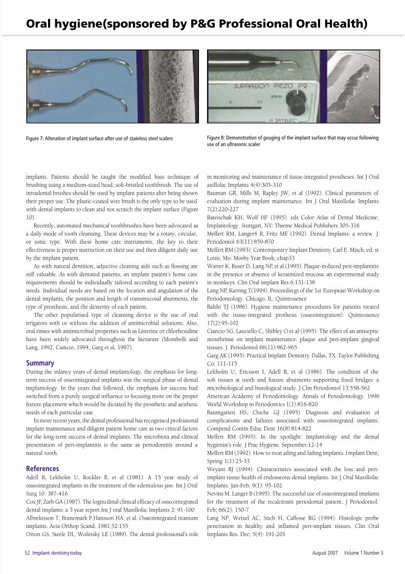
Figure 7: Alteration of implant surface after use of stainless steel scalers Figure 8: Demonstration of gouging of the implant surface that may occur followinguse of an ultrasonic scaler
Figure 9: Demonstration of alteration of the implant surface following application of an air polisher and baking soda. Note the change in surface texture
Figure 10: Plastic-coated interproximal brush applied around implant abutmentsand under the superstructure for plaque removal
I
van Steenberghe D, Klinge B, Linden U, Quirynen M, Herrmann I,
Garpland C (1993). Periodontal indices around natural and titanium
abutments: a longitudinal multicenter study.
J Periodontol. Jun; 64(6): 538-41
Quirynen M, van Steenberghe D, Jacobs R, Schotte A, Darius P (1991).
The reliability of pocket probing around screw-type implants. Clin Oral
Implants Res. Oct-Dec; 2(4): 186-92
Mombelli A, Marxer M, Gaberthuel T, Grunder U, Lang NP (1995).
The microbiota of osseointegrated implants in patients with a history of