Single-Molecule Investigations of RNA Dissociation Nicola H. Green, Philip M. Williams, Omar Wahab, Martyn C. Davies, Clive J. Roberts, Saul J. B. Tendler, and Stephanie Allen Laboratory of Biophysics and Surface Analysis, School of Pharmacy, University of Nottingham, University Park, Nottingham, United Kingdom ABSTRACT Given the essential cellular roles for ribonucleic acids (RNAs) it is important to understand the stability of three- dimensional structures formed by these molecules. This study aims to investigate the dissociation energy landscape for sim- ple RNA structures via atomic-force-microscopy-based single-molecule force-spectroscopy measurements. This approach provides details on the locations and relative heights of the energy barriers to dissociation, and thus information upon the relative kinetic stabilities of the formed complexes. Our results indicate that a simple dodecamer RNA helix undergoes a forced dissociation process similar to that previously observed for DNA oligonucleotides. Incorporating a UCU bulge motif is found to introduce an additional energy barrier closer to the bound state, and also to destabilize the duplex. In the absence of magnesium ions a duplex containing this UCU bulge is destabilized and a single, shorter duplex is formed. These results reveal that a bulge motif impacts upon the forced dissociation of RNA and produces an energy landscape sensitive to the presence of magnesium ions. Interestingly, the obtained data compare well with previously reported ensemble measurements, illustrating the potential of this approach to improve our understanding of RNA stability and dissociation kinetics. INTRODUCTION The RNA molecule plays a fundamental role in some of the most highly conserved cellular processes, demonstrating a versatility of form and function that is not seen for DNA (Saenger, 1984). RNA can relay genetic information to the ribosome for translation as well as acting as a catalyst at the center of this ribosome. In addition, certain RNAs can fold to form catalytic ribozymes, analogous to enzymes, which assist in RNA processing events whereas others facilitate in specific RNA, DNA, or protein interactions. Many of these roles are largely determined by the complex and specific three-dimensional structures adopted by functional RNA. When predicting the three-dimensional structures of RNA molecules it is often possible to assign a number of putative structures to one sequence, with current predictions often being based upon an understanding of the thermodynamic and/or kinetic stabilities of a range of potential structures. An improved knowledge of the forces and barriers that con- trol the kinetic stabilities of such structures would thus be valuable to this process, from both a biological and theoretical point of view. Unlike proteins, whose secondary structures usually depend on the global amino acid sequence, RNA molecules are currently thought to assemble in a hierarchical manner. As a result, RNA exhibits a modular structure with individual structural motifs demonstrating independent characteristics (Saenger, 1984; Brion and Westhof, 1997; Moore, 1999). This has facilitated, in previous theoretical and experimental studies, the investigation of complex RNA molecules through the detailed examination of individual structural motifs, and it is this property that is particularly suited to the approach described herein. Experimentally, researchers have typically studied RNA stability by dissociating/unfolding RNA molecules. Tradi- tionally, this has been done using heat to melt, or chemicals to denature the RNA. These methods are performed in relatively large volumes, require the averaging of data from considerable numbers of molecules, and are likely to involve a varied set of kinetic paths and transient states. Recently, however, single-molecule studies have begun to be em- ployed to elucidate the complex processes of RNA folding. The single-molecule approach provides access to individual molecules within an ensemble, allowing further insights into the folding pathways. Fluorescence microscopy, for exam- ple, has provided information into the folding kinetics of individual ribozymes, and revealed a number of folding pathways, including some not previously observed by ensemble methods (Zhuang et al., 2000). Optical tweezers, meanwhile, have been employed to apply an external force to the ends of long RNA molecules causing them to ‘‘unzip’’. In these experiments, when the molecules were held at a critical force they were also shown to undergo rapid folding and refolding events, facilitating a study of such events under near equilibrium conditions (Liphardt et al., 2001, 2002). More recent investigations of the T. thermophila ribozyme have identified the location of kinetic barriers to mechanical unfolding (Onoa et al., 2003). Atomic-force-microscopy-based force measurements have also been employed for the study of single nucleic acid molecules. Most of these studies have focused upon DNA, including investigations of the force-extension Submitted April 3, 2003, and accepted for publication January 28, 2004. Address reprint requests to Stephanie Allen, Laboratory of Biophysics and Surface Analysis, School of Pharmacy, University of Nottingham, University Park, Nottingham, UK NG7 2RD. Tel.: 44-0-115-9515050; Fax: 144-0-115-9515110; E-mail: [email protected]. Ó 2004 by the Biophysical Society 0006-3495/04/06/3811/11 $2.00 doi: 10.1529/biophysj.103.026070 Biophysical Journal Volume 86 June 2004 3811–3821 3811
Transcript
Single-Molecule Investigations of RNA Dissociation
Nicola H. Green, Philip M. Williams, Omar Wahab, Martyn C. Davies, Clive J. Roberts,Saul J. B. Tendler, and Stephanie AllenLaboratory of Biophysics and Surface Analysis, School of Pharmacy, University of Nottingham, University Park,Nottingham, United Kingdom
ABSTRACT Given the essential cellular roles for ribonucleic acids (RNAs) it is important to understand the stability of three-dimensional structures formed by these molecules. This study aims to investigate the dissociation energy landscape for sim-ple RNA structures via atomic-force-microscopy-based single-molecule force-spectroscopy measurements. This approachprovides details on the locations and relative heights of the energy barriers to dissociation, and thus information upon therelative kinetic stabilities of the formed complexes. Our results indicate that a simple dodecamer RNA helix undergoes a forceddissociation process similar to that previously observed for DNA oligonucleotides. Incorporating a UCU bulge motif is found tointroduce an additional energy barrier closer to the bound state, and also to destabilize the duplex. In the absence ofmagnesium ions a duplex containing this UCU bulge is destabilized and a single, shorter duplex is formed. These results revealthat a bulge motif impacts upon the forced dissociation of RNA and produces an energy landscape sensitive to the presence ofmagnesium ions. Interestingly, the obtained data compare well with previously reported ensemble measurements, illustratingthe potential of this approach to improve our understanding of RNA stability and dissociation kinetics.
INTRODUCTION
The RNA molecule plays a fundamental role in some of the
most highly conserved cellular processes, demonstrating
a versatility of form and function that is not seen for DNA
(Saenger, 1984). RNA can relay genetic information to the
ribosome for translation as well as acting as a catalyst at the
center of this ribosome. In addition, certain RNAs can fold
to form catalytic ribozymes, analogous to enzymes, which
assist in RNA processing events whereas others facilitate in
specific RNA, DNA, or protein interactions. Many of these
roles are largely determined by the complex and specific
three-dimensional structures adopted by functional RNA.
When predicting the three-dimensional structures of RNA
molecules it is often possible to assign a number of putative
structures to one sequence, with current predictions often
being based upon an understanding of the thermodynamic
and/or kinetic stabilities of a range of potential structures.
An improved knowledge of the forces and barriers that con-
trol the kinetic stabilities of such structures would thus
be valuable to this process, from both a biological and
theoretical point of view.
Unlike proteins, whose secondary structures usually
depend on the global amino acid sequence, RNA molecules
are currently thought to assemble in a hierarchical manner.
As a result, RNA exhibits a modular structure with
Biophysical Journal Volume 86 June 2004 3811–3821 3811
properties of DNA molecules of several hundreds to
thousands of basepairs in length (Rief et al., 1999;
Clausen-Schaumann et al., 2000), and the modification of
such properties by various agents, including the binding of
drugs (Krautbauer et al., 2002). In addition, various groups
have demonstrated that it is possible to record interaction
forces between individual complementary oligonucleotides,
as force transducers (e.g., AFM probe) and surfaces
functionalized with such molecules are brought into and
out of contact (Noy et al., 1997; Strunz et al., 1999; Grange
et al., 2001; Pope et al., 2001; Schumakovitch et al., 2002).
In this type of experiment (termed dynamic force spectros-
copy measurements if recorded over a range of loading rates;
Evans and Ritchie, 1997, 1999; Evans, 1998, 2001; Merkel
et al., 1999) a force is applied to the oligonucleotide complex
formed during probe-sample contact, and the thermally
induced dissociation event accelerated. Through this ap-
proach information upon energy barriers traversed during
force-induced processes have been provided, and recent
experiments have revealed how such data can be influenced
by the length of the duplex probed (Strunz et al., 1999) and
a change in the experimental environment (Schumakovitch
et al., 2002). The following studies were performed to further
extend this latter experimental approach through the in-
vestigation of the forced dissociation of a range of RNA
oligonucleotides. For the analysis of RNA stability, this
approach enables the modular nature of RNA to be exploited
and provides a method to investigate the dissociation kinetics
over located energetic barriers, and thus a means to explore
the relative kinetic stabilities of small structural units.
The presented studies focus upon the forced dissociation
of a 12-mer and 24-mer RNA double helix before and after
the insertion of a putative trinucleotide bulge motif. The
double helix itself is not considered a structural motif,
because the length is arbitrary and the conformation remains
unaltered by residue additions or deletions; however it is
a dominant element within RNA molecules, accounting for
as much as 50% of the residues in an average nonmessenger
RNA molecule (Saenger, 1984). The structural diversity of
RNA is provided by a combination of specific structural
motifs inserted within these helical stretches. The bulge
structure is one such motif, universally distributed through
all structurally functional RNAs. It consists of a small number
of unpaired residues in one strand, which may either extrude
from the duplex or be accommodated by stacking between
adjacent basepairs, and is known to play a biologically
significant role within RNA molecules as it creates unique
recognition sites within the RNA molecule.
The UCU bulge sequence studied in these experiments is
found within a stretch of purine residues in the HIV-1 TAR
(trans-activation response) element. This bulge has been the
subject of comprehensive study, being the recognition site
for the binding of the viral regulatory Tat protein (reviewed
by Aboul-ela et al., 1996). Structural analysis of HIV-1 TAR
has shown that the conformation of the molecule is
influenced by the presence of magnesium ions (Zacharias
and Hagerman, 1995; Ippolito and Steitz, 1998). Conse-
quently, the dissociation of a duplex containing the UCU
bulge structure was also studied in the presence and ab-
sence of magnesium ions to determine if changes in stability
could be detected and characterized by dynamic force
spectroscopy.
MATERIALS AND METHODS
All chemicals were obtained from Sigma Aldrich (Poole, UK) unless
otherwise stated. Care was taken to eliminate ribonuclease contamination
from the experiment by employing sterile techniques in the preparation of
samples. In addition, gloves were worn at all times and sterilized, disposable
plasticware was used where possible. All water used was of 18 MV cm
resistivity and was pretreated overnight with diethylpyrocarbonate (DEPC,
0.05% v/v) before being autoclaved for 30 min. All solutions were passed
through sterile 0.2 mm Minisart filters (Sartorius AG, Gottingen, Germany)
before use, all work surfaces including the AFM liquid cells were wiped with
an RNase inhibitor (RNaseZap), and the liquid cell was rinsed thoroughly
before use.
Functionalization of AFM surfaces
Short RNA oligonucleotides were synthesized with a ‘‘Phosphoramidite
18’’ linker (Oswell Research Products, University of Southampton, UK),
with a hexaethyleneglycol phosphoramidite-thiophosphate linker at the 5#terminus resulting in a free thiol at the 5# end of the molecule. The free thiol
functionality was included to facilitate covalent attachment to gold surfaces
and AFM probes, and the ethyleneglycol linker molecules to space the
oligonucleotide away from the underlying substrate and thus minimize its
impact upon the interaction (Hinterdorfer et al., 2000). Gold-coated surfaces
(Hegner et al., 1993) and AFM probes were then functionalized with these
oligonucleotides to produce the self-assembled monolayer architecture
described by Noy et al. (1997). Undecanol-thiol was incorporated into the
incubation mixture at 200-fold higher concentration to dilute the RNA
surface density and ensure a high probability for the formation of single
RNA duplexes.
To prepare the interacting surfaces, AFM cantilever probes (NanoProbes,
Veeco, Santa Barbara, CA; nominal spring constants 10–55 pN/nm), which
had been gold sputter-coated, and template-stripped gold substrates were
incubated overnight at room temperature in 1 mM oligonucleotide solutions,
200 mM undecanol-thiol, 75% (v/v) ethanol, 40 mM Tris buffer, pH 7. The
surfaces were then rinsed in 20 mM Tris buffer, pH 7, 10 mMMgCl2 for use
in force-spectroscopy experiments the same day. MgCl2 was omitted from
the wash for experiments performed in the absence of magnesium ions.
Four distinct duplexes were studied in the course of these experiments
(Table 1). The first required the tip to be functionalized with the 12-mer
sequence GCGUUUUUUGCG while the substrate was coated with its fully
complementary sequence. For the second series of experiments, the same
12-mer bulge duplex sequence was used on the tip and a 15-mer
oligonucleotide, based on the complementary sequence with a UCU
trinucleotide incorporated at positions 7–9, on the substrate. The third
duplex consisted of a 24-mer fully complementary sequence related to the
12-mer sequence, but with an extended run of adenine-uracil basepairs. The
fourth experimental configuration aimed to investigate the impact of the
UCU trinucleotide bulge (inserted at positions 13–15) upon the forced
dissociation of the 24-mer duplex. Again these experiments employed tips
functionalized with a 24-base oligomer, while the substrates were coated
with the complementary sequence containing the trinucleotide bulge
sequence.
The dodecamer sequence was chosen to promote the formation of a single
12-mer duplex, to avoid collecting data from a number of duplexes formed
3812 Green et al.
Biophysical Journal 86(6) 3811–3821
by partially overlapping strands. Examination of this sequence reveals that
the maximum number of interacting complementary basepairs within
a partial overlap is six, and it is anticipated that formation of this structure is
significantly less favorable than the fully complementary 12 mer. A similar
lack of partial overlapping is expected from the 12-mer bulge-containing
sequences. 24-mer sequences were chosen as to provide extended structures
with sequences related as closely as possible to that of the 12-mer and 12-
mer bulge sequences. Importantly, in none of the putative duplexes could the
UCU bulge nucleotides form complementary basepairs with the opposing
strand.
Force-distance experiments
Force-distance measurements were carried out using the Molecular Force
Probe (Asylum Research, Santa Barbara, CA). In most instances, experi-
ments were performed in 20 mM Tris, pH 7, 10 mM MgCl2. The exception
to this was when studying the effect of the absence of magnesium ions, when
the MgCl2 was omitted. In all cases an RNA-functionalized tip at the end of
a flexible cantilever was brought into and out of contact with an RNA-
modified surface. The gradient of the retract trace when the tip was in contact
with a hard surface enabled the measured detector signal to be converted into
actual deflection (Lee et al., 1994), allowing the horizontal distance axis to
be corrected from z-piezo displacement to tip-sample separation. The force
of interaction between tip and sample was then determined by Hooke’s law,
as a product of the cantilever spring constant and the cantilever deflection
distance. The spring constant of each cantilever used was ascertained
through thermal fluctuations of the cantilever (Hutter and Bechhoefer,
1993).
Applied loading rates were approximated as the product of the cantilever
spring constant and the piezo retract velocity (Evans, 1998), as our
measurements were dominated by cantilever loading. This approximation
was made because the linker molecules employed in our studies were short
(;2–3 nm per RNA molecule) and the cantilevers used were soft. Both
velocity and cantilever spring constant were varied during the course of the
study to provide the required range of rates. At least 800 force curves were
collected at each loading rate and the specificity of the tip-substrate
interaction confirmed by blocking the substrate with an excess of the
complementary sequence.
RESULTS
Analysis of force-distance data
Only force-distance curves attributed to specific interactions
between RNA molecules were analyzed within our experi-
ments. It should be noted that control experiments confirmed
that specific interactions did not occur between surfaces
functionalized only with undecanol-thiol spacer molecules,
whereas previous experiments have shown that specific
interactions are not observed when surfaces are functional-
ized with identical oligonucleotides (Pope et al., 2001). As
seen in Fig. 1, it was possible to separate specific interaction
events from the small number of curves showing nonspecific
interactions due to a change in the gradient of the retract
slope observed in force-distance curves derived from specific
interactions (Willemsen et al., 1998). This arose because
the specific interactions occurred with rupture lengths
TABLE 1 The duplex sequences studied by dynamic force
spectroscopy
Name Duplex sequence
12-merCGCAAAAAACGC
GCGUUUUUUGCG
12-merB
UCU
CGCAAA AAACGC
GCGUUU---UUUGCG
24-merCGCAAAAAAAAAAAAAAAAAACGC
GCGUUUUUUUUUUUUUUUUUUGCG
24-merB
UCU
CGCAAAAAAAAA AAAAAAAAACGC
GCGUUUUUUUUU---UUUUUUUUUGCG
FIGURE 1 Examples of the types of force curves observed between RNA
functionalized AFM surfaces and tips; each curve has been corrected to
show force against tip-sample displacement. In each example the approach
trace is shown in gray and the retract trace in black. (A) No interaction has
occurred between the tip and substrate. (B) A nonspecific interaction
between the two surfaces, with no observable change in the gradient of the
retraction slope. (C) A specific interaction between the two RNA
functionalized surfaces, showing a stretch of ;10 nm before the rupture
event.
Dissociation of Single RNA Molecules 3813
Biophysical Journal 86(6) 3811–3821
approximately equivalent to the length of the RNA molecule
plus twice the length of the linker, whereas nonspecific adhe-
sions occurred with a rupture length of zero. The combined
length of two fully extended hexaethyleneglycol linkers is
;4 nm (assuming a length of ;3.4 A per ethytleneglycol
monomer), and neglecting the geometry of the tip/sample
contact a 12-mer RNA duplex before extension would be
;3.4 nm and a 24-mer RNA duplex ;6.8 nm. Little varia-
tion in the mean rupture length was actually observed be-
tween all the experiments and a mean rupture length of 11.5
6 7.6 nm was calculated for all the force curves analyzed.
In each experiment, the proportion of force curves that
resulted in a specific interaction between the two surfaces was
determined. If they arose more frequently than one force-
distance curve in five the experiment was discarded because
the probability that multiple interactions were occurring
became too high (Williams and Evans, 2001). In the presented
study from between one in five and one in 12 of the curves
demonstrated a specific interaction. At the end of the ex-
periment, to verify the specificity of the probe-sample inter-
action, the surface was incubated with the complementary
RNA molecule, reducing further the number of specific
sites available for interactionwith the functionalized tip. After
this blocking stage the frequency of force curves showing
a specific interaction fell by a minimum of 50%, confirming
that the observed interactions were between complementary
RNA strands.
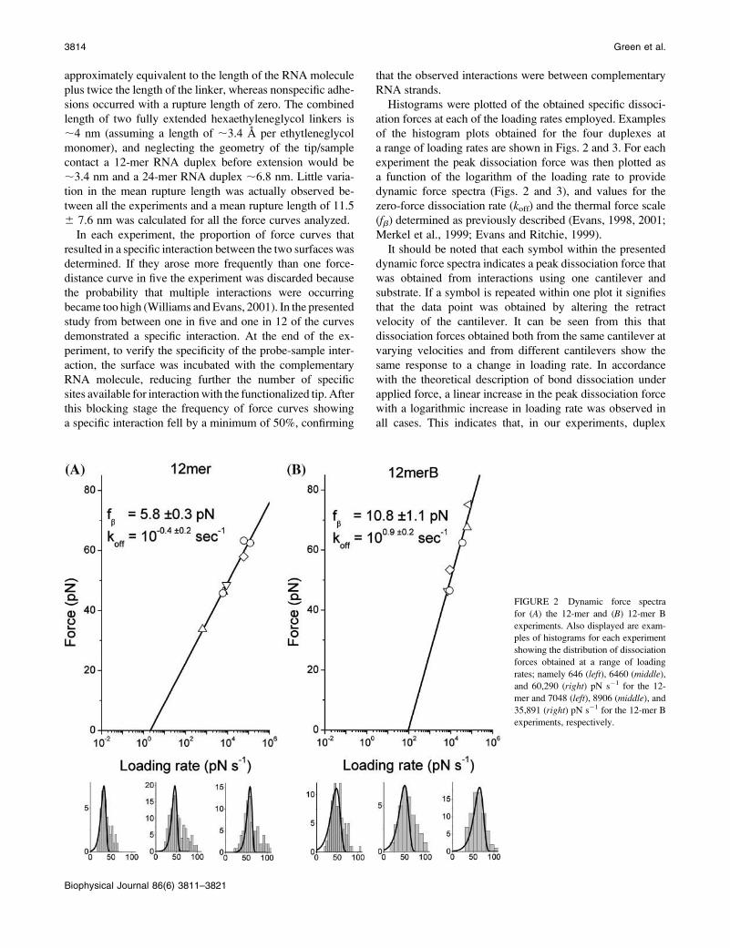
Histograms were plotted of the obtained specific dissoci-
ation forces at each of the loading rates employed. Examples
of the histogram plots obtained for the four duplexes at
a range of loading rates are shown in Figs. 2 and 3. For each
experiment the peak dissociation force was then plotted as
a function of the logarithm of the loading rate to provide
dynamic force spectra (Figs. 2 and 3), and values for the
zero-force dissociation rate (koff) and the thermal force scale
(fb) determined as previously described (Evans, 1998, 2001;
Merkel et al., 1999; Evans and Ritchie, 1999).
It should be noted that each symbol within the presented
dynamic force spectra indicates a peak dissociation force that
was obtained from interactions using one cantilever and
substrate. If a symbol is repeated within one plot it signifies
that the data point was obtained by altering the retract
velocity of the cantilever. It can be seen from this that
dissociation forces obtained both from the same cantilever at
varying velocities and from different cantilevers show the
same response to a change in loading rate. In accordance
with the theoretical description of bond dissociation under
applied force, a linear increase in the peak dissociation force
with a logarithmic increase in loading rate was observed in
all cases. This indicates that, in our experiments, duplex
FIGURE 2 Dynamic force spectra
for (A) the 12-mer and (B) 12-mer B
experiments. Also displayed are exam-
ples of histograms for each experiment
showing the distribution of dissociation
forces obtained at a range of loading
rates; namely 646 (left), 6460 (middle),and 60,290 (right) pN s�1 for the 12-
mer and 7048 (left), 8906 (middle), and
35,891 (right) pN s�1 for the 12-mer B
experiments, respectively.
3814 Green et al.
Biophysical Journal 86(6) 3811–3821
separation occurred within the thermally active regime and
that a single energy barrier was being probed in each
experiment (Evans and Ritchie, 1997), albeit over the very
narrow range of rates possible with the AFM.
Values for koff and fb (Table 2) were derived from each
dynamic force spectrum by least squares fitting through the
points, and these data used to predict the full distribution of
dissociation forces at the rates tested (Evans and Williams,
2001). Superimposed upon each data histogram is that
predicted for correlated bond failure, assuming no experi-
mental error. It can be seen that, particularly at the lower
range of dissociation forces, there is good agreement
between this model distribution and the experimental data;
however, at higher forces more frequent events are
sometimes observed in the experimental data than are
predicted by the model. We attribute these higher force
interactions to the low frequency incidence of multiple RNA
interactions between the tip and substrate. Finally, ranges for
the estimated koff and fb values (Table 2) were determined by
applying the ‘‘bootstrap’’ Monte Carlo resampling method
(Press et al., 1992) with 100 replacements to the peak dis-
sociation forces obtained at the varying loading rates.
Although there are several potential sources of error within
these measurements, the error associated with calibration of
the cantilever spring is likely have the most impact, with an
absolute uncertainty estimated at;10% (Florin et al., 1995).
The influence of this uncertainty is, however, reduced in our
experiments because data sets were obtained using both a
number of different cantilevers and a range of retract
velocities for the same cantilever, and in all instances the
experimentally obtained peak dissociation force demon-
strated a comparable linear response to the logarithm of the
loading rate. It should also be noted that the good agreement
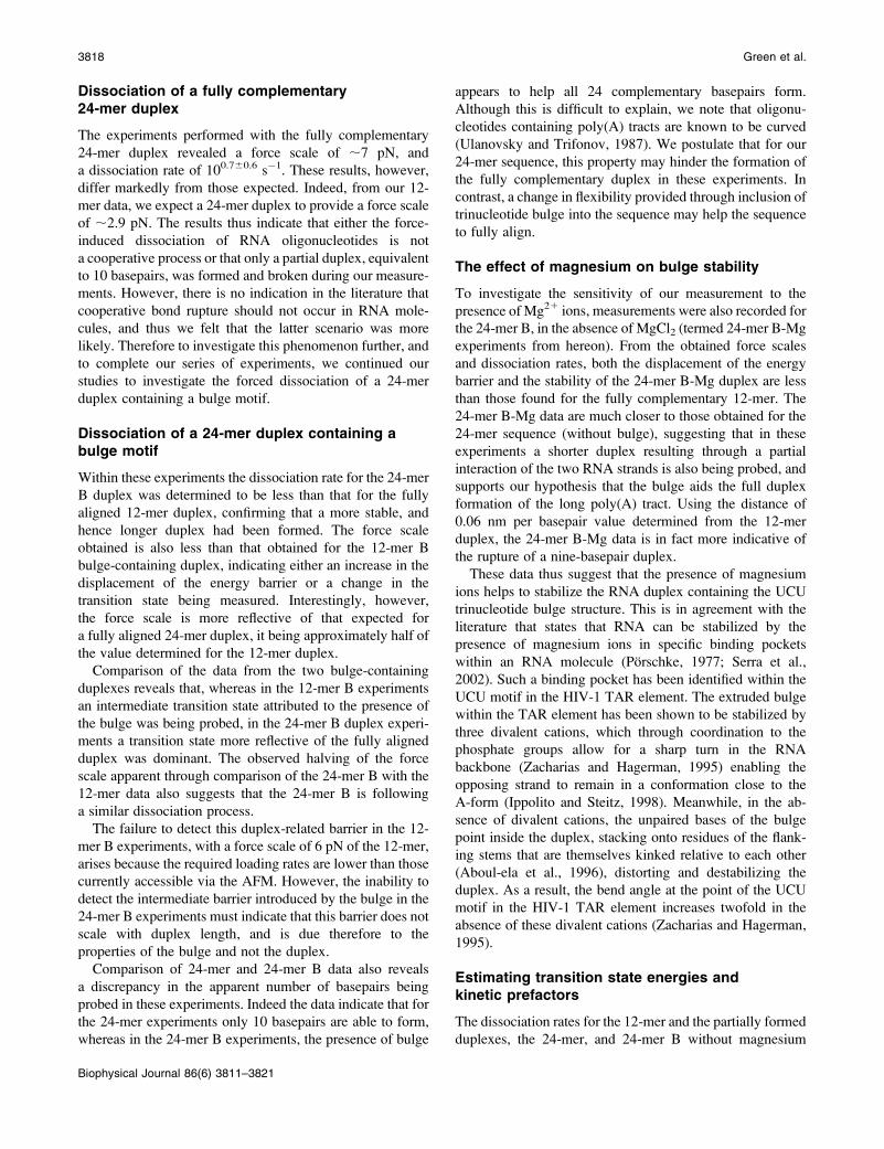
FIGURE 3 Dynamic force spectra for the (A) 24-mer, (B) 24-mer B, and (C) 24-mer B-Mg experiments. Example histograms for each experiment showing
the distribution of dissociation forces at a range of loading rates are also displayed; namely 7691 (left), 23,886 (middle), and 55,734 (right) pN s�1 for the
24-mer; 1391 (left), 1653 (middle), and 66,290 (right) pN s�1 for the 24-mer B; and 27,632 (left), 83,480 (middle), and 264,300 (right) pN s�1 for the 24-mer
B-Mg experiments, respectively.
TABLE 2 The dissociation rate (koff) and force scale (fb)
determined for all the duplexes studied
Name koff (s�1) fb (pN)
Approximate
koff/koff (12-mer)
Approximate
fb/fb (12-mer)
12-mer 10�0.460.2 5.8 6 0.3 1 1
12-merB 100.960.2 10.8 6 1.1 20 2
24-mer 100.760.6 7.0 6 1.4 13 1.2
24-merB 10�4.060.8 2.8 6 0.3 0.0003 0.5
24-merB-Mg* 101.160.1 7.7 6 0.3 32 0.8
Approximate factors, relative to the 12-mer data, for koff and fb are also
provided.
*Experiment performed in the absence of Mg21.
Dissociation of Single RNA Molecules 3815
Biophysical Journal 86(6) 3811–3821
between data obtained with different cantilevers, and their
response to loading rate, is further evidence that our
measurements are dominated by cantilever loading.
Dissociation of a 12-mer fullycomplementary duplex
Experiments were first performed using the two 12-mer fully
Consideration of the data obtained from the 12-mer duplex
indicates that the combined lifetime of the two 12-mer halves
within the 24-mer B bulge-containing duplex would be short
(koff ; 10�0.6s�1) compared to the total lifetime of the
24-mer B complex (koff ¼ 10�4.0 s�1). This in turn suggests
that the bulge would be the dominant contributor to stability
(koff ; 10�4.0 s�1). Because in this scenario, the lifetime of
the bulge should be independent of the lengths of the
surrounding helices, the stability of this bulge within the 24-
mer B duplex would be the same within the 12-mer B
duplex. However, it can be seen that the dissociation rate for
the complete 12-mer B duplex containing the same UCU
trinucleotide bulge (koff ¼ 100.9 s�1) is significantly faster
than the presumed lifetime of this bulge component. This
demonstrates that the lifetime of a complete bulge-contain-
ing duplex is not simply a sum of its components. Hence, this
indicates that there is no disruption to the cooperativity of
unbinding after insertion of the bulge motif; rather there is an
increase in the complexity of the dissociation landscape.
A comparison of the location of the energy barrier for the
12-mer and 12-mer B duplexes provides further information
regarding the dissociation landscape after the insertion of the
bulge. It can be seen that in the 12-mer B data, alongside an
increase in the complexity of the landscape, there is also
a doubling in the force scale. This change in barrier
displacement without an accompanying change in basepair
number reveals that the 12-mer B experiments must be
probing an additional barrier introduced with the insertion of
the bulge. The presence of this additional barrier after the
insertion of the bulge indicates that an unstable intermediate
structure must exist between the fully bound and unbound
states. It can also be seen that the location of this additional
transition state along the dissociation coordinate can be
related to the cooperative response from half the number of
basepairs within the duplex and thus may be associated with
the location of the bulge motif within the duplex, although
this relationship needs to be confirmed by further experi-
mentation.
Dissociation of Single RNA Molecules 3817
Biophysical Journal 86(6) 3811–3821
Dissociation of a fully complementary24-mer duplex
The experiments performed with the fully complementary
24-mer duplex revealed a force scale of ;7 pN, and
a dissociation rate of 100.760.6 s�1. These results, however,
differ markedly from those expected. Indeed, from our 12-
mer data, we expect a 24-mer duplex to provide a force scale
of ;2.9 pN. The results thus indicate that either the force-
induced dissociation of RNA oligonucleotides is not
a cooperative process or that only a partial duplex, equivalent
to 10 basepairs, was formed and broken during our measure-
ments. However, there is no indication in the literature that
cooperative bond rupture should not occur in RNA mole-
cules, and thus we felt that the latter scenario was more
likely. Therefore to investigate this phenomenon further, and
to complete our series of experiments, we continued our
studies to investigate the forced dissociation of a 24-mer
duplex containing a bulge motif.
Dissociation of a 24-mer duplex containing abulge motif
Within these experiments the dissociation rate for the 24-mer
B duplex was determined to be less than that for the fully
aligned 12-mer duplex, confirming that a more stable, and
hence longer duplex had been formed. The force scale
obtained is also less than that obtained for the 12-mer B
bulge-containing duplex, indicating either an increase in the
displacement of the energy barrier or a change in the
transition state being measured. Interestingly, however,
the force scale is more reflective of that expected for
a fully aligned 24-mer duplex, it being approximately half of
the value determined for the 12-mer duplex.
Comparison of the data from the two bulge-containing
duplexes reveals that, whereas in the 12-mer B experiments
an intermediate transition state attributed to the presence of
the bulge was being probed, in the 24-mer B duplex experi-
ments a transition state more reflective of the fully aligned
duplex was dominant. The observed halving of the force
scale apparent through comparison of the 24-mer B with the
12-mer data also suggests that the 24-mer B is following
a similar dissociation process.
The failure to detect this duplex-related barrier in the 12-
mer B experiments, with a force scale of 6 pN of the 12-mer,
arises because the required loading rates are lower than those
currently accessible via the AFM. However, the inability to
detect the intermediate barrier introduced by the bulge in the
24-mer B experiments must indicate that this barrier does not
scale with duplex length, and is due therefore to the
properties of the bulge and not the duplex.
Comparison of 24-mer and 24-mer B data also reveals
a discrepancy in the apparent number of basepairs being
probed in these experiments. Indeed the data indicate that for
the 24-mer experiments only 10 basepairs are able to form,
whereas in the 24-mer B experiments, the presence of bulge
appears to help all 24 complementary basepairs form.
Although this is difficult to explain, we note that oligonu-
cleotides containing poly(A) tracts are known to be curved
(Ulanovsky and Trifonov, 1987). We postulate that for our
24-mer sequence, this property may hinder the formation of
the fully complementary duplex in these experiments. In
contrast, a change in flexibility provided through inclusion of
trinucleotide bulge into the sequence may help the sequence
to fully align.
The effect of magnesium on bulge stability
To investigate the sensitivity of our measurement to the
presence of Mg21 ions, measurements were also recorded for
the 24-mer B, in the absence of MgCl2 (termed 24-mer B-Mg
experiments from hereon). From the obtained force scales
and dissociation rates, both the displacement of the energy
barrier and the stability of the 24-mer B-Mg duplex are less
than those found for the fully complementary 12-mer. The
24-mer B-Mg data are much closer to those obtained for the
24-mer sequence (without bulge), suggesting that in these
experiments a shorter duplex resulting through a partial
interaction of the two RNA strands is also being probed, and
supports our hypothesis that the bulge aids the full duplex
formation of the long poly(A) tract. Using the distance of
0.06 nm per basepair value determined from the 12-mer
duplex, the 24-mer B-Mg data is in fact more indicative of
the rupture of a nine-basepair duplex.
These data thus suggest that the presence of magnesium
ions helps to stabilize the RNA duplex containing the UCU
trinucleotide bulge structure. This is in agreement with the
literature that states that RNA can be stabilized by the
presence of magnesium ions in specific binding pockets
within an RNA molecule (Porschke, 1977; Serra et al.,
2002). Such a binding pocket has been identified within the
UCU motif in the HIV-1 TAR element. The extruded bulge
within the TAR element has been shown to be stabilized by
three divalent cations, which through coordination to the
phosphate groups allow for a sharp turn in the RNA
backbone (Zacharias and Hagerman, 1995) enabling the
opposing strand to remain in a conformation close to the
A-form (Ippolito and Steitz, 1998). Meanwhile, in the ab-
sence of divalent cations, the unpaired bases of the bulge
point inside the duplex, stacking onto residues of the flank-
ing stems that are themselves kinked relative to each other
(Aboul-ela et al., 1996), distorting and destabilizing the
duplex. As a result, the bend angle at the point of the UCU
motif in the HIV-1 TAR element increases twofold in the
absence of these divalent cations (Zacharias and Hagerman,
1995).
Estimating transition state energies andkinetic prefactors
The dissociation rates for the 12-mer and the partially formed
duplexes, the 24-mer, and 24-mer B without magnesium
3818 Green et al.
Biophysical Journal 86(6) 3811–3821
(24-mer B-Mg), permit an estimation of the average contribu-
tion of each basepair to the transition state energy, alongside
the corresponding estimation of the exponential prefactor
of the dissociation kinetics. Under cooperative failure the
duplex can be considered as the compound bond and,
neglecting any sequence dependence, a generic expression
for the dissociation rate can be written as
koffðNÞ � 1
NtDexp �NEb
kBT
� �; (1)
with each basepair adding Eb of energy to the transition state;
where N is the number of basepairs (where N ¼ (kBT / fb)/(xb per basepair, as determined from our 12-mer data), or
;70 / fb) and tD is the diffusional relaxation time. Using
loge½NkoffðNÞ� ¼ � Eb
kBTN1 loge
1
tD
� �; (2)
Fig. 4 shows the estimation of Eb and 1/tD of 1.1 kBT/b.p.
and 2 3 106 s�1, respectively. We are, of course, wary of
the considerable variance in these estimates. The 1.1 kBT/
b.p. contribution to the transition state energy may also at
first seem unreasonably low as it is similar to estimates of the
thermodynamic stability per basepair. However, under force
the molecule is likely to undergo some conformational
change (stretch) before dissociation and so here we are not
measuring the transition state energy relative to the ground
state but rather from a destabilized form (Pope et al., 2001).
We reiterate, therefore, that our measurements and predic-
tions of rate above are for dissociation under force. The
kinetic prefactor suggests a diffusional relaxation time of
a few hundred nanoseconds, and lying between the nano-
seconds for the diffusion of small ligands and the micro-
seconds of proteins (Schuler et al., 2002), it also seems
a reasonable estimate. It should be noted that this relaxation
time is comparable to the longest timescale currently
available for atomistic molecular simulation.
Plotted also in Fig. 4 are the data for the 12-mer B
(diamond) and 24-mer B with magnesium (triangle).Although the data for the sequences in which only simple
duplexes are formed (i.e., the 12-mer, 24-mer, and 24-mer
Mg data) lie on the same line, the 12-mer B dissociation
profile differs significantly, supporting our conclusion that
the transition state measured in this dissociation is of a bulge
formed in a 12-mer duplex and not dissociation of a partially
formed 6-mer. Using the values of Eb and 1/tD Eq. 1 permits
us to estimate how a 6-mer duplex would behave under force
and contrast this to that of the 12-mer B. The dissociation
rate of a 6-mer duplex is calculated to be;450 s�1, which is
60 times faster than we measured for the 12-merB. With
a force scale of ;12 pN, a 6-mer would require loading at
.30,000 pN s�1 to measure a force above 20 pN, and such
a study would therefore require the use of cantilevers with
much greater stiffness and consequently different immobi-
lization chemistries. The dissociation rate of the 12-mer B,
therefore, cannot be compared to the other duplexes as the
transition state being measured is clearly different.
In addition, the 24-mer B (in the presence of Mg21)
dissociates at a rate significantly faster than we extrapolate
for a 24-mer. The increase in rate is equivalent to a reduction
in the energy to dissociate of around 5 kBT. Nearest-neighbor
predictions (Mathews et al., 1999) have suggested that upon
insertion of a UCU bulge there is a drop in stability of ;7
kBT (with the inclusion of terminal AU penalty terms; Xia
et al., 1998), suggesting the duplex is only partly structured
in the transition state, if at all. This again is consistent with
the presence of a bulge-induced transition state on the
dissociation landscape, which is crossed before the final
dissociation event.
CONCLUSIONS
The presented study has revealed single-molecule dissocia-
tion data for RNA molecules of increasing structural
complexity. We have demonstrated that a simple RNA
duplex dissociates in a comparable manner to that previously
reported for DNA duplexes of similar length. With this
behavior in mind, we were able to show that the in-
corporation of a bulge motif adds complexity to the force-
induced dissociation landscape through the introduction of
an additional barrier. Interestingly, there has been some
FIGURE 4 Using Eq. 2, a plot of loge[Nkoff(N)] versusN reveals estimates
for the contribution of each basepair to the probed transition states (DE), andalso the exponential prefactor for dissociation kinetics (1/tD).
Dissociation of Single RNA Molecules 3819
Biophysical Journal 86(6) 3811–3821
published evidence for the existence of an intermediate in
the dissociation pathway of a bulge-containing structure in
solution experiments (Davis et al., 1998). Most significantly,
studies have demonstrated that many bulge-containing RNA
species do not melt in a two-state manner (Longfellow et al.,
1990).
The destabilizing effect by a bulge motif has been
observed in a number of solution melting studies (Long-
fellow et al., 1990; Leblanc and Morden, 1991; Zagorowska
and Adamiak, 1996). Through experiments performed with
longer 24-mer oligonucleotides we were able to estimate the
destabilizing effect of the introduced UCU bulge motif, and
also demonstrate its sensitivity to the presence of magnesium
ions. Importantly, through consideration of all of the pre-
sented data we could also provide estimates for the contribu-
tion of each basepair to the explored transition states, and for
the exponential prefactor for dissociation kinetics.
Our results thus demonstrate the potential of single-
molecule dynamic force measurements to profile the dis-
sociation landscapes of RNAmolecules and to also probe the
impact of specific motifs upon their kinetic stabilities. We
believe that such experiments, together with the other single-
molecule approaches currently under development, promise
to profoundly impact upon our understanding of RNA
stability and dissociation energetics.
Thanks go to Kelvin Chung for his help in obtaining the results in the
absence of magnesium ions.
N.H.G. thanks the BBSRC for funding and S.A. thanks Pfizer Global
Research and Development for funding her lectureship. P.M.W. is an
EPSRC Advanced Research Fellow.
REFERENCES
Aboul-ela, F., J. Karn, and G. Varani. 1996. Structure of HIV-1 TAR RNAin the absence of ligands reveals a novel conformation of thetrinucleotide bulge. Nucleic Acids Res. 24:3974–3981.
Brion, P., and E. Westhof. 1997. Hierarchy and dynamics of RNA folding.Annu. Rev. Biophys. Biomol. Struct. 26:113–137.
Clausen-Schaumann, H., M. Rief, C. Tolksdorf, and H. E. Gaub. 2000.Mechanical stability of single DNA molecules. Biophys. J. 78:1997–2007.
Conte, M. R., G. L. Conn, T. Brown, and A. L. Lane. 1997. Conforma-tional properties and thermodynamics of the RNA duplexr(CGCAAAUUUGCG)2: comparison with the DNA analogued(CGCAAATTTGCG)2. Nucleic Acids Res. 25:2627–2634.
Davis, T. M., L. McFail-Isom, E. Keane, and L. D. Williams. 1998. Meltingof a DNA hairpin without hyperchromism. Biochemistry. 37:6975–6978.
Evans, E. 1998. Energy landscapes of biomolecular adhesion and receptoranchoring explored with dynamic force spectroscopy. Faraday Discuss.Chem. Soc. 111:1–16.
Evans, E. 2001. Probing the relationship between force—lifetime—andchemistry in single molecular bonds. Annu. Rev. Biophys. Biomol. Struct.30:105–128.
Evans, E., and K. Ritchie. 1997. Dynamic strength of molecular adhesionbonds. Biophys. J. 72:1541–1555.
Evans, E., and K. Ritchie. 1999. Strength of a weak bond connectingflexible polymer chains. Biophys. J. 76:2439–2447.
Evans, E., and P. Williams. 2001. Dynamic force spectroscopy. I. Singlebonds. In Les Houches Session LXXV Physics of Bio-Molecules andCells. H. Flyvbjerg, F. Julicher, P. Ormos, and F. David, editors.Springer-Verlag, Berlin, Germany. 145–186.
Florin, E. L., M. Rief, H. Lehmann, M. Ludwig, C. Dornmair, V. T. Moy,and H. E. Gaub. 1995. Sensing specific molecular-interactions with theatomic-force microscope. Biosens. Bioelectron. 10:895–901.
Freier, S. M., R. Kierzek, J. A. Jaeger, N. Sugimoto, M. H. Caruthers,T. Neilson, and D. H. Turner. 1986. Improved free-energy parametersfor predictions of RNA duplex stability. Proc. Natl. Acad. Sci. USA.83:9373–9377.
Grange, W., T. Strunz, I. Schumakovitch, H.-J. Guntherodt, and M. Hegner.2001. Molecular recognition and adhesion of individual DNA strandsstudied by dynamic force spectroscopy. Single Mol. 2:75–78.
Hegner, M., P. Wagner, and G. Semenza. 1993. Ultralarge atomically flattemplate-stripped Au surfaces for scanning probe microscopy. Surf. Sci.291:39–46.
Hinterdorfer, P., F. Kienberger, A. Raab, H. J. Gruber, W. Baumgartner, G.Kada, C. Riener, S. Wielert-Badt, C. Borken, and H. Schindler. 2000.Poly(ethylene glycol): an ideal spacer for molecular recognition forcemicroscopy/spectroscopy. Single Mol. 1:99–103.
Hutter, J. L., and J. Bechhoefer. 1993. Calibration of atomic forcemicroscopy tips. Rev. Sci. Instrum. 64:1868–1873.
Ippolito, J. A., and T. A. Steitz. 1998. A 1.3 A resolution crystal structure ofthe HIV-1 trans-activation response region RNA stem reveals a metalion-dependent bulge conformation. Proc. Natl. Acad. Sci. USA. 95:9819–9824.
Krautbauer, R., L. H. Pope, T. E. Schrader, S. Allen, and H. E. Gaub. 2002.Discriminating drug-DNA binding modes by single molecule forcespectroscopy. FEBS Lett. 510:154–158.
Leblanc, D. A., and K. M. Morden. 1991. Thermodynamic characterizationof deoxyribooligonucleotide duplexes containing bulges. Biochemistry.30:4042–4047.
Lee, G. U., D. A. Kidwell, and R. J. Colton. 1994. Sensing discretestreptavidin biotin interactions with atomic force microscopy. Langmuir.10:354–357.
Liphardt, J., S. Dumont, S. B. Smith, I. Tinoco, Jr., and C. Bustamante.2002. Equilibrium information from nonequilibrium measurements in anexperimental test of Jarzynski’s equality. Science. 296:1832–1835.
Liphardt, J., B. Onoa, S. B. Smith, I. Tinoco, Jr., and C. Bustamante. 2001.Reversible unfolding of single RNA molecules by mechanical force.Science. 292:733–737.
Longfellow, C. E., R. Kierzek, and D. H. Turner. 1990. Thermodynamicand spectroscopic study of bulge loops in oligoribonucleotides. Bio-chemistry. 29:278–285.
Mathews, D. H., J. Sabina, M. Zuker, and D. H. Turner. 1999. Expandedsequence dependence of thermodynamic parameters improves predictionof RNA secondary structure. J. Mol. Biol. 288:911–940.
Merkel, R., P. Nassoy, A. Leung, K. Ritchie, and E. Evans. 1999. Energylandscapes of receptor-ligand bonds explored with dynamic forcespectroscopy. Nature. 397:50–53.
Moore, P. B. 1999. Structural motifs in RNA. Annu. Rev. Biochem. 68:287–300.
Mukerji, I., and A. P. Williams. 2002. UV resonance Raman and circulardichromism studies of a DNA duplex containing an A3T3 tract: evidencefor a pre-melting transition and three-centered H-bonds. Biochemistry.41:69–77.
Noy, A., D. V. Vezenov, J. F. Kayyem, T. J. Meade, and C. M. Lieber.1997. Stretching and breaking duplex DNA by chemical force. Chem.Biol. 4:519–527.
Onoa, B., S. Dumont, J. Liphardt, S. B. Smith, I. Tinoco, Jr., and C.Bustamante. 2003. Identifying kinetic barriers to mechanical unfoldingof the T. thermophila ribozyme. Science. 299:1892–1895.
Pope, L. H., M. C. Davies, C. A. Laughton, C. J. Roberts, S. J. B. Tendler,and P. M. Williams. 2001. Force-induced melting of a short DNA doublehelix. Eur. Biophys. J. 30:53–62.
3820 Green et al.
Biophysical Journal 86(6) 3811–3821
Porschke, D. 1977. Elementary steps of base recognition and helix-coiltransitions in nucleic acids. Mol. Biol. Biochem. Biophys. 24:191–218.
Press, W. H., S. A. Teukolsky, W. T. Vetterling, and B. P. Flannery. 1992.Numerical recipes in C. Cambridge University Press, New York.
Rief, M., H. Clausen-Schaumann, and H. E. Gaub. 1999. Sequencedependent mechanics of single DNA molecules. Nat. Struct. Biol. 6:346–349.
Saenger, W. 1984. Principles of Nucleic Acid Structure. Springer Verlag,New York.
Schuler, B., E. A. Lipman, and W. A. Eaton. 2002. Probing the free-energysurface for protein folding with single molecule fluorescence spectros-copy. Nature. 419:743–747.
Schumakovitch, I., W. Grange, T. Strunz, P. Bertoncini, H.-J. Guntherodt,and M. Hegner. 2002. Temperature dependence of unbinding forcesbetween complementary DNA strands. Biophys. J. 82:517–521.
Serra, M. J., J. D. Baird, T. Dale, B. T. Fey, K. Retatagos, and E. Westhof.2002. Effects of magnesium ions on the stabilization of RNA oligomersof defined structures. RNA. 8:307–323.
Strunz, T., K. Oroszlan, R. Schafer, and H.-J. Guntherodt. 1999. Dynamicforce spectroscopy of single DNA molecules. Proc. Natl. Acad. Sci.USA. 96:11277–11282.
Ulanovsky, L. E., and E. N. Trifonov. 1987. Estimation of wedgecomponents in curved DNA. Nature. 326:720–722.
Williams, P., and E. Evans. 2001. Dynamic force spectroscopy. II. Multiplebonds. In Les Houches Session LXXV Physics of Bio-Molecules andCells. H. Flyvbjerg, F. Julicher, P. Ormos, and F. David, editors.Springer-Verlag, Berlin. 187–204
Willemsen, O. H., M. M. E. Snel, K. O. van der Werf, B. G. de Grooth,J. Greve, P. Hinterdorfer, H. J. Gruber, H. Schindler, Y. van Kooyk, andC. G. Figdor. 1998. Simultaneous height and adhesion imaging ofantibody-antigen interactions by atomic force microscopy. Biophys. J.75:2220–2228.
Xia, T., J. SantaLucia, Jr., M. E. Burkard, R. Kierzek, S. J. Schroeder, X.Jiao, C. Cox, and D. H. Turner. 1998. Parameters for an expandednearest-neighbour model for formation of RNA duplexes with Watson-Crick pairs. Biochemistry. 37:14719–14735.
Zacharias, M., and P. J. Hagerman. 1995. The bend in RNA created by thetransactivation response element bulge of HIV is straightened by arginineand TAT-derived peptide. Proc. Natl. Acad. Sci. USA. 92:6052–6056.
Zagorowska, I., and R. W. Adamiak. 1996. 2-aminopurine labelled RNAbulge loops. Synthesis and thermodynamics. Biochimie. 78:123–130.
Zhuang, X., L. E. Bartley, H. P. Babcock, R. Russell, T. Ha, D. Herschlag,and S. Chu. 2000. A single-molecule study of RNA catalysis and folding.Science. 288:2048–2051.