4 JSAP International No.4 (July 2001) Abstract Epoch-making techniques for manipulat- ing a single biological macromolecule have been developed recently and used to measure directly the chemo-mechanical reactions of a single molecule of actomyosin, the molecular motor of muscle. The dynamic properties and the unique operation of actomyosin molecules, which are different from those of man-made machines, suggest that the mechanisms of mo- lecular machines are flexible and effective. 1. Introduction Actomyosin, a complex of actin filaments and myosin motor proteins, is responsible for muscle contraction. The sliding movement of actin filaments relative to myosin molecules is driven by the chemical energy of ATP hydroly- sis. A long-held model for this process hypoth- eses that one ATP molecule is hydrolyzed by a myosin head, causing the myosin head to change its structure and pull the actin filament by one step. 1) This model is analogous to the principle of man-made machines that operate deterministically at energies much higher than the thermal noise. The myosin molecule, how- ever, is nanometers in size and has a flexible structure, and it operates at an energy as small as the average energy of thermal noise. It is therefore very prone to thermal agitation. Ac- tomyosin motors can thus operate under the strong influence of thermal noise, with high chemo-mechanical energy conversion (40% maximum). The mechanism underlying the actomyosin motor must therefore be essentially different from the mechanism one would pre- dict by analogy to man-made machines. The working principle of the actomyosin motor can- (a) (b) Laser in Laser out Single-molecule imaging Biotinylated probe Nanometry S1 Actin bundle Manipulation Displacement Streptavidin Evanescent field Glass Laser Water Glass Oil Evanescent Field Probe θc=61.0 o θa=67.1 o Objective Lens N.A.=1.4 Fig. 1 Direct capture and manipulation of a single S1 molecule by a scanning probe. (a) Schematic drawing of the experiment. A single S1 molecule, biotinylated and fluorescently-labeled with Cy3, was attached (at its tail end, through the biotin-streptavidin system) to a scanning probe and observed by objective-type TIRFM. The displacement produced when the S1 molecule was brought into contact with an actin bundle bound to a glass surface was determined by measuring the position of the needle with nanometer accuracy. (b) Fluorescence images of single S1 molecules. The micrograph shows superimposed images of single S1 molecules either captured by the probe (arrowhead) or bound to actin bundles on the surface of the coverslip. The red and yellow spots respectively represent those seen in images before and after the stage was moved by a piezoelectric actuator. The captured S1 molecule (arrowhead) did not move with the stage but could be moved independently by piezoelectric scanners holding the needle. Bar: 5 µm. Kazuo Kitamura Single-Molecule Processes Project, JST, 2-4-14 Senba-Higashi, Mino, Osaka 562-0035, Japan Akihiko Ishijima Department of Applied Physics, School of Engineering, Nagoya University, Chikusa-ku, Nagoya, Aichi 464-8603, Japan Makio Tokunaga Structural Biology Center, National Institute of Genetics, Mishima, Shizuoka 411-8540, Japan Toshio Yanagida Department of Physiology & Biosignaling, Osaka University Medical School, 2-2 Yamadaoka, Suita, Osaka 565-0871, Japan Single-Molecule Nanobiotechnology Single-Molecule Nanobiotechnology Kazuo Kitamura, Akihiko Ishijima, Makio Tokunaga and Toshio Yanagida
Transcript
4 JSAP International No.4 (July 2001)
AbstractEpoch-making techniques for manipulat-
ing a single biological macromolecule have
been developed recently and used to measure
directly the chemo-mechanical reactions of a
single molecule of actomyosin, the molecular
motor of muscle. The dynamic properties and
the unique operation of actomyosin molecules,
which are different from those of man-made
machines, suggest that the mechanisms of mo-
lecular machines are flexible and effective.
1. IntroductionActomyosin, a complex of actin filaments
and myosin motor proteins, is responsible for
muscle contraction. The sliding movement of
actin filaments relative to myosin molecules is
driven by the chemical energy of ATP hydroly-
sis. A long-held model for this process hypoth-
eses that one ATP molecule is hydrolyzed by a
myosin head, causing the myosin head to
change its structure and pull the actin filament
by one step.1) This model is analogous to the
principle of man-made machines that operate
deterministically at energies much higher than
the thermal noise. The myosin molecule, how-
ever, is nanometers in size and has a flexible
structure, and it operates at an energy as small
as the average energy of thermal noise. It is
therefore very prone to thermal agitation. Ac-
tomyosin motors can thus operate under the
strong influence of thermal noise, with high
chemo-mechanical energy conversion (40%
maximum). The mechanism underlying the
actomyosin motor must therefore be essentially
different from the mechanism one would pre-
dict by analogy to man-made machines. The
working principle of the actomyosin motor can-
(a)
(b)
Laser in Laser out
Single-molecule imaging
Biotinylated probe
Nanometry
S1
Actin bundle
Manipulation
Displacement
Streptavidin
Evanescent field
Glass
Laser
WaterGlass
Oil
Evanescent Field
Probe
θc=61.0o
θa=67.1o
Objective Lens N.A.=1.4
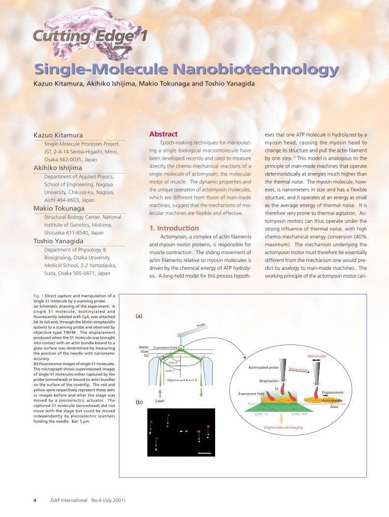
Fig. 1 Direct capture and manipulation of asingle S1 molecule by a scanning probe.(a) Schematic drawing of the experiment. Asingle S1 molecule, biotinylated andfluorescently-labeled with Cy3, was attached(at its tail end, through the biotin-streptavidinsystem) to a scanning probe and observed byobjective-type TIRFM. The displacementproduced when the S1 molecule was broughtinto contact with an actin bundle bound to aglass surface was determined by measuringthe position of the needle with nanometeraccuracy.(b) Fluorescence images of single S1 molecules.The micrograph shows superimposed imagesof single S1 molecules either captured by theprobe (arrowhead) or bound to actin bundleson the surface of the coverslip. The red andyellow spots respectively represent those seenin images before and after the stage wasmoved by a piezoelectric actuator. Thecaptured S1 molecule (arrowhead) did notmove with the stage but could be movedindependently by piezoelectric scannersholding the needle. Bar: 5 µm.
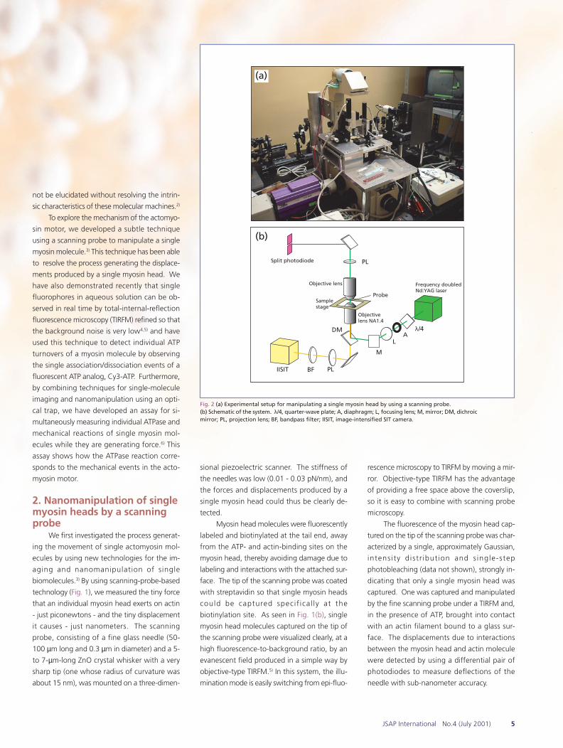
Fig. 2 (a) Experimental setup for manipulating a single myosin head by using a scanning probe.(b) Schematic of the system. λ/4, quarter-wave plate; A, diaphragm; L, focusing lens; M, mirror; DM, dichroicmirror; PL, projection lens; BF, bandpass filter; IISIT, image-intensified SIT camera.
not be elucidated without resolving the intrin-
sic characteristics of these molecular machines.2)
To explore the mechanism of the actomyo-
sin motor, we developed a subtle technique
using a scanning probe to manipulate a single
myosin molecule.3) This technique has been able
to resolve the process generating the displace-
ments produced by a single myosin head. We
have also demonstrated recently that single
fluorophores in aqueous solution can be ob-
served in real time by total-internal-reflection
fluorescence microscopy (TIRFM) refined so that
the background noise is very low4,5) and have
used this technique to detect individual ATP
turnovers of a myosin molecule by observing
the single association/dissociation events of a
fluorescent ATP analog, Cy3-ATP. Furthermore,
by combining techniques for single-molecule
imaging and nanomanipulation using an opti-
cal trap, we have developed an assay for si-
multaneously measuring individual ATPase and
mechanical reactions of single myosin mol-
ecules while they are generating force.6) This
assay shows how the ATPase reaction corre-
sponds to the mechanical events in the acto-
myosin motor.
2. Nanomanipulation of singlemyosin heads by a scanningprobe
3. Stepping motionWhen the myosin head was not associ-
ated with an actin filament, large thermal fluc-
tuations of the probe were apparent. Their
r.m.s. amplitude was about 13 nm. When the
myosin head bonded to actin the r.m.s. ampli-
tude of the fluctuations fell to less than 4.5 nm
(Fig. 3(a), upper trace). Probe motion caused
by actomyosin interactions could be clearly dis-
tinguished from thermal noise by monitoring
the increase in stiffness, calculated as the re-
ciprocal of the variance of the fluctuating probe
position (Fig. 3(a), lower trace). The concen-
tration of ATP was low in these experiments
Fig. 3 Displacement caused by singleS1 molecules.(a) Upper: typical recording of thedisplacements made by an S1molecule. Lower: stiffness calculatedfrom the variance of the probeposition.(b ) An example o f a s ing ledisplacement event and its risingphase on an expanded time scale.(c) Records of the rising phase ofdi sp lacements under var iousconditions: (i) 1 µM ATP, 20ºC; (ii) 0.1µM ATP, 20ºC; (iii) 1 µM ATP, 27ºC.Horizontal gridlines have beendrawn at a spacing of 5.5 nm. In thefalling phase of the displacements,s tepwise movement was notobserved (last traces in (i) and (iii)).
-40
0
40
0 1 2 3 4 50.0
0.5
1.0
1.5
Stif
fnes
s(p
N/n
m)
Dis
pla
cem
ent
(n
m)
Time (s)
(a)
10 nm0.1 s
5.5 nm
10 ms
(b) (i)
(iii)
(ii)
5 ms
5.5 nm
5 ms
5.5 nm
5 ms
5.5 nm
(c)
detachment
detachment
(0.1 or 1 µM), leading to prolonged actomyo-
sin interactions and thus enabling individual
mechanical events to be identified easily. When
the myosin head was attached to an actin mol-
ecule, the stiffness of the probe-myosin-actin
linkage was (at 20oC) 0.2-1.5 pN/nm, more than
ten times the stiffness of the probe. The high
degree of stiffness during the generation of dis-
placements greatly improved the temporal reso-
lution and signal-to-noise ratio, allowing us to
resolve the elementary processes generating the
displacements.
Displacements observed with a low tem-
poral resolution took place abruptly (Fig. 3(a)),
which appeared to be consistent with the con-
ventional model. On an expanded timescale,
however, we found that the displacements were
not actually abrupt but instead developed in mul-
tiple steps (Fig. 3(b)). The time course of the
rising and falling phases of displacements in the
presence of ATP at different concentrations and
temperatures is shown on an expanded
timescale in Fig. 3(c). The steps at 20oC are
clearer than those at 27oC because the dwell-
time between them was longer. In the falling
phase of the displacements (detachment of the
myosin head from actin following ATP binding),
the probe returned to the zero position with a
1/e time of 2-5 ms (Fig. 3(c), parts (i) and (iii),
last traces), which was similar to the settling time
of the free needle. No regular steps were evi-
dent during the falling phase. The size of steps
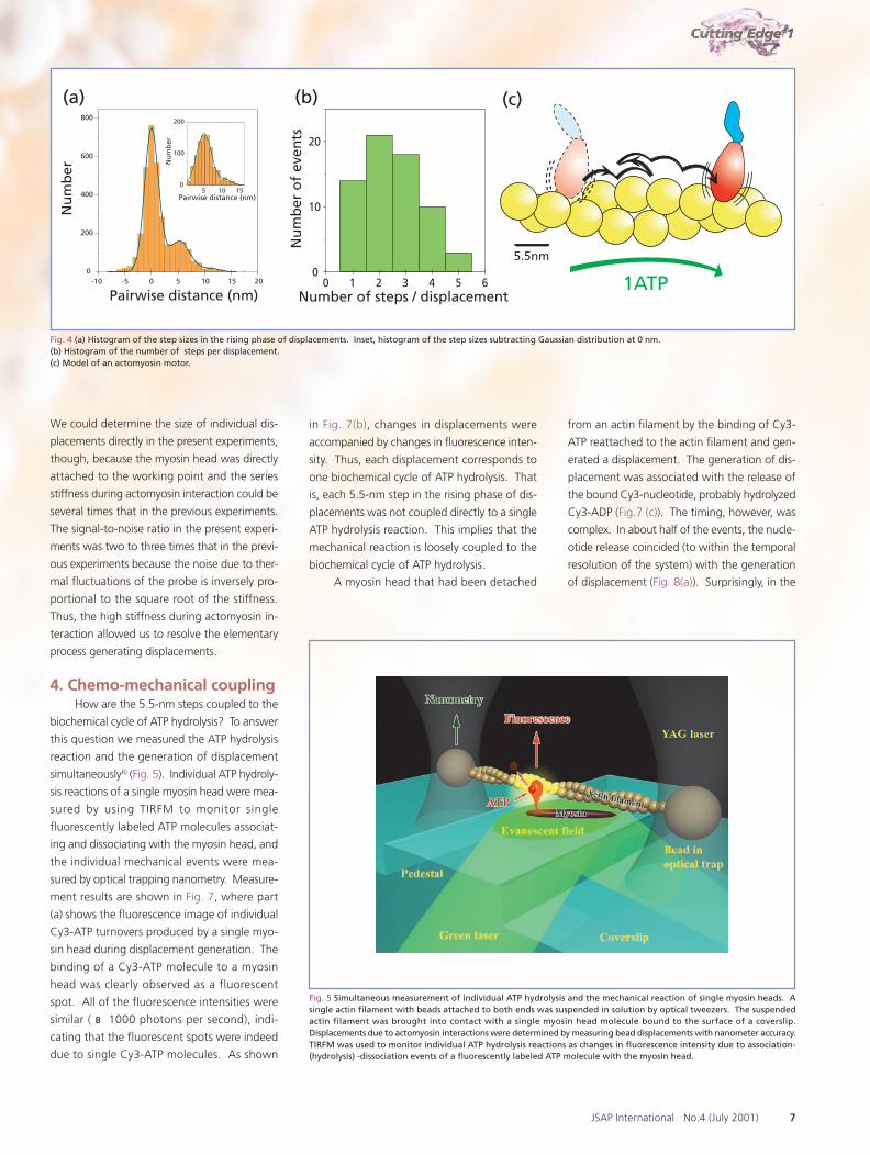
seen during the rising phase was regular and
consistent with the periodicity of adjacent actin
molecules in an actin filament, 5.5 nm (Fig. 4(a)).
The number of steps per displacement event
varied stochastically from one to five (Fig. 4(b)),
yielding overall displacements with sizes between
5 and 30 nm. Steps were not always forward
but were sometimes backward (10% of the to-
tal number of steps). The size of the backward
steps was also 5.5 nm. These patterns of steps
indicate that the myosin head steps back and
forth along the actin molecules in a filament by
Brownian motion rather than by changing shape
(Fig. 4(c)). Note that the individual displacements
4. Chemo-mechanical couplingHow are the 5.5-nm steps coupled to the
biochemical cycle of ATP hydrolysis? To answer
this question we measured the ATP hydrolysis
reaction and the generation of displacement
simultaneously6) (Fig. 5). Individual ATP hydroly-
sis reactions of a single myosin head were mea-
sured by using TIRFM to monitor single
fluorescently labeled ATP molecules associat-
ing and dissociating with the myosin head, and
the individual mechanical events were mea-
sured by optical trapping nanometry. Measure-
ment results are shown in Fig. 7, where part
(a) shows the fluorescence image of individual
Cy3-ATP turnovers produced by a single myo-
sin head during displacement generation. The
binding of a Cy3-ATP molecule to a myosin
head was clearly observed as a fluorescent
spot. All of the fluorescence intensities were
similar ( ~1000 photons per second), indi-
cating that the fluorescent spots were indeed
due to single Cy3-ATP molecules. As shown
Fig. 4 (a) Histogram of the step sizes in the rising phase of displacements. Inset, histogram of the step sizes subtracting Gaussian distribution at 0 nm.(b) Histogram of the number of steps per displacement.(c) Model of an actomyosin motor.
-10 -5 0 5 10 15 200
200
400
600
800
Pairwise distance (nm)
Nu
mb
er
5 10 150
100
200
Nu
mb
er
Pairwise distance (nm)
(a)
Nu
mb
er o
f ev
ents
Number of steps / displacement
(b)
0 1 2 3 4 5 60
10
20
1ATP
5.5nm
(c)
in Fig. 7(b), changes in displacements were
accompanied by changes in fluorescence inten-
sity. Thus, each displacement corresponds to
one biochemical cycle of ATP hydrolysis. That
is, each 5.5-nm step in the rising phase of dis-
placements was not coupled directly to a single
ATP hydrolysis reaction. This implies that the
mechanical reaction is loosely coupled to the
biochemical cycle of ATP hydrolysis.
A myosin head that had been detached
from an actin filament by the binding of Cy3-
ATP reattached to the actin filament and gen-
erated a displacement. The generation of dis-
placement was associated with the release of
the bound Cy3-nucleotide, probably hydrolyzed
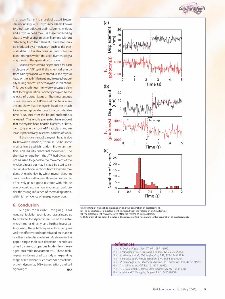
Cy3-ADP (Fig.7 (c)). The timing, however, was
complex. In about half of the events, the nucle-
otide release coincided (to within the temporal
resolution of the system) with the generation
of displacement (Fig. 8(a)). Surprisingly, in the
Fig. 5 Simultaneous measurement of individual ATP hydrolysis and the mechanical reaction of single myosin heads. Asingle actin filament with beads attached to both ends was suspended in solution by optical tweezers. The suspendedactin filament was brought into contact with a single myosin head molecule bound to the surface of a coverslip.Displacements due to actomyosin interactions were determined by measuring bead displacements with nanometer accuracy.TIRFM was used to monitor individual ATP hydrolysis reactions as changes in fluorescence intensity due to association-(hydrolysis) -dissociation events of a fluorescently labeled ATP molecule with the myosin head.
Fig. 6 (a) Setup for simultaneous measurement of individualATPase and mechanical reactions.(b) Schematic of the system. D, dichroic mirror; BF and BP,bandpass filter; BS, beam splitter cube.
Fig. 7 (a) Fluorescence imageof an association-(hydrolysis)-d i s s o c i a t i o n e v e n t o ffluorescent ATP molecules witha m y o s i n h e a d d u r i n ggeneration of displacements.(b ) Time cour se o f thegeneration of displacements(upper trace) and of changesin fluorescence intensity (lowertrace). Each displacementevent corresponds to onebiochemical cycle of ATPhydrolysis (c).
Fig. 8 Timing of nucleotide dissociation and the generation of displacement.(a) The generation of a displacement coincided with the release of Cy3-nucleotide.(b) The displacement was generated after the release of Cy3-nucleotide.(c) Histogram of the delay times from the release of Cy3-nucleatide to the generation of displacements.
0
10
20
Dis
pla
cem
ent
(
nm
)
0 1 2 3 4 52000
3000
4000
F.
I.(p
ho
ton
s/s)
Time (s)
Time lag
-10
0
10
20
30
Dis
pla
cem
ent
(
nm
)
0 1 2 3 4 5
2000
4000
F.
I.(p
ho
ton
s/s)
Time (s)
-1 -0.5 0 0.5 1 1.5 20
5
10
15
20
25
Nu
mb
er o
f ev
ents
Time (s)
(a)
(b)
(c)
in an actin filament is a result of biased Brown-
ian motion (Fig. 4(c)). Myosin heads are known
to bind two adjacent actin subunits in rigor,
and a myosin head may use these two binding
sites to walk along an actin filament without
detaching from the filament. Each step may
be produced by a mechanism such as the ther-
mal ratchet.7) It is also possible that conforma-
tional changes within the actin filament play a
major role in the generation of force.
Multiple steps would be produced for each
molecule of ATP split if the chemical energy
from ATP hydrolysis were stored in the myosin
head or the actin filament and released gradu-
ally during successive actomyosin interactions.
This idea challenges the widely accepted view
that force generation is directly coupled to the
release of bound ligands. The simultaneous
measurements of ATPase and mechanical re-
actions show that the myosin head can attach
to actin and generate force for a considerable
time (>100 ms) after the bound nucleotide is
released. The results presented here suggest
that the myosin head or actin filament, or both,
can store energy from ATP hydrolysis and re-
lease it productively in several packets of work.
If the movement of a myosin head is due
to Brownian motion, there must be some
mechanism by which random Brownian mo-
tion is biased into directional movement. The
chemical energy from the ATP hydrolysis may
not be used to generate the movement of the
myosin directly but may instead be used to se-
lect unidirectional motions from Brownian mo-
tions. A mechanism by which myosin does not
overcome but rather uses Brownian motion to
effectively gain a good distance with minute
energy could explain how myosin can walk un-
der the strong influence of thermal agitation,
with high efficiency of energy conversion.
6. ConclusionS i n g l e - m o l e c u l e i m a g i n g a n d
nanomanipulation techniques have allowed us
to evaluate the dynamic nature of the acto-
myosin motor directly, and further investiga-
tions using these techniques will certainly re-
veal the effective and sophisticated mechanism
of other molecular machines. As shown in this
paper, single-molecule detection techniques
unveil dynamic properties hidden from aver-
aged ensemble measurements. These tech-
niques are being used to study an expanding
range of life science, such as enzyme reactions,
protein dynamics, DNA transcription, and cell
signaling.8)
References1 ) R. Cooke, Physiol. Rev. 77, 671-697 (1997).2 ) T. Yanagida et al., Curr. Opin. Cell Biol. 12, 20-25 (2000).3 ) K. Kitamura et al., Nature (London) 397, 129-134 (1999).4 ) T. Funatsu et al., Nature (London) 374, 555-559 (1995).5 ) M. Tokunaga et al., Biochem. Biophys. Res. Commun. 235, 47-53 (1997).6 ) A. Ishijima et al., Cell 92, 161-171 (1998).7 ) R. D. Vale and F. Oosawa, Adv. Biophys. 26, 97-134 (1990).8 ) Y. Ishii and T. Yanagida, Single Mol. 1, 5-16 (2000).