+ All Categories
Home > Documents > Single-staged Treatment Using a Standardized Protocol Results in Functional Motion in the Majority...

Single-staged Treatment Using a Standardized Protocol Results in Functional Motion in the Majority...

Date post: 21-Dec-2016
Category:
Upload: daphne
View: 214 times
Download: 0 times
Share this document with a friend
9
SYMPOSIUM: TRAUMATIC ELBOW INSTABILITY AND ITS SEQUELAE Single-staged Treatment Using a Standardized Protocol Results in Functional Motion in the Majority of Patients With a Terrible Triad Elbow Injury Akash Gupta MD, David Barei MD, Ansab Khwaja BA, Daphne Beingessner MD Ó The Association of Bone and Joint Surgeons1 2014 Abstract Background Terrible triad injuries of the elbow, defined as elbow dislocation with associated fractures to the radial head and coronoid, are associated with stiffness, pain, and loss of motion. Studies to date have consisted of small sample sizes and used heterogeneous surgical techniques, which render comparisons difficult and unreliable. Questions/purposes In a group of patients treated under a standard surgical protocol, we sought to determine the early dislocation rate, the range of motion in those not undergoing secondary procedures, the frequency and types of secondary surgical interventions required, the difference in motion between those undergoing secondary surgery and those who did not, and the frequency of heterotopic ossi- fication and patient-reported stiffness. Methods Patients underwent a surgical protocol that involved fixing the coronoid, fixing the radial head if possible, otherwise performing radial head arthroplasty, and repairing the lateral ligamentous structures. Patients were excluded if ipsilateral upper extremity fractures from the humerus to the distal forearm were present. Fifty-two patients had a minimum followup of 6 weeks and were included for the early dislocation rate, and 34 of these (65%) had a minimum of 6 months followup and were included for the rest of the data. Eighteen of the 52 (35%) were considered lost to followup because they were seen for less than 6 months postsurgically and were excluded from further analysis. Chart review was performed to determine the presence of early dislocation within the first 6 weeks after surgery, range of motion in patients not requiring a secondary procedure, the frequency and types of secondary procedures required, the range of motion before and after a secondary procedure if it was required, and postoperative stiffness. Postoperative radiographs were analyzed to determine the presence and severity of het- erotopic ossification. Results One of 52 patients sustained a dislocation within the first weeks of surgery (1.9%). Those not undergoing a secondary procedure were able to achieve a flexion arc of 110° and a supination-pronation arc of 148°. Nine of 34 patients (26%) underwent a secondary surgical procedure with stiffness, heterotopic ossification, and ulnar neuropathy being the most common surgical indications. Before sec- ondary surgical procedures, patients had a flexion arc of 57° and a supination-pronation arc of 55°, which was less than those only requiring primary surgery alone (p \ 0.001). After secondary surgery, patients were able to achieve a flexion arc of 96° and a supination-pronation arc of 124°, which was not different from those who did not undergo reoperation (p = 0.09 and p = 0.08, respectively). Twenty- eight of 34 patients demonstrated evidence of heterotopic ossification on radiographs, whereas 20 patients, including all nine undergoing secondary procedures, reported stiffness at the elbow. Each author certifies that he or she, or a member of his or her immediate family, has no funding or commercial associations (eg, consultancies, stock ownership, equity interest, patent/licensing arrangements, etc) that might pose a conflict of interest in connection with the submitted article. All ICMJE Conflict of Interest Forms for authors and Clinical Orthopaedics and Related Research editors and board members are on file with the publication and can be viewed on request. Each author certifies that his or her institution approved the human protocol for this investigation, that all investigations were conducted in conformity with ethical principles of research, and that informed consent for participation in the study was obtained. A. Gupta, D. Barei, A. Khwaja, D. Beingessner (&) Harborview Medical Center, University of Washington, 325 9th Avenue, Box 359798, Seattle, WA 98104, USA e-mail: [email protected] 123 Clin Orthop Relat Res DOI 10.1007/s11999-014-3475-3 Clinical Orthopaedics and Related Research ® A Publication of The Association of Bone and Joint Surgeons®
Transcript

SYMPOSIUM: TRAUMATIC ELBOW INSTABILITY AND ITS SEQUELAE

Single-staged Treatment Using a Standardized Protocol Resultsin Functional Motion in the Majority of Patients With a TerribleTriad Elbow Injury

Akash Gupta MD, David Barei MD, Ansab Khwaja BA,

Daphne Beingessner MD

� The Association of Bone and Joint Surgeons1 2014

Abstract

Background Terrible triad injuries of the elbow, defined

as elbow dislocation with associated fractures to the radial

head and coronoid, are associated with stiffness, pain, and

loss of motion. Studies to date have consisted of small

sample sizes and used heterogeneous surgical techniques,

which render comparisons difficult and unreliable.

Questions/purposes In a group of patients treated under a

standard surgical protocol, we sought to determine the

early dislocation rate, the range of motion in those not

undergoing secondary procedures, the frequency and types

of secondary surgical interventions required, the difference

in motion between those undergoing secondary surgery and

those who did not, and the frequency of heterotopic ossi-

fication and patient-reported stiffness.

Methods Patients underwent a surgical protocol that

involved fixing the coronoid, fixing the radial head if

possible, otherwise performing radial head arthroplasty,

and repairing the lateral ligamentous structures. Patients

were excluded if ipsilateral upper extremity fractures from

the humerus to the distal forearm were present. Fifty-two

patients had a minimum followup of 6 weeks and were

included for the early dislocation rate, and 34 of these

(65%) had a minimum of 6 months followup and were

included for the rest of the data. Eighteen of the 52 (35%)

were considered lost to followup because they were seen

for less than 6 months postsurgically and were excluded

from further analysis. Chart review was performed to

determine the presence of early dislocation within the first

6 weeks after surgery, range of motion in patients not

requiring a secondary procedure, the frequency and types

of secondary procedures required, the range of motion

before and after a secondary procedure if it was required,

and postoperative stiffness. Postoperative radiographs were

analyzed to determine the presence and severity of het-

erotopic ossification.

Results One of 52 patients sustained a dislocation within

the first weeks of surgery (1.9%). Those not undergoing a

secondary procedure were able to achieve a flexion arc of

110� and a supination-pronation arc of 148�. Nine of 34

patients (26%) underwent a secondary surgical procedure

with stiffness, heterotopic ossification, and ulnar neuropathy

being the most common surgical indications. Before sec-

ondary surgical procedures, patients had a flexion arc of 57�and a supination-pronation arc of 55�, which was less than

those only requiring primary surgery alone (p \ 0.001).

After secondary surgery, patients were able to achieve a

flexion arc of 96� and a supination-pronation arc of 124�,

which was not different from those who did not undergo

reoperation (p = 0.09 and p = 0.08, respectively). Twenty-

eight of 34 patients demonstrated evidence of heterotopic

ossification on radiographs, whereas 20 patients, including

all nine undergoing secondary procedures, reported stiffness

at the elbow.

Each author certifies that he or she, or a member of his or her

immediate family, has no funding or commercial associations

(eg, consultancies, stock ownership, equity interest, patent/licensing

arrangements, etc) that might pose a conflict of interest in connection

with the submitted article.

All ICMJE Conflict of Interest Forms for authors and Clinical

Orthopaedics and Related Research editors and board members are

on file with the publication and can be viewed on request.

Each author certifies that his or her institution approved the human

protocol for this investigation, that all investigations were conducted

in conformity with ethical principles of research, and that informed

consent for participation in the study was obtained.

A. Gupta, D. Barei, A. Khwaja, D. Beingessner (&)

Harborview Medical Center, University of Washington,

325 9th Avenue, Box 359798, Seattle, WA 98104, USA

e-mail: [email protected]

123

Clin Orthop Relat Res

DOI 10.1007/s11999-014-3475-3

Clinical Orthopaedicsand Related Research®

A Publication of The Association of Bone and Joint Surgeons®

Conclusions Using a standardized surgical protocol, a

low early dislocation rate was observed, although stiffness

remains a challenge. Many patients who initially do not

attain functional range of motion can usually attain this

after secondary procedures aimed at removing the hetero-

topic ossification.

Level of Evidence Level IV, therapeutic study. See

guidelines for authors for a complete description of levels

of evidence.

Introduction

An elbow dislocation with associated radial head and

coronoid fractures is referred to as a ‘‘terrible triad’’ injury

and has a reputation for being a challenging condition to

treat [7], because it can cause stiffness, instability, post-

traumatic arthrosis, and pain [23]. Better results have been

reported using a biomechanically sound standard treatment

protocol [1] that includes coronoid repair, radial head open

reduction and internal fixation (ORIF) or replacement, and

lateral collateral ligament (LCL) repair. Medial collateral

ligament (MCL) repair and/or external fixation are required

for those with persistent instability [19].

The terrible triad is a relatively rare injury [27] and most

published series are small. Although outcomes have

improved with standardized protocols, the infrequency and

complexity of this injury type [27] has left published series

with small sample sizes and heterogeneous surgical

approaches. A recent review article summarized 137 cases

from five studies with the largest single study reporting on

36 patients [9, 11, 17, 23, 30, 31].

We therefore sought to evaluate a single institution’s

results using a standard protocol. Specifically, we evaluated

(1) the early dislocation rate after surgical intervention; (2)

the ROM achieved in those who did not undergo secondary

procedures; (3) the frequency of reoperation and the types of

surgical interventions used in secondary procedures; (4) the

difference in ROM achieved between patients undergoing

secondary surgical procedures and those who did not; and

(5) the frequency of radiographic heterotopic ossification

and patient-reported stiffness.

Patients and Methods

Clinical Algorithm, Operative Technique, and

Aftercare

Between January 1, 1995, and March 1, 2013, four fellow-

ship-trained orthopaedic trauma surgeons at one institution

treated 52 patients with 52 terrible triad injuries, defined as

elbow dislocations with associated coronoid and radial head

fractures. During that time, all were managed surgically. Of

those, 52 (100%) had adequate radiographic and clinical

followup at 6 weeks to ascertain the early redislocation rate,

which was defined as dislocation within 6 weeks of the

initial surgical procedure. Of these patients, however,

18 (35%) did not have followup longer than 6 months and

were considered lost to long-term followup and excluded

from all other data points. A total of 34 (65%) had adequate

radiographic and clinical followup at 6 months to evaluate

our other study endpoints, including the use of secondary

procedures, ROM, and patient-perceived stiffness.

Patients who underwent surgery were managed simi-

larly (Figs. 1, 2, 3). Large coronoid fractures were treated

with screws, whereas small fragments were treated with

suture repair of the anterior capsule. The radial head was

treated with primary repair or arthroplasty in all elbows.

The primary indication for radial head arthroplasty was to

avoid tenuous radial head fixation in comminuted frac-

tures, which would predispose to recurrent dislocation

should the repair fail [1, 3]. The determination of a tenuous

repair was established by the treating surgeon in each

instance but was uniformly considered to be radial head

fixation that was not anticipated to allow physiologic loads

through a functional arc of motion without a substantial

risk of early dislocation [29]. The radial head was evalu-

ated first and if arthroplasty was chosen as treatment, the

neck cut was performed to improve the access to the

coronoid and then the coronoid was fixed. If repair was

chosen, the fragments were carefully moved out of the way

taking care not to disrupt the periosteum, and the coronoid

was fixed first. The LCL complex and common extensor

origin were then repaired. If, after this sequence, ulnohu-

meral instability was not achieved, then repair of the

MCL was also performed. External fixation was never

used. The elbow was taken through a ROM and stability

was assessed both clinically and using fluoroscopy.

Extension using gravity was assessed but the elbows were

not forced into terminal extension so that the coronoid

repair would not be disrupted. Valgus instability was

not an indication for medial repair. Subluxation at the

ulnohumeral joint at any point during the range was an

indication for further intervention.

Postoperative rehabilitation protocols were similar in all

patients. Patients were initially placed in a posterior plaster

splint. The splint was removed on the first or second post-

operative day, and active and active-assisted motion was

started. Full ROM was allowed with the forearm in prona-

tion or neutral with supination occurring with the elbow at

90� of flexion or greater. Terminal extension in supination

was avoided for approximately 3 weeks. No patient received

postoperative heterotopic ossification prophylaxis.

Gupta et al. Clinical Orthopaedics and Related Research1

123

Data Collection

All injuries involving a fracture or dislocation of the elbow

were reviewed to identify terrible triad injuries of the elbow.

Radiographs were reviewed to confirm the preoperative

presence of an ulnohumeral dislocation, radial head fracture,

and coronoid fracture. Those with associated ipsilateral

upper extremity injuries including distal humerus fractures,

Monteggia and Monteggia variant fractures, ulnar or radial

shaft fractures, distal radius or ulna fractures, and distal ra-

dioulnar joint disruption were excluded. Cases with

ipsilateral carpal pathology such as scaphoid fractures were

included. Those without at least one postoperative radio-

graph were also excluded.

Of the 52 patients identified, 39 were men and 13 were

women with an average age of 44 years (SD 13; range, 19–

65 years). Sixteen were smokers. Twenty-four and 28

patients sustained right and left elbow injuries, respectively.

Patients underwent surgical intervention at an average of

4 days from injury (range, 0–15 days). Radial head

arthroplasty was performed for 40 patients whose fractures

were deemed too comminuted to be adequately fixed and

radial head fixation was performed in 12 cases. The coronoid

was repaired in 45 cases and was treated without fixation in

seven cases. In 49 of the 52 cases, the LCL complex was

repaired. In one patient, the MCL was also repaired. Two

patients required repair of the common extensor origin and

one required repair of a triceps tendon avulsion.

The 34 patients with followup longer than 6 months were

divided into two groups: those requiring primary surgery only

and those undergoing secondary surgery. The need for sec-

ondary surgery was defined as any subsequent surgical

procedure on the ipsilateral extremity as a sequela of the initial

injury and not related to new trauma. For the primary surgery

only group, ROM at the last clinical followup date was

recorded, including flexion, extension, supination, pronation,

and the respective flexion-extension and supination-pronation

arcs. For the secondary surgery group, the same data were

collected at the last clinical visit before the secondary surgery

as well as the last clinical visit after secondary surgery. For this

group, the indications and diagnoses for secondary surgery

and types of secondary surgery performed were gathered from

preoperative documentation and operative reports. The dif-

ference in ROM after secondary surgery compared with

before secondary surgery was then calculated as well as the

difference between the final ROMs between the two groups.

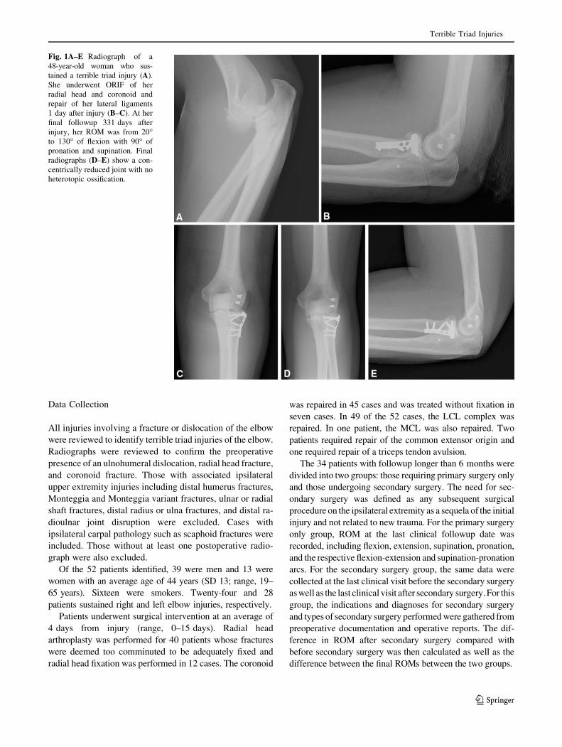
Fig. 1A–E Radiograph of a

48-year-old woman who sus-

tained a terrible triad injury (A).

She underwent ORIF of her

radial head and coronoid and

repair of her lateral ligaments

1 day after injury (B–C). At her

final followup 331 days after

injury, her ROM was from 20�to 130� of flexion with 90� of

pronation and supination. Final

radiographs (D–E) show a con-

centrically reduced joint with no

heterotopic ossification.

Terrible Triad Injuries

123

Fig. 2A–I Radiographs of a 35-year-old man who sustained a terrible

triad injury (A–B). He underwent radial head arthroplasty, coronoid

fixation, and lateral ligament repair 11 days after injury (C–E). He

showed early signs of heterotopic ossification 3 weeks after repair.

The patient’s elbow became ankylosed at 90� of flexion and neutral

rotation (F–G) by 8 weeks. He underwent resection of heterotopic

bone, synovectomy, ulnar neurolysis, and lateral ligament repair

114 days after his injury. At final followup 1 year after release, his

motion was from 45� to 125� of flexion with 50� each of pronation

and supination (H–I).

Gupta et al. Clinical Orthopaedics and Related Research1

123

Clinical stiffness was considered to be present once the

chart reflected that the symptoms were problematic for

patients at their 6-week postoperative visit or later. Any

clinically reported stiffness before this was considered part

of the normal recovery process. Radiographs were ana-

lyzed until the patient’s last clinic visit, which was at an

average of 57 weeks after surgery, and included presence

of heterotopic ossification and the number of days from

surgery to the first appearance of heterotopic ossification.

Statistical Analysis

Analysis was performed using statistical software (SPSS

Version 21; SPSS Inc, Chicago, IL, USA). Group t-tests

were used to evaluate statistical differences between the

primary surgery only group and the secondary surgery

group both preoperatively and postoperatively. Paired

t-tests were used to evaluate the differences in ROM from

preoperatively to postoperatively in the secondary surgery

group.

Results

Early Dislocation Rate

Fifty-two patients with a minimum of 6 weeks of followup

were analyzed for the presence of an early dislocation after

surgery. Only one patient (1.9% of the 52) was found to

Fig. 3 Flowchart depicts the

surgical protocol sequence.

Terrible Triad Injuries

123

have a postoperative redislocation within the first 6 weeks

after surgery. This occurred at 33 days and was a result of a

fall directly onto the affected extremity resulting in failure

of the LCL repair. The patient underwent secondary sur-

gery, which included LCL reconstruction and removal of

heterotopic ossification. This patient, however, was lost to

followup and was excluded from subsequent analysis.

ROM in the Primary Surgery Only Group

Of the 34 patients with at least 6 months of followup, 25

(74%) did not undergo a secondary surgical procedure.

These patients had a mean flexion of 128� and a mean

extension of 17� (mean flexion-extension arc, 110�; SD

23�). Mean supination was 72� and mean pronation was 77�(mean supination-pronation arc, 148�; SD 37�; Table 1).

Secondary Surgery Group

Secondary surgeries were performed in nine of the 34

patients (26%). In all of these patients, stiffness and het-

erotopic ossification were the major indications for

reoperation. One patient developed stiffness requiring

release but underwent a total elbow arthroplasty instead as a

result of a supracondylar fracture that had occurred from a

fall before her planned release. Excision of heterotopic

ossification was performed in all nine secondary procedures,

and synovectomy was performed in three. Ulnar neurolysis

and transposition were performed in seven patients. Lateral

collateral ligament repair was performed in one elbow after

contracture release. Other procedures performed once each

included revision radial head arthroplasty, median nerve

neurolysis, and biceps and triceps tenolysis.

Before the secondary procedure, the patients in this group

had a mean flexion of 108� and a mean extension of 51� (mean

flexion arc, 57�; SD 44�). Mean supination measured 27� and

mean pronation was 28� (mean supination-pronation arc, 55�;

SD 54�). Both the mean flexion arc and mean supination-

pronation arc were inferior to those in the primary surgery-

only group (p \ 0.001 for both). After secondary surgery,

patients achieved a mean flexion of 122� and mean extension

of 26� (mean flexion-extension arc, 96�; SD 11�) at a mean

followup of 84 weeks (range, 32–143 weeks). Patients

attained a mean pronation of 57� and mean supination of 68�(mean supination-pronation arc, 124�; SD 23). Secondary

surgery improved the mean flexion-extension arc by 39�(p = 0.02) and the mean supination-pronation arc by 69�(p \ 0.003). After secondary surgery, with the numbers

available, patients had a comparable flexion-extension arc

(p = 0.09) and supination-pronation arc (p = 0.08) as those

in the primary surgery only group (Table 1).

Heterotopic Ossification and Stiffness

Twenty-eight of the 34 patients demonstrated radiographic

evidence of heterotopic ossification with the first signs

appearing on radiographs at an average of 64 days post-

operatively (Table 2). Ten patients demonstrated both

MCL and LCL calcifications. Four demonstrated isolated

MCL calcification and two demonstrated isolated LCL

calcification. Twenty of the 34 patients (59%), including all

nine patients undergoing secondary procedures, reported

stiffness at the elbow.

Table 1. ROM in the primary surgery group and the secondary

surgery group before the second intervention and after the second

intervention

Variable Primary

surgery

only

group

Secondary

surgery

group (before

secondary

procedure,

after the index

procedure)

Secondary

surgery

group (after

the secondary

procedure[s])

Flexion (degrees) 128 108 122

Extension (degrees) 17 51 26

Supination (degrees) 72 27 68

Pronation (degrees) 78 28 57

Flexion-extension

arc (degrees)

110 57* 96�

Supination-pronation

arc (degrees)

148 55* 124�

* Statistically significant difference compared with the primary sur-

gery only group (p \ 0.05); �no statistically significant difference

compared with the primary surgery only group (p [ 0.05).

Table 2. Radiographic analysis in both primary and secondary sur-

gery groups

Variable Primary surgery

only group

Secondary

surgery group

Last radiograph analyzed

(number of days)

276 625

Number of cases with

radiographic HO (%)

19/25 (76%) 9/9 (100%)

First radiographic appearance of

HO (number of days)

68 55

Combined MCL/LCL

calcification (%)

7/25 (28%) 3/9 (33%)

Isolated MCL calcification (%) 3/25 (12%) 1/9 (11%)

Isolated LCL calcification (%) 1/25 (7%) 1/9 (11%)

HO = heterotopic ossification; MCL = medial collateral ligament;

LCL = lateral collateral ligament.

Gupta et al. Clinical Orthopaedics and Related Research1

123

Discussion

Terrible triad injuries of the elbow have historically been

challenging injuries to treat and although initial treatment

protocols were limited because of small sample sizes and

high complication rates, recent studies have shown more

predictable results [11, 16, 18, 22, 30]. The aim of this

study was to evaluate the algorithm used at one high-vol-

ume trauma center, which relies on biomechanical and

clinical principles of functional anatomy and how it relates

to postoperative stability [1–6, 12, 13, 24] and early ROM.

The results demonstrated that the early dislocation rate was

very low, and those who did not undergo secondary pro-

cedures were able to obtain good functional ROM

postoperatively. Although stiffness and heterotopic ossifi-

cation were quite common and indicated secondary surgery

in a large proportion of patients, most of those patients

reliably gained functional ROM after the secondary

surgery.

There are several limitations to this study that must be

considered. First, this is a retrospective review, which

limits the ability to control factors such as followup,

standard motion measurements, and standardized timing

for radiographs. The most important limitation of this study

was the relatively high proportion of patients who were lost

to followup (35%, 18 of 52 at 6 months or longer). Thus,

the data presented here likely represent that of patients with

a higher degree of compliance with postoperative protocols

than those who did not return, and those who do not follow

postoperative rehabilitation are expected to have less ROM

and function. Given the long period of data collection,

there is some inevitable variation in intraoperative tech-

nique (including radial head prosthetic design, suture

material/technique, and perhaps others). Despite this, on

careful review of all charts, it is apparent that a consistent

general protocol (emphasizing the biomechanical impor-

tance of radial head repair or replacement, coronoid

assessment and stabilization, and LCL complex integrity)

was used with variations owing to the surgeon trying to

tailor recovery to the patients’ ability, motivation, and

specific complaints. The retrospective nature of this study

also prevented the collection of validated outcomes mea-

suring tools such as the Disability of the Arm, Shoulder,

and Hand Questionnaire, Mayo Elbow Performance Index,

or the Broberg and Morrey rating system. Collection of

these data would certainly enhance the standardization of

classifying outcomes. Another limitation is that data were

collected only within the study period and several of the

patients in the primary surgery group were still being fol-

lowed in the clinic and potentially could undergo

secondary procedures for stiffness if monitored for a longer

period of time. Even so, however, we believe these results

to be valid because there was still a large population that

was followed to completion of postoperative treatment.

Also, the majority of patients in the region that require

surgery for elbow stiffness are seen at our institution so it is

unlikely that further surgery was performed on those with

limited followup.

The results of this study show that early dislocation with

initial definitive fixation is low (2%, one of 52 patients) and

that the case in which this happened was likely a result of

direct trauma from a fall. This result is similar to other

studies such as that published by Egol et al that showed

resubluxation in one of 29 patients [9]. In a study by Lin-

denhovius et al [17], 14 patients presented to them after

3 weeks postinjury with subluxation or dislocation of the

ulnohumeral joint. Of these, five were treated operatively

before presentation to their institution and two were treated

with radial head excision. These results reinforce recent

biomechanical literature that demonstrate that joint stability

can be achieved with early fixation of the coronoid [4, 5, 25],

repair or replacement of the radial head [8, 13–15, 28, 30],

and repair of the LCL complex [20, 30].

Functional results from this study show that an aver-

age flexion-extension arc of 110� and a supination-

pronation arc of 148� can be achieved in approximately

74% of patients (25 of 34 patients) but the remainder

required secondary procedures for stiffness. Other pub-

lished results similarly demonstrate an average flexion-

extension arc of 100� to 119� and a supination-pronation

arc of 128� to 141� [9, 11, 17, 23, 31]. The functional

ROM of the elbow for most activities of daily living is

thought to be 100� of flexion-extension and 100� of

supination-pronation [22] and in a large majority of

patients, this can adequately be achieved with a single

surgical intervention.

There were several indications for secondary surgery for

symptomatic patients with the most common procedures

performed being excision of heterotopic ossification for

stiffness (26%, nine of 34 patients) and ulnar neurolysis

with transposition for ulnar neuropathy (21%, seven of 34

patients). Previous reports show that most common com-

plications with these injuries include stiffness, instability,

heterotopic ossification, and ulnar neuropathy [11, 18, 21–

23, 25] (15%–25% [22]). The rate of secondary surgery in

this study (26%) compares similarly to other studies, which

publish a secondary surgical rate of 15% to 25% [9, 11, 17,

22, 23]. Other studies have noted ulnar neuritis requiring

secondary ulnar nerve decompression at similar rates as

well [9, 11, 17], although prophylactic release during the

index procedure was variably performed. Routine decom-

pression during the index procedure was not performed in

our patients; however, with similar rates of secondary

surgery compared with other studies in which prophylactic

decompression was performed, it suggests that ulnar neu-

ritis is more likely related to heterotopic ossification

Terrible Triad Injuries

123

postinjury. As such, we do not recommend routine initial

prophylactic decompression of the ulnar nerve.

Patients requiring secondary surgery had less ROM

before secondary surgery compared with those who only

underwent the index procedure. After secondary surgery,

however, the ROM achieved was not statistically different

and also was within the functional ROM required for

activities of daily living. These results are similar to pre-

viously published data regarding elbow ROM after

capsulectomy for stiffness, which showed restoration of

flexion-extension arcs to 103� to 109� [10, 26]. Although

secondary surgical intervention must be tailored to the

individual patient, these data suggest that patients who

have vastly diminished ROM that impairs activities of

daily living should be considered for secondary surgical

procedures, which appear to significantly improve function.

Heterotopic ossification remains a challenging and

inconclusive problem in the treatment of elbow trauma.

Although present in many patients, it was not clinically

relevant in most of them. Similarly, all patients had a

similar injury pattern and some patients did not develop

any heterotopic bone at all underscoring our lack of

understanding of which patients are at risk and therefore

should be treated. Our experience is that many patients are

noncompliant with treatment and that it is difficult to

predict which patients would benefit the most from it and

we therefore do not give prophylaxis for heterotopic ossi-

fication. With regard to patient complaints of stiffness, this

also was seen quite frequently throughout the course of

care but again most of them had achieved a functional arc

of motion. This reinforces the notion that a ‘‘normal’’

elbow is rarely achieved but that reasonable results within

functional ROMs are attainable [22]. Patients can be

counseled that even several months into postoperative

recovery, stiffness may persist, but a good functional out-

come that does not require further surgery is possible in the

majority of cases.

Overall, this study represents a large collection of ter-

rible triad elbow fracture-dislocations treated with a

standard protocol consisting of radial head repair or

replacement, coronoid or anterior capsular repair, LCL

complex repair, early postoperative ROM, and no hetero-

topic ossification prophylaxis. No patients underwent

external fixation, and MCL repair was rarely performed.

The rate of recurrent instability is extremely low. The

majority of patients reliably attained functional ROM arcs

with this treatment protocol. Although the use of secondary

surgery for contracture remains higher than desired, con-

tracture release provides near functional ROM arcs in most

patients who undergo it.

Acknowledgments We thank Julie Agel for the assistance with the

statistical analysis in this article.

References

1. Beingessner DM, Dunning CE, Beingessner CJ, Johnson JA,

King GJ. The effect of radial head fracture size on radiocapitellar

joint stability. Clin Biomech (Bristol, Avon). 2003;18:677–681.

2. Beingessner DM, Dunning CE, Gordon KD, Johnson JA, King GJ.

The effect of radial head excision and arthroplasty on elbow kine-

matics and stability. J Bone Joint Surg Am. 2004;86:1730–1739.

3. Beingessner DM, Dunning CE, Gordon KD, Johnson JA, King

GJ. The effect of radial head fracture size on elbow kinematics

and stability. J Orthop Res. 2005;23:210–217.

4. Beingessner DM, Dunning CE, Stacpoole RA, Johnson JA, King

GJ. The effect of coronoid fractures on elbow kinematics and

stability. Clin Biomech (Bristol, Avon). 2007;22:183–190.

5. Beingessner DM, Stacpoole RA, Dunning CE, Johnson JA, King GJ.

The effect of suture fixation of type I coronoid fractures on the

kinematics and stability of the elbow with and without medial col-

lateral ligament repair. J Shoulder Elbow Surg. 2007;16:213–217.

6. Chanlalit C, Shukla DR, Fitzsimmons JS, An KN, O’Driscoll SW. The

biomechanical effect of prosthetic design on radiocapitellar stability in

a terrible triad model. J Orthop Trauma. 2012;26:539–544.

7. Chemama B, Bonnevialle N, Peter O, Mansat P, Bonnevialle P.

Terrible triad injury of the elbow: how to improve outcomes?

Orthop Traumatol Surg Res. 2010;96:147–154.

8. Doornberg JN, Parisien R, van Duijn PJ, Ring D. Radial head

arthroplasty with a modular metal spacer to treat acute traumatic

elbow instability. J Bone Joint Surg Am. 2007;89:1075–1080.

9. Egol KA, Immerman I, Paksima N, Tejwani N, Koval KJ.

Fracture-dislocation of the elbow functional outcome following

treatment with a standardized protocol. Bull NYU Hosp Jt Dis.

2007;65:263–270.

10. Ehsan A, Huang JI, Lyons M, Hanel DP. Surgical management of

posttraumatic elbow arthrofibrosis. J Trauma Acute Care Surg.

2012;72:1399–1403.

11. Forthman C, Henket M, Ring DC. Elbow dislocation with intra-

articular fracture: the results of operative treatment without repair of

the medial collateral ligament. J Hand Surg Am. 2007;32:1200–1209.

12. Garrigues GE, Wray WH 3rd, Lindenhovius AL, Ring DC, Ruch

DS. Fixation of the coronoid process in elbow fracture-disloca-

tions. J Bone Joint Surg Am. 2011;93:1873–1881.

13. Johnson JA, Beingessner DM, Gordon KD, Dunning CE, Stac-

poole RA, King GJ. Kinematics and stability of the fractured and

implant-reconstructed radial head. J Shoulder Elbow Surg.

2005;14(Suppl S):195S–201S.

14. Josefsson PO, Gentz CF, Johnell O, Wendeberg B. Dislocations

of the elbow and intraarticular fractures. Clin Orthop Relat Res.

1989;246:126–130.

15. Judet T, Garreau de Loubresse C, Piriou P, Charnley G. A

floating prosthesis for radial-head fractures. J Bone Joint Surg Br.

1996;78:244–249.

16. Leigh WB, Ball CM. Radial head reconstruction versus

replacement in the treatment of terrible triad injuries of the

elbow. J Shoulder Elbow Surg. 2012;21:1336–1341.

17. Lindenhovius AL, Jupiter JB, Ring D. Comparison of acute

versus subacute treatment of terrible triad injuries of the elbow.

J Hand Surg Am. 2008;33:920–926.

18. Mathew PK, Athwal GS, King GJ. Terrible triad injury of the elbow:

current concepts. J Am Acad Orthop Surg. 2009;17:137–151.

19. McKee MD, Pugh DM, Wild LM, Schemitsch EH, King GJ.

Standard surgical protocol to treat elbow dislocations with radial

head and coronoid fractures. Surgical technique. J Bone Joint

Surg Am. 2005;87(Suppl 1):22–32.

20. McKee MD, Schemitsch EH, Sala MJ, O’Driscoll SW. The

pathoanatomy of lateral ligamentous disruption in complex elbow

instability. J Shoulder Elbow Surg. 2003;12:391–396.

Gupta et al. Clinical Orthopaedics and Related Research1

123

21. O’Driscoll SW, Morrey BF, Korinek S, An KN. Elbow sublux-

ation and dislocation. A spectrum of instability. Clin Orthop

Relat Res. 1992;280:186–197.

22. Pugh DM, McKee MD. The ‘terrible triad’ of the elbow. Tech

Hand Up Extrem Surg. 2002;6:21–29.

23. Pugh DMW, Wild LM, Schemitsch EH, King GJW, McKee MD.

Standard surgical protocol to treat elbow dislocations with radial head

and coronoid fractures. J Bone Joint Surg Am. 2004;86:1122–1130.

24. Reichel LM, Milam GS, Hillin CD, Reitman CA. Osteology of the

coronoid process with clinical correlation to coronoid fractures in

terrible triad injuries. J Shoulder Elbow Surg. 2013;22:323–328.

25. Ring D. Fractures of the coronoid process of the ulna. J Hand

Surg Am. 2006;31:1679–1689.

26. Ring D, Adey L, Zurakowski D, Jupiter JB. Elbow capsulectomy for

posttraumatic elbow stiffness. J Hand Surg Am. 2006;31:1264–1271.

27. Ring D, Jupiter JB, Zilberfarb J. Posterior dislocation of the

elbow with fractures of the radial head and coronoid. J Bone Joint

Surg Am. 2002;84:547–551.

28. Ring D, King G. Radial head arthroplasty with a modular metal

spacer to treat acute traumatic elbow instability. Surgical tech-

nique. J Bone Joint Surg Am. 2008;90(Suppl 2):63–73.

29. Ring D, Quintero J, Jupiter JB. Open reduction and internal fix-

ation of fractures of the radial head. J Bone Joint Surg Am.

2002;84:1811–1815.

30. Rodriguez-Martin J, Pretell-Mazzini J, Andres-Esteban EM,

Larrainzar-Garijo R. Outcomes after terrible triads of the elbow

treated with the current surgical protocols. A review. Int Orthop.

2011;35:851–860.

31. Zeiders GJ, Patel MK. Management of unstable elbows following

complex fracture-dislocations–the ‘terrible triad’ injury. J Bone

Joint Surg Am. 2008;90(Suppl 4):75–84.

Terrible Triad Injuries

123


Recommended