Sinusitis: The Great Masquerader of Eye Strain JAMES L. FANELLI, O.D., F.A.A.O. [email protected]South Dakota Optometric Society September 2017 ‘Tis the Season Educational Objectives • Anatomy of Para Nasal Air Sinuses • In Office Diagnostic Evaluation • Management of Sinusitis Case #1 • 23 year old college student • c/o periocular headache, worse on computer, blurred vision, itchy eyes • Currently wearing soft contact lenses -3.00 OD -3.25OS • Meds: Nasonex, Ortho-Tricyclene Lo NKDA • Manifest: -2.25-0.50X090 -2.25-1.00X090 • SLEX: mild GPC INTERNAL: normal Case #2 • 37 year old electrical engineer • c/o blurred vision eyestrain and headaches after reading or detailed close work • Current Rx: (NVO, 6 months old) • +1.25-0.50X 090 +0.75 sph • Meds: Singulair, Flonase, Ambien PRN NKDA • Manifest: +0.75-0.50X080 +0.75-0.25X095 • UCVA: 20/20 OD,OS BCVA 20/20 OD, OS • SLEX: normal INTERNAL: normal
• 58 year old OD• c/o periocular headaches, varied times of onset, most often
worse in AM• Exascerbated by reading, but only when headache already present
• d/c CL wear X 18 yrs due to decreased tolerance• S/P LASIK X 18 years
• Meds: ibuprofen 600mg prn• Manifest: -0.75 sph - 1.25-0.50X 015• SLEX: normal INTERNAL: normal
The Problem:
• Patient complaints of periorbital headache pain and discomfort
• With or without significant ophthalmic or refractive findings, sinusitis must be considered
Contributing Factor
• We are all eye doctors who are:• Busy
• Tend to get behind schedule
• See patients all the time who have headaches that are refractive in origin
• Unless the patient presents with disc edema, we ‘default’ to the refractive status as the source of the problem
• We are programmed to think of what we can do for the eye to alleviate symptoms
The Solution:
• Proper diagnosis of the condition• by the patient's health care provider
• family practitioner, internist, allergist etc....
• primary eye care provider
• by the patient, family, friends, employees and co-workers.
A Closer Look: Patient #1
• Our slightly overminused college student
• “Always have strain when reading”
• Wearing -3.00 and -3.25 SCL
• Manifest: -2.25-0.50X090 -2.25-1.00X090
• Meds tell us she has allergies (Nasonex)
• Clinical exam tells us she has allergies (GPC)
A Closer Look: Patient #1
• Further refractive findings: Convergence Insufficiency
• Evaluation of Paranasal Air sinuses: Normal
• ASSESSMENT: GPC, CI, CMA
• PLAN: • d/c CLS (or decrease wt)
• Proper spherocylindrical Rx
• VT
• NO MEDS!
A Closer Look: Patient #2
• The hyperopic engineer with headaches and eyestrain after reading.
• Refractive findings generally match up with NVO RX, yet headaches and strain persist.
• Long standing history of nasal congestion and recurrent URI.
• Symptoms are better when wearing Rx, but persist.
A Closer Look: Patient #2
• Evaluation of Paranasal Air Sinuses:• Bilateral maxillary and frontal congestion
• ASSESSMENT:• Hyperopia/astigmatism
• Sinusitis
• PLAN:• Spectacle Rx
• Oral antibiotics and decongestants
A Closer Look: Patient #3
• 58 year old optometrist with complaints of headache, pressure and periocular discomfort throughout the day
• S/P LASIK with residual myopic undercorrection (planned)
• History of nasal congestion since moving to NC 30+ years ago
A Closer Look: Patient #3
• Evaluation of Paranasal Air Sinuses:• In office evidence of sinus disease
• ASSESSMENT:• Myopia, presbyopia
• Chronic sinusitis
• PLAN:• Designer Italian acetate frame with poly, Crizal Alize, Physio, Transition lenses
• PRN oral sinus therapy
The Solution:
• Proper diagnostic work up of the patient with headache
• Determination of etiology of headache
• Clinical exam should include evaluation of the sinus system
Sinusitis
• Incidence• Approx 30 million cases per year
• Sinusitis refers to inflammation of the sinus
• Once inflammed, sinuses become clogged with mucous, and are prone to microbial overgrowth
Paranasal Air Sinuses
• Function:• Warm and moisturize air entering the respiratory tract
• Filtration of air
• Voice resonance
• Lightening of the skull
Sinusitis
• Types:• allergic sinusitis
• infectious based sinusitis• bacterial sinusitis
• fungal sinusitis
• mucosal abnormalities
• obstructive abnormalities
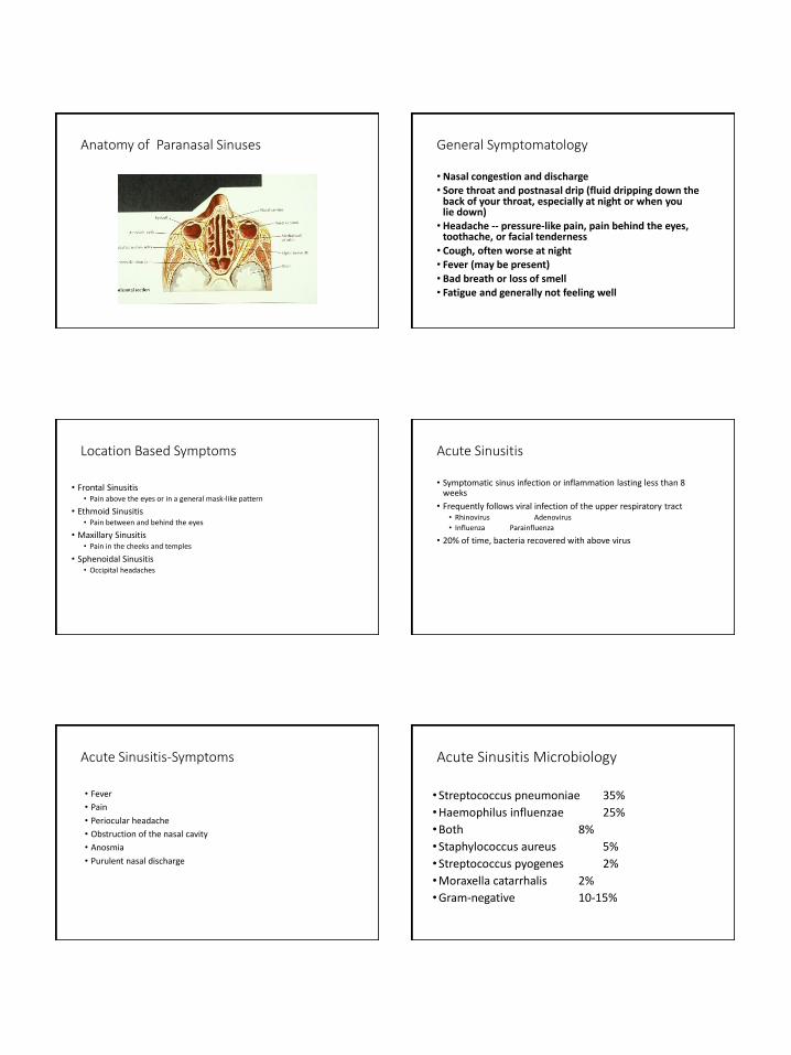
Anatomy of Paranasal Sinuses
• Cavities within facial skeleton that communicate with the nose
• Lined by ciliated respiratory epithelium
• Maxillary and ethmoid sinuses are present at birth
• Expansion of ethmoid labrynth above orbital rim gives rise to frontal sinuses
Anatomy of Paranasal Sinuses
• Unilateral agenesis of one frontal sinus is common• 4% of population has complete agenesis of frontal sinus
• Sphenoid sinus is last to develop and is not mature until early 20’s
• Mastoid Air Cells/Sinuses also late to develop• Unusual for mastoid sinus to be involved alone, without other sinus involvement
Anatomy of Paranasal Sinuses Anatomy of Paranasal Sinuses
Anatomy of Paranasal Sinuses General Symptomatology
• Nasal congestion and discharge • Sore throat and postnasal drip (fluid dripping down the
back of your throat, especially at night or when you lie down) • Headache -- pressure-like pain, pain behind the eyes,
toothache, or facial tenderness
• Cough, often worse at night • Fever (may be present) • Bad breath or loss of smell • Fatigue and generally not feeling well
Location Based Symptoms
• Frontal Sinusitis• Pain above the eyes or in a general mask-like pattern
• Ethmoid Sinusitis• Pain between and behind the eyes
• Maxillary Sinusitis• Pain in the cheeks and temples
• Sphenoidal Sinusitis• Occipital headaches
Acute Sinusitis
• Symptomatic sinus infection or inflammation lasting less than 8 weeks
• Frequently follows viral infection of the upper respiratory tract• Rhinovirus Adenovirus
• Influenza Parainfluenza
• 20% of time, bacteria recovered with above virus
Acute Sinusitis-Symptoms
• Fever
• Pain
• Periocular headache
• Obstruction of the nasal cavity
• Anosmia
• Purulent nasal discharge
Acute Sinusitis Microbiology
•Streptococcus pneumoniae 35%
•Haemophilus influenzae 25%
•Both 8%
•Staphylococcus aureus 5%
•Streptococcus pyogenes 2%
•Moraxella catarrhalis 2%
•Gram-negative 10-15%
Chronic Sinusitis
• Multiple etiologies• Allergic
• Anatomic (deviated septum, fx, trauma)
• Mucous abnormalities
• Persistent signs and symptoms despite continuous treatment• Post nasal drainage
• Facial pain
• Pressure within face or eyes
Chronic Sinusitis-Clinical Signs
• Thickening of the sinus mucosa on plain film and CT
• Anaerobic bacteria more common than in acute sinusitis
• Chronic presence of thickened nasal or post nasal discharge• Waxes and wanes
• Headache pain worse in AM
Chronic Sinusitis Microbiology
• AEROBIC• Streptococcus pyogenes
• alpha-hemolytic streptococci
• Staphylococcus
• S.pneumoniae
• H.flu
• Strep. viridans
ANAEROBIC
– Bacteriodes
– Peptococcus
– Propionobacterium
– Fusobacterium
– Veilonella
– Corynebacterium
Clinical Examination
• Complete Medical History
• Visualization of the Oropharynx
• Visualization of the Nares
• Plain Film X-Rays
• Computed Axial Tomography
• MRI ?
Clinical Examination In-Office Pearls
• Articulation of facial bones
• Sinus percussion
• Sinus transillumination
Articulation of Facial Bones
• an assessment of the relative pain or discomfort level associated with movement of the mucosal sinus linings.
• movement created by slight shifting of maxillary, frontal and nasal bone articulations.
• thumbs are placed on the vertical aspect of the maxillary bones, with the head supported posteriorly, and pressure is exerted to retroplace the maxillary bones.
Sinus Percussion
• a measure of vibratory sensation in the frontal and right and left maxillary sinuses.
• the middle or index finger of the non-dominant hand is placed over the sinus to be tested.
• the index and middle fingers of the dominant hand are used to ‘tap’ the finger laying across the sinus.
• a positive result is indicated by the presence of a painful, ‘reflected’ sensation radiating posteriorly through the tested sinus.
Sinus Percussion
Sinus Percussion Sinus Percussion
Sinus Transillumination
• Easy, inexpensive way to view frontal and maxillary sinuses
• Must be performed in a completely darkened room
• Shows us what can be seen in plain film and CT imaging
Conventional Radiography
• LATERAL VIEW
• SUBMENTOVERTICAL VIEW
• WATER’S VIEW
• CALDWELL’S VIEW
-obtain views with patient in upright position to facilitate sinus fluid
settling.
LATERAL VIEW: best evaluation of the sphenoid sinus, which is
occasionally obstructed by the floor of the middle cranial fossa.
SUBMENTOVERTICAL VIEW: a cumbersome view since beam is
aimed from below the neck upward, but visualizes well the spheniodal
sinuses and the posterior lateral walls of the antrum.
Water’s View
• AKA: chin-nose position, or occipitomeatal view.
• patient’s head is upward with the chin and nose against the film surface and the x-ray tube behind the head.
• gives best view of the maxillary sinuses.
• also used to evaluate the orbit for orbital floor blowout fractures.
Water’s View
Caldwell’s View
• AKA: forehead-nose position
• nose and forehead are placed against the film cassette, and the beam is directed from posteriorly 15 degrees below the horizontal.
• gives the best view of the ethmoidal sinuses and the nasal cavity.
• also useful in evaluation of nasal orbital wall fractures.
Caldwell’s View
CT EXAMINATION
• excellent visualization of all the sinuses.
• useful in obtaining information about adjacent structures, such as the orbit, cavernous sinus and pituitary fossa.
• 5mm sections are adequate for sinus evaluation.
• disadvantage: $$
• for our evaluation of sinusitis, this is preferred over NMR, primarily due to cost factors.
CT- Frontal Sinus
CT-Maxillary and Ethmoid Sinuses CT-Sphenoidal Sinus
CT-Mastoid Sinus Significant Sinus Invasion
Sinus Transillumination
• Easy to perform in primary eye care office
• Quick
• Relative ease in result interpretation
• Must be performed in a completely darkened examination room.
Sinus Transillumination
• Frontal Sinus Transillumination
• the tip of the transilluminator is placed beneath the orbital rim portion of the frontal bone and the light is directed upward toward the frontal sinus.
• the patient’s head is tilted back far enough to see the palate, and the light source is placed on the orbital portion of the maxillary bone and aimed downward.
Maxillary Sinus Transillumination
Maxillary Sinus Transillumination Management of Sinusitis
Antihistamines have no direct role in the treatment of sinusitis, and may perpetuate it by thickening mucous. They are useful in treating related allergy symptoms only!!!
Decongestants
• help promote drainage and decongestion of swollen sinus mucous membranes.
• conversely, they can excessively dry nasal mucosa, thereby impairing the mucociliary transport system.
• increase blood pressure
• agitation
• mucous membrane drying
• pseudoephedrine
• phenylephrine
Decongestants and Sinusitis
• Are beneficial in the initial treatment of sinusitis
• Rx’s for 5-7 days
• More difficult to get OTC now due to illegal usage• Typically are an adjunct to Tylenol & Motrin or Claritin-D, Zyrtec-D, Allegra-D
Remember the Etiology
• Mucosal swelling leading to mucosal infection.
• Treatment aimed at reduced swelling and reduced infection
Mucosal Nasal Anti-Inflammatories
• fluticasone (Flonase)
• mometasone (Nasonex)
• triamcinolone (Nasacort AQ).
• Nasal saline sprays
Antibiotics
•Antibiotics are the mainstay of treatment of acute and chronic sinusitis. •Acute treatment: 10-14 days•Chronic: 30 days
•Generally, sinusitis is managed with both antibiotics and decongestants (for 7 days max) and nasal sprays PRN
Remember…Acute Sinus Pathogens
•Streptococcus pneumoniae 35%
•Haemophilus influenzae 25%
•Both 8%
•TOTAL ~70%
Antibiotics of Choice
• ACUTE:• Amoxicillin-clavulanate (Augmentin)
500/125 TID
• *EES-400 TID*
• *ECN/Sulfisoxazole (Pediazole)* 5cc BID-TID
• Trimethoprim-sulfamethoxazole (Septra DS) BID
• Cephalexin (Keflex) 500 TID
•Acute and Chronic:
• *Clarithromycin (Biaxin) *500 BID
• *Azithromycin (Zithromax)* 250BID day one then QD X 4D
A Word of Caution
• sedation has been the major complaint with traditional antihistamines, such as Benadryl (diphenhydramine).
• newer antihistamines have significantly reduced incidences of sedation
• also require less frequent dosing (QD to BID).
2nd Gen Antihistamines
• TAVIST (TAVIST-D)• BID dosage
• OTC
• CLARITIN (loratadine):• 10mg QD
• OTC
• Used alone or with decongestant
• CLARINEX (desloratadine)• 10mg QD
• ZYRTEC (citirizine):• 10mg QD
• no reported cardiac interactions
• SINGULAIR • 10mg QD
• not an antihistamine
• inhibits leukotrienes
**Precaution**
• Macrolide antibiotics and certain antifungal agents in conjunction with Seldane (terfenadine) and Hismanal (astemizole) have been reported to increase levels of the antihistamine to toxic levels, causing cardiac arrythmias and death• Torsades des Pointes
Take Home on Sinusitis Management
• In office evaluation of the paranasal sinus is quick and easy
• Oral antibiotics are treatment of choice for both acute and chronic sinusitis
• Decongestants and perhaps antihistamines are useful in treating ancillary symptoms
• Avoid prolonged macrolide AB use and antihistamines