13
www.caymanchem.com Customer Service 800.364.9897 Technical Support 888.526.5351 1180 E. Ellsworth Rd · Ann Arbor, MI · USA SIRT3 Direct Fluorescent Screening Assay Kit Item No. 10011566

www.caymanchem.comCustomer Service 800.364.9897Technical Support 888.526.53511180 E. Ellsworth Rd · Ann Arbor, MI · USA
SIRT3 Direct Fluorescent Screening Assay Kit
Item No. 10011566

3GENERAL INFORMATION
TABLE OF CONTENTS GENERAL INFORMATION 3 Materials Supplied
4 Safety Data
4 Precautions
5 If You Have Problems
5 Storage and Stability
5 Materials Needed but Not Supplied
INTRODUCTION 6 Background
8 About This Assay
PRE-ASSAY PREPARATION 10 Reagent Preparation
ASSAY PROTOCOL 12 Plate Set Up
14 Performing the Assay
ANALYSIS 16 Calculations
17 Performance Characteristics
18 Interferences
RESOURCES 20 Troubleshooting
21 References
22 Plate Template
23 Notes
23 Warranty and Limitation of Remedy
GENERAL INFORMATION
Materials Supplied
Item Number Item Quantity
10012540 Assay Buffer (10X) 1 vial
10012541 SIRT3 (human recombinant) 2 vials
10012542 SIRT3 Direct Peptide 2 vials
10012543 NAD+ 1 vial
10012544 Nicotinamide 1 vial
10012545 Developer 1 vial
10012546 Fluorophore 1 vial
10011288 Half Volume 96-Well Plate (white) 1 plate
400012 96-Well Cover Sheet 1 cover
If any of the items listed above are damaged or missing, please contact our Customer Service department at (800) 364-9897 or (734) 971-3335. We cannot accept any returns without prior authorization.Sold under license from CycLex® Co., Ltd (Patent Nos. EP 1243658, US 7033778, US 7256013, and JP 4267043).
4 GENERAL INFORMATION 5GENERAL INFORMATION
! WARNING: THIS PRODUCT IS FOR RESEARCH ONLY - NOT FORHUMAN OR VETERINARY DIAGNOSTIC OR THERAPEUTIC USE.
Safety DataThis material should be considered hazardous until further information becomes available. Do not ingest, inhale, get in eyes, on skin, or on clothing. Wash thoroughly after handling. Before use, the user must review the complete Safety Data Sheet, which has been sent via email to your institution.
PrecautionsPlease read these instructions carefully before beginning this assay.
If You Have ProblemsTechnical Service Contact Information
Phone: 888-526-5351 (USA and Canada only) or 734-975-3888Fax: 734-971-3641Email: [email protected]: M-F 8:00 AM to 5:30 PM EST
In order for our staff to assist you quickly and efficiently, please be ready to supply the lot number of the kit (found on the outside of the box).
Storage and StabilityThis kit will perform as specified if stored at -80°C and used before the expiration date indicated on the outside of the box.
Materials Needed But Not Supplied1. A fluorometer with the capacity to measure fluorescence using an excitation
wavelength of 350-360 nm and an emission wavelength of 450-465 nm.2. Adjustable pipettes and a repeating pipettor.3. A source of pure water; glass distilled water or HPLC-grade water is
acceptable.
6 INTRODUCTION 7INTRODUCTION
INTRODUCTION
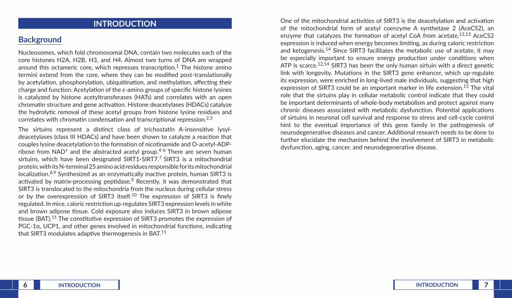
BackgroundNucleosomes, which fold chromosomal DNA, contain two molecules each of the core histones H2A, H2B, H3, and H4. Almost two turns of DNA are wrapped around this octameric core, which represses transcription.1 The histone amino termini extend from the core, where they can be modified post-translationally by acetylation, phosphorylation, ubiquitination, and methylation, affecting their charge and function. Acetylation of the ε-amino groups of specific histone lysines is catalyzed by histone acetyltransferases (HATs) and correlates with an open chromatin structure and gene activation. Histone deacetylases (HDACs) catalyze the hydrolytic removal of these acetyl groups from histone lysine residues and correlates with chromatin condensation and transcriptional repression.2,3 The sirtuins represent a distinct class of trichostatin A-insensitive lysyl-deacetylases (class III HDACs) and have been shown to catalyze a reaction that couples lysine deacetylation to the formation of nicotinamide and O-acetyl-ADP-ribose from NAD+ and the abstracted acetyl group.4-6 There are seven human sirtuins, which have been designated SIRT1-SIRT7.7 SIRT3 is a mitochondrial protein, with its N-terminal 25 amino acid residues responsible for its mitochondrial localization.8,9 Synthesized as an enzymatically inactive protein, human SIRT3 is activated by matrix-processing peptidase.9 Recently, it was demonstrated that SIRT3 is translocated to the mitochondria from the nucleus during cellular stress or by the overexpression of SIRT3 itself.10 The expression of SIRT3 is finely regulated. In mice, caloric restriction up-regulates SIRT3 expression levels in white and brown adipose tissue. Cold exposure also induces SIRT3 in brown adipose tissue (BAT).11 The constitutive expression of SIRT3 promotes the expression of PGC-1α, UCP1, and other genes involved in mitochondrial functions, indicating that SIRT3 modulates adaptive thermogenesis in BAT.11
One of the mitochondrial activities of SIRT3 is the deacetylation and activation of the mitochondrial form of acetyl coenzyme A synthetase 2 (AceCS2), an enzyme that catalyzes the formation of acetyl CoA from acetate.12,13 AceCS2 expression is induced when energy becomes limiting, as during caloric restriction and ketogenesis.14 Since SIRT3 facilitates the metabolic use of acetate, it may be especially important to ensure energy production under conditions when ATP is scarce.12,14 SIRT3 has been the only human sirtuin with a direct genetic link with longevity. Mutations in the SIRT3 gene enhancer, which up-regulate its expression, were enriched in long-lived male individuals, suggesting that high expression of SIRT3 could be an important marker in life extension.15 The vital role that the sirtuins play in cellular metabolic control indicate that they could be important determinants of whole-body metabolism and protect against many chronic diseases associated with metabolic dysfunction. Potential applications of sirtuins in neuronal cell survival and response to stress and cell-cycle control hint to the eventual importance of this gene family in the pathogenesis of neurodegenerative diseases and cancer. Additional research needs to be done to further elucidate the mechanism behind the involvement of SIRT3 in metabolic dysfunction, aging, cancer, and neurodegenerative disease.

8 INTRODUCTION 9INTRODUCTION
Ac-Gln-Pro-Lys-Lys(Ac)-AMC
Ac-Gln-Pro-Lys-Lys-AMC
Fluorescence
Niconamide + O-Ac-ADP-ribose
Developer
SIRT3
NAD+
Ac-Gln-Pro-Lys-Lys + AMC
Figure 1. Assay scheme
About This AssayCayman’s SIRT3 Direct Fluorescent Screening Assay Kit provides a convenient fluorescence-based method for screening SIRT3 inhibitors or activators. The procedure requires only two easy steps, both performed in the same microplate (Figure 1, on page 9). In the first step, the substrate, which comprises the p53 sequence Gln-Pro-Lys-Lys(ε-acetyl)-AMC, is incubated with human recombinant SIRT3 along with its cosubstrate NAD+. Deacetylation sensitizes the Substrate such that treatment with the Developer in the second step releases a fluorescent product. The Fluorophore can be analyzed with an excitation wavelength of 350-360 nm and an emission wavelength of 450-465 nm.

10 PRE-ASSAY PREPARATION 11PRE-ASSAY PREPARATION
PRE-ASSAY PREPARATION
Reagent Preparation
1. Assay Buffer (10X) - (Item No. 10012540)Dilute 3 ml of Assay Buffer concentrate with 27 ml of HPLC-grade water. This final Buffer (50 mM Tris-HCl, pH 8.0, containing 137 mM NaCl, 2.7 mM KCl, and 1 mM MgCl2) should be used in the assay and for diluting reagents. When stored at 4°C, this diluted Buffer is stable for at least six months.
2. SIRT3 (human recombinant) - (Item No. 10012541)Each vial contains 50 µl of human recombinant SIRT3. Thaw the enzyme on ice, add 200 µl of diluted Assay Buffer to the vial, and vortex. The diluted enzyme is stable for four hours on ice. One vial of enzyme is enough SIRT3 to assay 50 wells. Use the additional vial if assaying the entire plate.
3. SIRT3 Direct Peptide - (Item No. 10012542)Each vial contains 100 µl of a 5 mM peptide solution comprising amino acids 317-320 of human p53 conjugated to aminomethylcoumarin (AMC). It is ready to use to make the Substrate Solution.
4. NAD+ - (Item No. 10012543)The vial contains 500 µl of a 50 mM solution of NAD+. It is ready to use to make the Substrate Solution.
5. Nicotinamide - (Item No. 10012544)The vial contains 500 µl of a 50 mM solution of nicotinamide, a sirtuin inhibitor. It is ready to use to make the Stop/Developing Solution.
6. Developer - (Item No. 10012545)The vial contains 100 mg of the SIRT3 developer.
7. Fluorophore - (Item No. 10012546)The vial contains 50 µl of 10 mM 7-amino-4-methylcoumarin in dimethylsulfoxide (DMSO). The Fluorophore can be used to assay for interference (see page 18).
12 ASSAY PROTOCOL 13ASSAY PROTOCOL
ASSAY PROTOCOL
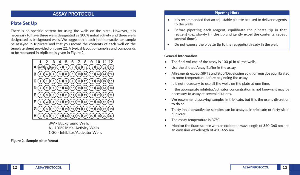
Plate Set UpThere is no specific pattern for using the wells on the plate. However, it is necessary to have three wells designated as 100% initial activity and three wells designated as background wells. We suggest that each inhibitor/activator sample be assayed in triplicate and that you record the contents of each well on the template sheet provided on page 22. A typical layout of samples and compounds to be measured in triplicate is given in Figure 2.
BW - Background WellsA - 100% Inial Acvity Wells1-30 - Inhibitor/Acvator Wells
A
B
C
D
E
F
G
H
1 2 3 4 5 6 7 8 9 10 11 12BW
A
1
2
3
4
5
6 6
5
4
3
2
1
A
BW
14
13
12
11
10
9
8
7
14
13
12
11
10
9
8
7
14
13
12
11
10
9
8
7
22
21
20
19
18
17
16
15
22
21
20
19
18
17
16
15
22
21
20
19
18
17
16
15
30
29
28
27
26
25
24
23
30
29
28
27
26
25
24
23 23
30
29
28
27
26
25
24
BW
A
1
2
3
4
5
6
Figure 2. Sample plate format
Pipetting Hints
• It is recommended that an adjustable pipette be used to deliver reagents to the wells.
• Before pipetting each reagent, equilibrate the pipette tip in that reagent (i.e., slowly fill the tip and gently expel the contents, repeat several times).
• Do not expose the pipette tip to the reagent(s) already in the well.
General Information• The final volume of the assay is 100 µl in all the wells.• Use the diluted Assay Buffer in the assay.• All reagents except SIRT3 and Stop/Developing Solution must be equilibrated
to room temperature before beginning the assay.• It is not necessary to use all the wells on the plate at one time.• If the appropriate inhibitor/activator concentration is not known, it may be
necessary to assay at several dilutions.• We recommend assaying samples in triplicate, but it is the user’s discretion
to do so.• Thirty inhibitor/activator samples can be assayed in triplicate or forty-six in
duplicate.• The assay temperature is 37°C.• Monitor the fluorescence with an excitation wavelength of 350-360 nm and
an emission wavelength of 450-465 nm.

14 ASSAY PROTOCOL 15ASSAY PROTOCOL
Performing the Assay1. Preparation of Substrate Solution - To one of the thawed SIRT3 peptide vials
(Item No. 10012542), add 240 µl of NAD+ Solution (Item No. 10012543), and 850 µl of diluted Assay Buffer. One vial of peptide will make enough Substrate Solution for 79 wells. The Substrate Solution is stable for six hours. The addition of 15 µl to the assay yields a final concentration of 125 µM peptide and 3 mM NAD+. Note: The Km values for the peptide and NAD+ are 323 and 241 µM, respectively.
2. 100% Initial Activity Wells - add 25 µl of Assay Buffer, 5 µl of diluted SIRT3, and 5 µl of solvent (the same solvent used to dissolve the inhibitor/activator) to three wells.
3. Background Wells - add 30 µl of Assay Buffer and 5 µl of solvent (the same solvent used to dissolve the inhibitor/activator) to three wells.
4. Inhibitor/Activator Wells - add 25 µl of Assay Buffer, 5 µl of diluted SIRT3, and 5 µl of inhibitor/activator* to three wells.
5. Initiate the reactions by adding 15 µl of Substrate Solution to all the wells being used.
6. Cover the plate with the plate cover and incubate on a shaker for 45 minutes at 37°C.
7. Preparation of Stop/Developing solution - Weigh 30 mg of Developer (Item No. 10012545) into a vial that will hold 5 ml then add 200 µl of Nicotinamide (Item No. 10012544) and 4.8 ml of diluted Assay Buffer. Vortex until the Developer is into solution. This is enough Stop/Developing solution for the entire plate. The Stop/Developing solution is stable for four hours on ice.
8. Remove the plate cover and add 50 µl of Stop/Developing solution to each well. Cover the plate with the plate cover and incubate for 30 minutes at room temperature.
9. Remove the plate cover and read the plate using an excitation wavelength of 350-360 nm and an emission wavelength of 450-465 nm. It may be necessary to adjust the gain setting on the instrument to allow for the measurement of all the samples. The fluorescence is stable for 30 minutes.
*Inhibitors/activators can be dissolved in Assay Buffer, methanol, or DMSO and should be added to the assay in a final volume of 5 µl. In the event that the appropriate concentration of inhibitor/activator needed for SIRT3 inhibition or activation is completely unknown, we recommend that several concentrations of the inhibitor/activator be assayed.
16 ANALYSIS 17ANALYSIS
ANALYSIS
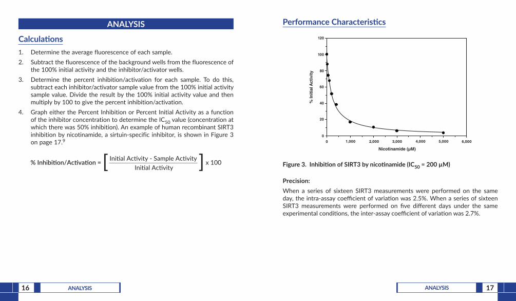
Calculations1. Determine the average fluorescence of each sample.2. Subtract the fluorescence of the background wells from the fluorescence of
the 100% initial activity and the inhibitor/activator wells.3. Determine the percent inhibition/activation for each sample. To do this,
subtract each inhibitor/activator sample value from the 100% initial activity sample value. Divide the result by the 100% initial activity value and then multiply by 100 to give the percent inhibition/activation.
4. Graph either the Percent Inhibition or Percent Initial Activity as a function of the inhibitor concentration to determine the IC50 value (concentration at which there was 50% inhibition). An example of human recombinant SIRT3 inhibition by nicotinamide, a sirtuin-specific inhibitor, is shown in Figure 3 on page 17.9
% Inhibi�on/Ac�va�on = Ini�al Ac�vity - Sample Ac�vityIni�al Ac�vity ][ x 100
Performance Characteristics
60
80
100
120
% In
itia
l Act
ivit
y
Nicotinamide (µM)
0 1,000 2,000 3,000 4,0000
20
40
6,0005,000
Inhibition of SIRT3 by Nicotinamide (IC50 = 200 µM)
Figure 3. Inhibition of SIRT3 by nicotinamide (IC50 = 200 µM)
Precision:When a series of sixteen SIRT3 measurements were performed on the same day, the intra-assay coefficient of variation was 2.5%. When a series of sixteen SIRT3 measurements were performed on five different days under the same experimental conditions, the inter-assay coefficient of variation was 2.7%.
18 ANALYSIS 19ANALYSIS
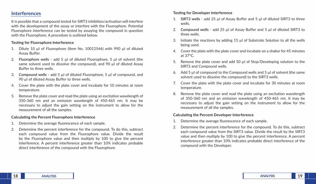
InterferencesIt is possible that a compound tested for SIRT3 inhibition/activation will interfere with the development of the assay or interfere with the Fluorophore. Potential Fluorophore interference can be tested by assaying the compound in question with the Fluorophore. A procedure is outlined below.
Testing for Fluorophore Interference1. Dilute 10 µl of Fluorophore (Item No. 10012546) with 990 µl of diluted
Assay Buffer.2. Fluorophore wells - add 5 µl of diluted Fluorophore, 5 µl of solvent (the
same solvent used to dissolve the compound), and 90 µl of diluted Assay Buffer to three wells.
3. Compound wells - add 5 µl of diluted Fluorophore, 5 µl of compound, and 90 µl of diluted Assay Buffer to three wells.
4. Cover the plate with the plate cover and incubate for 10 minutes at room temperature.
5. Remove the plate cover and read the plate using an excitation wavelength of 350-360 nm and an emission wavelength of 450-465 nm. It may be necessary to adjust the gain setting on the instrument to allow for the measurement of all the samples.
Calculating the Percent Fluorophore Interference1. Determine the average fluorescence of each sample.2. Determine the percent interference for the compound. To do this, subtract
each compound value from the Fluorophore value. Divide the result by the Fluorophore value and then multiply by 100 to give the percent interference. A percent interference greater than 10% indicates probable direct interference of the compound with the Fluorophore.
Testing for Developer Interference1. SIRT3 wells - add 25 µl of Assay Buffer and 5 µl of diluted SIRT3 to three
wells.2. Compound wells - add 25 µl of Assay Buffer and 5 µl of diluted SIRT3 to
three wells.3. Initiate the reactions by adding 15 µl of Substrate Solution to all the wells
being used.4. Cover the plate with the plate cover and incubate on a shaker for 45 minutes
at 37°C.5. Remove the plate cover and add 50 µl of Stop/Developing solution to the
SIRT3 and Compound wells.6. Add 5 µl of compound to the Compound wells and 5 µl of solvent (the same
solvent used to dissolve the compound) to the SIRT3 wells.7. Cover the plate with the plate cover and incubate for 30 minutes at room
temperature.8. Remove the plate cover and read the plate using an excitation wavelength
of 350-360 nm and an emission wavelength of 450-465 nm. It may be necessary to adjust the gain setting on the instrument to allow for the measurement of all the samples.
Calculating the Percent Developer Interference1. Determine the average fluorescence of each sample.2. Determine the percent interference for the compound. To do this, subtract
each compound value from the SIRT3 value. Divide the result by the SIRT3 value and then multiply by 100 to give the percent interference. A percent interference greater than 10% indicates probable direct interference of the compound with the Developer.

20 RESOURCES 21RESOURCES
RESOURCES
Troubleshooting
Problem Possible Causes Recommended Solutions
Erratic values; dispersion of duplicates/triplicates
A. Poor pipetting/technique
B. Bubble in the well(s)
A. Be careful not to splash the contents of the wells
B. Carefully tap the side of the plate with your finger to remove bubbles
No fluorescence detected above background in any of the wells
Either SIRT3, developer, or other key solution was not added to the wells
Make sure to add all the components to the wells and re-assay
The fluorometer exhibited ‘MAX’ values for the wells
The GAIN setting is too high Reduce the GAIN and re-read
No inhibition/activation seen with compound
A. The compound concentration is not high enough
B. The compound is not an inhibitor/activator of the enzyme
Increase the compound concentration and re-assay
References1. Grunstein, M. Nature 389, 349-352 (1997).2. Strahl, B.D. and Allis, D. Nature 403, 41-45 (2000).3. Cheung, W.L., Briggs, D.B., and Allis, C.D. Curr. Opin. Cell Biol. 12, 326-333
(2000).4. Imai, S.-I., Armstrong, C.M., Kaeberlein, M., et al. Nature 403, 795-800
(2000).5. Tanner, K.G., Landry, J., Sternglanz, R., et al. Proc. Natl. Acad. Sci. USA 97(26),
14178-14182 (2000).6. Tanny, J.C., Moazed, D. Proc. Natl. Acad. Sci. USA 98(2), 415-420 (2001).7. Frye, R.A. Biochem. Biophys. Res. Commun. 273, 793-798 (2000).8. Onyango, P., Celic, I., McCaffery, J.M., et al. Proc. Natl. Acad. Sci. USA 99(21),
13653-13658 (2002).9. Schwer, B., North, B.J., Frye, R.A., et al. J. Cell Biol. 158(4), 647-657 (2002).10. Scher, M. B., Vaquero, A., Reinberg, D. Genes Dev. 21, 920-928 (2007).11. Shi, T., Wang, F., Stieren, E., et al. J. Biol. Chem. 280(14), 13560-13567
(2005).12. Hallows, W. C., Lee, S., Denu, J.M. Proc. Natl. Acad. Sci. USA 103(27),
10230-10235 (2006).13. Fujino, T., Kondo, J., Ishikawa, M., et al. J. Biol. Chem. 276(14), 11420-11426
(2001).14. Schwer, B., Bunkenborg, J., Verdin, R. O., et al. Proc. Natl. Acad. Sci. USA
103(27), 10224-10229 (2006).15. Bellizzi, D., Rose, G., Cavalcante, P., et al. Genomics 85, 258-263 (2004).

22 RESOURCES 23RESOURCES
A B C D E F G H
12
34
56
78
910
1112
NOTES
Warranty and Limitation of RemedyBuyer agrees to purchase the material subject to Cayman’s Terms and Conditions.Complete Terms and Conditions including Warranty and Limitation of Liability information can be found on our website.This document is copyrighted. All rights are reserved. This document may not, in whole or part, be copied, photocopied, reproduced, translated, or reduced to any electronic medium or machine-readable form without prior consent, in writing, from Cayman Chemical Company.©08/18/2016, Cayman Chemical Company, Ann Arbor, MI, All rights reserved. Printed in U.S.A.


![Vaccinia-relatedKinase1(VRK1)IsanUpstreamNucleosomal ... · mM NaCl, 1 mM DTT, 50 M ATP, and 5 Ci of (0.1 M) [ -32P]ATP (33).The CHK2 kinase assay was performed in buffer containing](https://static.documents.pub/doc/80x56/5f85ce92fe1faa1c4f0db0df/vaccinia-relatedkinase1vrk1isanupstreamnucleosomal-mm-nacl-1-mm-dtt-50-m.jpg)


