53
Sistanizad M. Pharm D, BCCP SBMU

Sistanizad M. Pharm D, BCCP
SBMU

Definition
Anemia is a reduction in red cell mass
The term anemia is not a diagnosis, but rather an
objective sign of a disease
2

Causes of Anemia- Pathophysiologic
Blood Loss
Acute: trauma, ulcer, hemorrhoids
Chronic: ulcer, vaginal bleeding, aspirin ingestion
Inadequate Red Blood Cell Production
Nutritional deficiency: B12, folic acid, iron
Erythroblast deficiency: bone marrow failure (aplastic anemia,
irradiation, chemotherapy, folic acid antagonists) or bone marrow
infiltration (leukemia, lymphoma, myeloma, metastatic solid tumors)
Chronic disease: renal, liver, infection, granulomatous, collagen vascular
Excessive Red Blood Cell Destruction
Intrinsic factors: hereditary (G6PD), abnormal hemoglobin synthesis
Extrinsic factors: autoimmune reactions, drug reactions, infection
(endotoxin)
3

Classifications of Anemia-Morphologic Macrocytic
Megaloblastic: pernicious (vitamin B12 deficiency), folic
acid deficiency
Normochromic, normocytic
Recent blood loss
Hemolysis
Chronic disease
Renal failure
Autoimmune
Microcytic, hypochromic
Iron deficiency
Genetic abnormalities: sickle cell, thalassemia
4

Detection Signs and Symptoms:
Vary with the degree of RBC reduction
History
Physical Examination:
Pallor (conjunctiva), nail beds
Postural hypotension and tachycardia
Neurologic findings
Jaundice
5

Detection- cont. Laboratory:
a full laboratory evaluation is necessary to confirm the
diagnosis, establish its severity, and determine its cause
Can provide sufficient information to distinguish between
the most common forms of anemia
6

Routine Laboratory Evaluation for
Anemia Workup Complete blood count (CBC): Hgb, Hct, RBC count, red cell
indices (MCV, MCH, MCHC), WBC count (and differential)
Platelet count
Red cell morphology
Reticulocyte count
Bilirubin and LDH
Serum iron, TIBC, serum ferritin, transferrin saturation
Peripheral blood smear examination
Stool examination for occult blood
Bone marrow aspiration and biopsya
7

Laboratory Test
Pediatric Adult
1–15 yr Male Female
RBC (× 106/mm3) 4.7±6 5.4±0.7 4.8±6
Hgb (g/dL) 13±2 16±2 14±2
Hct (%) 40±5 47±5 42±2
MCV (µm3) 80±5 87±7 90±9
MCH (pg/cell) 33.5±2 29±2 34±2
MCHC (g/dL) 31±3.6 31±3.6 31±3.6
Erythropoietin (mU/mL) 4–26 4–26 4–26
Reticulocyte count (%) 0.5–1.5 0.5–1.5 0.5–1.5
TIBC (mg/dL) 250–400 250–400 250–400
Fe (mg/dL) 50–120 50–160 40–150
Folate (ng/mL) 7–25 7–25 7–25
Fe/TIBC (%) 20–30 20–40 16–38
Vitamin B12 (pg/mL) >200 >200 >200
Ferritin (ng/mL) 7–140 15–200 12–150 8
Normal Hematology Values

Iron Deficiency Anemia-Iron Stores
The body contains approximately 3.5 g of iron, of which
2.5 g are found in Hgb
About 400 mg exists as iron-containing proteins such as
myoglobin and cytochromes.
Another 3 to 7mg of iron is bound to transferrin in
plasma(100-150mg/dL)
The remaining iron exists as storage iron in the form of
ferritin or hemosiderin:
Men: 600 – 1200mg
Women: 100 – 400mg
9

Iron Deficiency Anemia-Iron Lost
Only about 0.5 to 1 mg/day of iron is lost from urine,
sweat, and intestinal cells that contain ferritin
Menstruating , pregnancy and lactation are other common
sources of iron loss
10
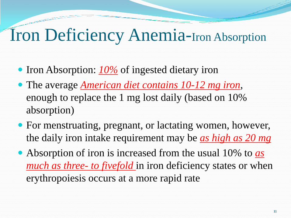
Iron Deficiency Anemia-Iron Absorption
Iron Absorption: 10% of ingested dietary iron
The average American diet contains 10-12 mg iron,
enough to replace the 1 mg lost daily (based on 10%
absorption)
For menstruating, pregnant, or lactating women, however,
the daily iron intake requirement may be as high as 20 mg
Absorption of iron is increased from the usual 10% to as
much as three- to fivefold in iron deficiency states or when
erythropoiesis occurs at a more rapid rate
11

Iron Deficiency Anemia-Causes
Anemia caused by iron deficiency is the most common
nutritional deficiency worldwide
Blood loss is considered one of the more common.
Each milliliter of whole blood contains 0.5 mg of iron
12

Iron Deficiency Anemia-Causes Blood Loss
Menstruation, gastrointestinal (e.g., peptic ulcer), trauma
Decreased Absorption
Medications, gastrectomy, regional enteritis
Increased Requirement
Infancy, pregnant/lactating women
Impaired Utilization
Hereditary, Iron use
13
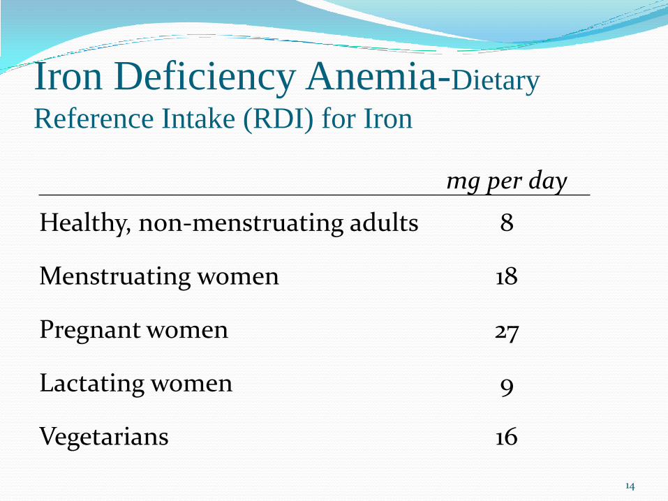
Iron Deficiency Anemia-Dietary
Reference Intake (RDI) for Iron
mg per day
Healthy, non-menstruating adults 8
Menstruating women 18
Pregnant women 27
Lactating women 9
Vegetarians 16
14

Iron Deficiency Anemia-Supplements
Necessary for pregnant and lactating mothers
Infants 6 months to 3 years of age experience rapid growth and
a threefold increase in blood volume [1 to 2 mg/kg/day (not to
exceed 20 mg/day)]
Premature infants have reduced iron stores (10 to 15 mg/day
for up to the first year of life)
15
Diagnosis History
Blood loss (melena, menorrhagia, frequent blood donation).
Physical Examination
Decreased exercise tolerance,Weakness, Palpitation,
Tachycardia
16

Diagnosis Laboratory Studies
Hypochromic microcytic
Iron testing
low serum iron (<50 mcg/dL) and ferritin (<12-15 mcg/dL) levels
and a high TIBC (>400 mcg/dL) .
Serum ferritin level of <10 ng/mL in women or 20 ng/mL in men
most sensitive)
17

Treatment: Iron Dose
Iron is poorly absorbed from vegetables, grain products,
dairy products, and eggs; it is best absorbed from meat.
In patients with IDA, it is generally recommended that
approximately 200 mg of elemental iron be administered
daily, usually in two or three divided doses to maximize
tolerability
18

Treatment: Iron Dose 0.25 g/dL/day is the maximal rate of hemoglobin
regeneration
Elemental iron (mg/day)= (0.25 g Hg/100ml) * (5000
ml/100ml) * (3.4 mg Fe/1 g Hgb) = 40 mg/day
40 mg/day /20% (Approximate absorption in iron deficiency) =
200 mg/day
200 /20% (Ferrous sulfate contains 20% elemental iron) = 1000
mg Ferrous sulfate/day
19

Absorption of Iron Gastric acid and other dietary components such as
ascorbic acid (doses>1g) increase the absorption of
nonheme iron
Dietary components that form insoluble complexes with
iron (tannates) decrease absorption.
20
Absorption of Iron Calcium inhibits absorption of both heme and nonheme
iron. Epidemiologic studies show a correlation between
milk intake and prevalence of iron deficiency.
Because gastric acid improves iron absorption, patients
who have undergone a gastrectomy or have achlorhydria
will have decreased iron absorption.
21

Patient Information Childproof container
Oral iron therapy produces dark stools.
Patient should try to take her iron on an empty stomach
because food, especially dairy products, decreases the
absorption by 40% to 50%
Gastric side effects, which occur in 5% to 20% of patients,
include nausea, epigastric pain, constipation, abdominal
cramps, and diarrhea.
22

Toxicity Acute elemental iron ingestions of <20 mg/kg are usually
nontoxic, 20 to 60 mg/kg doses result in mild to moderate
toxicity, and >60 mg/kg doses are severe and potentially
fatal
23

Patient Information Potential drug interactions that can occur with iron
therapy
PPIs: Patient should be advised to take her iron at least 1
hour before or 3 hours after the proton pump inhibitor dose
Antacids can increase stomach pH and certain anions
(carbonate and hydroxide) also are thought to form
insoluble complexes when combined with iron.
Tetracycline: the iron should be taken 3 hours before or 2
hours after the tetracycline dose as well.
24

Iron Salt–Drug Interactions
25

Treatment: Follow Up Hematologic response is usually seen in 2 to 3 weeks with
a 1 g/dL increase in hemoglobin
Regardless of the form of oral therapy used, treatment
must be continued 3 to 6 months after the anemia is
resolved to allow for repletion of iron stores and to avoid
relapse.
26

PARENTERAL IRON THERAPY When there is evidence of iron malabsorption or
intolerance to orally administered iron, or when long-term
noncompliance is a problem, parenteral iron therapy may
be warranted.
27
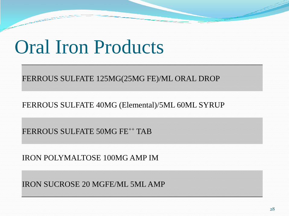
Oral Iron Products
28
FERROUS SULFATE 125MG(25MG FE)/ML ORAL DROP
FERROUS SULFATE 40MG (Elemental)/5ML 60ML SYRUP
FERROUS SULFATE 50MG FE++ TAB
IRON POLYMALTOSE 100MG AMP IM
IRON SUCROSE 20 MGFE/ML 5ML AMP
Megaloblastic Anemias
29

Megaloblastic Anemia Megaloblastosis results from impaired DNA synthesis in
replicating cells, which is signaled by a large immature
nucleus
Can have several causes:
a) Vitamin B12 deficiency
b) Folic acid deficiency
c) Metabolic or inherited defects associated with decreased
ability to utilize vitamin B12 or folic acid
30

Vitamin B12 Deficiency Anemia The daily requirement of approximately 2 mcg
Vitamin B12 deficiency can result from
i. Decreased intake, absorption, transport, and utilization
ii. Increased requirements, metabolic consumption,
destruction, and excretion.
Strict vegetarians most frequently present with signs and
symptoms of vitamin B12 deficiency
31
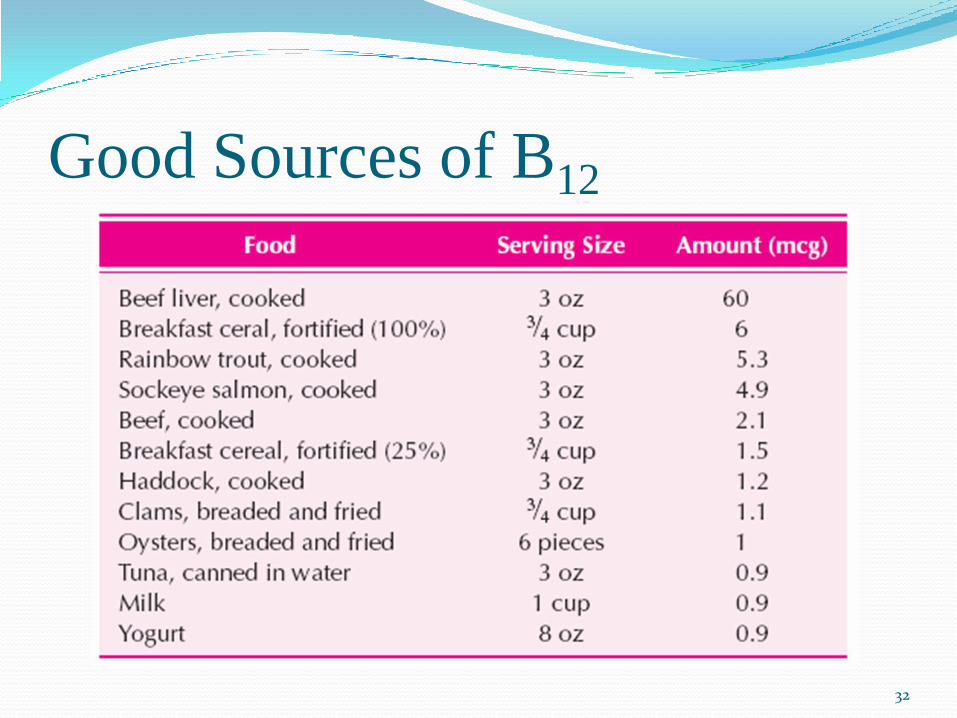
Good Sources of B12
32
Causes Pernicious anemia
Helicobacter pylori
Elderly subjects
Intestinal disorders
Dietary intake
HIV infection
Hereditary causes
Nitrous oxide exposure:
(N2O) inactivates cobalamin and its use in anesthesia or inhalant abuse may precipitate RAPID hematologic and neuropsychiatric deterioration in Cbl-deficient subjects
33
Pernicious anemia Develops from a lack of gastric intrinsic factor
production, which causes vitamin B12 malabsorption and,
ultimately, vitamin B12 deficiency
Pernicious anemia occurs commonly in patients with
thyrotoxicosis, Hashimoto's thyroiditis, vitiligo,
rheumatoid arthritis, or gastric cancer.
Anti-intrinsic factor antibodies have been observed in the
serum of some patients with pernicious anemia.
Partial or total gastrectomy often results in anemia,
particularly pernicious anemia
34

Pernicious anemia Patients generally do not feel well for 6 to 12 months and
often complain of at least two of the following triad of
symptoms:
I. Weakness
II. Sore tongue
III. Symmetric numbness or tingling in the extremities
Neurologic symptoms: tinnitus, vertigo, headache
35

Pernicious anemia- Laboratory Evaluation
In general, the serum vitamin B12 level reliably reflects
vitamin B12 tissue stores
Hgb
MCV
36

Pernicious anemia- Treatment
In a dose sufficient to provide not only the daily
requirement of approximately 2 mcg, but also the amount
needed to replenish tissue stores (about 2,000 to 5,000
mcg; average, 4,000 mcg
37

Pernicious anemia- Treatment
100 mcg of cyanocobalamin daily for 1 week, then 100
mcg every other day for 2 weeks, followed by 100 mcg
every 3 to 4 days for 2 to 3 weeks. A monthly maintenance
dose of cyanocobalamin (100 mcg) would then be
required for the remainder of life.
Another treatment option may be cyanocobalamin (1,000
mcg) once a week for 4 to 6 weeks followed by 100
mcg/mo for lifetime maintenance therapy
38

Pernicious anemia- Treatment
IM or deep SC administration provides sustained release
of vitamin B12 with better utilization compared with rapid
IV infusion.
An oral or intranasal cyanocobalamin gel is also
available for maintenance therapy, after the patient has
achieved hematologic remission.
With adequate vitamin B12 therapy neurologic symptoms
should improve within 24 hours.
39

Pernicious anemia- Treatment
Hematologic parameters should begin to improve within
the first few days.
The bone marrow becomes normoblastic within 48 hours
The reticulocyte count should peak around day 5 of therapy
The Hct should return to normal in 1 to 2 months.
Serum potassium should be monitored and potassium
supplementation provided as necessary
40

Oral Vitamin B12 Approximately 5 mcg of vitamin B12 is absorbed daily from the
average American diet.
The percentage of vitamin B12 absorbed decreases with
increasing doses.
About 50% of a 1 to 2 mcg dose of vitamin B12 is absorbed,
whereas only about 5-10% of a 20 mcg dose is absorbed. Doses
>100 mcg must be ingested to absorb 5 mcg of vitamin B12.
Oral therapy for pernicious anemia using high dosages of oral
cyanocobalamin (1,000 to 2,000 mcg)
Patients can be given 1,000 to 2,000 mcg/day for 1 month,
followed by 125 to 500 mcg/day as maintenance treatment.
41

Folic Acid Deficiency Anemia Folate is abundant in virtually all food sources, especially
fresh green vegetables, fruits, yeast, and animal protein
Excessive or prolonged cooking (>15 minutes) in large
quantities of water destroys a high percentage of the folate
that is contained in food
Requirements: generally 3 mcg/kg/day
42

Folic Acid Deficiency Anemia A daily intake of 200 mcg is recommended.
Folate requirements are increased in conditions in which
the metabolic rate and rate of cellular division are
increased (e.g., pregnancy, infancy, infection,
malignancies, hemolytic anemia).
The following are estimates of daily folate requirements
based on age and growth demands:
children, 80 mcg
infants, 65 mcg
pregnant or lactating women, 400 to 800 mcg
43

Causes Nutritional
The most common cause
Increased requirements
Drugs:
Trimethoprim
Pyrimethamine
MTX:
Treatment usually consists of FA in a dose of 1 mg/day although up to 5 mg/day may be required
Phenytoin:
blocks FA absorption and increases utilization of FA by an unknown mechanism
44

Folic Acid Deficiency Anemia Folate deficiency is most commonly associated with
Alcoholism
rapid cell turnover
dietary deficiency
45

Folic Acid Deficiency Anemia-Diagnosis and Management
Serum folate concentrations
Because the estimated total body folate store is only about
5 to 10 mg, 1 mg of folic acid given daily for 2 to 3 weeks
should be more than adequate to replace her storage pool
of folate.
Higher dosages (up to 5 mg) may be needed, however, if
absorption is compromised by alcohol or other factors
Once anemia is corrected, 0.1 mg of folate as a nutritional
supplement should be adequate for maintenance treatment
46

Anemia of Chronic Disease
47

Anemia of Chronic Disease ACD refers to a mild to moderate anemia associated with
a number of chronic disorders (e.g., rheumatoid arthritis
[RA], systemic lupus erythematosus, chronic infections,
chronic renal failure, acquired immunodeficiency
syndrome [AIDS], neoplastic disease).
The second most common anemia behind iron deficiency.
Most often, ACD is a normochromic, normocytic anemia
48

ACD-Pathogenesis
Inflammatory cytokines:
Competition for EPO receptors by IFN-γ and TNF-α may
possibly lead to EPO resistance
IL-1 and TNF-α also inhibit hepatic and renal expression of
EPO messenger RNA (mRNA)
49

ACD-Management
Focuses on the underlying disease process.
Unless a concurrent deficiency of vitamin B12, folate, or
iron exists, administration of vitamin supplements is not of
value.
Recombinant human EPO (rhEPO) has been used
successfully to treat ACD in patients with RA, AIDS,
some neoplastic diseases, and chronic kidney disease
50

Therapeutic Uses and Regimens for Recombinant
Human Erythropoietin (rhEPO)
Dose (U/kg) Frequency
Overall
Response Rate
(%)
Acquired immunodeficiency syndrome
(AIDS)100 17–35
Chemotherapy-induced malignancy 150 Once a week32–61
48–83
Renal insufficiency 50–100 Once a week 90–97
51

Anemia of Chronic Disease
52

?????
53


