0 UNIVERSITATEA POLITEHNICA DIN BUCUREȘTI ȘCOALA DOCTORALĂ CHIMIE APLICATĂ ȘI ȘTIINŢA MATERIALELOR Sisteme cu eliberare țintită de substanțe farmaceutic active Targeted delivery systems for active pharmaceutical ingredients SUPERVISOR Prof.dr.eng. Daniela Berger AUTHOR Eng. Doina Georgescu BUCHAREST 2017
Transcript
0
UNIVERSITATEA POLITEHNICA DIN BUCUREȘTI ȘCOALA DOCTORALĂ CHIMIE APLICATĂ ȘI ȘTIINŢA
MATERIALELOR
Sisteme cu eliberare țintită de substanțe farmaceutic active
Targeted delivery systems for active pharmaceutical ingredients
SUPERVISOR
Prof.dr.eng. Daniela Berger AUTHOR
Eng. Doina Georgescu
BUCHAREST 2017
1
Keywords: drug delivery systems; targeted delivery systems; magnetite; cobalt ferrite; magnetic composites; magnetite-silica; cobalt ferrite-mesoporous silica composite; SBA-15-type silica-titania composites
Content
Part I. Literature data 7 I.1 Introduction 7 I.2 Synthesis and properties of mesoporous silica with hexagonal pore array 9 I.3 Applications of mesoporous silica in biomedicine 12 I.3.1 Influence of mesoporous material textural parameters on the adsorption of biologically
active molecules 15
I.3.2 Multifunctional mesoporous silica-based materials 17 I.3.3 Examples of mesoporous silica carriers 19 I.4 Importance of magnetic particles for biomedicine 30 I.4.1 Magnetic properties of materials 30 I.4.2 Applications of magnetic nanoparticles and magnetic composite materials 35 I.4.3 Synthesis methods of magnetic nanoparticles 42 I.4.4 Encapsulation of magnetic nanoparticles 47 I.5 Silica-titania mesoporous composite materials 49 I.5.1 Synthesis and characterization of silica-titania mesoporous composite materials 49 I.5.2 Effect of titanium incorporation on the hydrophilic nature of TiSBA-15 composite material
surface 56
I.5.3 Applications of silica-titania composite materials 57 References 60 Part II. Original contributions 76 II.1 Justification of theme 76 II.2 Investigation methods of synthesized compounds 77 II.3 Synthesis and characterization of magnetic nanoparticles 80 II.3.1 Obtaining and characterization of mesoporous ferrites in the presence of copolymer P123 80 II.3.2 Obtaining and characterization of ferrite nanoparticles in the presence of oleic acid 87 II.3.3 Synthesis of magnetite in the presence of CTAB 91 II.4 Encapsulation of ferrite nanoparticles in mesoporous silica 94 II.4.1 Synthesis and characterization of magnetic composite materials 96 II.4.1.1 Studies on cellular toxicity of magnetic composite materials 103 II.4.2 Synthesis and characterization of Fe3O4@MCM-41 magnetic composites in the presence of
CTAB 104
II.4.3 Conclusions on the synthesis and properties of the magnetic materials 109 II.5 Antibiotics delivery systems 112 II.5.1 Synthesis and characterization silica-titania mesoporous composite materials 112 II.5.2 Evaluation of silica-titania mesoporous composite materials in drug delivery systems 118 II.5.2.1 Synthesis and characterization of oxytracycline-loaded silica-titania composites 120 II.5.2.2 In vitro release profiles of oxytetracycline from silica-titania mesoporous composites 123 II.5.2.3 Antimicrobial activity of oxytetracycline delivery systems 127 II.5.3 Mesoporous titania as carrier in drug delivery systems 128 II.5.3.1 Synthesis and characterization of mesoporous titania 128 II.5.3.2 Testing of mesoporous titania as a carrier for oxytetracycline 136 II.5.3.3 In vitro release profile of oxytetracycline 138 II.5.4 Conclusions on the application of silica-titania mesoporous composites as carriers of
biologically active molecules 139
II.6 Doxorubicin targeted delivery systems 141 II.6.1 Synthesis of magnetic nanoparticles obtained from Fe(acac)3 144 II.6.2 Synthesis and characterization magnetic carriers 147 II.6.3 Synthesis and characterization doxorubicin targeted delivery sistems 157 II.6.4 In vitro doxorubicin release studies from magnetite-mesoporous silica-type carriers 161 II.6.5 Toxicity assays on doxorubicin targeted delivery systems 165 II.6.6 Partial conclusions about doxorubicin targeted delivery systems 167
2
II.7 Final remarks 168 References 174
Part II. Original contributions
II.1 Justification of theme
This PhD thesis, Targeted delivery systems for active pharmaceutical ingredients, falls within an interdisciplinary field of nanotechnology applied in biomedicine. Because cancer is the main cause of mortality, the development of modern therapies by developing antitumor targeted delivery systems, which can lead to increase the life expectancy by improving treatment efficiency is very important. Currently, many interdisciplinary research groups work in the field of drug delivery systems. The purpose of this PhD thesis was the development of antitumor targeted delivery systems based on inorganic carriers containing pure and functionalized mesoporous silica. As citostatic agent, doxorubicin, which has a high toxicity being used in the chemotherapy of leukemia, Hodgkin's lymphoma and stomach, breast, lungs, ovaries cancer, etc was chosen as model drug for the development of targeted drug delivery systems. In order to achieve the aim of this PhD thesis, the following objectives were established:
Synthesis and characterization of superparamagnetic ferrite nanoparticles Encapsulation of ferrite nanoparticles in pristine and functionalized mesoporous
silica to obtain magnetic carriers that can accommodate enough amount of therapeutic agent
Characterization of magnetic carriers by various techniques: X-ray diffraction, FTIR spectroscopy, scanning and transmission electron microscopy, N2 adsorption-desorption isotherms, magnetic measurements at room temperature.
Encapsulation of doxorubicin in magnetic inorganic carriers. Characterization of targeted doxorubicin delivery systems based on magnetic
carriers. Determination of in vitro doxorubicin release profiles from magnetic carriers.
Another objective of this thesis was the obtaining and characterization of mesostructured SBA-15-type silica-titania composites, which can be used as carriers for various active pharmaceutical ingredients. These mesoporous composites were compared to SBA-15 silica and mesoporous titania. In this study, as a model molecule was chosen oxytetracycline, a broad spectrum antibiotic from the tetracyclines class.
II.3 Synthesis and characterization of magnetic nanoparticles II.3.1 Obtaining and characterization of mesoporous ferrite in the presence of copolymer P123
In the PhD thesis, the ferrite nanoparticles were obtained by co-precipitation method in the presence of different stabilizers and through the decomposition of complex compounds were. Among stabilizers, Pluronic P123 triblock copolymer, which also acts as a pore-forming agent, was used. For the preparation of magnetite nanoparticles, 0.1875 g of Pluronic P123 was dissolved under magnetic stirring in 30 mL 1:1 (vol) water : ethanol. Iron salts were dissolved, (4.9819 g iron (III) chloride and 2.5668 g iron (II) sulfate) in a minimum volume of water, after which the solution of iron salts was added to the Pluronic P123 solution. The resulting mixture was maintained at 40 °C under argon atmosphere to avoid oxidation of divalent iron ions under mechanical agitation. After 1 hour, 20 mL of precipitating agent, 25% (wt) aqueous ammonia solution were poured into the reaction mixture containing iron ions. The
3
reaction mixture was mechanical stirred for 1 h and the resulted black solid was recovered and washed with a mixture of 1:1 (vol) water : ethanol by magnetic decantation [6]. Cobalt ferrite nanoparticles were obtained similarly by using as precursors 2.1968 g cobalt chloride, 4.9819 g iron (III) chloride and 10 mL 10 M aqueous solution NaOH as precipitating agent. Synthesis of the cobalt ferrite was performed in the presence of the same stabiliser, Pluronic P123 triblock copolymer (0.1875 g Pluronic P123 dissolved in 30 mL of 1:1 (vol) water : ethanol mixture), as in the case of magnetite. In the case of cobalt ferrite a long ageing step at 60°C for 20 h was necessary to obtained crystalline oxide powder [6].
Fig.II.2.XRD patterns of magnetite and cobalt ferrite nanopowders obtained in the presence of Pluronic
P123 Both magnetite and cobalt ferrite powders presented inverse spinel structure with cubic
symmetry demonstrated by powder X-ray diffraction analysis (Fig.II.2). Magnetite and cobalt ferrite were indexed according ICDD 79-0418 and ICDD 22-1086, respectively. The unit cell parameter of magnetite was 7.0137 Å, while for CoFe2O4, a lower value was obtained, 6.9813 Å. The crystallite size was computed using Rigaku PDXL software based on Sherrer’s equation from all diffraction peaks. The crystallite size was smaller for magnetite (9 nm) than for CoFe2O4 (12 nm) [6] . The oxide nanopowders were also investigated by FTIR spectroscopy in order to assess the formation of inverse spinel structure characteristic for ferrites and the interactions between oxide nanoparticles and the polymer used as capping agent. It has been demonstrated that the interactions between Pluronic copolymer and the surface of inverse spinel structure ferrite are not strong as the stabiliser t has been removed during the isolation and washing of samples, the main role of the polymer being the formation of interparticle mesopores.
The characteristic stretching vibrations of Fe-O bonds in FTIR spectrum of Fe3O4 can be noticed at 450, 580 and 630 cm-1 and correspond to Fe(II)-O and Fe(III)-O bonds in both octahedral and tetrahedral geometries [7-9]. The polymer bands used as porogen agent are hardly visible in the FTIR spectrum for Fe3O4 [6].
The morphology of magnetite and cobalt ferrite samples was investigated by scanning electron microscopy. The samples presented similar morphology having nanosized spherical particles, with narrow size distribution having the tendency to form spherical agglomerates (Fig. II.4). TEM investigation of CoFe2O4 nanopowder showed the presence of small particles with the diameter in the range of 8-16 nm. One can notice that the porosity of the sample resulted due to interparticle pores (Fig. II.5) [6].
4
Fig.II.4 SEM micrographs of Fe3O4 (left) and CoFe2O4 (right)
Fig.II.5 TEM micrograph of CoFe2O4
The N2 adsorption-desorption isotherms for both ferrites synthesized in the presence of Pluronic P123 are type IV with H1 hysteresis loop typical for mesoporous materials (Fig.II.6). The textural parameters of ferrite samples (specific surface area, SBET, total pore volume, Vp and average pore diameter, dBJH) are listed in Table II.1. The total pore volumes were determined at relative pressure, P/P0=0.9900, specific surface area values were computed using Brunauer–Emmett–Teller (BET) method from multi-point regression in the 0.05-0.30 relative pressure range, while the pore size distribution curves were computed by Barrett-Joyner-Halenda model based on Kelvin equation from the isotherm desorption branch. Both samples exhibited unimodal pore size distribution, lower value of average pore diameter (8.34 nm) being determined for CoFe2O4 than for magnetite (11.42 nm). Both samples have the pores dimension in the range of mesopores. Also, Fe3O4 and CoFe2O4 have high values of specific surface area (120 and 155 m2/g, respectively).
Fig.II.6 N2 adsorption-desorption isotherms of Fe3O4 and CoFe2O4 nanopowders
Table II.1 Properties of synthesized spinel ferrites
Probă Ms (emu/g)
gx106
Mr/Ms SBET
(m2/g) Vp
(cm3/g) dBJH
(nm) Fe3O4 49.73 970.26 0 120 0.24 11.42 CoFe2O4 42.47 583.30 0.257 155 0.24 8.34 The magnetic properties of magnetite and cobalt ferite were studied by measuring the
magnetization as function of applied magnetic field in the range of -10000 – +10000 Oe. At a given applied magnetic field, the magnetization depends on the magnetic moment, which is influenced by the particle size. The total magnetization is a sum of three components: ferromagnetic (hysteretic magnetization), superparamagnetic (fitted by a Langevin function) and paramagnetic [1,2]. It can be noticed that either magnetite or cobalt ferrite did not reach the
5
saturation magnetization, these values being determined by fitting the experimental data considering the ferromagnetic and paramagnetic contributions for cobalt ferrite that exhibited a ferrimagnetic behavior and the superparamagnetic component for Fe3O4. The magnetization loop of magnetite shows zero coercivity and zero remanence proving its superparamagnetic behavior (Fig II.8). The saturation magnetization of Fe3O4 (49.73 emu/g) is lower than for magnetite obtained similarly by coprecipitation in the presence of trimethylhexadecylammonium bromide (59.81 emu/g) [10]. Cobalt ferrite capped in P123 has high coercivity, Hc=468.87 Oe, and a remnant magnetization of 10.93 emu/g, demonstrating a ferrimagnetic behavior (Table II.1), its saturation magnetization being 42.47 emu/g, lower value than for cobalt ferrite capped in oleic acid (52.33 emu/g), having the average particle size of 13 nm [11].
Fig.II.8 Magnetization versus applied filed for mesoporous ferrite nanopowders
II.3.2 Obtaining and characterization of ferrite nanoparticles in the presence of oleic acid
Magnetite and cobalt ferrite were similarly synthesized, also by coprecipitation, but oleic acid was used as stabilizer. In comparison with the synthesis of magnetic nanoparticles in the presence of Pluronic P123 at 40 °C, the preparation of magnetite and cobalt ferrite nanoparticles using oleic acid was carried aout at room temperature and 90 °C, respectively. For the synthesis of magnetite, the metal precursors were FeCl36H2O and FeCl24H2O, while for cobalt ferrite, FeCl36 H2O and CoCl26H2O were used. Moreover, the same precipitation agents were used, in the case of magnetite, e 25%(wt.) concentrated ammonia solution [14], and for cobalt ferrite, 10 M NaOH aqueous solution. In order to obtain crystalline cobalt ferrite, a long aging step of formed precipitate was necessary, the time and temperature at which the process was carried out being similar to the cobalt ferrite obtained in the presence Pluronic P123. Briefly, 30 ml of deionized water was purjed with argon for 10 min and then the solid salts (for the preparation of the magnetite, iron trichloride and iron dichloride and for cobalt ferrite, iron trichloride, and cobalt dichloride), as well as oleic acid were added in the reaction mixture under inert atmosphere and vigurous mechanical stirring. In the case of magnetite preparation, the inert atmosphere contributed to avoiding the formation of the maghemite phase, -Fe2O3. The metallic ions:oleic acid molar ratio was 1:0.0965. The reaction mixtures were heated at 90 °C, for 30 min and then the precipitation agent was added [14]. The reaction mixtures were heated again at 90°C, for 2.5 h. Cobalt ferrite precipitate was aging at 60°C, for 20 h. The black solid precipitates were filtered off and intensively washed with water and ethanol [11]. The ferrite nanopowders were characterized by wide-angle X-ray diffraction, FTIR spectroscopy, transmission electron microscopy and magnetic measurements at room temperature.
The wide-angle XRD patterns of magnetite and cobalt ferrite nanoparticles (Fig.II.9 și Fig.II.10) obtained in presence of oleic acid showed the formation of the inverse spinel structure with cubic symmetry (spacegroup Fd3m), matching with ICDD 79-0418 and ICDD 22-1086.
6
The average crystallite size was 8 nm for magnetite, smaller value than for cobalt ferrite, 15 nm, and unit cell parameter, a, was 8.478 Å for magnetite, higher than for CoFe2O4, 8.370 Å .
10 20 30 40 50 60 700
500
1000
ICDD 76-1849
(440)
(511)
(422)
(400)
(222)
(111)
(220)
(111)
Inte
nsity
(cps
)
2(CuK
CoFe2O4-AO,60°C
Fig.II.9 Wide-angle XRD pattern of magnetite obtained in the presence of oleic acid
Fig. II.10 Wide-angle XRD pattern of cobalt ferrite obtained in the presence of oleic acid
In the FTIR spectra of magnetite and cobalt ferrite samples obtained in the presence of oleic acid one can observe stronger interactions between magnetite NPs and the capping agent than in the case of cobalt ferrite, being presented the characteristic bands of oleate groups in the range of 2800-2900 cm-1 (asymmetric and symmetric stretching vibrations of the C-H from the methylene groups), as well as the vibrations of carboxyl groups in the range of 1350-1700 cm-1. In both spectra one can observe the intense bands at 580 cm-1 and 630 cm-1 of Fe(II)-O/Co(II)-O și Fe(III)-O bonds in both octahedral and tetrahedral geometries.
The ferrite powders with inverse spinel structure synthetized in the presence of oleic acid were investigated by SEM and TEM. In Fig.II.12 it can be observed that the magnetite present small spherical nanoparticles with the tendency to interact each other through magnetic forces and form spherical agglomerates. The TEM investigation of cobalt ferrite nanopowder (Fig.II.13) showed the formation of polyhedral NPs with narrow size distribution and an average dimension of 12 2.5 nm (Fig.II.13 inset).
The magnetic behavior of synthesized ferrite samples was evaluated at room temperature. Fe3O4 sample exhibited a typical superparamagnetic behavior, while CoFe2O4 presented ferromagnetic properties, the magnetization curve having a hysteresis loop (Fig.II.14)), which corresponds to a remnant magnetization and coercivity of 12.63 emu/g and 369.5 Oe, respectively. CoFe2O4 did not reach the saturation at applied magnetic field of 10 kOe.
Fig.II.12 SEM image of magnetite powders obtained in the presence of oleic acid
Fig.II.13 TEM image of CoFe2O4 NPs and their size distribution (inset)
The calculated saturation magnetization values were 57.10 emu/g for Fe3O4 and 52.33
emu/g for CoFe2O4, respectively. As it was expected, the saturation magnetization values were
7
lower than for the bulk Fe3O4 [12] and CoFe2O4 due to nanosized particles. It is well known that the loss of the saturation magnetization is due to the small particle surface effect (spin canting effect) and the presence of magnetic dead layer on the small particles surface [15].
The superparamagnetic behavior is related to magneto-crystalline anisotropy that is determined from spin-orbital coupling strength. The superparamagnetic state of CoFe2O4 is difficult to obtain because among spinel ferrites, it has the highest value of spin-orbital coupling and anisotropy constant and its superparamagnetic critical particle size is very small, much lower than for magnetite [16].
It was reported for cobalt ferrite NPs with a diameter around 25 nm, prepared by coprecipitation in the presence of oleic acid, lower values for saturation magnetization and coercivity of 46.2 emu/g and 147 Oe, respectively [1] than for our cobalt ferrite sample [11]. The determination of specific surface area values for spinel oxide nanopowders by BET method, determined in range of partial pressure, p/p0, 0,01-0,25, revealed a higher value for CoFe2O4 (119 m2/g) than for Fe3O4 (21 m2/g).
Fig.II.14 Room temperature magnetization curves of ferrite samples.
II.3.3 Synthesis of magnetite in the presence of CTAB
The preparation of magnetite nanoparticles in the presence of CTAB was performed using different precursors. Iron (II) dichloride or iron (II) sulfate were used as Fe (II) precursor. FeCl2 was added in 20% excess in comparison with the magnetite stoichiometry, because is more unstable at oxidation than iron (II) sulfate. As Fe3+ source was used iron trichloride in both cases. The preparation magnetite NPs from iron dichloride and iron trichloride was carried out by “one-step method”, magnetite nanoparticles were not isolated before their coating with mesoporous MCM-41-type silica layer. In this chapter, only the preparation of magnetite NPs is presented. 2.5 mL and 3.5 mL of 0.5 M aqueous solutions of iron dichloride and iron trichloride, respectively, were added to 54 mL water containing 1.2 g CTAB under mechanical stirring. To avoid the formation of the maghemite phase, FeCl2 was added in 20% excess in comparison with the magnetite stoichiometry. Then, 8 mL of 25% (wt.) concentrated aqueous ammonia solution was poured into the reaction mixture at room temperature and a black precipitate was formed [11].
The wide-angle XRD patterns demonstrated the formation of the magnetite phase with inverse spinel crystalline structure and cubic symmetry (space group Fd3m), matching with ICDD 79-0418. Another synthesis of magnetite NPs using CTAB was performed by using iron (II) sulfate and iron trichloride. In the first step, magnetite nanopowder was synthesized and isolated and in the second step, magnetite NPs were coated with MCM-41-type mesoporoussilica. Fe3O4 were obtained in the presence of CTAB through coprecipitation method using FeCl3 and FeSO4.To a solution prepared by dissolution of 2.73 g CTAB in 60 mL ultrapure water, 18 mL 0.5 M FeCl3
aqueous solution and 24 mL 0.25 M FeSO4 were added under mechanical stirring and inert atmosphere, at 40 °C. After 1 h, 20 mL of 25% (wt.) aqueous ammonia solution were poured into the solution containing Fe2+ and Fe3+ ions under argon atmosphere. The reaction mixture was stirred for 1 h at 40 °C and the resulting nanoparticles were isolated and washed with water and ethanol by magnetic decantation [10].
The wide-angle XRD pattern of Fe3O4 NP shows the formation of the cubic magnetite phase with inverse spinel structure (ICDD 76-1849) as the only crystalline phase (Fig.II.15). The
8
unit cell parameter for magnetite phase computed from all diffraction peaks was 0.836 nm and corespond to the value obtained from N2 adsorbtion-desorbtion isotherm. The crystallite size of 11 nm was calculated using Rigaku PDXL software based on Scherrer’s equation from all Bragg reflections for magnetite. The magnetite NP’s specific surface aria obtained from N2 adsorbtion-desorbtion isotherm is 67,4 m2/g [10].
10 20 30 40 50 60 700
200
400
600
800
1000
(440)
(511)
(422)
(400)
(220)
(311)
Inte
nsity
(cps
)
2(CuK
Fe 3O 4 - C TAB, Ar
(111)
IC DD 76-1849
Fig.II.15 Wide-angle XRD pattern of magnetite powder obtained in the presence of CTAB
The morphology of the synthesized powder was investigated through SEM analysis. The Fe3O4 consists of spherical particles with nanometric dimensions, which tend to form spherical agglomerates with a diameter in the range of 50-80 nm (Fig.II.17 (A)) [10]. Magnetite sample synthesized in the presence of CTAB showed typical superparamagnetic behavior at room temperature (Fig.II.18), demonstrating the nanosize dimension of particles. The saturation magnetization value of magnetite NPs calculated by fitting the magnetization data with a Langevin function is lower than for the bulk material being 59.81 emu/g. It can be noticed that the magnetization did not reach saturation at the maximum value of applied magnetic field [10].
Fig.II.17 SEM image of Fe3O4 Fig.II.18 Magnetization curve versus applied field at room temperature for Fe3O4 NPs synthesized in the presence of CTAB
II.4 Encapsulation of ferrite nanoparticles in mesoporous silica
Magnetic nanoparticles can be coated with organic substances (polymers or surfactants) [18-22], inorganic materials (silica [23], carbon [24], precious metals such as gold or silver [25-27]), or a layer of oxide that is formed through an oxidation process [28]. A special case is core-shell composite materials, in which the magnetite nanoparticles are dispersed or coated in or with a matrix of polymer, silica or carbon. By coating the magnetic nanoparticles and because of the protective coating and they can form stable colloidal solution. Among the methods for the
9
protection of magnetic nanoparticles, the mesoporous silica coating with magnetite-silica composite materials formation was applied during the PhD studies. Mesoporous magnetic composites based on cobalt ferrite and magnetite coated with MCM-41 type silica with an ordered hexagonal pore network were synthesized and characterized. In the literature, studies have been reported on the preparation of mesoporous magnetic composites based on magnetite-silica [36,37]. II.4.1 Synthesis and characterization of magnetic composite materials For the synthesis of ferrite-silica mesoporous composite materials, magnetic nanoparticles with inverse spinel structure and cubic symmetry obtained by the coprecipitation method using CTAB as surfactant and magnetite and cobalt-ferrite nanoparticles with a spinel structure that were also prepared in aqueous solution, by coprecipitation method, but using oleic acid as stabilizer. Magnetite and cobalt ferrite nanoparticles prepared in aqueous solution by the co-precipitation method, using oleic acid as stabilizer, and metallic chlorides as precursors. In order to encapsulate the magnetic nanoparticles in MCM-41-type mesoporous silica matrix, two approaches were used: one pot for the preparation of Fe3O4-MCM-41 composite material without isolation of magnetite nanoparticles and two steps procedure, in which magnetite nanoparticles or cobalt ferrite were isolated, then dispersed in chloroform and coated with mesoporous silica. In both cases, MCM-41 silica coatings were obtained by the sol-gel method, assisted by hydrothermal treatment [11]. For MCM-41 silica coating of magnetite nanoparticles by the one-step method, magnetite NP were prepared in the presence of CTAB without isolation and then the formed NPs were encapsulated into the silica matrix by dropping the silica precursor, tetraethylortosilicate, into the suspension of magnetite NPs. The reaction mixture was mechanically stirred at 40 °C for 24 h followed by a hydrothermal treatment carried out at 100 °C for 24 hours. The formed precipitate was isolated by magnetic separation and washed with water and ethanol to remove the surfactant molecules. The sample was dried at room temperature, calcined at 550 °C for 5h for complete removal of the surfactant and labeled as Fe3O4-MCM(A) [11]. For the preparation of Fe3O4-MCM-41 and CoFe2O4-MCM-41 composites in two steps, magnetite, Fe3O4 and cobalt ferrite CoFe2O4 nanoparticles prepared in the presence of oleic acid were used. 0.045 g of isolated magnetic NPs, Fe3O4 or CoFe2O4, capped with oleic acid, were dispersed into 2.5 mL of chloroform in an ultrasonic bath. The obtained suspension was added to 60 mL distilled water containing 1.2 g CTAB. The reaction mixtures were kept at 60 °C for 10 min. into the ultrasonic bath for the chloroform evaporation, which contributes also to a good dispersion of the magnetic NPs. Then, concentrated ammonia aqueous solution (3 mL) was poured into the reaction mixtures, followed by the addition of 2.95 mL TEOS. The reaction mixtures were mechanically stirred at 40 ° C, 2 4h and then hydrothermally treated at 100 °C for 24 hours.The solids were isolated by magnetic separation and washed with water and ethanol to remove the surfactant molecules. The samples were dried at room temperature and then calcined at 550 °C for 5 hours to completely remove the structure directing agent. The samples were labeled Fe3O4-MCM(B) and CoFe2O4-MCM [11].
The wide-angle XRD paterns proved the preservation of ferrite NPs structure and the formation of amorphous silica phase (Fig.II.21 c, d și e), while small-angle XRD patterns demonstrated the formation of mesostructured silica shell, presenting the characteristic Bragg reflections (100), (110) and (200) of MCM-41 material (Fig.II.22).
10
Fig.II.21 Wide-angle XRD patterns of ferrite NPs (a and b) and composite samples (c-e)
Fig.II.22 Small-angle XRD patterns of magnetic composites
Fig.II.23 FTIR spectra of ferrite NPs (a and b) and magnetic composite samples (c-e)
In the FTIR spectra of composite samples (Fig.II.23), one can observe the characteristic
vibrations of silica matrix, νas,s(Si-O-Si) =1080 and 800 cm-1, νas(Si-OH)=950 cm-1; δ(Si-O)=463 cm-1, the bands of adsorbed water molecules at 1650 cm-1, the large stretching band of OH groups in the range of 3400-3600 cm-1, as well as the stretching vibration of M-O at 570 cm-1 and 630 cm-1 for Fe3O4 and 600 cm-1 for CoFe2O4 ((Fig. II. 23 c, d, e). The presence of the silica vibrations in the FTIR spectra of all composites demonstrated the encapsulation of ferrite NPs in the silica matrix. In the FTIR spectrum of Fe3O4 sample it can be also noticed the vibrations of oleate groups (Fig. II.23 b) as result of stronger interactions between magnetite NPs and the capping agent than in the case of cobalt ferrite (Fig.II.23 a).
Table II.2 lists the textural properties (the specific surface area, SBET, the volume of pores smaller than 10 nm, Vp, and the average pore size, dBJH calculated using the Barett Joyner Halenda model), the saturation magnetization values, as well as the ferrite NPs content in the prepared composites determined by ICP-MS analysis.
11
Fig. II.24 a N2 adsorption-desorption isotherms of magnetic composite samples
Fig.II.24 b Pore size distribution curves of magnetic composite samples calculated using BJH model for ferrite-silica mesoporous composites
The magnetic composites present type IV nitrogen adsorption-desorption isotherms characteristic for MCM-41 materials (Fig.II.24 a). All the composites have relatively high specific surface area and mesopores volume values that are necessary in drug delivery applications (Table II.2). The highest value of specific surface area was determined for CoFe2O4-MCM sample, whose isotherm presents also a fraction of macropores that corresponds with the sharp increase of adsorbed gas volume at relative pressure, P/P0, higher than 0.9 corresponding to magnetic interparticle voids. The pore size distribution curve for all samples is unimodal and the average pore size, ranging between 2.52 nm for CoFe2O4-MCM and 2.67 nm for the Fe3O4-MCM(B), is characteristic for MCM-41-type materials (Fig. II.24 b).
The TEM investigation of magnetic composites proved the encapsulation of ferrite NPs into an ordered MCM-41mesoporous silica matrix. The Fe3O4-MCM(A) sample obtained by one-pot approach has small magnetite NPs, with narrow size distribution and an average size of 6.51.5 nm, dispersed in mesoporous silica matrix (Fig.II.25 a). Fe3O4-MCM(B) sample showed at TEM investigation that magnetite NPs are embedded in the mesostructured silica matrix having narrow size distribution and an average size of 71.7 nm (Fig.II.25 b) [11].
CoFe2O4 NPs with a tendency to interact each other to form agglomerates of two or three
nanoparticles are embedded into the mesoporous silica matrix forming spherical particles with an average diameter of 160 nm and an ordered pore array, in agreement with the small-angle XRD pattern (Fig.II.25 c, d). Some smaller particles with an average size of 27 nm are also observed.
The magnetic behavior of all composites was measured by VSM at room temperature. There was no hysteresis detected for any composite samples containing Fe3O4, indicating superparamagnetic properties, while CoFe2O4-MCM material showed a ferrimagnetic behavior (Fig.II.26). The remnant magnetization and coercivity values of silica coated cobalt ferrite were 0.38 emu/g and 427 Oe, respectively.
12
Fig. II.25 TEM images of magnetic composites: Fe3O4-MCM(A) sample (a), Fe3O4-MCM(B) material (b) and CoFe2O4-MCM sample (c and d). Inset, the particle size distribution of CoFe2O4-MCM.
Fig.II.26 Magnetization curves of magnetic composite materials
The saturation magnetization value of 1.26 emu/g for synthesized CoFe2O4-MCM-41
composite with 3.10% (wt.) magnetic phase is higher than 0.6 emu/g reported in literature for the composite obtained by impregnation of mesoporous silica with cobalt ferrite nanoparticles having a spinel phase content of 6.4% (wt.) [39].
It was recently proved based on Mössbauer spectra that amorphous silica coated CoFe2O4 particles with the crystallite size in the range of 2.2–4.3 nm were not superparamagnetic, but were paramagnetic, this property being specific for magnetic nanoparticles isolated by another nonmagnetic material [40].
II.4.1.1 Studies on cellular toxicity of magnetic composite materials
For the cytotoxicity assays of magnetic mesoporous composite samples was used OK cell line (adherent epithelial opossum kidney cells). The OK cell line was exposed 48 h to 50, 100 and 200 μg/mL Fe3O4-MCM(B) și CoFe2O4-MCM magnetic composites nanoparticles as well as CoFe2O4 NPs. The mitochondrial activity of cells (mitochondria is a cytoplasmic organism made
13
up of proteins, lipids and enzymes that provide energy for the cell) was evaluated through the reduction of a tetrazolium-type compound to a formazan product in metabolically active cells [41,42]. The toxicity was quantified by measuring the absorbance of the samples at 490 nm.
The absorbance of the formazan derivative that is produced in an active cell is directly proportional with the number of viable cells. The cellular proliferation was not affected even at high concentrations (Fig.II.27) that were sometimes reported as toxic in the literature. For example, it was reported that CoFe2O4 NPs are cytotoxic to human pancreatic and ovarian cancer cells at concentration higher than 0.95 m/mL [43], while cell viability assays showed low toxicity of CoFe2O4 NPs of 4 nm on lung cancer cells in the concentration range of 0.2-0.8 mg/mL [44].
Fig. II.27 Toxicity after 48 h exposure to magnetic samples.
II.4.2 Synthesis and characterization of Fe3O4@MCM-41 magnetic composites in the presence of CTAB
Fe3O4@MCM-41 nanocomposite were also synthesized, in a two-step procedure, but the magnetite nanoparticles were synthesized by the co-precipitation method using precursors, iron (III) chloride, FeCl3 · 6H2O 0.5 M and iron (II) sulphate, 0.25 M FeSO4 · 7H2O, in the presence of CTAB at 40 °C in an inert atmosphere by precipitation with concentrated ammonia solution (25% wt.). The magnetite nanoparticles were washed with a mixture of water and alcohol (vol.) 50:50 and recovered by magnetic decantation. For coating Fe3O4 NP with MCM-41 type silica, 0.046 g Fe3O4 NP (0.2 mmol) were dispersed by ultrasonic treatment into a solution containing 0.600 g CTAB (1.64 mmol) and 2.5 mL ammonia in 30 mL water. 2.5 mL TEOS (11.2 mmol) were added and the reaction mixture was mechanically stirred at 40 °C for 24 h. The reaction mixture was hydrothermally treated at 150 °C for 24 h, followed by the solid separation and surfactant removal through extraction in a 2% (wt) ammonium nitrate alcoholic solution. The sample was labeled Fe3O4@MCM-41E[10].
The wide-angle XRD pattern (Fig.II.28) demonstrated that magnetite nanoparticles were encapsulate in MCM-41 type silica. For the Fe3O4@MCM-41E sample an additional broad peak between 15-35° 2θ, corresponding to amorphous silica phase formation can be noticed.
The crystallite size of magnetite phase (11 nm) was not altered after the silica coating procedure or the composite extraction [10].
The small-angle diffraction pattern (Fig.II.29) of Fe3O4@MCM-41 composite material presents three characteristic peaks, indexed as the (100), (110) and (200) Bragg reflections of the hexagonal space group P6m[10].
14
10 20 30 40 50 60 70
Fe3O4@MCM-41
Fe3O4 Inte
nsity
(a.u
.)
2 (°) 2 3 4 5 6
(200)(110)
(100)
Inte
nsity
(a.u
.)
2 (°)
Fe3O4@MCM-41
Fig.II.28 Wide-angle XRD patterns of Fe3O4 NP and Fe3O4-MCM-41E silica-magnetite composites
Fig.II.29 Small-angle XRD pattern of Fe3O4@MCM-41E
The nitrogen adsorption-desorption isotherms (Fig.II.30) for Fe3O4@MCM-41E composite material is type IV, characteristic for mesoporous materials [45] and presents H1-type hysteresis, characteristic for independent pores with hexagonal arrangement. The pore size distribution curve is calculated from the desorption branch of the isotherm by Barrett Joyner Halenda (BJH) method [46]. This approach is suitable for non-intersecting mesopores as the case of MCM-41-type materials.
0.0 0.2 0.4 0.6 0.8 1.0
100
200
300
2 4 6 8 10 12 14 16 18 200
1
2
3
4
dV(lo
gd)
Pore size (nm)
Volu
me
of a
dsor
bed
gas
(cm
3 /g)
P/P0
Fe3O4@MCM-41
Fig.II.30 N2 adsorption–desorption isotherm and the corresponding pore size distribution curve for
Fe3O4@MCM-41E
Table II.3 Unit cell parameter, specific surface area, total pore volume and average pore diameter values for the Fe3O4 nanoparticles and Fe3O4@MCM-41E magnetite-silica material
*a0=2d100//3 for mesoporous silica with hexagonal pore array
Unit cell parameter, a0, specific surface area, SBET, total pore volume Vpore, and average pore diameter values, dpore, for the Fe3O4 nanoparticles and Fe3O4@MCM-41E magnetite-silica material are presented in Table II.3.
The morphology of the synthesized materials was investigated through SEM analyses (Fig. II.17 (A) și II.31 B,C). The Fe3O4@MCM-41E composite (Fig.II.31 B) consists of large spherical particles, polydisperse in size, with a diameter between 200 nm and 500 nm. A very good dispersion of the magnetite nanoparticles (bright spots) into the MCM-41 framework can be noticed for Fe3O4@MCM-41E (Fig.31 B) in the back scatter electrons (BSE) detection mode from SEM image [10]. The EDX map of Fe3O4@MCM-41E (Fig.II.31 C) also shows a good dispersion of Fe3O4 nanoparticles in the silica matrix. The ratio Fe/Si was found using EDX
15
quantitative analyses in at least five different areas of each sample (Table II.4) and it is lower than the initial ratio used in the synthesis. For Fe3O4@MCM-41, the content of magnetite phase computed from EDX analysis was 7.2% (wt) [10].
FT-IR spectroscopy was used to assess the various chemical bonds present in the obtained materials (Fig.II.32). The characteristic stretching vibrations associated with Fe-O bonds for the Fe3O4 NPs are observed at 450 cm-1, 580 cm-1 and 630 cm-1 and correspond to the Fe(II) and Fe(III) ions in both octahedral and tetrahedral geometries [7]. These bands can be also noticed in the silica-magnetite materials, proving the successful incorporation of magnetite in mesoporous silica matrix, even though the 450 cm-1 Fe-O in octahedral geometry is superimposed with the 465 cm-1 Si-O bending vibration.
Fig.II.31 SEM images of Fe3O4@MCM-41E and Fe3O4@MCM-41E(C) with elements distribution
4000 3500 3000 2500 2000 1500 1000 500
}Fe-
O58
063
046
5Si
-OH
Si-O
HSi
-O-S
i
}Si-O
-Si
800
960
1090
1220
H2O
1640
}C-H
2850
2925
O-H
Fe3O4@MCM-41
Fe3O4 NP
Tran
smitt
ance
(a.u
.)
Wavenumber (cm-1)
~345
0
-10000 -5000 0 5000 10000
-10
-5
0
5
10
Fe3O4@MCM-41
Mag
netis
atio
n (e
mu/
g)
Magnetic field (Oe)
B
Fig. II.32 FT-IR spectra of magnetite nanoparticles and magnetite-silica composite
Fig.II.33 Magnetization versus applied field curves for Fe3O4@MCM-41 at room temperature
The presence of silica in the composite materials is indicated by its characteristic vibrations: Si-O-Si asymmetric stretching at ~1090 cm-1, Si-O-Si symmetric stretching at ~800 cm-1 and Si-OH deformation vibrations at ~960 cm-1. Samples contain physisorbed water and surface hydroxyl groups, as evidenced by the 1640 cm-1 water bending vibration and the broad 3450 cm-1 O-H stretching vibration, respectively. Lastly, the existence of surfactant molecules for Fe3O4 NP can be inferred through the C-H stretching bands between 2850-2925 cm-1 and the C-C and C-N vibrations in the 1200-1500 cm-1 range [10].
The magnetic susceptibility measurements at room temperature of both magnetite NPs and magnetite-mesoporous silica material show a typical superparamagnetic behavior, with anhysteretic behavior (Fig.II.33). The magnetization (M) versus applied magnetic field (H) curves present a sharp increase in magnetization between -3000 and 3000 G for both samples,
16
reaching saturation at around 4000 G. This behavior is characteristic for magnetite [36] and demonstrates the preservation of Fe3O4 phase in the nanocomposite samples. A high saturation magnetization is desired for the magnetite-silica composite for targeted drug delivery or catalysis. As expected, the saturation magnetization of Fe3O4 nanoparticles (59.8 emu/g) is lower than that of bulk magnetite (~95 emu/g) [12] due to the nanometer size of Fe3O4 NP [10]. Table II.4 Saturation magnetization (Ms), magnetic susceptibility (χg ) and Fe3O4 content computed from magnetic measurements and EDX analysis Sample Ms
From table II.4, one can observed that the magnetic susceptibility and saturation magnetization values are lower for Fe3O4@MCM-41E sample than for the magnetite nanoparticles due to the formation of the non-magnetic mesoporous silica layer [10]. II.5 Antibiotics delivery systems II.5.1 Synthesis and characterization silica-titania mesoporous composite materials
The mesostructured silica–titania composites with up to 30% (mol) titania and 1D pore array were obtained by sol–gel method assisted by hydrothermal treatment, using tetraethyl orthosilicate and titanium oxydichloride as silica and titania precursor, respectively, in the presence of triblock copolymer, Pluronic P123, as structure directing agent. The silica–titania supports for various active pharmaceutical ingredients were compared with a SBA–15-type silica. Titanium oxydichloride used as titania precursor was freshly prepared from titanium tetraisopropoxide and concentrated hydrochloric acid according literature data [47]. The small-angle XRD patterns (Fig.II.35) of all synthesized supports present three characteristic diffraction peaks corresponding to (100), (110) and (200) Bragg reflections of SBA-15-type materials, proving the formation of an ordered hexagonal pore array for all samples. The increase of titanium content led to the enlargement of a0 lattice parameter (Table II.5) [48]. The wide-angle XRD patterns (Fig.II.36) of silica-titania composite samples show the diffraction peaks characteristic for the anatase phase and the broad signal corresponding to the amorphous silica. The only exception is the Ti4.5SBA-15 sample for which the diffraction peaks of anatase phase are hardly detectable, suggesting that titanium is mostly incorporated into the silica framework, in agreement with literature data [49,50].
The crystallite dimension generally increases with the titanium content, except the values recorded for Ti10SBA-15 and Ti20SBA-15, which are very close (Tabelul II.5) [48].
Fig.II.35 Small-angle X-ray diffraction patterns of SBA-15 silica and mesoporous silica-titania composite materials.
The electron microscopy images of titania-silica supports revealed the formation of fibre or rope-like aggregates characteristic for SBA-15 silica, with a certain degree of ordering (straighter and longer fibres) for Ti10SBA-15 powder (Fig.II.38 b). The fibres-like arrangement appears more distorted with the increase of titanium content. The EDX elemental mapping shows an even distribution of titanium in the silica framework for all titania-silica composite samples (Fig.II.38 a și Fig.II.39), while crystalline titania nanoparticles are noticed in both SEM and TEM images (Fig. II.38 b, c and d). All samples present type IV N2 adsorption/desorption isotherms, characteristic of SBA-15-type materials ((Fig.II.40 a) and unimodal pore size distribution curves (Fig.II.40 b). Larger pore size values were obtained for pristine SBA-15 than for titania-silica samples (Tabelul II.5) suggesting a contraction of the mesoporous framework due to the presence of the titanium dopant. Although a slight increase in the pore size is noticed with increasing titania content, the wall thickness values remains relatively constant, indicating the saturation of the mesostructure in titanium. Notably, the textural properties of the Ti10SBA-15 and Ti20SBA-15 samples, as well as titania crystallite size for both samples are similar, despite of the difference in a0 lattice parameter [48].
Fig..II.38 Electron microscopy images of titania-silica supports: (a) SEM image of Ti4.5SBA-15 sample with EDX elemental mapping; (b) SEM image of Ti10SBA-15; (c) TEM image of Ti20SBA-15 sample;
(d) SEM image of Ti30SBA-15 samples.
Fig.II.40 a Nitrogen adsorption/desorption isotherms of SBA-15 type silica and mesoporous silica-titania composite materials.
Fig.II.40 b Pore size distribution curves of SBA-15 type silica and mesoporous silica-titania composite materials obtained with BJH model determined from the desorption branch of nitrogen adsorption/desorption isotherms.
18
Table II.5 Structural and textural parameters of mesoporous silica-titania composites [48]
* pore diameter determined from the desorption branch of isotherms ** Vpt / Vp – total pore volume including mesopores and macropores vs. mesopores volume of pores with diameter smaller than 10 nm *** TiO2 crystallite size computed from (200) diffraction peak (2~48°) of anatase phase
II.5.2 Evaluation of silica-titania mesoporous composite materials in drug delivery systems The oxytetracycline–loaded materials with a drug content of 25% (wt.) or 33% (wt.) were
obtained by incipient wetness impregnation method using a concentrated aqueous solution of antibiotic. In brief, 100 mg mesoporous support was added to the corresponding volume of freshly prepared oxytetracycline hydrochloride aqueous solution with a concentration of 100 mg/mL. The support and antibiotic solution were mixed for homogenization and then the mixture was vacuum dried in dark conditions for 12 h [48].
The pristine SBA-15 silica and silica-titania composite materials loaded with oxytetracycline were characterized by wide-angle XRD, FT-IR spectroscopy and nitrogen adsorption-desorption isotherms. The FTIR spectra of all oxytetracycline-loaded samples evidence the presence of antibiotic into the inorganic matrices. In Fig.II.42 can be notised FTIR spectra oxytetracycline–loaded materials based on inorganic mesoporous matrix compared with the antibiotic spectrum.
The characteristic vibrations of oxytetracycline are identified in the range of 2850-2950 cm-1, ascribed to the methylene groups, 1590-1650 cm-1, attributed to the amide groups, and 1310-1410 cm-1, assigned to the phenol moieties [54]. The oxytetracycline–mesoporous inorganic matrix were analysed by wide-angle XRD. In the wide-angle XRD patterns of the oxytetracycline–loaded materials with a drug content of 25% (wt.) or 33% (wt.), one can observe no diffraction peaks of the drug, and only those characteristic of anatase phase (Fig.II.44). Correlating the FTIR spectra with the wide-angle XRD patterns, one can conclude that the oxytetracycline molecules were adsorbed into the pores of support in amorphous state [48].
Similar to the supports, all drug-loaded samples exhibit type IV N2 adsorption/desorption isotherms, but with narrow hysteresis loop (Fig.II.45) indicating that not all the pores are completely filled with drug molecules. However, the samples showed lower porosity than the corresponding carriers, more pronounced in the case of materials containing 33% (wt) drug (Table II.6) and reduced values for both specific surface area and total pore volume. On the other hand, the pore size values decrease for the drug-loaded SBA-15 silica samples, while for the materials containing titania-silica composites they remain almost unchanged. This indicates a different mechanism of the drug hosting into the support mesopores, namely, by adsorption onto the inner pore surface in the case of pristine SBA-15 and a loading that involves the pore clogging being more likely for the titanium-doped silica network. The origin of this opposite behaviour could lay on the different wetting properties of silica and titania-silica composite materials [48].
19
Fig. II.42 FTIR spectra of oxytetracycline-loaded silica-titania mesoporous composites.
Fig.II.44 Wide-angle XRD patterns of oxytetracycline-loaded samples: with 33% (wt) oxytetracycline.
Fig.II.45 Nitrogen adsorption/desorption isotherms of representative oxytetracycline-mesoporous inorganic matrix
Fig.II.46 Pore size distribution curves of oxytetracycline-mesoporous inorganic matrix.
* pore diameter determined from the desorption branch of isotherm; ** Vpt / Vp – total pore volume including mesopores and macropores vs. pore volume calculated for pores with diameter up to 10 nm.
II.5.2.2 In vitro release profiles of oxytetracycline from silica-titania mesoporous composites
The oxytetracycline release experiments were performed in 0.2 M saline phosphate buffer solution (PBS) as simulated body fluid, at 37 °C, under magnetic stirring (150 rpm). An amount of drug–loaded material containing 12.5 mg antibiotic was added to 90 mL PBS. At predetermined time intervals, set amounts of simulated body fluid were withdrawn, diluted and analyzed by UV–Vis spectroscopy to determine the antibiotic release amount on the basis of the previously determined calibration curve.
20
The release profiles (Fig.II.47 a și b) show a decrease of antibiotic release from inorganic mesoporous supports, which containes crystalline anatase nanopoarticles: Ti10SBA-15, Ti20SBA-15 and Ti30SBA-15, while from Ti4.5SBA-15 the profiles are similar with those recorded for the drug-loaded SBA-15 samples, presenting even a higher burst effect (Fig.II.48). The crystalline titania, located on the silica surface, positively contribute to the decrease of the drug delivery rate. The lowest sustained antibiotic release rates were noticed for oxy25@Ti10SBA-15 (Fig.II.48 a) and oxy33@Ti10SBA-15 (Fig.II.48 b) samples, an expected performance as the longest fibre-like structures and mesopore channels were observed for Ti10SBA-15 support. The experimental drug release profiles were fitted with a three-parameter theoretical kinetic model.
Fig.II.47a Oxytetracycline release profiles for samples containing 25% (wt) drug. Symbols and continuous lines denote experimental and fitted data, respectively.
Fig.II.47 b Oxytetracycline release profiles for samples containing 33% (wt) drug. Symbols and continuous lines denote experimental and fitted data, respectively.
Equations of the three parameter model
Assuming that the adsorption and desorption processes both follow the first order kinetics, with the rate constants kon and koff, respectively, the molecular free enthalpy can be written as:
= − ∙ ln ( / ) where kB represents Boltzman’s constant and T, temperature. The equation used to fit the experimental release data is given by :
( )(0) = 2( − 2)
+ ( 1− 2) 1 − − 1 + 1( 1− )+ ( 1− 2) 1 − − 2 ,
where m(t) and m(0) denote the cumulative drug release at time t and total drug amount and
, = ( + + ) ± + + − 4 /2
This model assumes that there is an equilibrium between drug molecules adsorbed onto the carrier surface and desorbed molecules inside the mesopores [55-57]. The molecules are transported through the mesochannels into the release medium by diffusion, which is assumed to follow a first order kinetics.
A correlation study between the kinetic release parameters and the textural properties of the carriers has been considered (Tabelul II.8). The correlation coefficients take values between -1 and 1, with values close to ±1 denoting either direct or inverse correlation and values close to 0 indicating no correlation. The results show that titanium content is strongly correlated with the unit cell parameter (a0) of mesophase. These two parameters also show an inverse correlation with the free enthalpy ΔG for both 25% and 33% drug loadings. Thus increasing the titanium content is a promising strategy to design oxytetracycline delivery systems.
21
Table II.7 Kinetics release parameters for oxytetracycline-loaded samples
II.5.2.3 Antimicrobial activity of oxytetracycline delivery systems
The antimicrobial activity of oxytetracycline-containing samples with 25% (wt) drug content was assessed against gram positive Staphylococcus aureus ATCC 25923, gram negative E. coli ATCC 25922 and Pseudomonas aeruginosa strains. After 24 h incubation, all prepared samples demonstrated good bactericidal activity similar to the drug powder (Fig.II.49). The best antimicrobial activity was observed against Staphylococcus aureus strain. The measured growth inhibition zone diameters for tested antibiotic-loaded samples were: 342 mm versus 361 mm of the positive control for S. aureus, 302 mm versus 322 mm for antibiotic in the case of E. coli and 212 mm versus 221 mm of control for Pseudomonas aeruginosa strain.
II.5.4 Conclusions on the application of silica-titania mesoporous composites as carriers of biologically active molecules
The presence of titania nanoparticles on the silica surface can create stronger host-guest interactions between the support and oxytetracycline molecules because of donor-acceptor bonds formation in which titanium ions are involved. Doping the silica network with titanium ions modified the structural and pore wall surface properties of support, inducing a burst effect in the first stage of oxytetracycline delivery profiles, while the presence of the crystalline anatase on the silica surface contribute to a sustained drug release in the last stage, both altering the release mechanism. Thereby, controlling the distribution and content of titanium between the silica network and anatase phase is a promising strategy to develop oxytetracycline delivery systems with desired drug release profile, without affecting the bactericidal activity.
II.6 Doxorubicin targeted delivery systems The development of doxorubicin targeted delivery systems is very important considering its high toxicity. The magnetic composite materials used as carriers for doxorubicin, were obtained by coating the magnetite nanoparticles previously prepared by reducing Fe(III) tris(acetylacetonate) with NaBH4, at room temperature followed by their coating with pristine and aminopropyl functionalized MCM-41 mesoporous silica (samples labelled Fe3O4@MCM-41E and Fe3O4@MCM-APTES, respectively). The coating of Fe3O4 NPs with functionalized mesoporous silica was carried out by co-condensation. Subsequently,
22
Fe3O4@MCM-APTES sample was modified with folate groups for targeting the tumor cell receptors, the synthesized material being labelled Fe3O4@MCM-APTES-folate. Another carrier tested was Fe3O4@SBA-16. The doxorubicin targeted delivery systems with a drug content of 5% (wt.) or 10% (wt.) were obtained by incipient wetness impregnation method using a concentrated aqueous solution of cytostatic agent.
II.6.2 Synthesis and characterization magnetic carriers
2 3 4 5 6
0
5000
10000
15000
10 20 30 40 50 60 70
200
400
600*
Inte
nsity
( a.
u.)
2
Fe3O4@MCM-41
550°C
Precursor
*
Inte
nsity
( a.
u.)
2
Fe3O4
Fig.II.65 Small-angle XRD patterns of Fe3O4@MCM-41E (black curve), Fe3O4-MCM-41 calcined at 550⁰C (red curve), respectivelly. Inset, the wide-angle XRD pattern of calcinated sample.
From the small-angle XRD patterns of magnetite nanoparticles coated with MCM-41 type silica can be noticed the formation of an ordered, hexagonal pore network, characteristic of this type of silica presenting the characteristic Bragg reflections (100), (110) and (200) (Fig.II. 65). The wide-angle XRD patterns proved the preservation of magnetite crystalline structure and the formation of amorphous silica phase (Fig.II.65 inset). The textural parameters of Fe3O4@MCM-41E composite material were determined by N2 adsorption-desorption isotherm recorded at 77 K. The adsorption-desorption isotherm is type IV, characteristic for mesoporous MCM-41-type materials, completely reversible with narrow pore size distribution. The textural parameters were calculated from the adsorption-desorption isotherm: SBET=988 m2/g, dBJH=2.81 nm and Vp=0.99 cm3/g, the synthesized composite material exhibiting a narrow pore size distribution and a high porosity. The synthesized sample exhibited a superparamagnetic behavior due to nanometric particle size. The saturation magnetization value for the Fe3O4@MCM-41E sample was 1.4 emu/g, lower than the value for the used Fe3O4 NPs for obtaining the magnetic composite material (50.04 emu /g). Enapsulation of magnetite nanoparticles in MCM-APTES and MCM-APTES-folate silica matrix as well as in the SBA-16 type silica The magnetization curve of Fe3O4@MCM-APTES material demonstrates also a superparamagnetic behaviour, the saturation magnetization value of 5.15 emu/g being higher than the one for Fe3O4@MCM-41E sample. Folate groups were linked on Fe3O4@MCM-APTES material by reaction between amine groups and folic acid in dimethylsulfoxide. The resulted material was further denoted Fe3O4@MCM-APTES-folate. This sample exhibited the highest saturation magnetization value (5.54 emu/g) from all the samples because of its high magnetite content. The encapsulation of magnetite nanoparticles obtained by decomposing the Fe(acac)3 in mesoporous SBA-16 silica matrix was performed according to the procedure reported in the literature using the Pluronic P123 triblock copolymer as structure directing agent. [10]. Table II.10 list the textural parameters determined from the N2 adsorption-desorption isotherms for the synthesized magnetic supports.
Ms – saturation magnetization; k/ - magnetic moment divided by Boltzmann’s constant; SPP - superparamagnetic behavior II.6.3 Synthesis and characterization doxorubicin targeted delivery sistems In this PhD thesis, it is used as vehicle for targeted delivery sistems beside the carriers presented in this chapter, Fe3O4-MCM-41, a magnetic composite material whose synthesis and characterization is presented in chapters II.3.2 and II.4.1 to which the surfactant was removed by termal treatment at 550 °C. The Fe3O4-MCM-41 (0.4) material has a magnetic particles content of 4.7 (wt.%). Doxorubicin targeted delivery systems containing 5% and 10% cytostatic agent respectively were obtained by incipient wetness impregnation method using a concentrated aqueous solution of antibiotic.
FT-IR spectroscopy was used to assess the various functional groups bounded on mesoporous silica, and also doxorubicin molecules (Fig.II.74). The FTIR spectra of the mesoporous composite materials based on doxorubicin contained the characteristic vibrations of silica (1080 cm-1, 960 cm-1, 800 cm-1, 460 cm-1), the band characteristic of Fe(III)-O bonds at 640 cm-1 , the bands of adsorbed water at 1650 cm-1 and the bands of drug in the range of 2840-3000 cm-1, 1725 cm-1, 1290 cm-1. Also, in the FTIR spectra can be noticed characteristic vibrations for the methylene groups in the range of 2850-3000 cm-1, N-H symmetric and asymmetric stretching vibrations at 3300-3200 cm-1 proving the successful mesoporous silica functionalization by co-condensation method with 3-aminopropyl groups (green curve). FTIR spectroscopy revealed the presence of the cytostatic agent in the mesoporous magnetic-silica transporters. Wide-angle XRD analysis (Fig. II.76) of doxorubicin-loaded materials showed only the diffraction peaks characteristic to magnetite, along with the broad one in the range of 20-40° 2θ attributed to the amorphous silica phase. In wide-angle XRD patterns, the diffraction peaks associated with doxorubicin cannot be observed, this being present in amorphous phase into the pores of mesoporous support. Table II.12 lists the textural parameters for doxorubicin-loaded materials obtained from N2 absorption/desorption isotherms.
24
Fig.II.74 FTIR spectra of doxorubicin, carriers and doxorubicin-loaded samples
Fig.II.76 Wide-angle XRD patterns of doxorubicin and doxorubicin-loaded samples
Table II.12 Textural parameters of sample containing doxorubicin compared to the corresponding magnetic vehicles Sample Doxorubicin-loadeed carrier Magnetic mesoporous carrier
II.6.4 In vitro doxorubicin release studies from magnetite-mesoporous silica-type carriers Doxorubicin release profiles from magnetic vehicles were determined in saline phosphate buffer solution (PBS) pH 5.7, at 37 °C, simulating the tumoral cells environment. For the cytostatic agent, two concentrations in PBS were used, 0.033 and 0.016 g DOX /L PBS. At high tested DOX concentration, its dissolution reaches about 65% (wt) (Fig. II.79 A.DOX curve), while for low concentration of 0.016 g/L, the entire amount of drug (B.DOX curve) was dissolved. Thus, the method B using 0.016 g of DOX / L medium is the optimum procedure for determination of in vitro drug release profiles. The aminopropyl functionalization of silica layer led to an increase of the doxorubicin release rate in the first stage, but also to a decrease in the sustained release step (after ~ 8 hours).
Fig.II.79 Doxorubicin release profiles from Fe3O4@MCM-41E vehicle at different drug concentration in PBS in comparison with the drug dissolution curves.
Fig. II.80 Comparison of doxorubicin release profiles for Fe3O4@MCM-41 with a different drug content of 5% and 10%
This observation could be explained by the existence of repulsive electrostatic interactions between positively charged amino groups linked to the carrier pore surface and partially protonated doxorubicin. Unlike the MCM-41 material, SBA-16 silica has a cubic framework of interconnected pores [10], which favors the drug diffusion. A faster release kinetics for DOX-Fe3O4@SBA-16 sample similar to doxorubicin dissolution curve can be observed.
All experimental drug release data were fitted using the three-parameter theoretical kinetic model. This model assumes the existence of the both organic molecules, which are strongly adsorbed on the mesopores surface (associated molecules) and the free molecules into the mesochannels (dissociated molecules). Between these two types of molecules there is an equilibrium, characterized by kon and koff rate constants, corresponding to the association and dissociation processes. It is assumed that both processes follow a first order kinetics. Dissociated molecules can be released into the simulated body fluid through a diffusion process, with the rate constant ks, which is a first order kinetics process. Doxorubicin release kinetics from Fe3O4@MCM-APTES-folate material (Fig.II.82) is slow both in the burst step and sustained release stage, 44.7% and about 60% from encapsulated drug being released in the first 24 hours and after 48 hours, respectively.
Fig.II.81 Comparison of doxorubicin release profiles for Fe3O4@MCM-41, Fe3O4@MCM-APTES and Fe3O4@SBA-16 carriers
Fig. II.82 Doxorubicin release profile for the Fe3O4@MCM-APTES-folate carrier
The release rate is directly proportional with the diffusion constant, ks, due to the dissociated drug molecules, which are released in the first stage because the weak interactions between doxorubicin and the carrier. After the dissociated molecules release, the limiting process becomes the dissociation of associated molecules, and the sustained release rate (second stage of the release process) is directly proportional to the dissociation constant, koff. Negative values of ΔG indicates a small amount of drug released in the first release stage, desirable to minimize the cytostatic agent side effects. The best carriers are Fe3O4@MCM-APTES and Fe3O4@MCM-APTES-folate.
II.6.5 Toxicity assays on doxorubicin targeted delivery systems
A selection of the doxorubicin-targeted delivery systems developed in this PhD thesis was tested on a human lymphocytic cell line. The cell viability was analyzed using CellTiter 96®AQueous one solution (MTS kit) from Promega. DOX-Fe3O4@MCM-41 and DOX-Fe3O4MCM-APTES-folate samples with 5% (wt) doxorubicin were chosen for the cytotoxicity evaluation. One can notice lower toxicity of doxorubicin encapsulated in a magnetic mesoporous carrier than the case when cells were treated with doxorubicin aqueous solution in the same concentration as in the release experiments. The cytoxicity of either the samples containing encapsulated cytostatic agent, or doxorubicin is slightly higher after 48 hours than after 24 hours (Fig.II.83).
Fig. II.83 Cell viability of selected doxorubicin targeted delivery systems in comparison with doxorubicin II.7 Final remarks
In this PhD thesis were synthesized and characterized ferrite-silica MCM-41-type and silica-titania SBA-15-type composites. The model drug molecules, which were used for the development of drug delivery systems were oxytetracycline and doxorubicin. For the obtained drug delivery systems were studied in vitro release profiles by UV-vis spectroscopy. Both synthesized carriers and drug delivery systems obtained during the PhD studies were characterized by various techniques: FTIR spectroscopy, small- and wide-angle XRD, nitrogen adsorption-desorption isotherms, scanning and transmission electron microscopy, magnetic
27
measurements at room temperature etc. in order to determine their structure and properties and to understand the interactions between biologically active molecules and synthesized vehicles. To fulfill the aim of this PhD thesis, the development of drug-targeted delivery systems, several activities have been carried out which have contributed to the achievement of the assumed objectives: (i)Synthesis of magnetite and cobalt ferrite nanoparticles with inverse spinel structure and cubic symmetry by co-precipitation or decomposition of iron(III) tris(acetylacetonate), using different compounds as stabilizer, oleic acid, triblock copolymer Pluronic P123, or cetyltrimethyl ammonium bromide. For the first time it was obtained crystalline cobalt ferrite at low temperature, 60°C directly in the reaction medium. All synthesized magnetite samples showed superparamagnetic behavior, with a single magnetic domain, while cobalt ferrite nanoparticles exhibited ferromagnetic properties. The highest specific surface area values of ferrite nanoparticles were obtained in the presence of Pluronic P123, magnetite and cobalt ferrite being mesoporous materials. The saturation magnetization value (52.33 emu/g) was higher for ferrite obtained in the presence of oleic acid. The highest value of saturation magnetization (59.81 emu/g) was obtained for magnetite synthesized in the presence of CTAB. (ii) Mesoporous ferrite-silica composites were synthesized using two approaches: one-step method applied for the preparation of Fe3O4-MCM-41 composite material without the isolation of the magnetite nanoparticles (further applied as vehicle for doxorubicin) and the two steps procedure, in which magnetite or the cobalt ferrite nanoparticles were first synthesized and isolated, then re-dispersed in water and coated with mesoporous MCM-41 silica. The coating procedure was performed by sol-gel method, assisted by hydrothermal treatment at 100 °C, 24 h. In the first case, magnetite was prepared in the presence of CTAB, and in the second case, magnetite or cobalt ferrite nanoparticles were prepared using oleic acid as stabilizer. The magnetisation curves of magnetic composite materials containing magnetite did not have hysteresis exhibiting superparamagnetic properties, while CoFe2O4-MCM sample presented ferrimagnetic properties. The remnant magnetization and coercivity of CoFe2O4-MCM material were 0.38 emu/g and 427 Oe, respectively. For synthesized CoFe2O4-MCMcomposite with 3.10% (wt.) magnetic phase the saturation magnetization value was 1.26 emu/g. The cytoxicity of ferrite-mesoporous silica composite materials at different concentrations (50, 100 and 200 μg / mL) was evaluated on oposum kidney cell line and the cell viability was not altered significantly, demonstrating no toxicity at tested nanoparticles concentration. Fe3O4@MCM-41E composite material was obtained by coating the magnetite nanoparticles previously obtained in the presence of CTAB with MCM-41 silica. Magnetite nanoparticles are well dispersed into MCM-41 silica matrix. Fe3O4@MCM-41E composite exhibited the highest saturation magnetisation value from the obtained Fe3O4@MCM-41-type composite materials, but its specific surface area and pore volume values were lower than for the other samples. (iii) Mesostructured silica-titania SBA-15-type composites containing 4,5, 10, 20 and 30% (mol) titanium were obtained in the presence of Pluronic P123 as structure directing agent, by sol-gel technique combined with a hydrothermal treatment, characterized and further explored as carriers for a model molecule, oxytetracycline. All synthesized silica-titania composites showed an ordered mesophase and the presence of anatase phase, except for Ti4.5SBA-15 material for which the anatase phase is hardly detectable because of its low titanium content, suggesting that titanium is mostly incorporated into the silica matrix by replacing silicon atoms. Silica-titania composites were compared with SBA-15 silica and mesoporous titania. (iv) Oxytetracycline delivery systems based on inorganic silica-titania carriers with a drug content of 25% (wt.) or 33% (wt.) were obtained by incipient wetness impregnation method using a concentrated aqueous solution of antibiotic. The oxitetracycline release profiles were
28
determined in PBS solution pH 5.7, at 37 ° C. The crystalline titania phase, located on the silica surface, led to a slower release kinetcs of antibiotic molecules. The lowest sustained release rate was noticed for silica-titania composite with 10% (mol) titanium content mainly due to particles morphology. For Ti4.5SBA-15 carrier for both drug concentrations, the drug release profiles are similar to those from SBA-15 silica, with the most pronounced burst effect. For samples having a lower drug content, a slower release rate of antibiotic molecules was observed in the first step of delivery profile. The presence of anatase nanoparticles on the silica surface led to a reduction of the release rate, which was more pronounced with the increase of titanium content. All tested oxytetracycline delivery systems showed good bactericidal activity similar to antibiotic, demonstrating that the antimicrobial activity was not affected by drug adsorption into inorganic carrier mesopores. Designing of mesoporous silica-titania carriers with optimal titanium content is required to obtained effective drug delivery systems. (iv) Elaboration of doxorubicin targeted delivery systems by using magnetite-mesoporous MCM-41-type silica vehicles with adequate absorbent capacity and superparamagnetic properties. All synthesized and characterized magnetite coated with MCM-41-type silica carriers exhibited superparamagnetic behavior and enough porosity to accomodate the required amount of cytostatic agent. The highest values of saturation magnetization were obtained for functionalized carriers, Fe3O4@MCM-APTES-folate (5.54 emu/g) and Fe3O4@MCM-APTES (5.15 emu/g). A high saturation magnetization value for the carrier is necessary to deliver drug molecules into the targeted tissue. The doxorubicin release profiles were determined in saline phosphate buffer solution pH 5.7 at 37°C, simulating tumor cell environment. Two encapsulated drug content in 5% and 10% (wt) into magnetic carriers were studied. The effective encapsulation of cytostatic agent in the proposed magnetic vehicles was demonstrated, especially in the case of Fe3O4@MCM-APTES material, the drug release profiles being very slow, which would to target tumor tissue by applying a magnetic field or passive accumulation through folate groups linked on silica surface. The cytotoxicity of selected doxorubicin targeted delivery systems was tested on a healthy human lymphocyte cell line. The tests demonstrated a lower toxicity of doxorubicin when it is encapsulated in mesoporous silica-magnetite carriers than when it is applying as an aqueous solution in the same concentration.
Original contributions Synthesis and characterization of drug delivery systems containing SBA-15-type
mesostructured silica-titania composites. Comparison of the properties of mesostructured silica-titania composites with those of SBA-15 silica and mesoporous titania. In this case, the model drug was oxytetracycline.
Obtaining for the first time crystalline cobalt ferrite by coprecipitation method, directly from aqueous solution, at low temperature, 60 °C.
Cytotoxicity evaluation of synthesized cobalt ferrite/magnetite coated with MCM-41-type mesoporous silica depending on their concentration.
Encapsulation of doxorubicin into magnetic vehicles, magnetite-mesoporous silica functionalized with APTES or folate groups for passive accumulation in tumor tissue with reduced toxic effects compared with the citostatic agent adminstered as an aqueous solution.
Perspectives Development of mesoporous silica coated cobalt ferrite materials, which demonstrated low
cytotoxicity, for other applications. Testing of mesostructured silica-titania composites as carriers for other drug molecules or
photocatalysis. Cytotoxicity studies on different healthy and tumor cell lines of doxorubicin targeted
delivery systems. Development of other drug delivery systems based on mesoporous titania.
29
PUBLISHED ARTICLES 1/ R.-A. Mitran, D. Georgescu, N. Stănică, C. Matei, D. Berger, Acoperirea nanoparticulelor de magnetită cu un strat de silice mezoporoasă cu pori de dimensiuni și geometrii dierite/Coating magnetite nanoparticles with mesostructured silica shell of different pore and geometry, Revista Romȃnă de Materiale/Romanian Journal of Materials, 46 (2016), 296-302. 2/ D.Berger, D.Georgescu, L.Băjenaru, A.Zanfir, N.Stănică, C.Matei, Properties of mesostructured silica coated CoFe2O4 versus Fe3O4-silica composites, Journal of Alloys and Compounds, 708 (2017) 278-284. 3/ D. Georgescu, C. Matei, N. Stănică, D. Berger, Mesoporous ferrite obtained in the presence of pluronic copolymer for potential biomedical applications, UPB Scientific Bulletin, Series B, 79 (2017) 13-22. 4/ D.Georgescu, A.-M.Brezoiu, R.–A. Mitran, D. Berger, C. Matei, B. Negreanu–Pirjol Mesostructured silica–titania composites for improved oxytetracycline delivery systems, Comptes Rendus Chimie, 20 (2017) 1017-1025.
SCIENTIFIC MANIFESTATIONS 1/ Faculty of Chemistry Conference, „Alexandru Ioan Cuza” University Days, Iași, 29-31 Octomber 2015 Poster presentation „Magnetic mesoporous inorganic composites with biomedical applications”, authors D. Georgescu, A. Zanfir, N. Stănică, R.A. Mitran, C. Munteanu, C. Matei, D. Berger. Obtained results: DIPLOMĂ PENTRU REZULTATE DEOSEBITE ȊN ACTIVITATEA DE CERCETARE ȘTIINȚIICĂ, Iași 29-31 Octombrie 2015. 2/National Institute for Research&Development in Chemistry&Petrochemistry ICECHIM, International symposium Priorities of Chemistry for a Sustainable Development PRIOCHEM 12 th Edition Bucharest 27-29 octombrie 2016. Poster presentation „OXYTETRACYCLINE DELIVERY SYSTEMS CONTAINING MESOPOROUS SILICA-TITANIA COMPOSITES”, authors D. Georgescu, A.M. Brezoiu, R.A. Mitran, B. Negreanu-Pirjol, C. Matei, D. Berger.
30
References (selection)
[1] J.C.Cezar, M.Knobel, H.C.N.Tolentino, Magnetic properties of Cu-Permalloy granular alloy, Journal of Magnetism and Magnetic Materials, 226-230 (2001) 1519-1521. [2] M.Kuźmiński, A.Ślawska-Waniewska, H.K.Lachowicz, M.Knobel, The effect of particle size and surface-to-volume ratio distribution on giant magnetoresistance (GMR) in melt-spun Cu-Co alloys, Journal of Magnetism and Magnetic Materials, 205 (1999) 7-13. [6] D.Georgescu, C.Matei, N.Stănică, D.Berger, Mesoporous ferrite obtained in the presence of pluronic copolymer for potential biomedical applications, UPB Scientific Bulletin, Series B, 79 (2017) 13-22. [7] P.E.G.Casillas, C.A.M.Pérez, C.A.R.Gonzalez (2012), Infrared Spectroscopy – Materials Science, Engineering and Technology, INTECH Open Access Publisher, (2012) 405-420. [8] A.B.Salunkhe, V.M.Khot, N.D.Thorat, M.R.Phadatare, C.I.Sathish, D.S.Dhawale, S.H. Pawar, Polyvinyl alcohol functionalized cobalt ferrite nanoparticles for biomedical applications, Journal Applied surface science, 264 (2013) 598– 604. [9] C.I.Covaliu, C.Matei, A.C.Ianculescu, I.Jitaru, D.Berger, Fe3O4, CoFe2O4 nanoparticles stabilized in sodium alginate polymer, UPB Scientific Bulletin, Series B: Chemistry and Materials Science, 71 (2009) 53-60. [10] R.A.Mitran, D.Georgescu, N.Stănică, C.Matei, D.Berger, Coating magnetite nanoparticles with mesostructured silica shell of different pore size and geometry, Revista Română de Materiale / Romanian Journal of Materials, 46 (2016) 437 – 443. [11] D.Berger, D.Georgescu, L.Băjenaru, A.Zanfir, N.Stănică, C.Matei, Properties of mesostructured silica coated CoFe2O4 versus Fe3O4-silica composites, Journal of Alloys and Compounds, 708 (2017), 278-284. [14] J.Kim, J.E.Lee, J.Lee, J.Ho Yu, B.C.Kim, K.An, Y.Hwang, C.-H.Shin, J.-G.Park, J.Kim, T.Hyeon, Magnetic fluorescent delivery vehicle using uniform mesoporous silica spheres embedded with monodisperse magnetic and semiconductor nanocrystals, Journal of the American Chemical Society, 128 (2006) 688-689. [15] R.A.Bohara, N.D.Thorat, H.M.Yadav, S.H.Pawar, One-step synthesis of uniform and biocompatible amine functionalized cobalt ferrite nanoparticles: a potential carrier for biomedical applications, New Journal of Chemistry, 38 (2014) 2979-2986. [16] P.Pradhan, J.Giri, G.Samanta, H.D.Sarma, K.P.Mishra, J.Bellare, R.Banerjee, D.Bahadur, Comparative evaluation of heating ability and biocompatibility of different ferrite-based magnetic fluids for hyperthermia application, Journal of Biomedical Materials Research Part B: Applied Biomaterials, 81B (2007) 12-22. [17] G.Aygar, M.Kaya, N.Özkan, S.Kocabıyık, M.Volkan, Preparation of silica coated cobalt ferrite magnetic nanoparticles for the purification of histidine-tagged proteins, Journal of Physics and Chemistry of Solids, 87 (2015) 64-71. [18] L.E.Euliss, S.G.Grancharov, S.O’Brien, T.J.Deming, G.D.Stucky, C.B.Murray, G.A.Held, Cooperative assembly of magnetic nanoparticles and block copolypeptides in aqueous media, Nano Letters, 3 (2003) 1489-1493. [19] X.Liu, Y.Guan, Z.Ma, H.Liu, Surface modification and characterization of magnetic polymer nanospheres prepared by miniemulsion polymerization, Langmuir, 20 (2004) 10278-10282. [20] R.Hong, N.O.Fischer, T.Emrick, V.M.Rotello, Surface PEGylation and Ligand Exchange Chemistry of FePt Nanoparticles for Biological Applications, Chemistry of Materials, 17 (2005) 4617-4621. [21] Y.Sahoo, H.Pizem, T.Fried, D.Golodnitsky, L.Burstein, C.N.Sukenik, G.Markovich, Alkyl Phosphonate/Phosphate Coating on Magnetite Nanoparticles: A Comparison with Fatty Acids, Langmuir, 17 (2001) 7907-7911. [22] M.Kim, Y.Chen, Y.Liu, X.Peng, Super-Stable, High-Quality Fe3O4 Dendron–Nanocrystals Dispersible in Both Organic and Aqueous Solutions, Advanced Materials, 17 (2005) 1429-1432. [23] Y.Kobayashi, M.Horie, M.Konno, B.Rodriguez- González, L.M.Liz- Marzán, Preparation and Properties of Silica-Coated Cobalt Nanoparticles, J. Phys. Chem. B, 107 (2003) 7420-7425. [24] A.-H.Lu, W.-C.Li, N.Matoussevitch, B.Spliethoff, H.Bönnemann, F.Schüth, Highly stable carbon-protected cobalt nanoparticles and graphite shells, Chemical Communication, 0 (2005) 98-100. [25] N.S.Sobal, M.Hilgendorff, H.Möhwald, M.Giersig, M.Spasova, T.Radetic, M.Farle, Synthesis and Structure of Colloidal Bimetallic Nanocrystals: The Non-Alloying System Ag/Co Nano Letters, 2 (2001) 621–624.
31
[26] Q.Liu, Z.Xu, J.A.Finch, R.Egerton, A Novel Two-Step Silica-Coating Process for Engineering Magnetic Nanocomposites, Chemistry of Materials, 10 (1998) 3936-3940. [27] J.Lin, W.Zhou, A.Kumbhar, J.Wiemann, J.Fang, E.E.Carpenter, C.J.O’Connor, Gold-Coated Iron (Fe@Au) Nanoparticles: Synthesis, Characterization, and Magnetic Field-Induced Self-Assembly, Journal of Solid State Chemistry, 2001, 159 (2001) 26-31. [28] N.O.Núñez, P.Tartaj, M.P.Morales, P.Bonville, C. J. Serna, Yttria-Coated FeCo Magnetic Nanoneedles, Chemistry of Materials, 16 (2004) 3119-3124. [36] H.H.P.Yiu, M.A.Keane, Z.A.Lethbridge, M.R.Lees, A.J.El Haj, J.Dobson, Synthesis of novel magnetic iron metal–silica (Fe–SBA-15) and magnetite–silica (Fe3O4 –SBA-15) nanocomposites with a high iron content using temperature-programed reduction, Nanotechnology, 19 (2008) 255606. [37] X.Zang, W.Xie, Enzymatic interesterification of soybean oil and methyl stearate blends using lipase immobilized on magnetic Fe3O4/SBA-15 composites as a biocatalyst, Journal of Oleo Science, 63 (2014), 1027-1034. [39] H.R.Emamian, A.Honarbakhsh-Raouf, A.Ataie, A.Yourdkhani, Synthesis and magnetic characterization of MCM-41/CoFe2O4 nano-composite, Journal of Alloys and Compouds, 480 (2009) 681–683. [40] I.S.Lyubutin, S.S.Starchikov, N.E.Gervits, N.Yu.Korotkov, T.V.Dmitrieva, C.R.Lin, Y.T.Tseng, K.Y.Shih, J.S.Lee, C.C.Wang, Canted spin structure and the first order magnetic transition in CoFe2O4 nanoparticles coated by amorphous silica, Journal of Magnetism and Magnetic Materials, 415 (2016) 13-19. [41] M.V.Berridge, A.S.Tan, Characterization of the cellular reduction of 3-(4,5-dimethylthiazol-2-yl)-2,5-diphenyltetrazolium bromide (MTT): subcellular localization, substrate dependence, and involvement of mitochondrial electron transport in MTT reduction. Archives of Biochemistry and Biophysics, 303 (1993) 474-482. [42] T.Bernas, J.W.Dobrucki, The role of plasma membrane in bioreduction of two tetrazolium salts, MTT and CTC, Archives of Biochemistry and Biophysics, 380 (2000) 108–116. [43] V.Pašukonienė, A.Mlynska, S.Steponkienė, V.Poderys, M.Matulionytė, V.Karabanovas, U.Statkutė, R.Purvinienė, J.A.Kraśko, A.Jagminas, M.Kurtinaitienė, M.Strioga, R.Rotomskis, Accumulation and biological effects of cobalt ferrite nanoparticles in human pancreatic and ovarian cancer cells, Medicina 50 (2014) 237-244. [44] S.Sabale, V.Jadhav, V.Khot, X.Zhu, M.Xin, H.Chen, Superparamagnetic MFe2O4 (M=Ni, Co, Zn, Mn) nanoparticles: synthesis, characterization, induction heating and cell viability studies for cancer hyperthermia applications, Journal of Materials Science: Materials in Medicine, 26 (2015) 127. [45] K.S.W.Sing, Reporting physisorption data for gas/solid systems with special reference to the determination of surface area and porosity (Recommendations 1984), Pure and Applied Chemistry, 57 (1985) 603-619. [46] J.C.Groen, L.A.A.Peffer, J.Pérez-Ramı́rez, Pore size determination in modified micro- and mesoporous materials. Pitfalls and limitations in gas adsorption data analysis, Microporous and Mesoporous Materials, 60 (2003), 1-17. [47] S.Y.Chen, T.Mochizuki, Y.Abe, M.Toba, Y.Yoshimura, Ti-incorporated SBA-15 mesoporous silica as an efficient and robust Lewis solid acid catalyst for the production of high-quality biodiesel fuels, Applied Catalysis B: Environmental, 148–149 (2014) 344-356. [48] D.Georgescu, A.-M.Brezoiu, R.–A.Mitran, D.Berger, C.Matei, B.Negreanu–Pirjol Mesostructured silica–titania composites for improved oxytetracycline delivery systems, Comptes Rendus Chimie, 20 (2017) 1017-1025 . [49] M.M.Wan, X.D.Sun, S.Liu, J.Ma, J.H.Zhu, Versatile drug releaser derived from the Ti-substituted mesoporous silica SBA-15, Microporous and Mesoporous Materials, 199 (2014) 40-49. [50] L.Yang, Z.Jiang, S.Lai, C.Jiang, H.Zhon, Synthesis of Titanium Containing SBA-15 and Its Application for Photocatalytic Degradation of Phenol, International Journal of Chemical Engineering, 2014 (2014) 691562 7 pagini.

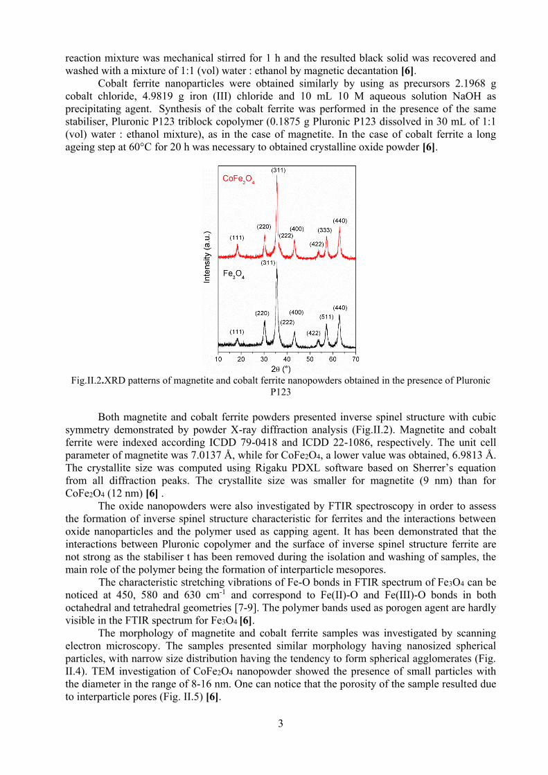

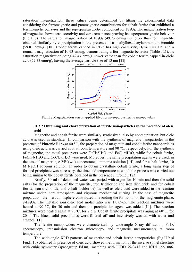
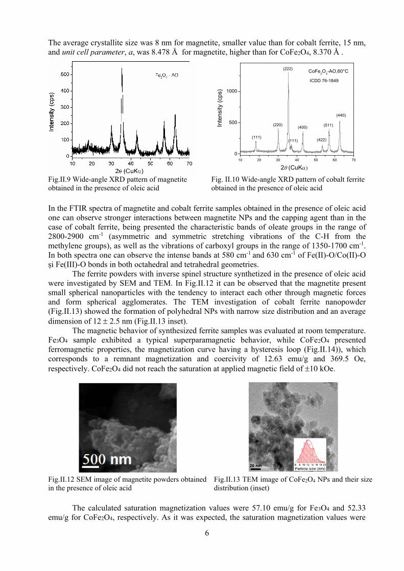
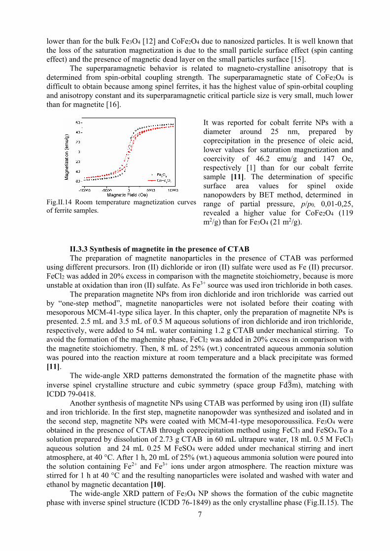
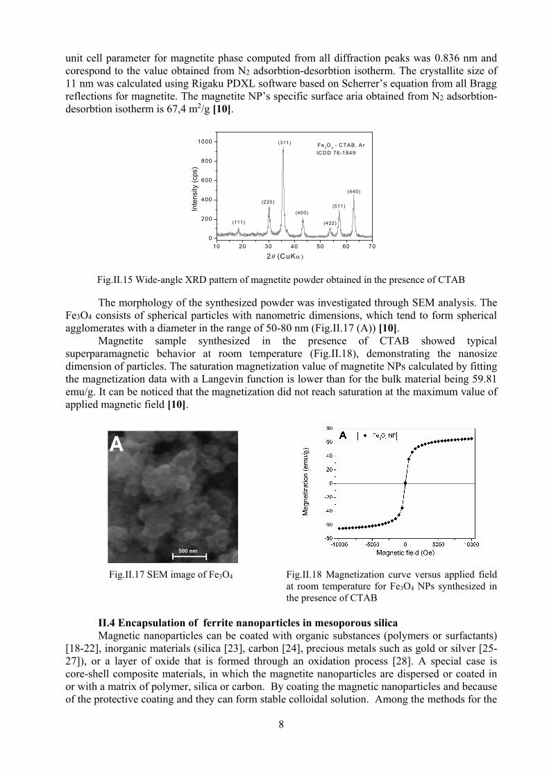
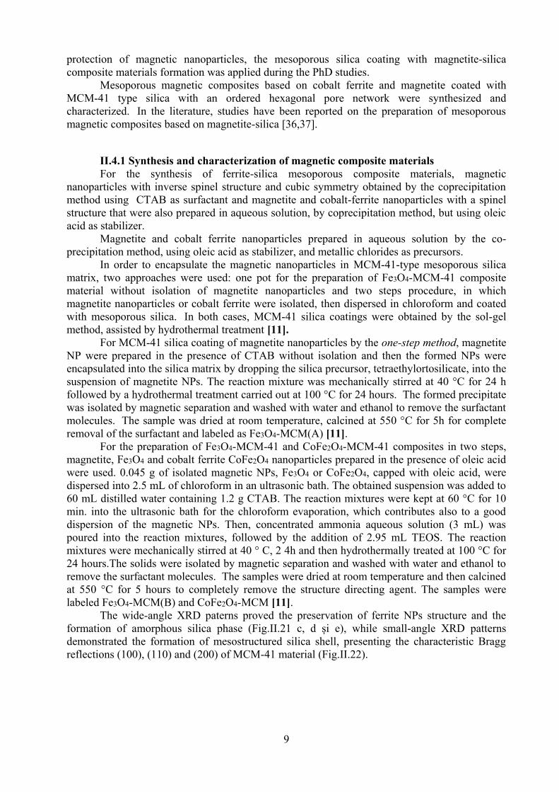

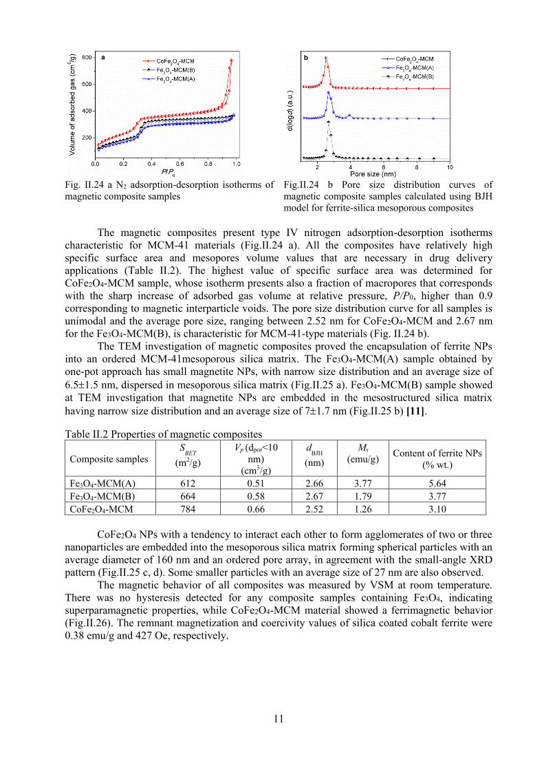
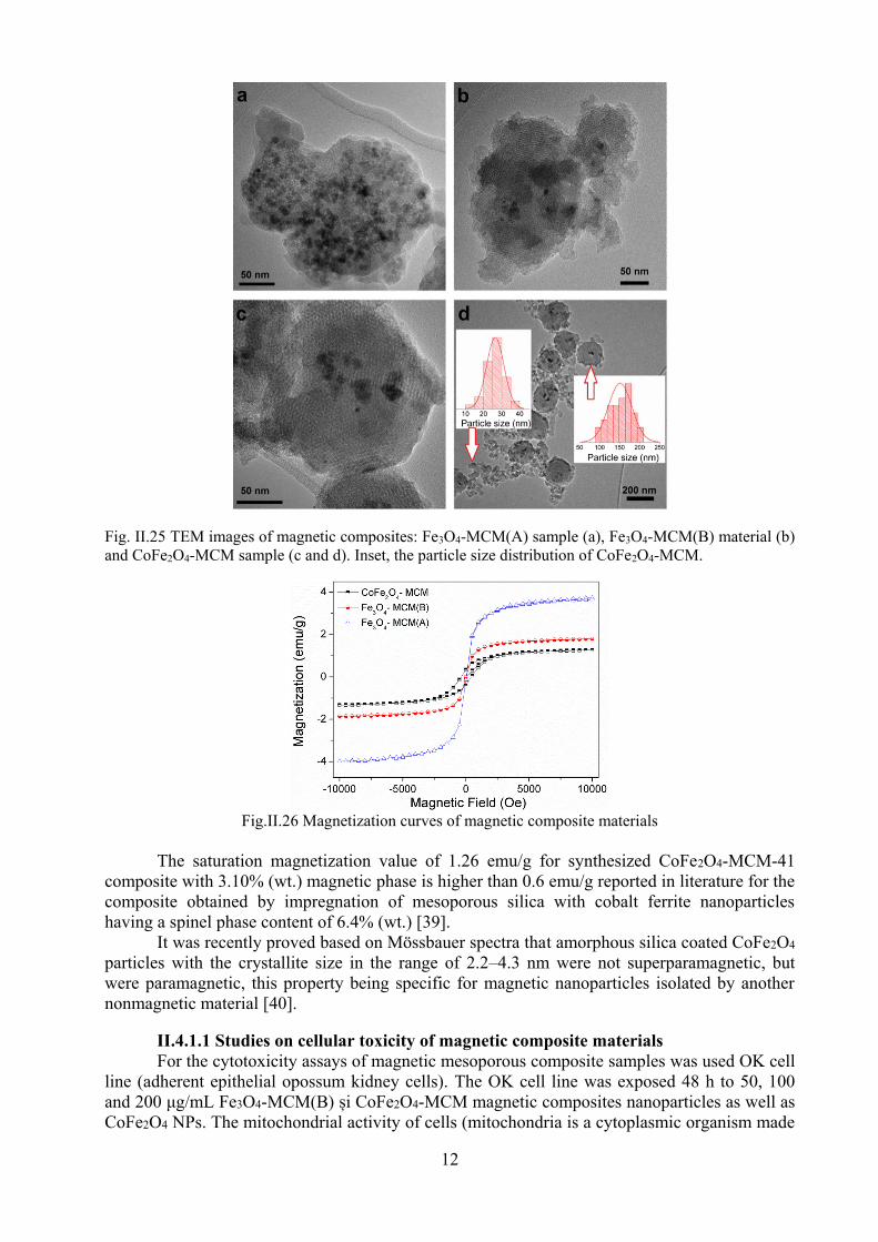
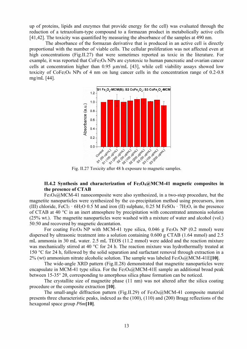
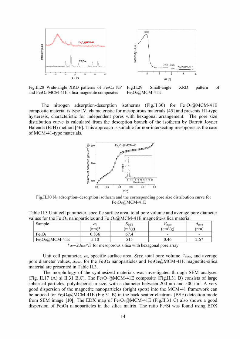
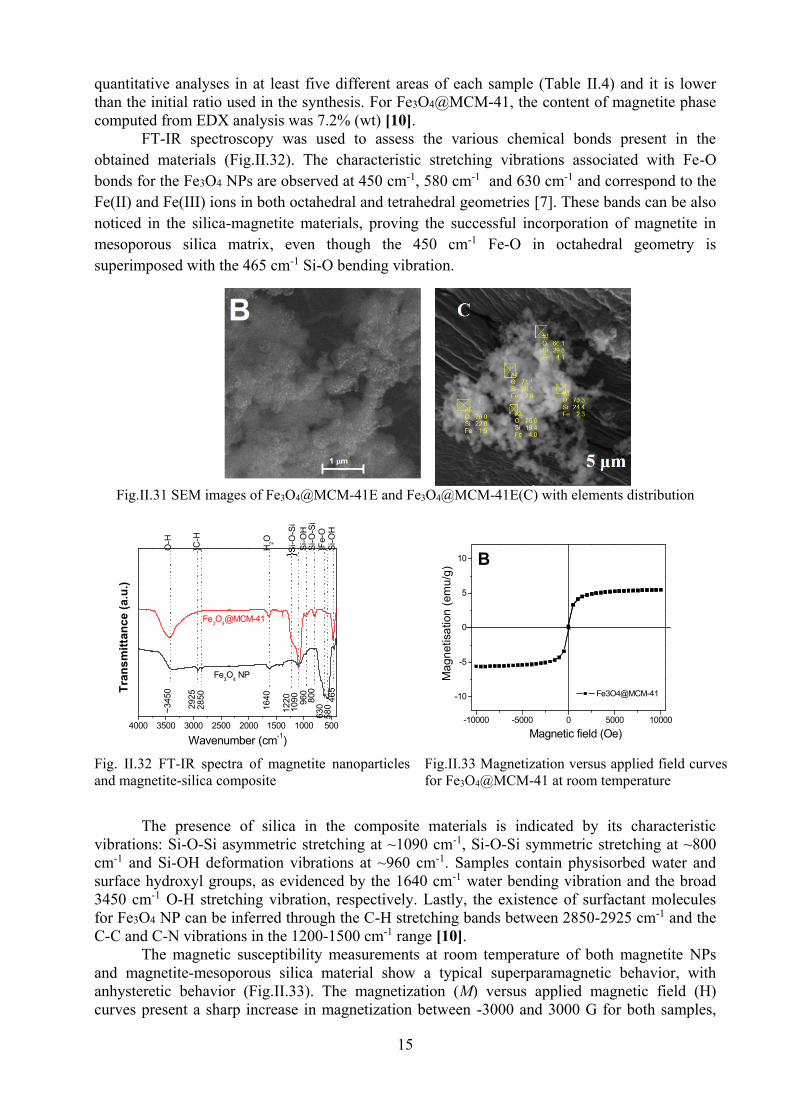

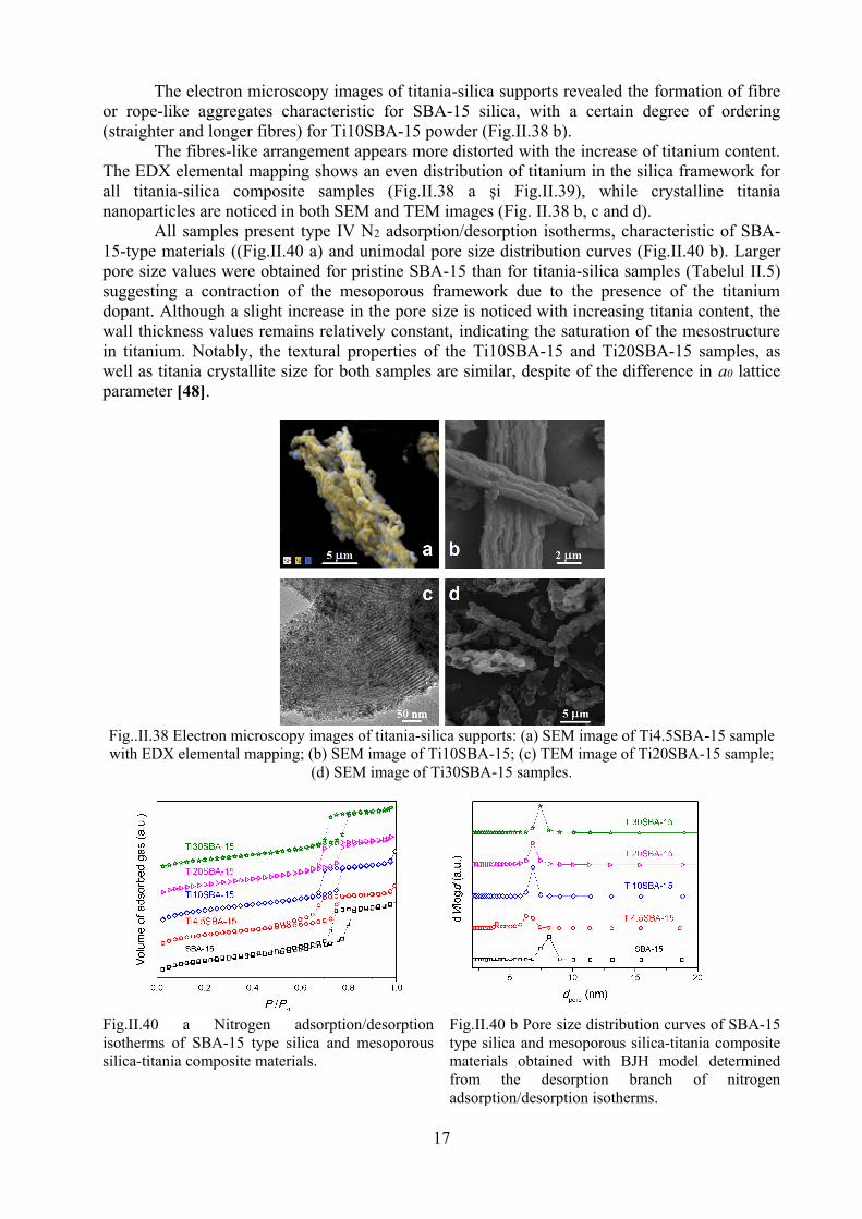

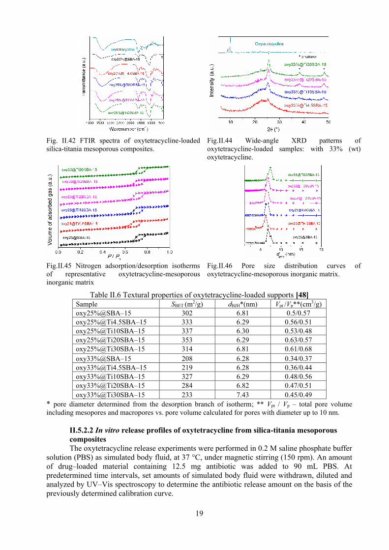
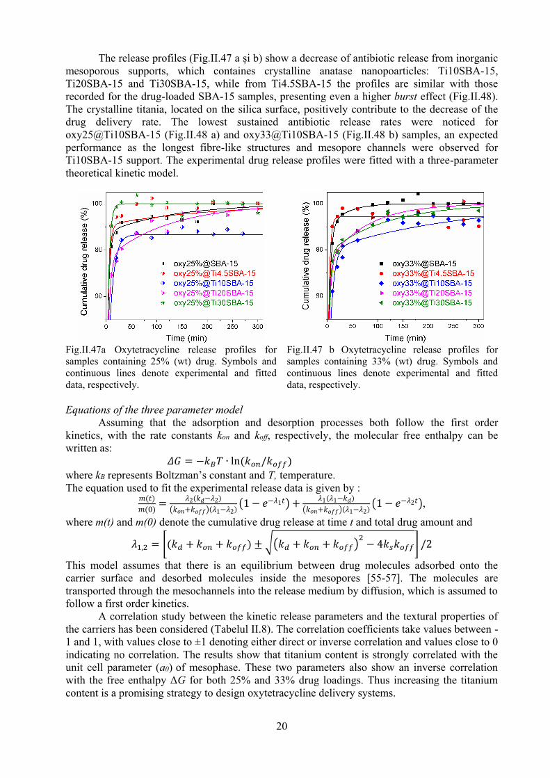
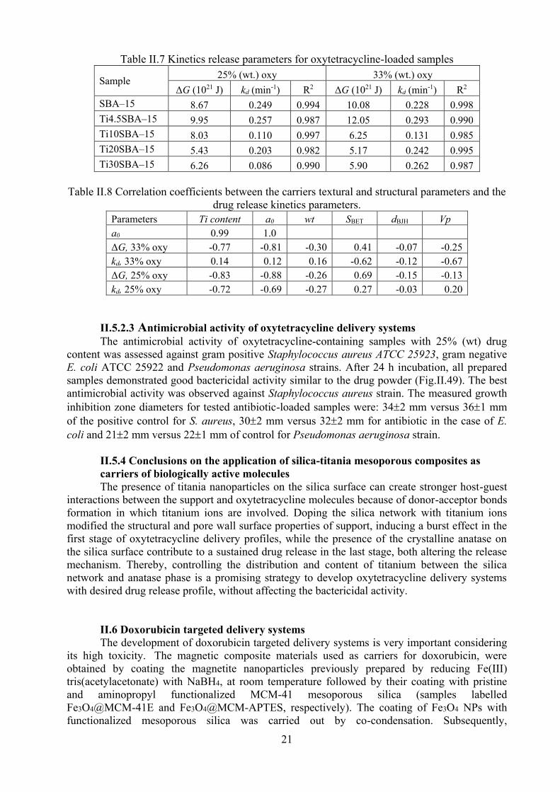
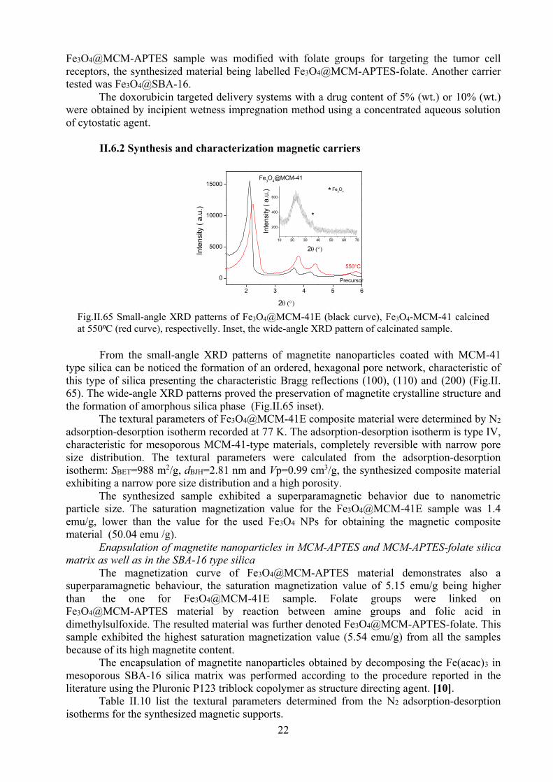
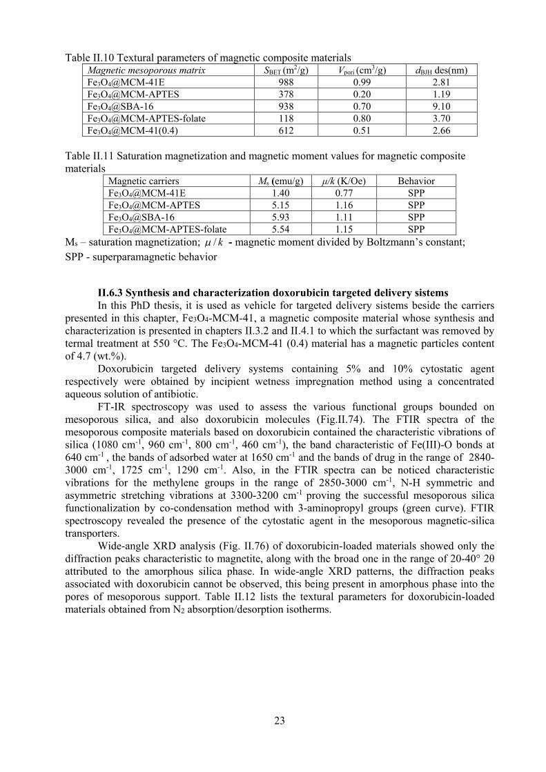
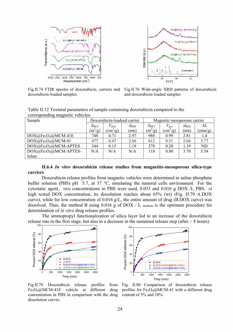
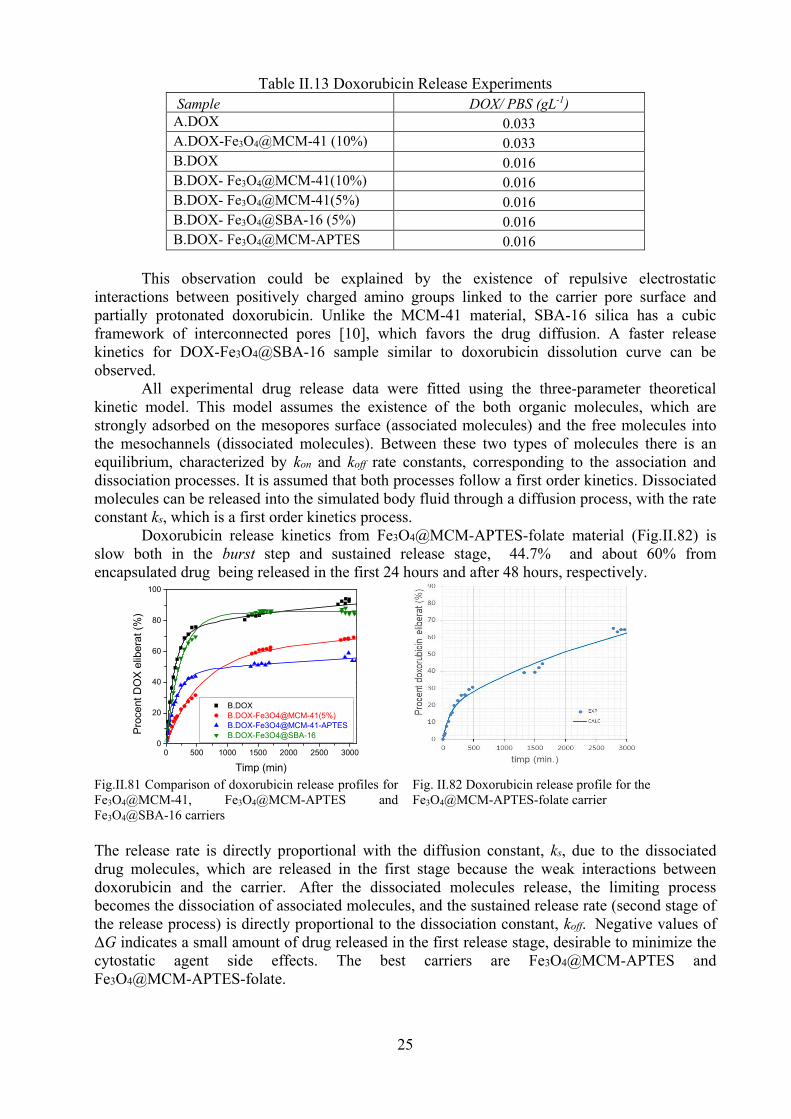
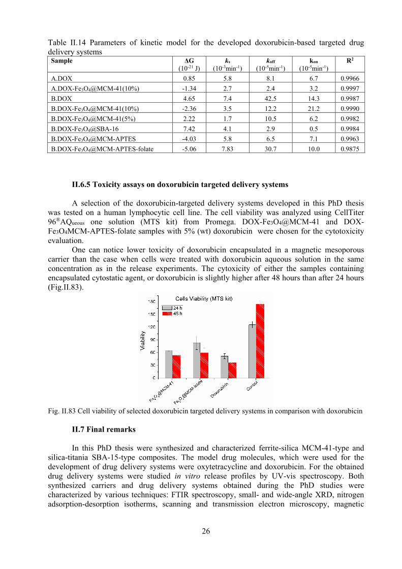


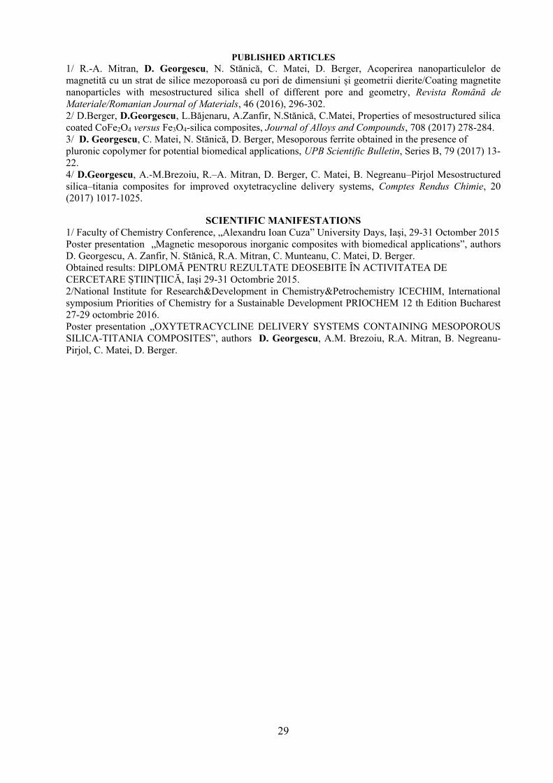
![Sisteme Monetare.[Conspecte.md]](https://static.documents.pub/doc/80x56/577cdc5f1a28ab9e78aa6855/sisteme-monetareconspectemd.jpg)


