64
Skeletal Consideration for Movement Kinesiology RHS 341 Lecture 2 Prepared by: Mrs. Lulu Al Rashed Dr. Einas Al Eisa; Dr. Amaal Ibrahim

Skeletal Consideration for Movement
Kinesiology RHS 341 Lecture 2
Prepared by:
Mrs. Lulu Al Rashed Dr. Einas Al Eisa; Dr. Amaal Ibrahim

The Skeletal System
Objectives • List and describe the two major divisions of the
skeletal system.
• List and describe the major functions of the skeletal system.
• Describe the types of various bone tissues, their locations, and functions.
• List, describe and give specific examples of the types of bones.

Objectives
• List and describe different types of joints and their functions.
• Degrees of freedom (joint motion).
• Close-packed versus Loose-packed position.
• List and describe various disorders and diseases of the skeletal system.
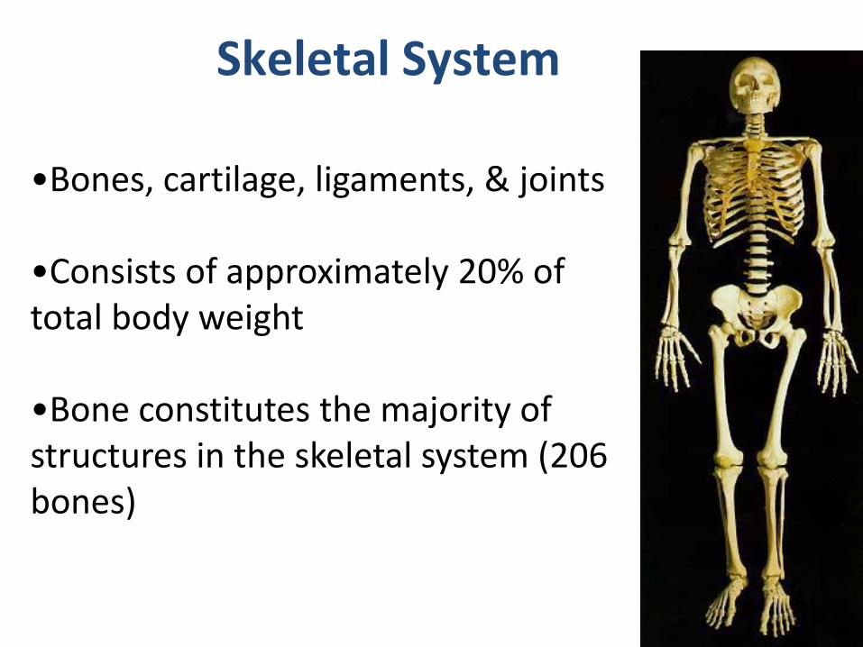
Skeletal System
•Bones, cartilage, ligaments, & joints •Consists of approximately 20% of total body weight •Bone constitutes the majority of structures in the skeletal system (206 bones)

Divisions of the Skeleton
• The Skeleton is divided into two major regions:
a. Axial Skeleton
b. Appendicular Skeleton
• The Axial Skeleton forms the longitudinal part of the body
80 bones (head (29), thorax (51)
• Divided into three parts
– Skull
– Vertebral column
– Bony thorax

• The Appendicular skeleton is composed of the appendages and the joints which attach them to the axial skeleton
126 bones
upper (32)
lower (31)
Girdles
- Upper & Lower Limbs (appendages)
- Shoulder girdle (shoulder, scapula, and clavicle)
-Pelvic girdle (pelvis)
Divisions of the Skeleton
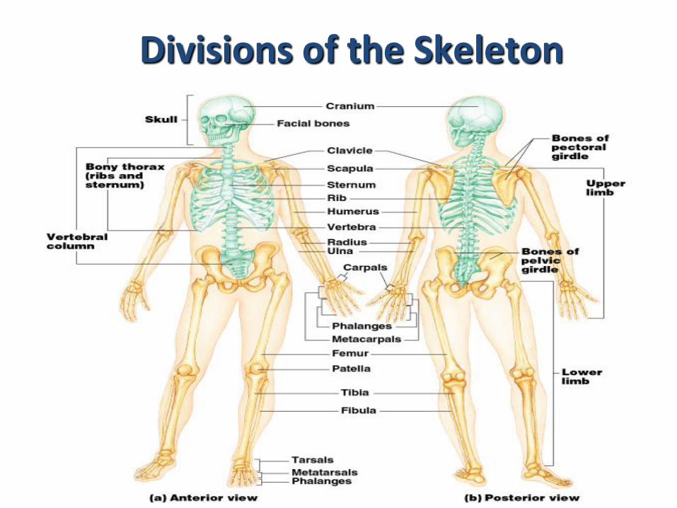
Divisions of the Skeleton

Functions of Skeletal System
• SUPPORT: Hard framework that supports and anchors the soft organs of the body.
• PROTECTION: Surrounds organs such as the brain and spinal cord.
• LEVERAGE : attachment for muscles to produce movement.
• STORAGE: Minerals and lipids are stored within bone material.
• BLOOD CELL FORMATION: The bone marrow is responsible for blood cell production.

Leverage
• Lever = a simple machine that magnifies the force and/or speed of movement
• The long bones act as the levers about which the muscular system generates the movements.
• Morphology = the shape & structural arrangement of the bones & articulations
determine movement

Two basic types of bone tissue
1) Compact bone
2) Cancellous (spongy) bone
Architecture of bone (osseous tissue)

1.Cortical (compact) bone
• The exterior dense layer of the bone
• Consists of hollow tubes called lamellae(collagen fibers that are arranged in layers and run in different directions)
• A series of lamellae form an osteon or haversian system
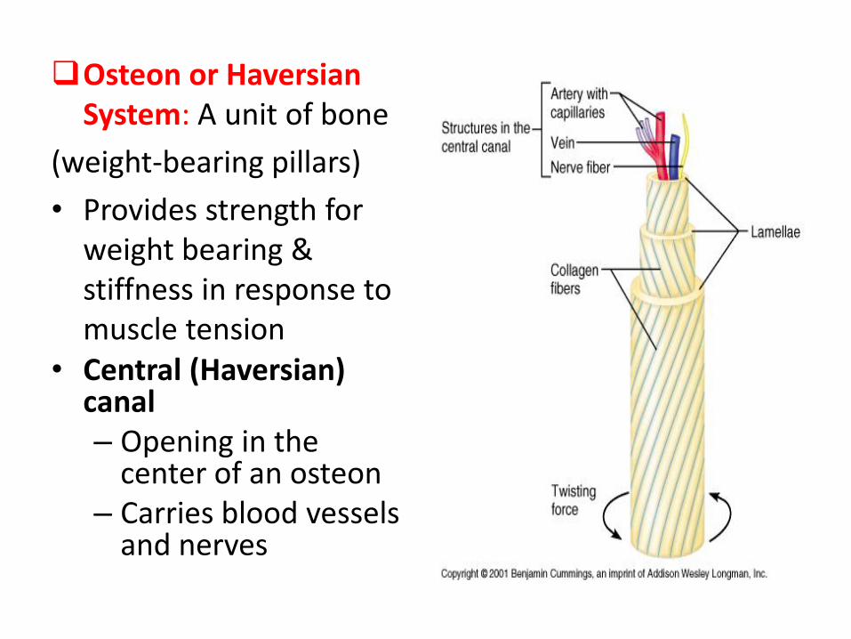
Osteon or Haversian System: A unit of bone
(weight-bearing pillars)
• Provides strength for weight bearing & stiffness in response to muscle tension
• Central (Haversian) canal – Opening in the
center of an osteon – Carries blood vessels
and nerves

Architecture of bone (osseous tissue)
2. Cancellous (spongy) bone : - interior to cortical bone - consists of flat pieces of bone
called trabeculae (collagen runs along the axis of the trabeculae
- Provides energy absorption & stress distribution in response to loads
- Not as strong as cortical bone (risk of fracture in the elderly)

Bone Structure
• Periosteum – hard outer covering
– Cells for growth and repair
• Compact bone – hard strong layer
– Bone cells, blood vessels, protein with Ca and P
• Spongy bone – at ends of long bones
– Has small open spaces to lighten weight
• Marrow cavity – hollow in middle of long bones

Bone Marrow
• Red marrow – produces blood cells and clotting factors
– Found in humerus, femur, sternum, ribs, vertebrae, pelvis
– Produces RBC 2 million per second
• Yellow marrow – stores fat
– Found in many bones

Classification of Bones by Shape
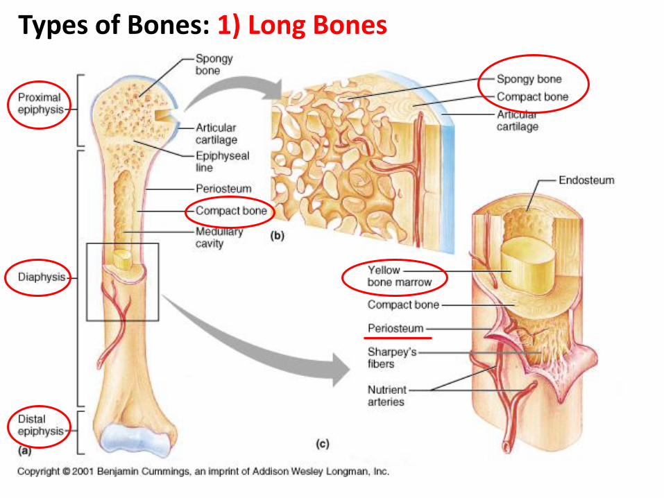
Types of Bones: 1) Long Bones

Long Bones
• Consist of: a shaft called diaphysis (made of compact bone), Which broadens out into the epiphysis (made up
of spongy bone inside a thin layer of compact bone)
• Function: support and leverage
• Example: humerus, radius, ulna, femur, tibia, fibula, metacarpals, metatarsals
Einas Al-Eisa; KSU

Gross Anatomy of a Long Bone
• Diaphysis – Shaft – Composed of compact bone
• Epiphysis – Ends of the bone – Composed mostly of spongy bone
• Medullary cavity – Cavity of the shaft – Contains yellow marrow (mostly fat) in
adults – Contains red marrow (for blood cell
formation) in infants
• Articular cartilage – Covers the external surface of the
epiphyses – Made of hyaline cartilage – Decreases friction at joint surfaces
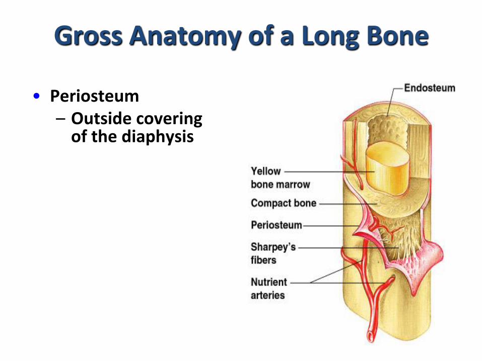
Gross Anatomy of a Long Bone
• Periosteum – Outside covering
of the diaphysis

Types of Bone

Types of Bone
• 2- Short bones • Consist of spongy bone
covered with a thin layer of compact bone
• Play an important role in shock absorption and transmission of forces
• Example: carpals of the hand and the tarsals of the foot

Types of Bone
• 3- Flat bones
• Flat , plate-like bones.
• Consist of: two layers of compact bone with spongy bone in between.

Types of Bone
3- Flat bones • Function: protect
internal structures and offer broad surfaces for muscle attachments
• Example: ribs, illium, sternum, scapula


Types of Bone
4- Irregular bones • Consist of: spongy bone and thin exterior layer of
compact bone
• Specialized functions such as: supporting the weight, protecting the spinal cord, dissipating loads
• Example: vertebrae, ischium, pubis
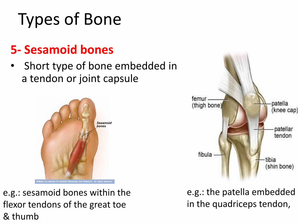
Types of Bone
5- Sesamoid bones • Short type of bone embedded in
a tendon or joint capsule
e.g.: the patella embedded in the quadriceps tendon,
e.g.: sesamoid bones within the flexor tendons of the great toe & thumb

Types of Bone
5- Sesamoid bones Function: Increase the tendon’s
mechanical effect • The presence of the sesamoid
bone holds the tendon slightly farther away from the center of the joint and thus increases its moment arm.
Protect the tendon • prevent the tendon from flattening
into the joint as tension increases
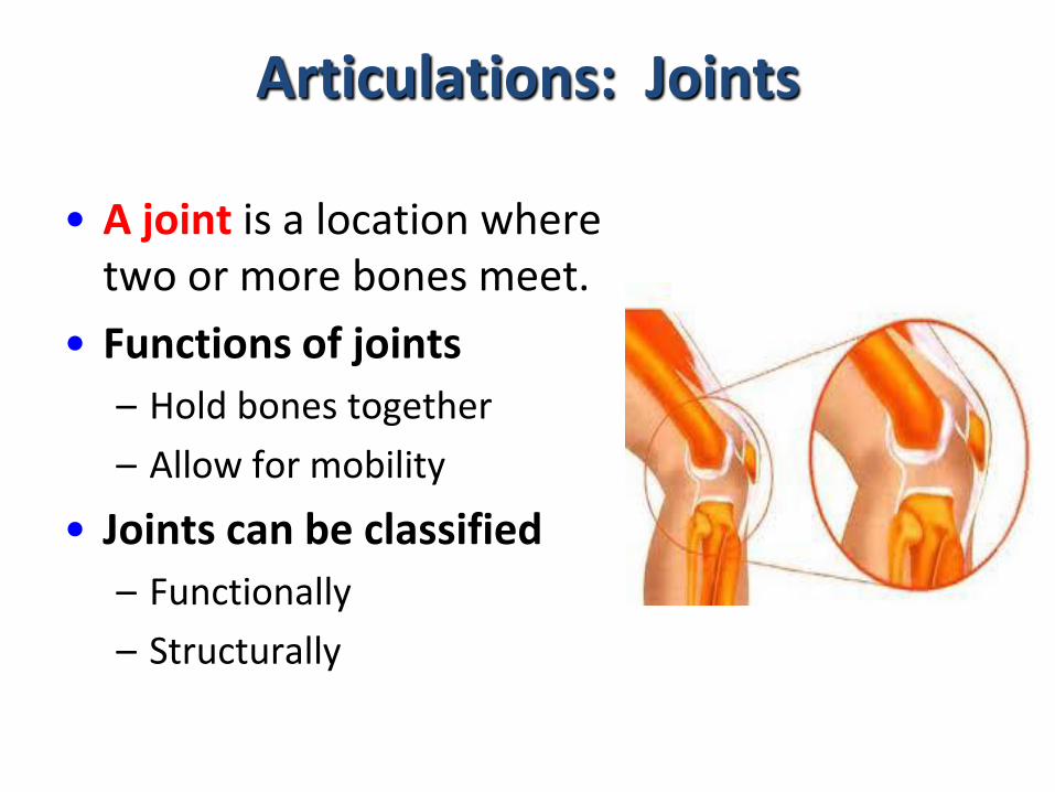
Articulations: Joints
• A joint is a location where two or more bones meet.
• Functions of joints
– Hold bones together
– Allow for mobility
• Joints can be classified
– Functionally
– Structurally

Types of Joints
Synovial
Diarthroidal Freely movable
e.g.: hip joint
Cartilagenous
Amphiarthroidal Slightly movable
e.g.: intervertebral
discs
Fibrous
Synarthroidal
Immovable
e.g.: Sutures of the skull
Structural
Functional

Characteristics of Synovial Joints
• Articular end plate = a thin layer of
compact bone over the spongy bone
(covering the ends of the bones)
• Articular (hyaline) cartilage for shock
absorption, stability, improved fit for the
surfaces, lubrication
Covered by:


Characteristics of Synovial Joints
• Joint capsule = a fibrous connective tissue that surround the bony ends forming the joint
• Synovial membrane = loose, vascularized connective tissue that secretes synovial fluid into the joint cavity for lubrication
Lined with:

Synovial joint

Characteristics of Synovial Joints
• Where additional support is needed, the joint capsule is thickened to form tough, non-elastic ligaments to provide additional support.
• Stability of a synovial joint is provided by: the capsule, ligaments, muscles & tendons spanning the joint, and the congruency of the bone surfaces.

Types of synovial joints
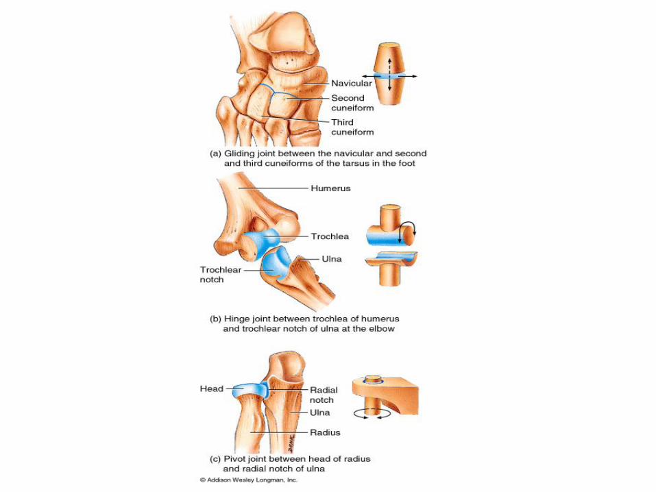
1) Plane (gliding) joint: consists of two flat
surfaces that glide over each other rather
than around an axis (nonaxial)
Example: carpals & tarsals (radial & ulnar
deviation, foot pronation & supination)

Plane (gliding) Joint


Types of synovial joints
2) Hinge joint: allow movement in one
plane (flexion / extension) around a single
axis (uniaxial)
Example: interphalangeal joints (hand),
ulnohumeral joint (elbow)

Hinge Joint

Types of synovial joints
3) Pivot Joint: allows a rotational movement around
a long axis (movement in one plane, uniaxial)
Example: superior & inferior radioulnar joint
(pronation / supination),
atlantoaxial joint at the
base of the skull (rotation)

Pivot Joint
Types of synovial joints
4) Condyloid joint: allows movement in two
planes (flexion / extension and abduction
/adduction) without rotation (biaxial).
Example: metacarpophalangeal joints

Condyloid Joint

Types of synovial joints
5) Saddle joint: allows two planes of
movement (flexion / extension, abduction /
adduction) which makes it biaxial.
Example:only found at the carpometacarpal
joint of the thumb.

Saddle Joint
Types of synovial joints
6) Ball-and-socket joint: allows movement
in all three planes (multiaxial:
flexion/extension, abduction/adduction, &
rotation)
Example: the hip and shoulder joints.

Ball-and-socket Joint

• Compound Joints: made up of several
joints between a number of different bones.
• The bones articulate with one another in
different ways, allowing for a variety of
movements such as the set of joints which
operate the movement of the skull on the
vertebral column.
• The condyles at the base of the skull fit into
the facets of the atlas, allowing for the
nodding movement of the head.
• While one moves one's head, the atlas is
able to rotate around the odontoid
process of the axis, allowing the head to
turn from side to side. There are also other
articulating surfaces, where the atlas and
axis meet.
• All these joints together make a compound
joint with its many possible movements in
the neck region.


Degrees of freedom
• Movement in a plane can be described as a single degree of freedom.
• Degree of freedom = the terminology used to describe the amount of movement structurally allowed by the joint
• Example: a uniaxial joint has one degree of freedom, ball and socket joints have 3 degrees of freedom

Joint Position
• Loose packed (resting) position = the
position at which the joint is under the
least amount of stress (capsule, ligaments,
bone contact).
• Close packed position = the position in
which the majority of joint structures are
under maximum tension.

Diseases and Disorders of the Skeletal System
• A fracture is a break in a bone
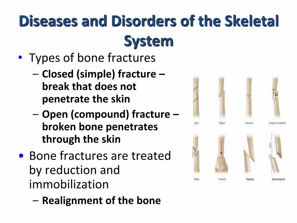
Diseases and Disorders of the Skeletal System
• Types of bone fractures – Closed (simple) fracture –
break that does not penetrate the skin
– Open (compound) fracture – broken bone penetrates through the skin
• Bone fractures are treated by reduction and immobilization – Realignment of the bone
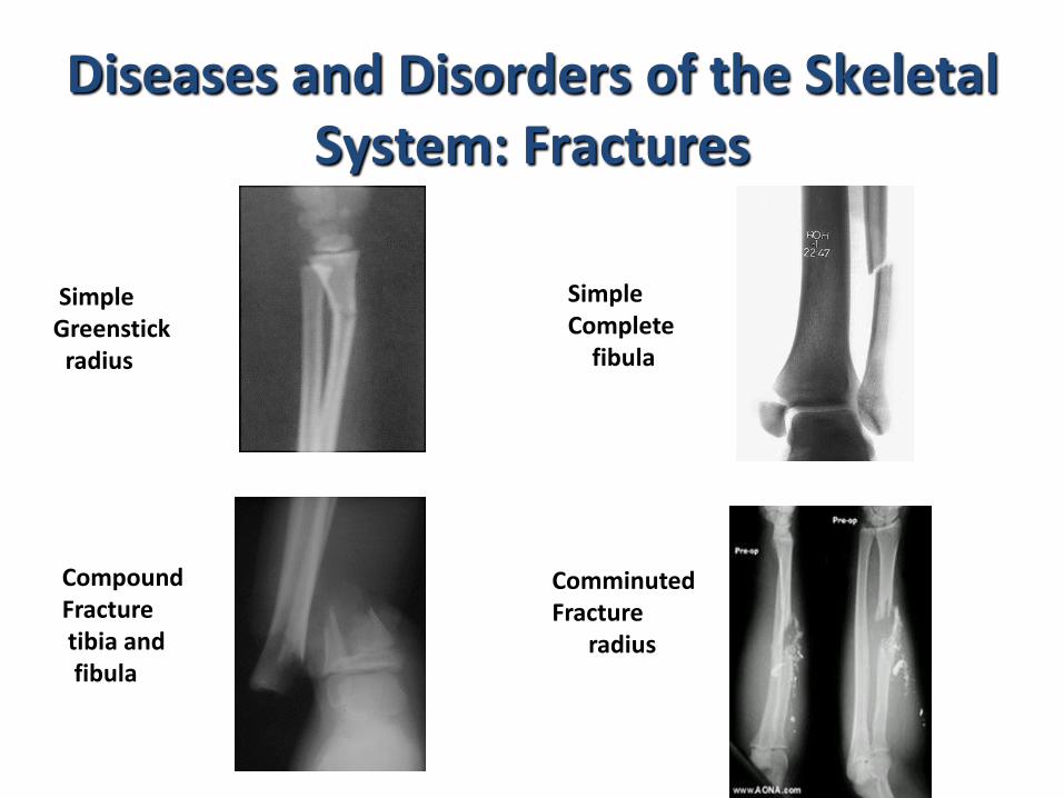
Diseases and Disorders of the Skeletal System: Fractures
Simple Greenstick radius
Simple Complete fibula
Compound Fracture tibia and fibula
Comminuted Fracture radius

Diseases and Disorders of the Skeletal System
• Osteoporosis:
affect both men and women but it is most common in post menopausal women.
The bone tissue becomes brittle and breaks easily with little applied stress; due to loss of calcium from the bone matrix.
Diseases and Disorders of the Skeletal System
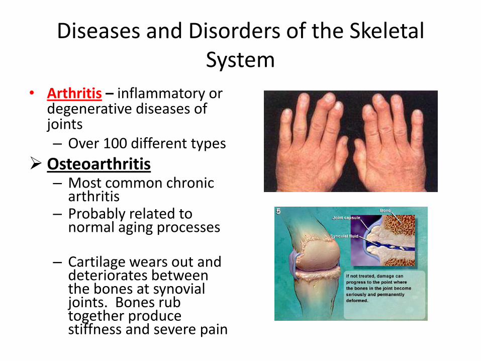
• Arthritis – inflammatory or degenerative diseases of joints – Over 100 different types
Osteoarthritis – Most common chronic
arthritis – Probably related to
normal aging processes – Cartilage wears out and
deteriorates between the bones at synovial joints. Bones rub together produce stiffness and severe pain

Diseases and Disorders of the Skeletal System
Rheumatoid arthritis – An autoimmune disease
– the immune system attacks the joints
– Symptoms begin with bilateral inflammation of certain joints
– Often leads to deformities
– Can appear at any age


