140
SKELETAL SYSTEM Anatomy and Physiology
| Date post: | 15-Jul-2015 |
| Category: |
Education |
| Upload: | tia-hohler |
| View: | 124 times |
| Download: | 1 times |

SKELETAL SYSTEM Anatomy and Physiology

FUNCTIONS OF BONE AND SKELETAL SYSTEM 1. Support 2. Protection 3. Assisting in movement

4. Mineral homeostasis 5. Production of blood cells 6. Triglyceride storage

TYPES OF BONES Long Bones- greater length than width and consist of a shaft and variable types of ends, arms and legs Short bones- cube-shaped, wrist and ankle bones

Flat bones- thin, afford considerable protection, skull and ribs Irregular bones- complex shapes, backbone and some facial bones


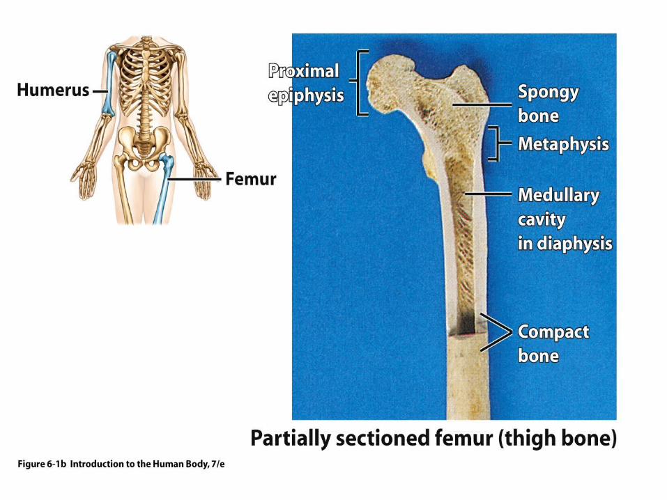
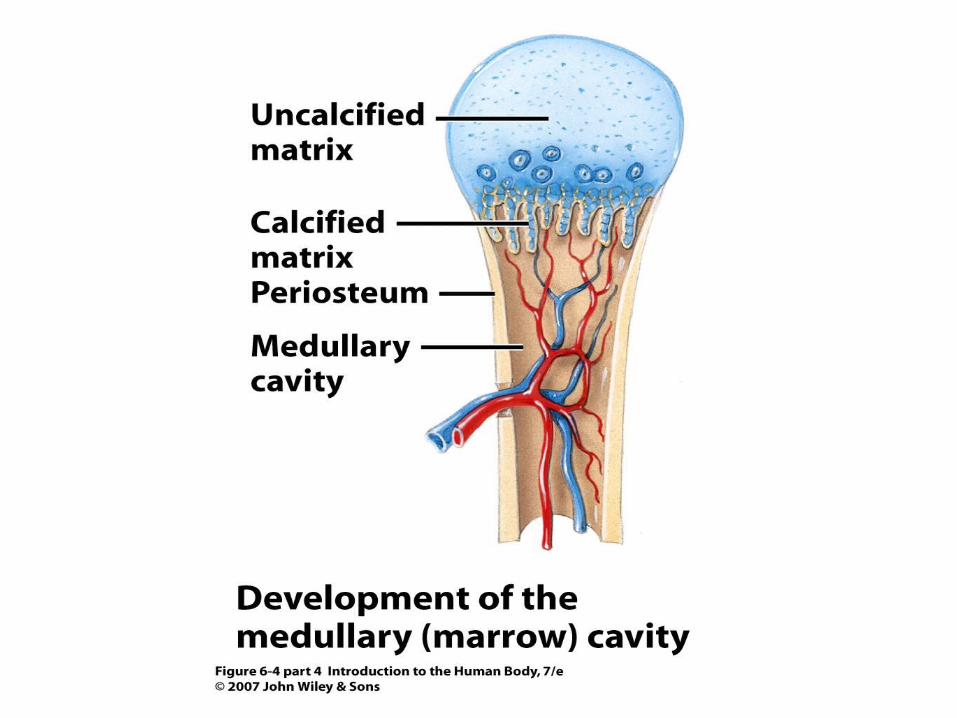
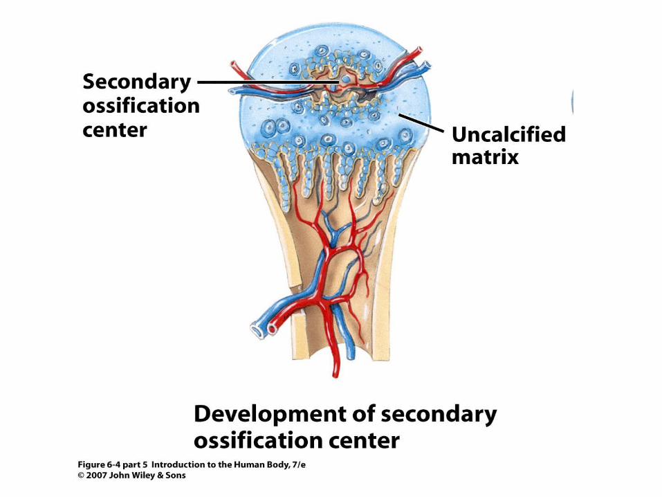
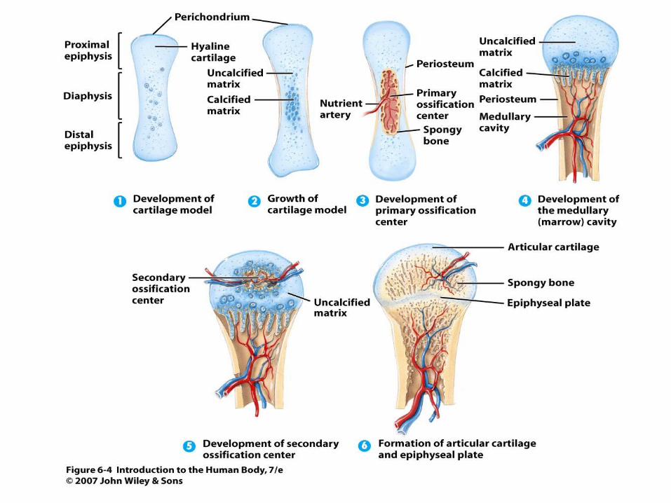
STRUCTURE OF BONE A typical long bone consists of: Diaphysis- bone’s shaft or body, long main portion Epiphyses- ends of the bone Metaphyses- area where the diaphysis joins the epiphyses, contains red bone marrow, area of growth plates

Articular cartilage- hyaline cartilage covering part of the epiphysis where a joint can form Periosteum- tough sheath of dense irregular connective tissue on the bone’s surface Medullary cavity- contains fatty yellow bone marrow Endosteum- membrane lining the medullary cavity containing bone forming cells.


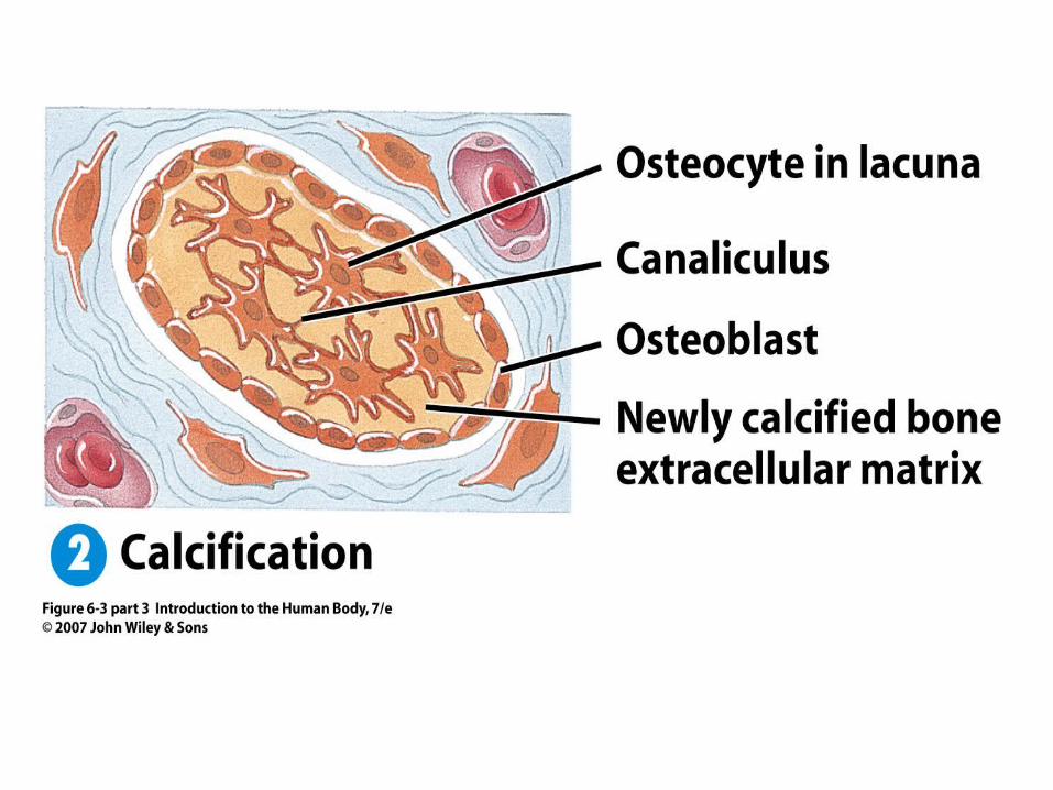
TYPES OF BONE CELLS Osteoblasts- synthesize and secrete collagen fibers and build bone matrix. Turn into osteocytes later. Osteocytes- main cells in bone tissue, maintain daily metabolism and nutrient/waste exchange Osteoclast- release enzymes that digest protein and mineral components of the bone’s matrix. Resorption is part of normal bone repair and growth.

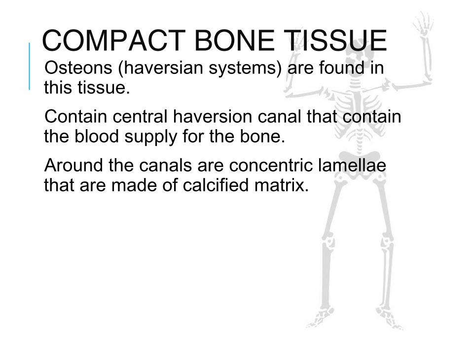
COMPACT BONE TISSUE Osteons (haversian systems) are found in this tissue. Contain central haversion canal that contain the blood supply for the bone. Around the canals are concentric lamellae that are made of calcified matrix.

Between the lamellae are small spaces called lacunae which contain osteocytes. Radiating from the lacunae are tiny canaliculi which allow nutrient and waste diffusion.


SPONGY BONE TISSUE Does not contain osteons. Consist of trabeculae which are an irregular network of lattice like tubes that contain red bone marrow and osteocytes. Only source of blood formation in adults.
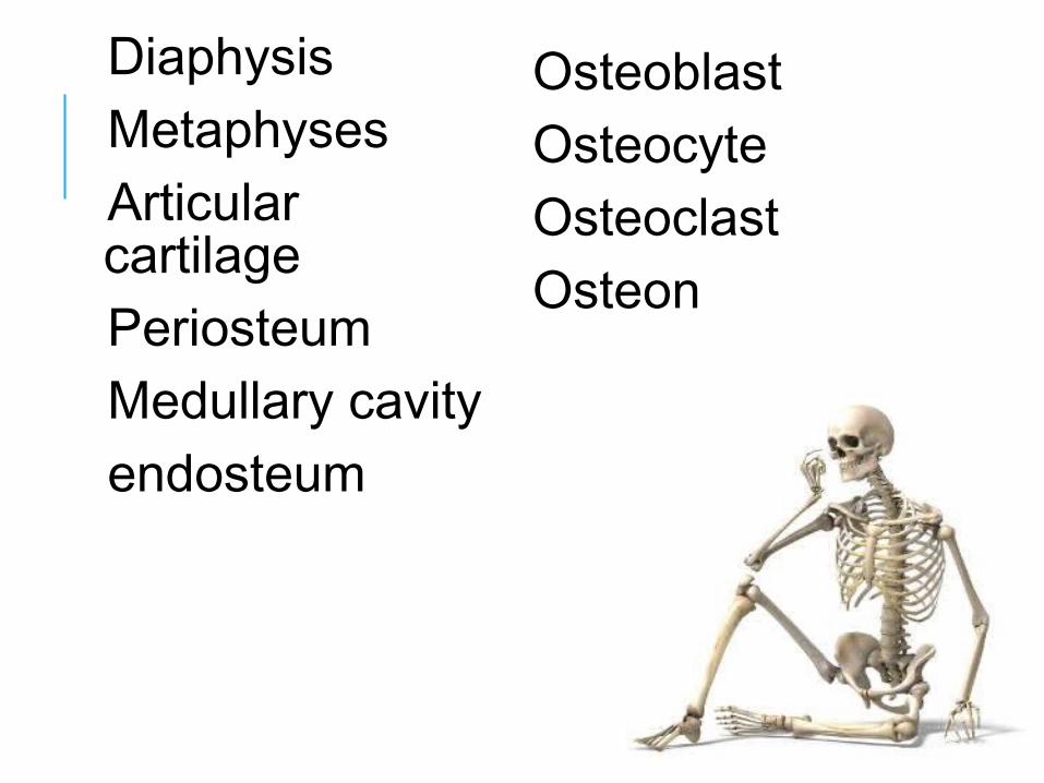
Diaphysis Metaphyses Articular cartilage Periosteum Medullary cavity endosteum
Osteoblast Osteocyte Osteoclast Osteon

OSSIFICATION: BONE FORMATION Intramembranous ossification- flat bones of the skull and mandible, fetal “soft spots” are replaced this way Bone forms directly on or within the loose fibrous connective tissue membranes.

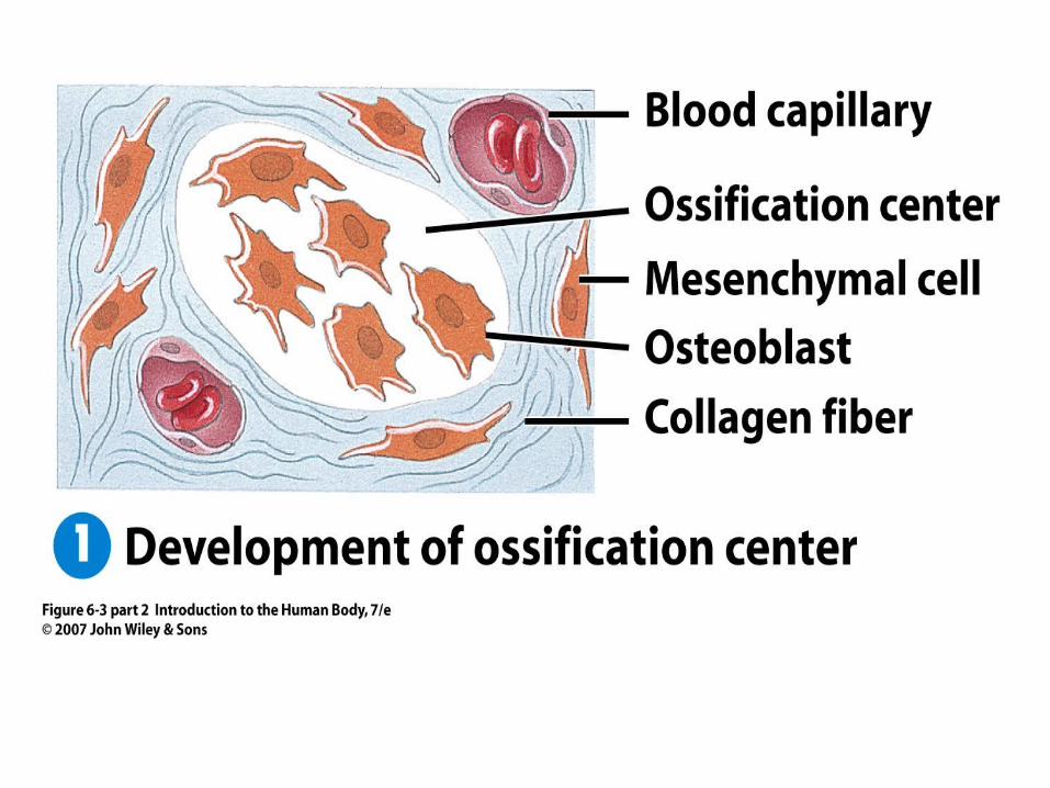


OSSIFICATION: BONE FORMATION Endochondral Ossification- most bones are made this way The bones form from within hyaline cartilage


HOMEOSTASIS OF BONE Bone continually renews itself. Bone remodeling occurs as old bone is broken down and replaced by new bone. Osteoclasts are responsible for the resorption of old bone but a delicate balance must exist between osteoclasts which break bone and osteoblasts which create new bone. Bone contains 99% of the body’s calcium.

Small changes in blood calcium can be deadly. The parathyroid gland secretes PTH, a hormone which signals the osteoclasts to increase bone resorption to increase blood calcium levels via a negative feedback system.
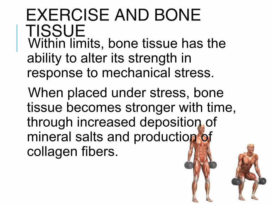
EXERCISE AND BONE TISSUE Within limits, bone tissue has the ability to alter its strength in response to mechanical stress. When placed under stress, bone tissue becomes stronger with time, through increased deposition of mineral salts and production of collagen fibers.
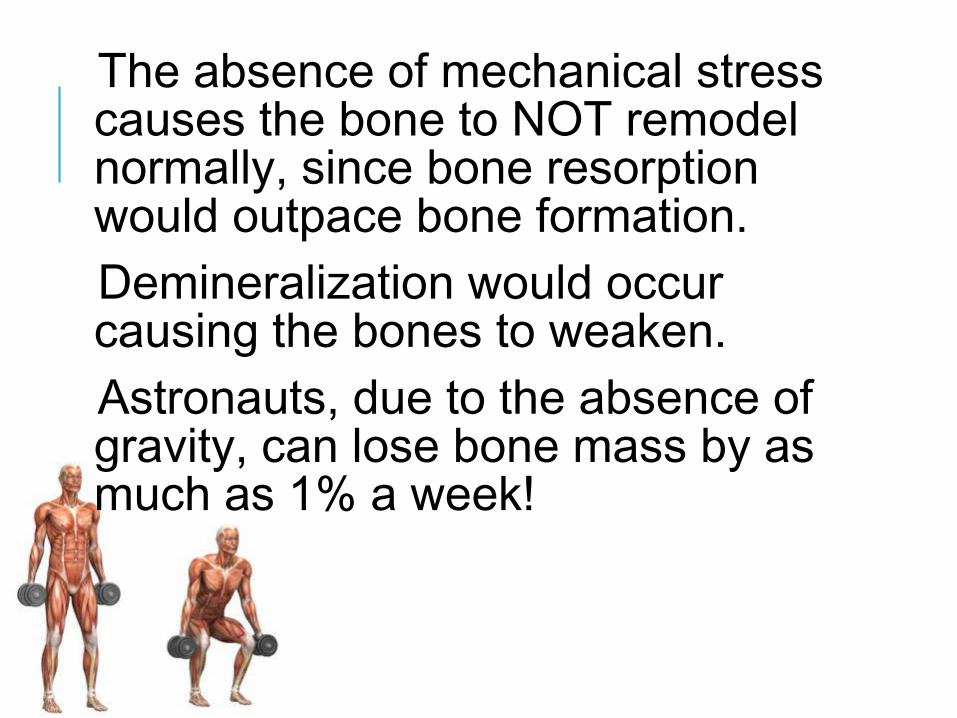
The absence of mechanical stress causes the bone to NOT remodel normally, since bone resorption would outpace bone formation. Demineralization would occur causing the bones to weaken. Astronauts, due to the absence of gravity, can lose bone mass by as much as 1% a week!

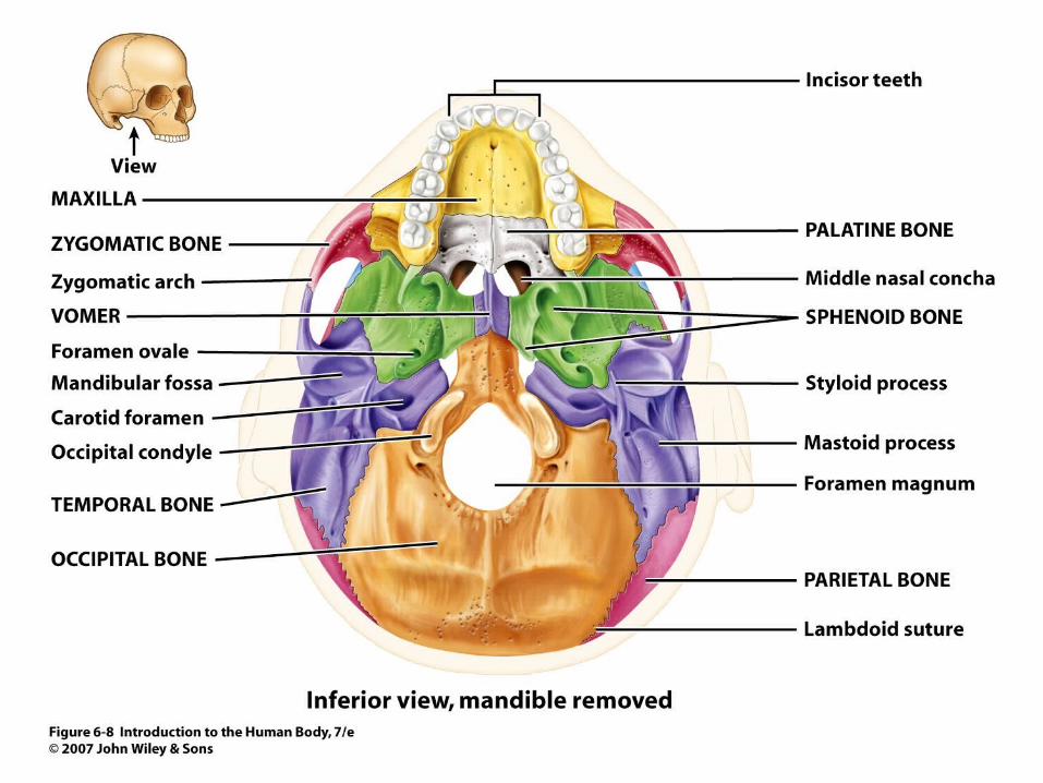
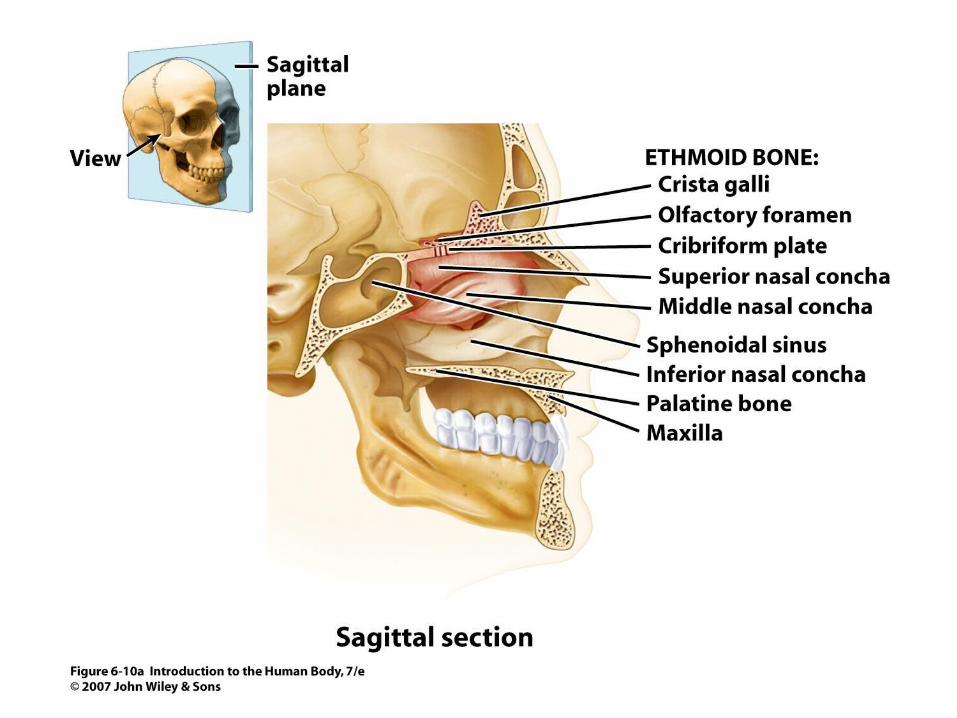
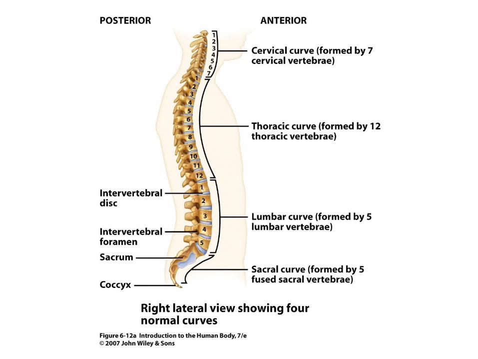
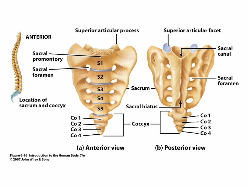
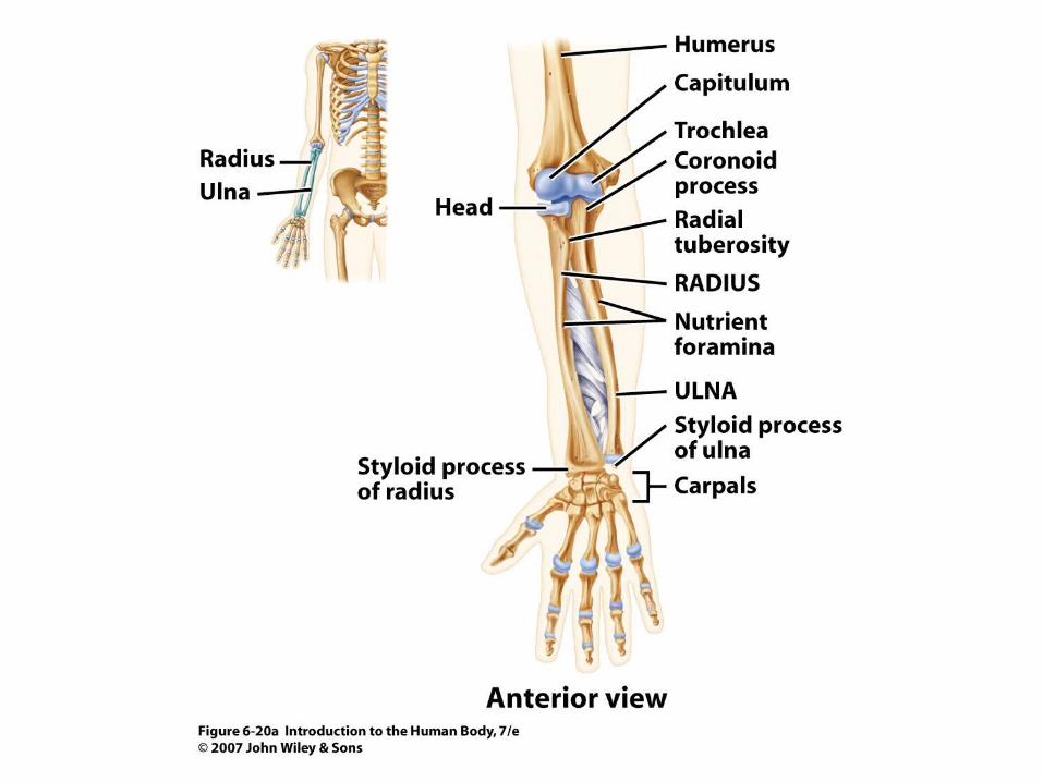
DIVISIONS OF THE SKELETAL SYSTEM Axial Skeleton- skull, face, hyoid, vertebral column, sternum, ribs Appendicular skeleton- clavicle, scapula, upper limbs, pelvic girdle, lower limbs


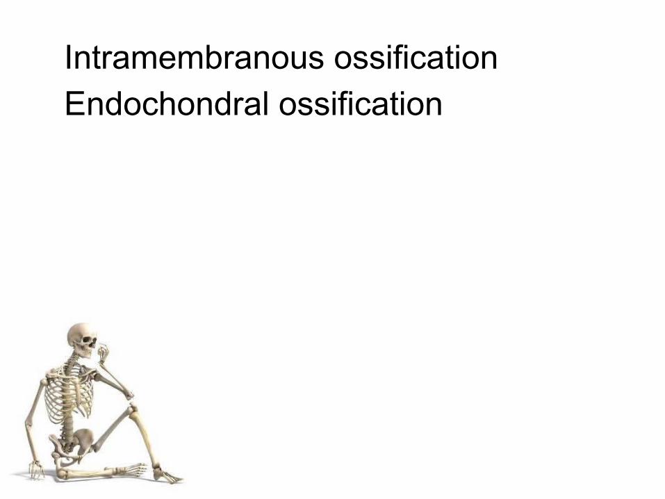
Intramembranous ossification Endochondral ossification

Joints

3 Types
Fibrous Cartilaginous Synovial

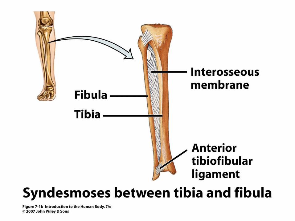
Fibrous Joints Suture- thin layer, immovable, skull Syndesmosis- light movement, in between bones like the tibia and fibula. Gomphosis- cone-shaped peg into socket, teeth into jaw, immovable.


Cartilaginous Joints Synchondrosis- hyaline, epiphyseal plate in long bone, when growth stops, it turns into bone, immovable Symphysis- ends of articulating bones are covered in hyaline cartilage but the bones themselves are connected by a broad, flat disc of fibrocartilage, pubic area and vertebrae


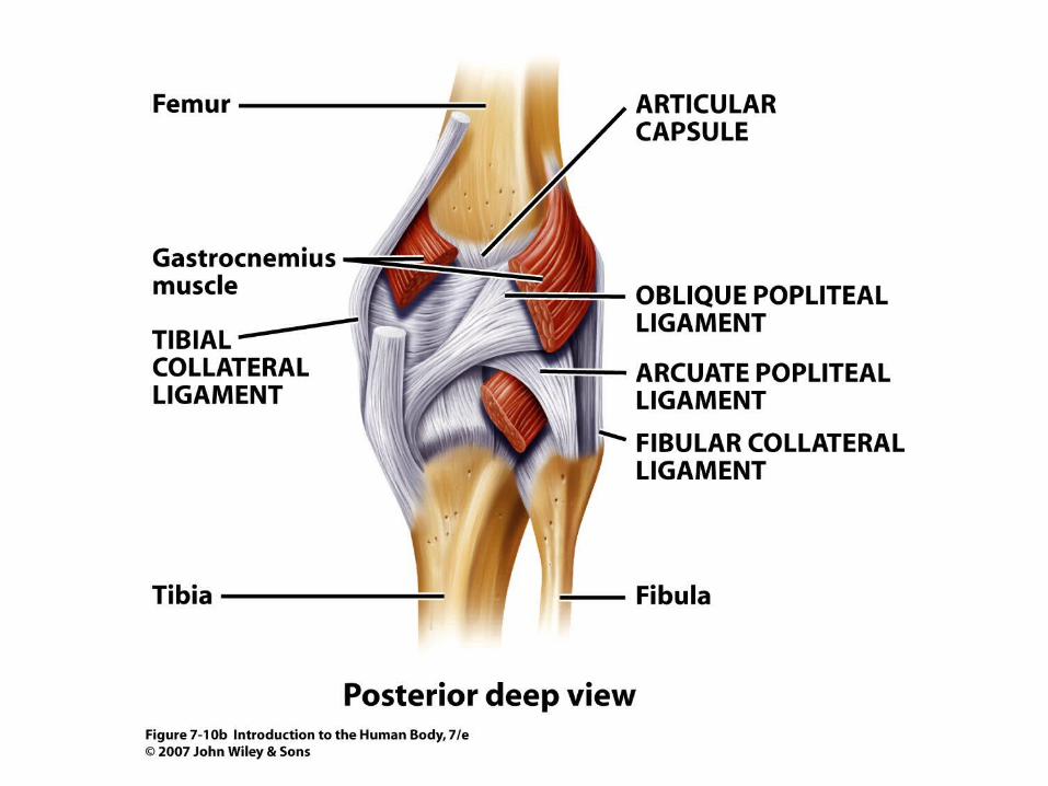
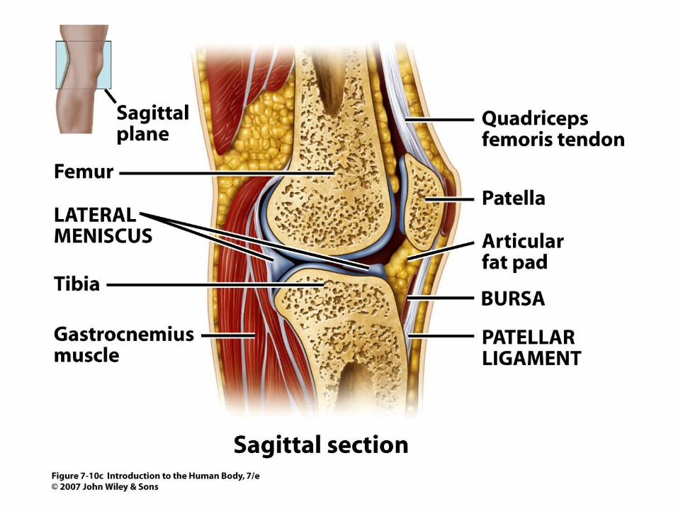
Synovial Joints This joint is unique because it has some special characteristics. These joints contain a synovial cavity between the articulating bones. This cavity allows these joints to be freely movable. The bones are covered in articular cartilage to reduce friction and absorb shock.
A sleevelike articular capsule surrounds the joint and contains two layers: the fiberous capsule and the synovial membrane. Some fiber bundles are called ligaments that hold the bones together and resist strain. Articular fat pads of adipose tissue help with shock absorption.

The synovial membrane secretes synovial fluid that reduces friction by lubricating the joint. It also supplies nutrients and removes wastes from the chondrocytes in the cartilage.Accessory ligaments sometimes lie outside the capsule.Inside some joints like the knee, a disc called a meniscus is attached to the fibrous capsule. This disc maintains stability.


Types of Synovial Joints Planar-side to side and back and forth gliding movements

Types of Synovial Joints Hinge- angular open and close motion

Types of Synovial Joints Pivot-allows rotation, contains accessory ligaments

Types of Synovial Joints Condyloid- up and down, side to side movements, oval shapes fit together

Types of Synovial Joints Saddle- side to side, up and down, like a rider in a saddle

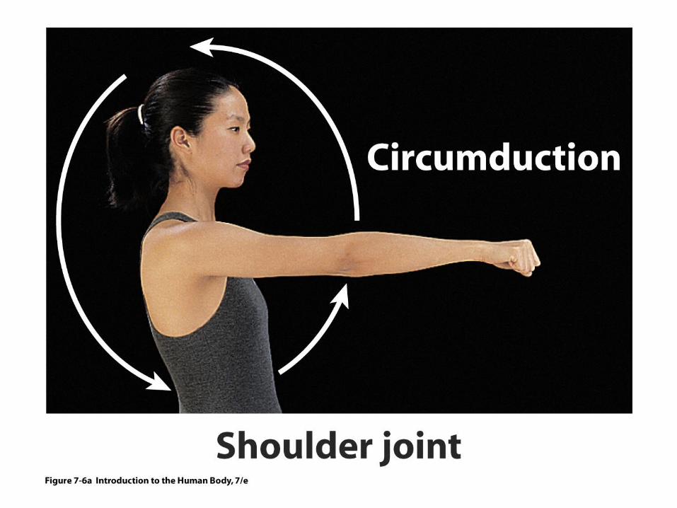
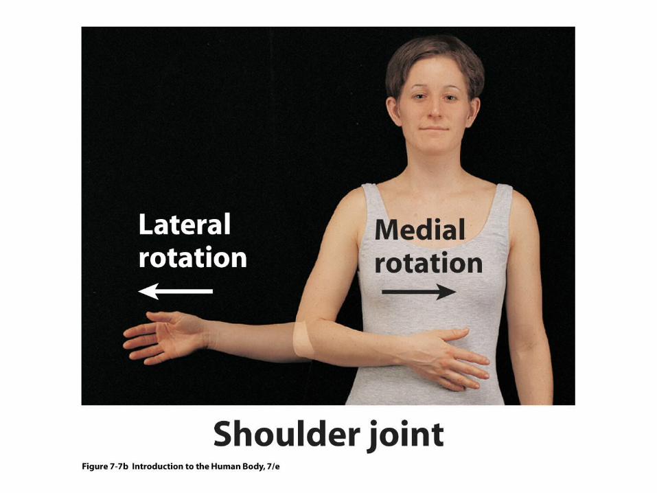
Types of Synovial Joints Ball and socket- multi directional movement, ball into cuplike depression, shoulder and hip only


Suture Syndesmoses Gomphosis Synchondrosis Symphysis Synovial
Planar Hinge Pivot Condyloid Saddle Ball and socket

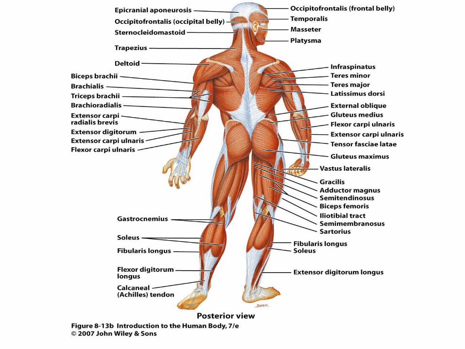
MUSCULAR SYSTEM Anatomy and Physiology

Types of Muscular Tissue Skeletal Muscle- striated, voluntary Cardiac muscle- striated, involuntary Smooth Muscle- non-striated, involuntary
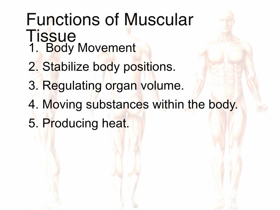
Functions of Muscular Tissue 1. Body Movement 2. Stabilize body positions. 3. Regulating organ volume. 4. Moving substances within the body. 5. Producing heat.

What’s in a Skeletal Muscle? Each skeletal muscle contains thousands of elongated, cylindrical cells called muscle fibers. Each muscle fiber is covered by a membrane called the sarcolemma. Transverse tubules (T tubules) pass through the muscle fiber . The sarcoplasm contains many mitochondria for ATP generation.

The sarcoplasmic reticulum stores calcium for muscle contractions. Myoglobin resides in the sarcoplasm and store oxygen until needed by the mitochondria. Myofibrils extend throughout the muscle fiber and contain two types of protein filaments, thick and thin. The filaments overlap each other in patterns to form compartments called sarcomeres.
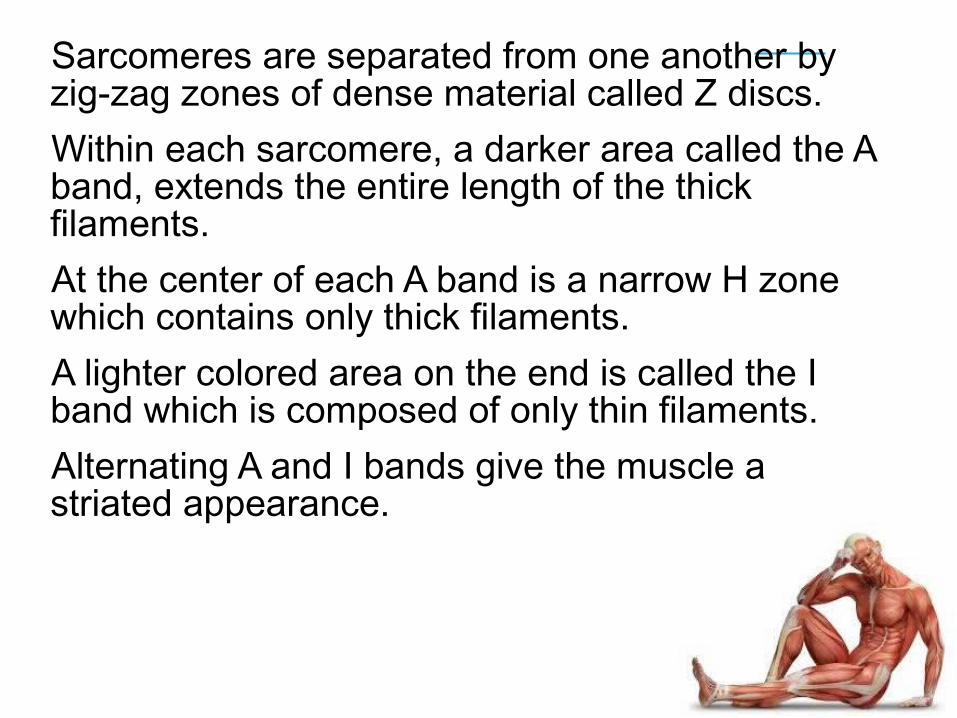
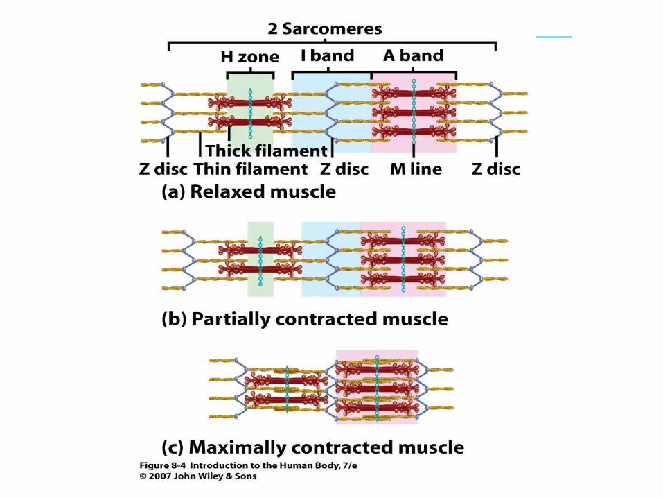
Sarcomeres are separated from one another by zig-zag zones of dense material called Z discs. Within each sarcomere, a darker area called the A band, extends the entire length of the thick filaments. At the center of each A band is a narrow H zone which contains only thick filaments. A lighter colored area on the end is called the I band which is composed of only thin filaments. Alternating A and I bands give the muscle a striated appearance.

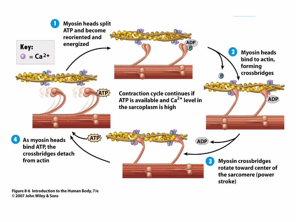
Thick filaments are composed of the protein myosin. Mysoin contains heads and tails and look like golf clubs. Thin filaments are anchored to the Z discs and are composed of actin. Thin filaments also contain the proteins tropomysoin and troponin that cover mysoin binding sites on relaxed muscle fibers.


Contraction and Relaxation of Skeletal Muscle
The sliding filament mechanism explains how muscles contract and relax.
During muscle contraction, myosin heads of the thick filaments pull on the thin filaments causing them to slide toward the center.

The thin filaments slide past the thick filaments because the myosin heads move like the oars of a boat pulling on the actin molecules in the thin filaments. Although the sarcomere shortens when the muscle contracts, the length of the filaments do not change.
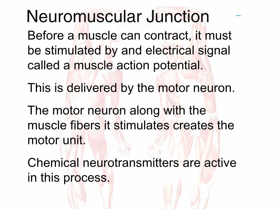
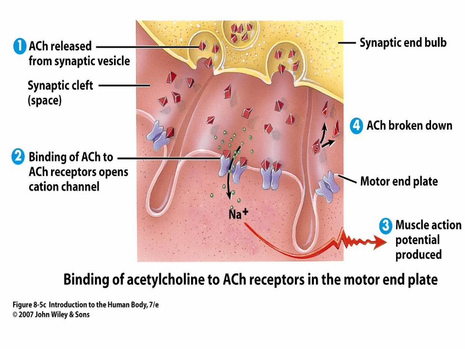
Neuromuscular JunctionBefore a muscle can contract, it must be stimulated by and electrical signal called a muscle action potential.
This is delivered by the motor neuron.
The motor neuron along with the muscle fibers it stimulates creates the motor unit.
Chemical neurotransmitters are active in this process.

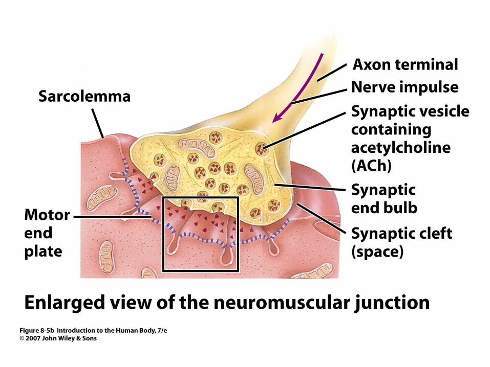

How It Works1. The nerve impulse from the motor neutron
releases acetylcholine (ACh).
2.The ACh binds to it’s receptor and activates ion channels to allow sodium ions to flow across the membrane.
3.The inflow of sodium ions generates the action potential that travels along the T tubules
4.The ACh breaks down unless more sodium generates more action potentials.


How Skeletal Muscles Produce Movement Skeletal Muscles are not attached directly to bones. They produce movement by pulling on tendons. Most skeletal muscles cross at least one joint. Not all muscles move when the bone moves however.
The bone the muscle is connected to that is stationary is the origin.
The bone that the muscle actually moves is the insertion.
This is where the muscle and tendon is actually connected for movement.


Skeletal muscle Cardiac muscle Smooth muscle Sarcolemma Sarcoplasmic reticulum Myoglobin Sarcomere
Z disc H Zone I band Myosin Actin Neuromuscular junction (NMJ)
37 total cards


