28

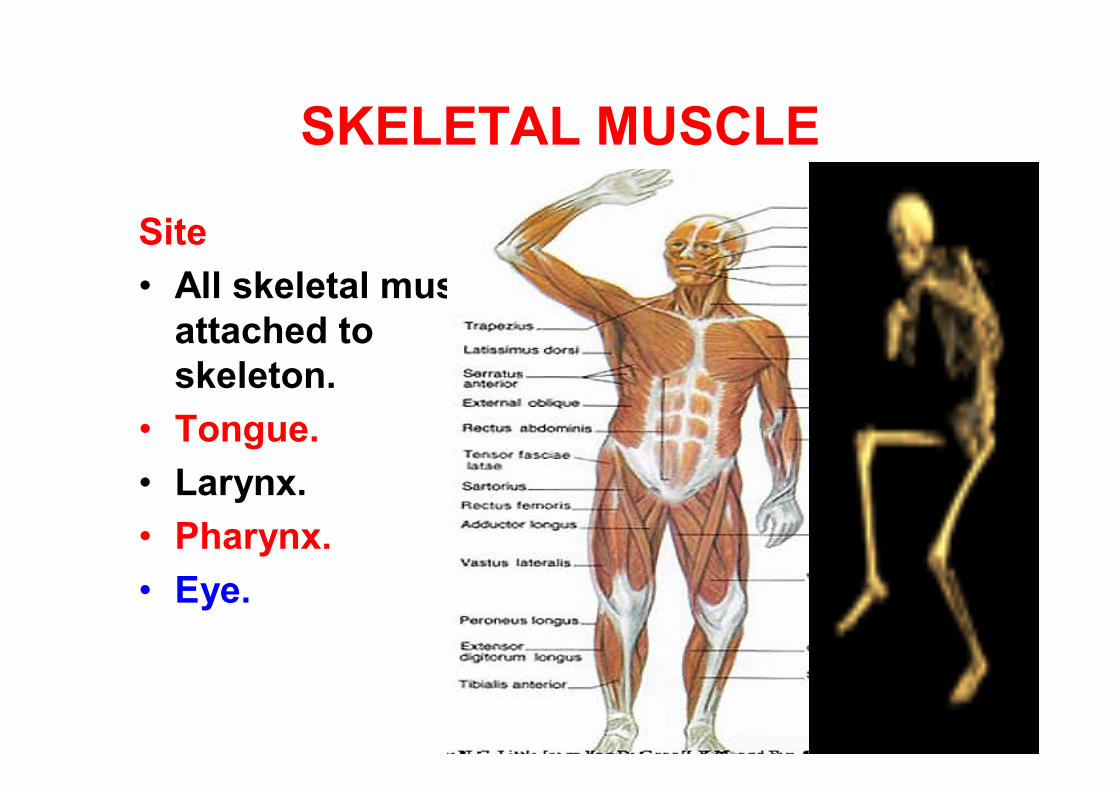
SKELETAL MUSCLE
Site • All skeletal muscle
attached to skeleton.
• Tongue.• Larynx.• Pharynx.• Eye.
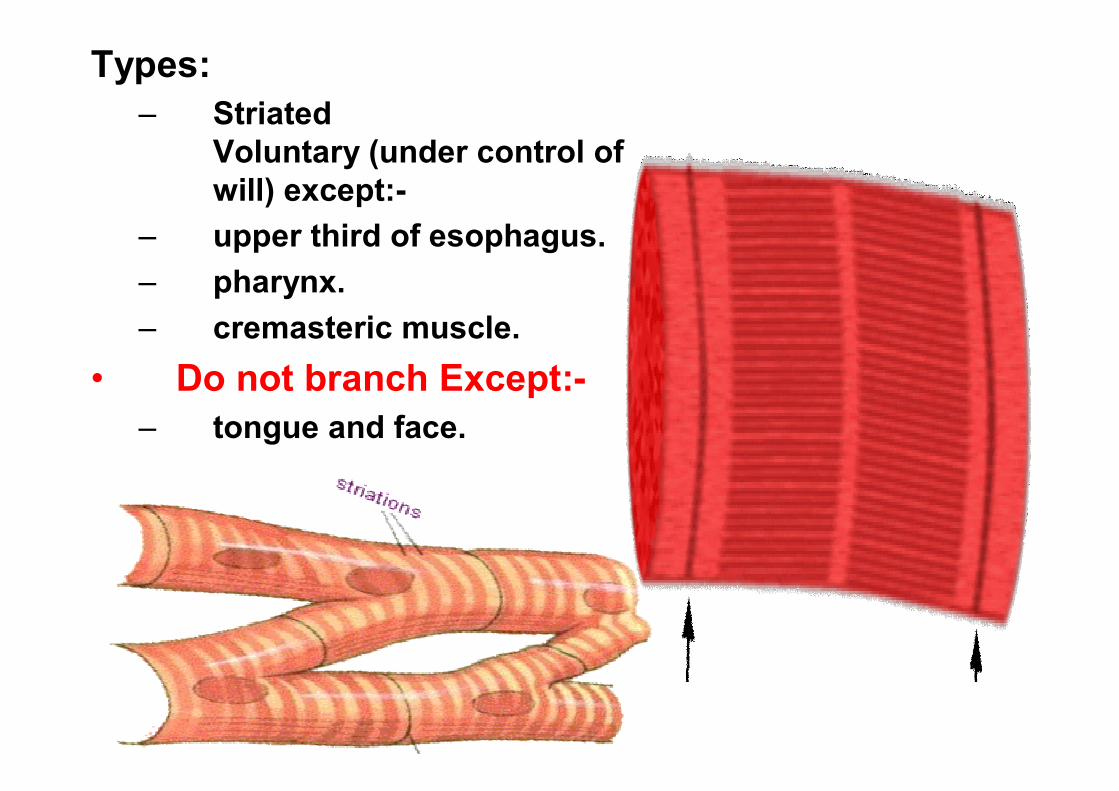
Types:– Striated
Voluntary (under control of will) except:-
– upper third of esophagus.– pharynx. – cremasteric muscle.
• Do not branch Except:-– tongue and face.
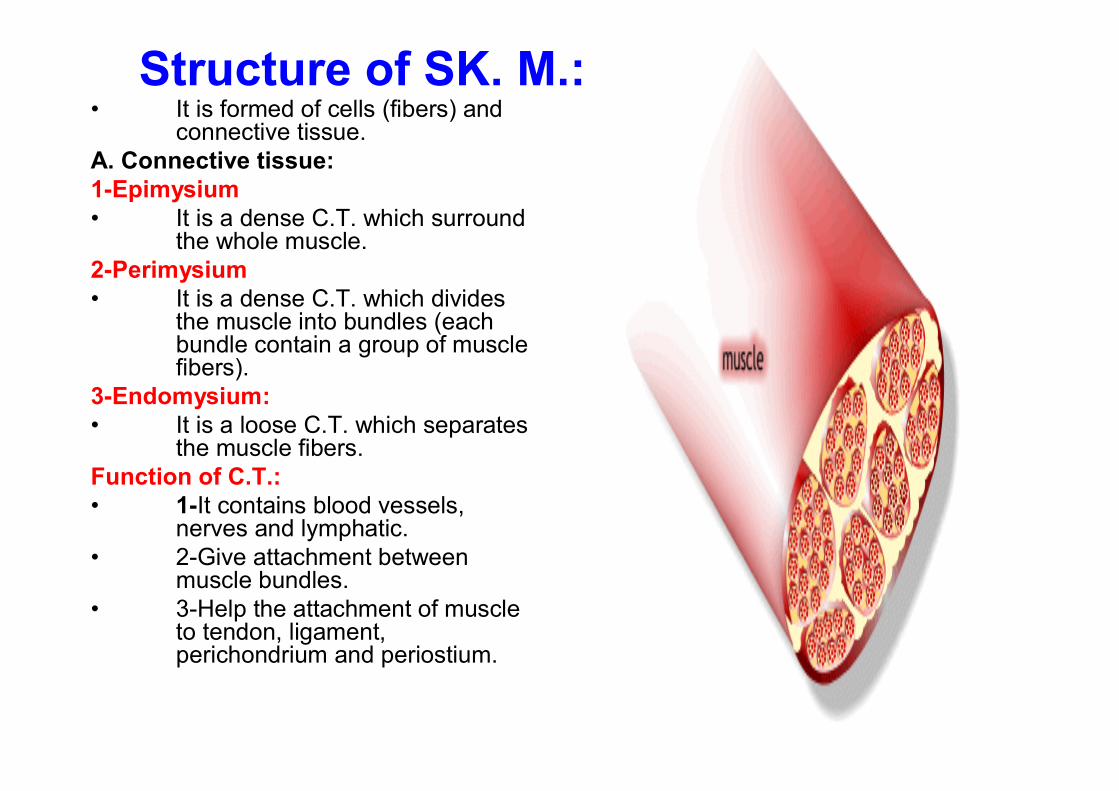
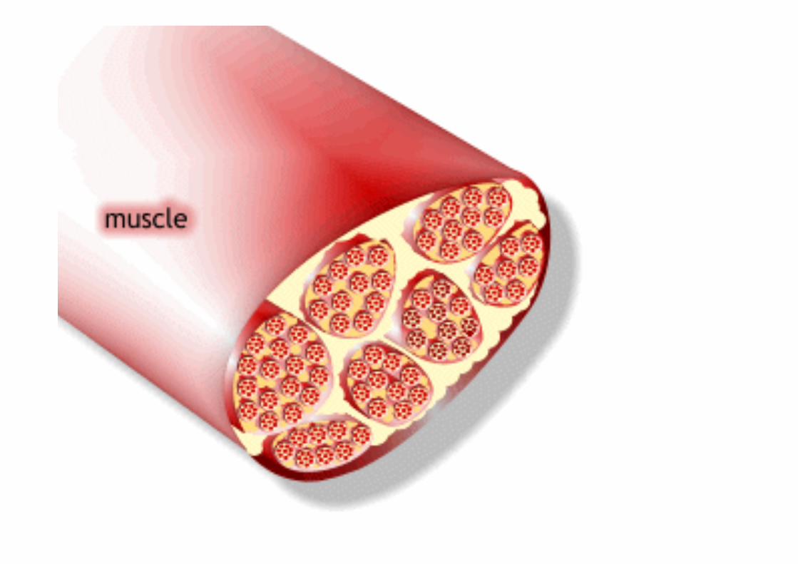
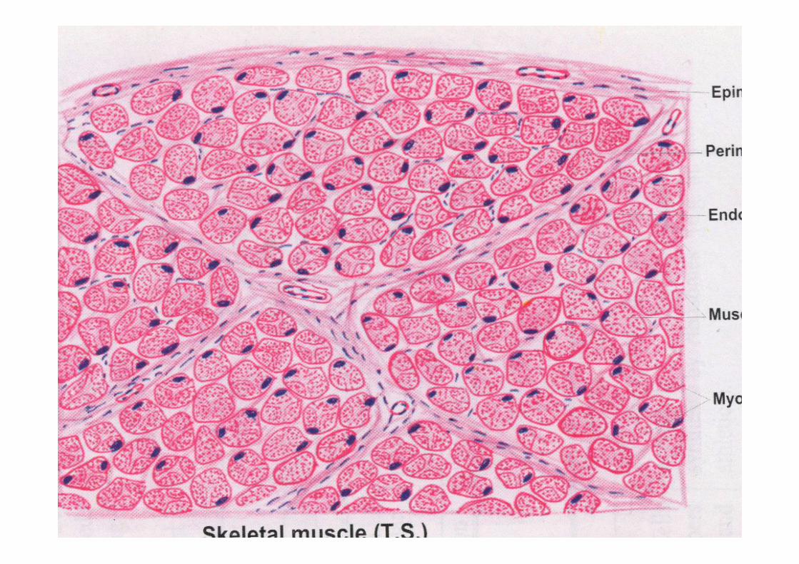
Structure of SK. M.:• It is formed of cells (fibers) and
connective tissue.A. Connective tissue:1-Epimysium• It is a dense C.T. which surround
the whole muscle.2-Perimysium• It is a dense C.T. which divides
the muscle into bundles (each bundle contain a group of muscle fibers).
3-Endomysium:• It is a loose C.T. which separates
the muscle fibers.Function of C.T.:• 1-It contains blood vessels,
nerves and lymphatic.• 2-Give attachment between
muscle bundles.• 3-Help the attachment of muscle
to tendon, ligament, perichondrium and periostium.
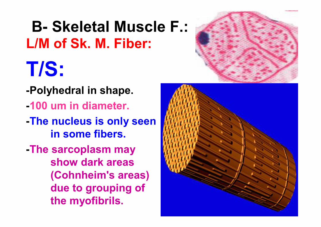
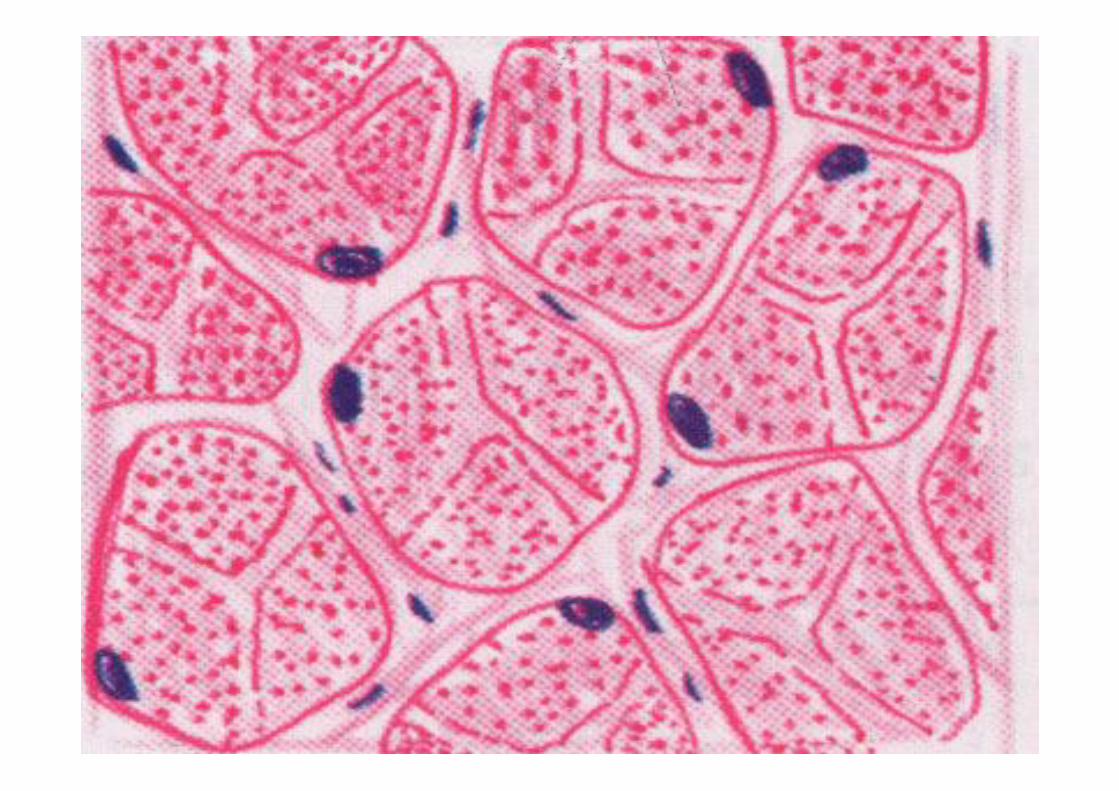
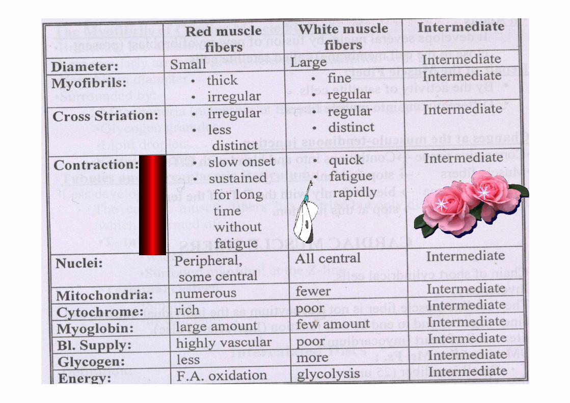
B- Skeletal Muscle F.:L/M of Sk. M. Fiber:
T/S:-Polyhedral in shape.-100 um in diameter.-The nucleus is only seen
in some fibers.-The sarcoplasm may
show dark areas (Cohnheim's areas) due to grouping of the myofibrils.
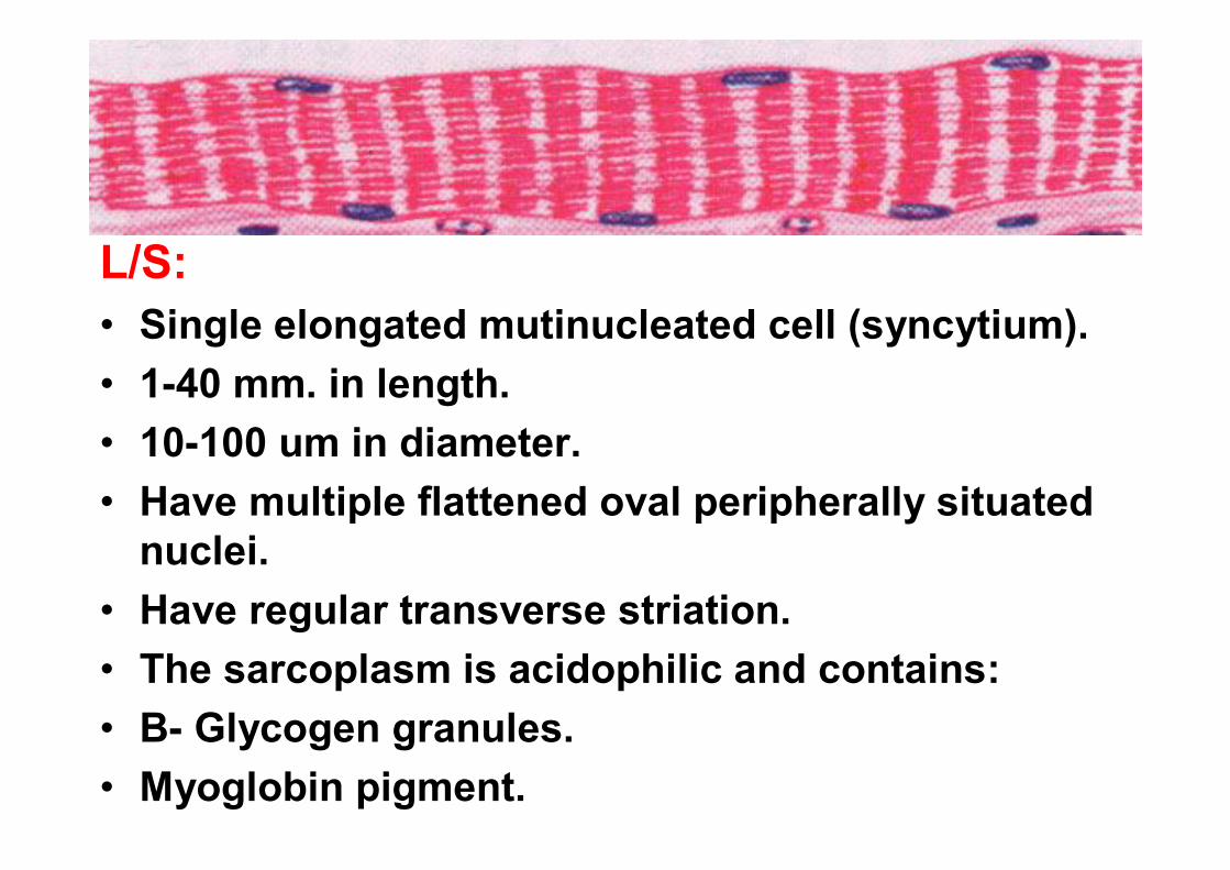
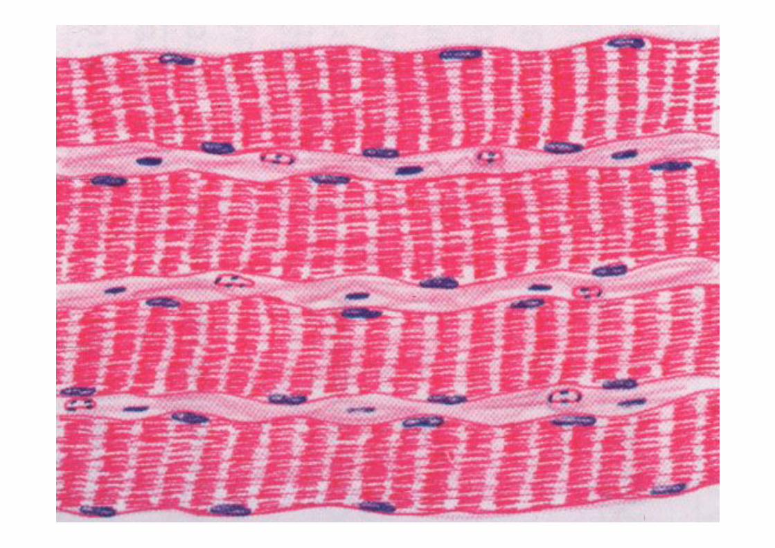
L/S:• Single elongated mutinucleated cell (syncytium).• 1-40 mm. in length.• 10-100 um in diameter.• Have multiple flattened oval peripherally situated
nuclei.• Have regular transverse striation.• The sarcoplasm is acidophilic and contains:• B- Glycogen granules.• Myoglobin pigment.
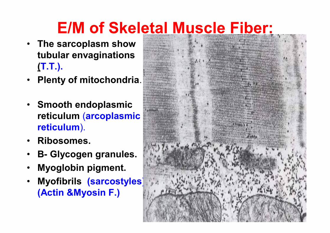
E/M of Skeletal Muscle Fiber:• The sarcoplasm show
tubular envaginations(T.T.).
• Plenty of mitochondria.
• Smooth endoplasmic reticulum (arcoplasmicreticulum).
• Ribosomes.• B- Glycogen granules.• Myoglobin pigment.• Myofibrils (sarcostyles)
(Actin &Myosin F.)
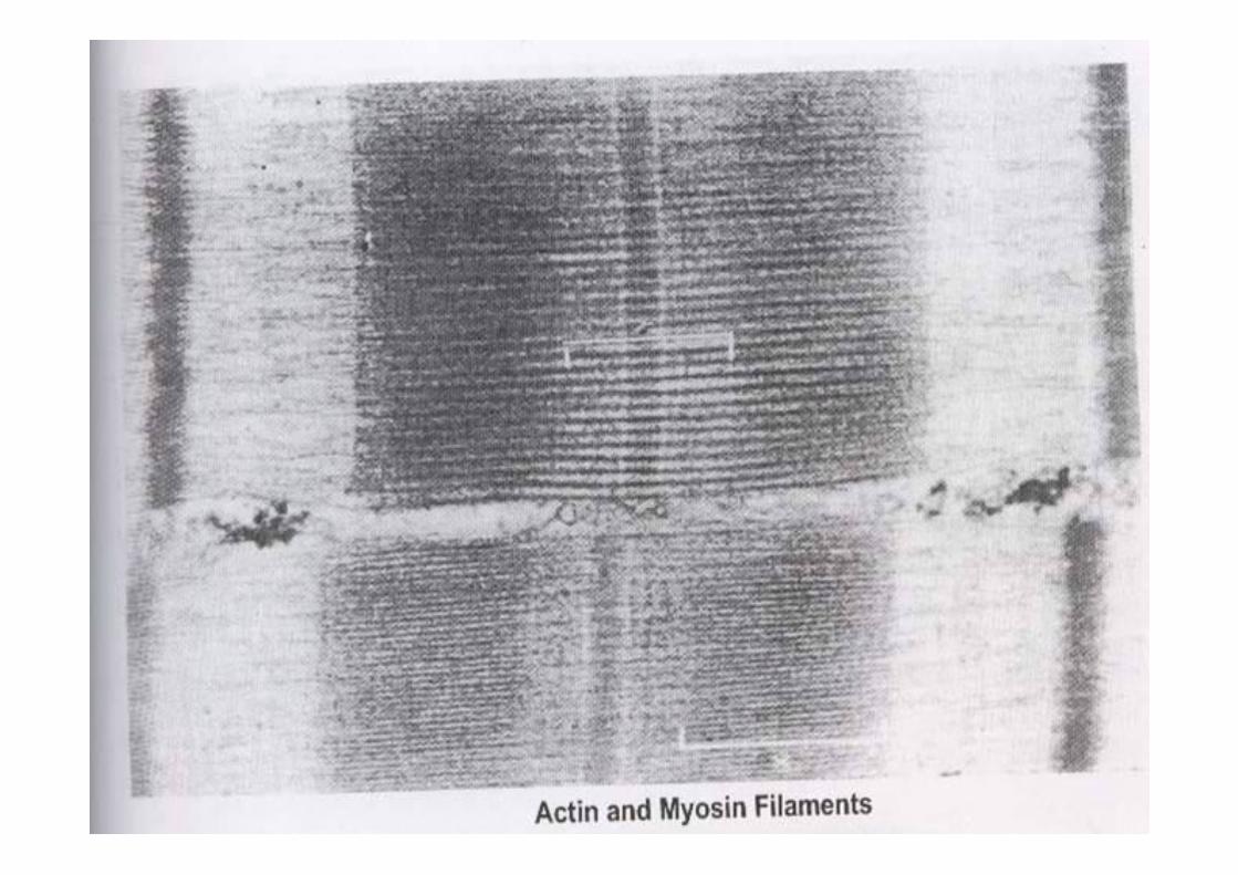
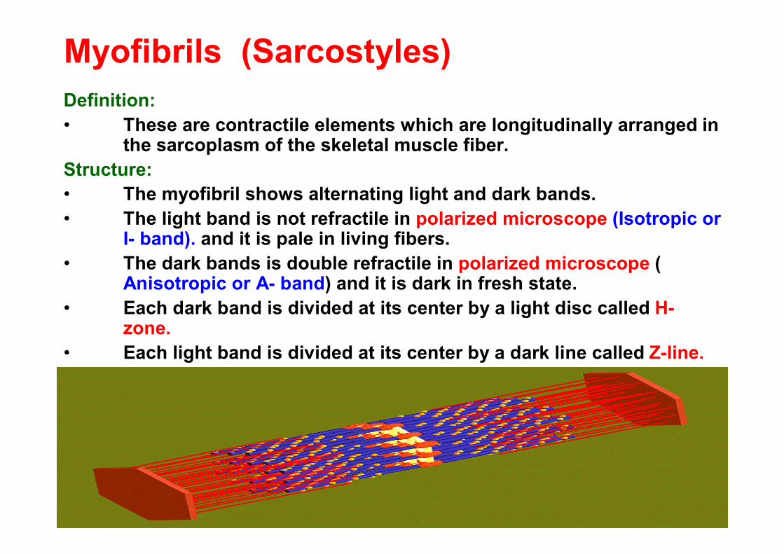
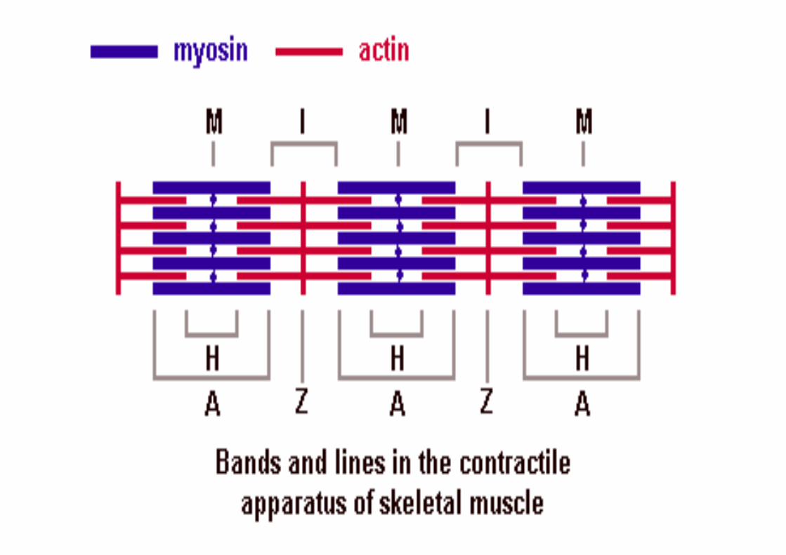
Myofibrils (Sarcostyles)Definition:• These are contractile elements which are longitudinally arranged in
the sarcoplasm of the skeletal muscle fiber.Structure:• The myofibril shows alternating light and dark bands.• The light band is not refractile in polarized microscope (Isotropic or
I- band). and it is pale in living fibers. • The dark bands is double refractile in polarized microscope (
Anisotropic or A- band) and it is dark in fresh state.• Each dark band is divided at its center by a light disc called H-
zone.• Each light band is divided at its center by a dark line called Z-line.
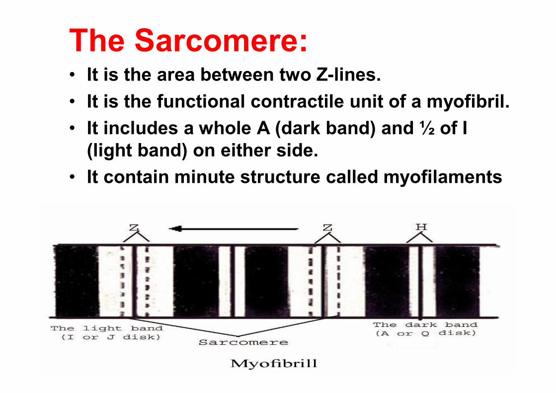
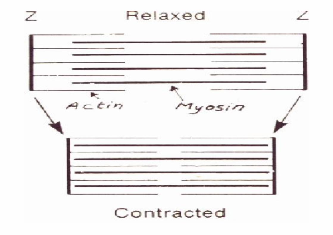
The Sarcomere:• It is the area between two Z-lines.• It is the functional contractile unit of a myofibril.• It includes a whole A (dark band) and ½ of I
(light band) on either side.• It contain minute structure called myofilaments
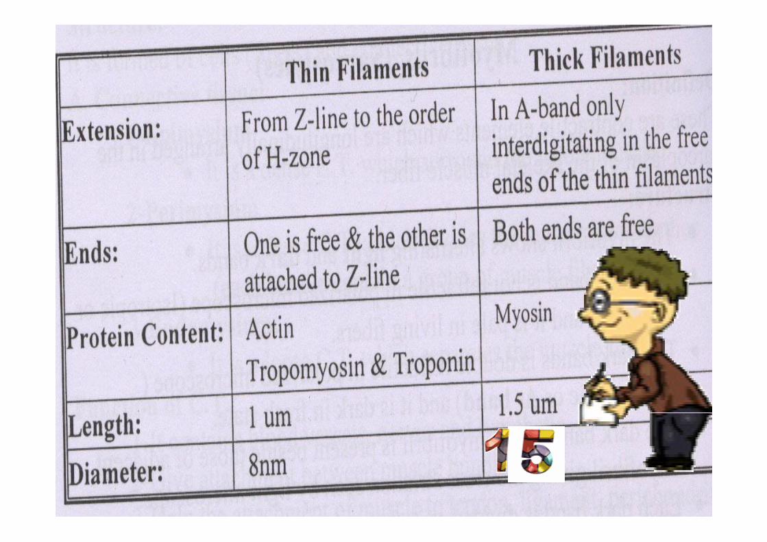
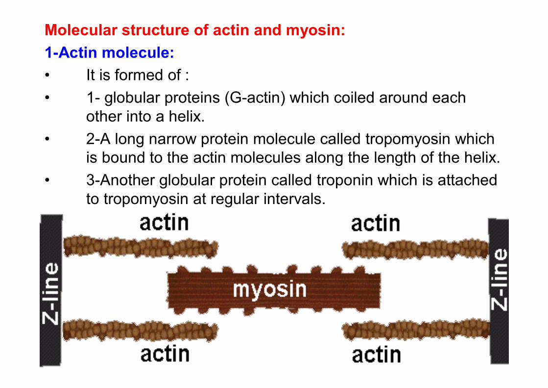
Molecular structure of actin and myosin:1-Actin molecule:• It is formed of :• 1- globular proteins (G-actin) which coiled around each
other into a helix. • 2-A long narrow protein molecule called tropomyosin which
is bound to the actin molecules along the length of the helix. • 3-Another globular protein called troponin which is attached
to tropomyosin at regular intervals.
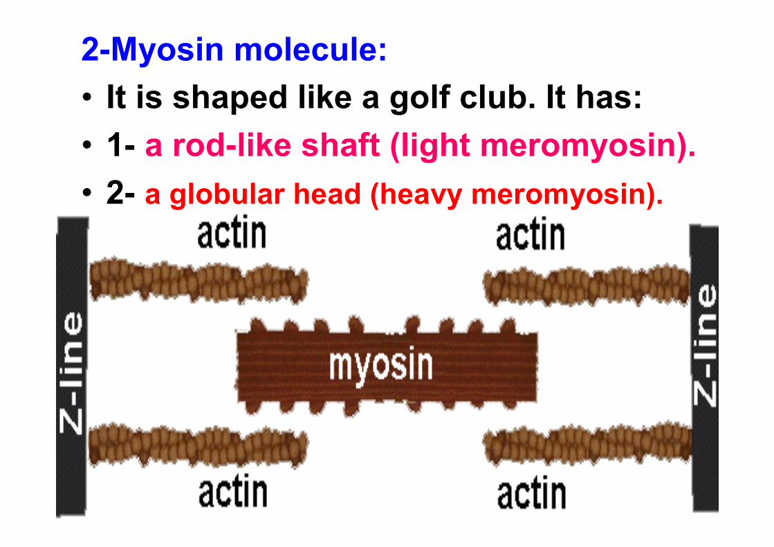
2-Myosin molecule:• It is shaped like a golf club. It has:• 1- a rod-like shaft (light meromyosin).• 2- a globular head (heavy meromyosin).
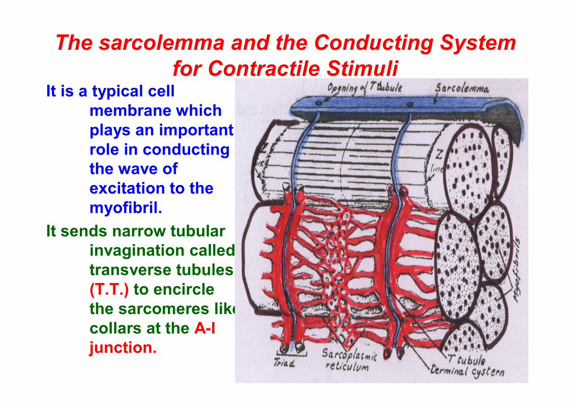
The sarcolemma and the Conducting System for Contractile Stimuli
It is a typical cell membrane which plays an important role in conducting the wave of excitation to the myofibril.
It sends narrow tubular invagination called transverse tubules (T.T.) to encircle the sarcomeres like collars at the A-I junction.
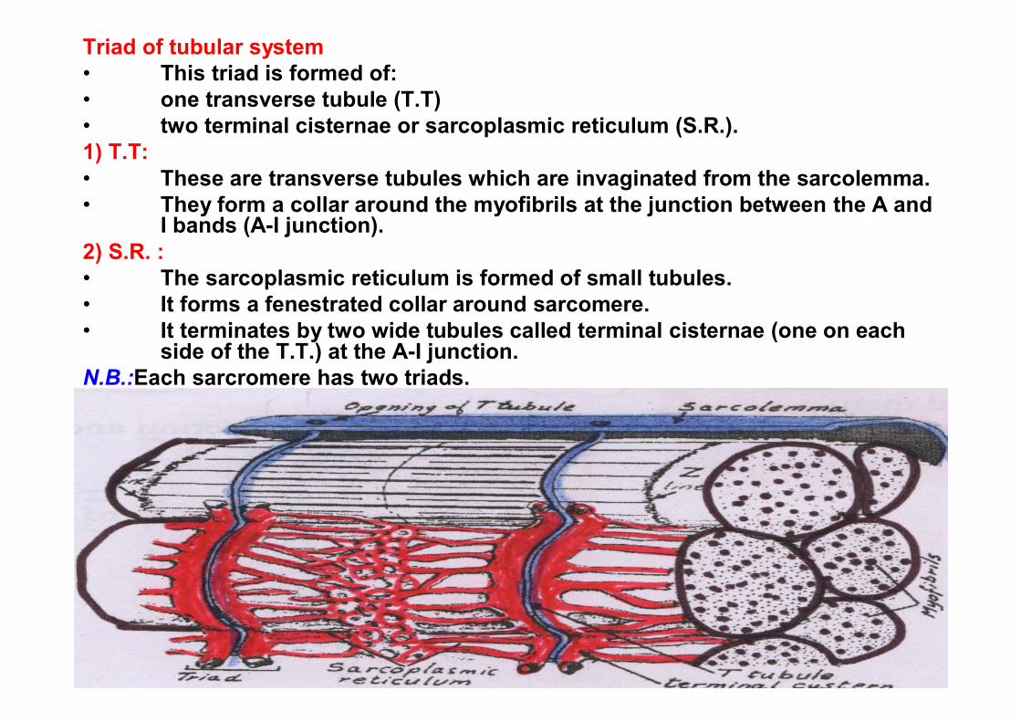
Triad of tubular system• This triad is formed of:• one transverse tubule (T.T)• two terminal cisternae or sarcoplasmic reticulum (S.R.).1) T.T:• These are transverse tubules which are invaginated from the sarcolemma.• They form a collar around the myofibrils at the junction between the A and
I bands (A-I junction).2) S.R. :• The sarcoplasmic reticulum is formed of small tubules.• It forms a fenestrated collar around sarcomere.• It terminates by two wide tubules called terminal cisternae (one on each
side of the T.T.) at the A-I junction.N.B.:Each sarcromere has two triads.
Role of this triad in contraction:• When a nerve impulses are transmitted to the
Sarcoplasm, It is transmitted to the sarcoplasmicreticulum via the T.T of the triads.
• The SR will pump Ca ions into the myofibrils.• The energy rich-ATP of the muscle is converted
into ADP with the release of energy.• This energy allows the actin to interact with
myosin and cause gliding of thin filaments over thick filaments and ensure a proper union between them.
• The thin filaments slide towards the middle of the sarcomere, pulling the 2 ''Z''- lines behind them.
• This allow shortening of sarcomere and lead to muscle contraction and loss of H- zone.
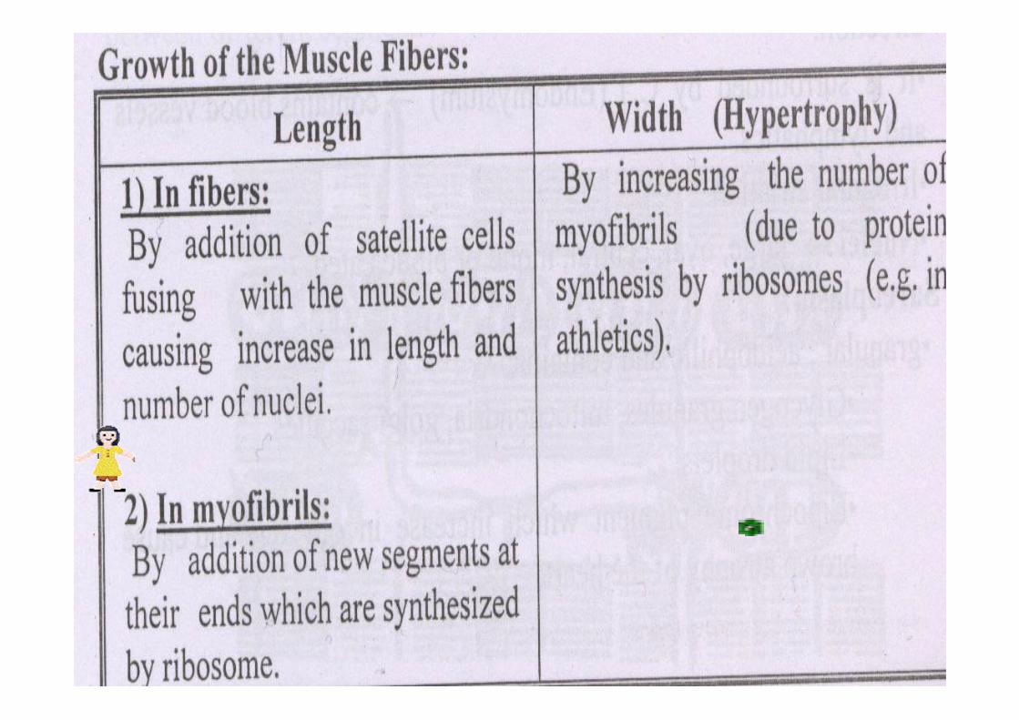
Development of the muscle fibers:
In Embryo • The muscle fibers
is formed by fusion of mono nucleated cells (Myoblasts).
In Adults• It develops several
nuclei by fusion of myofibroblast
)present close to the cell membrane )
called satellite cells.
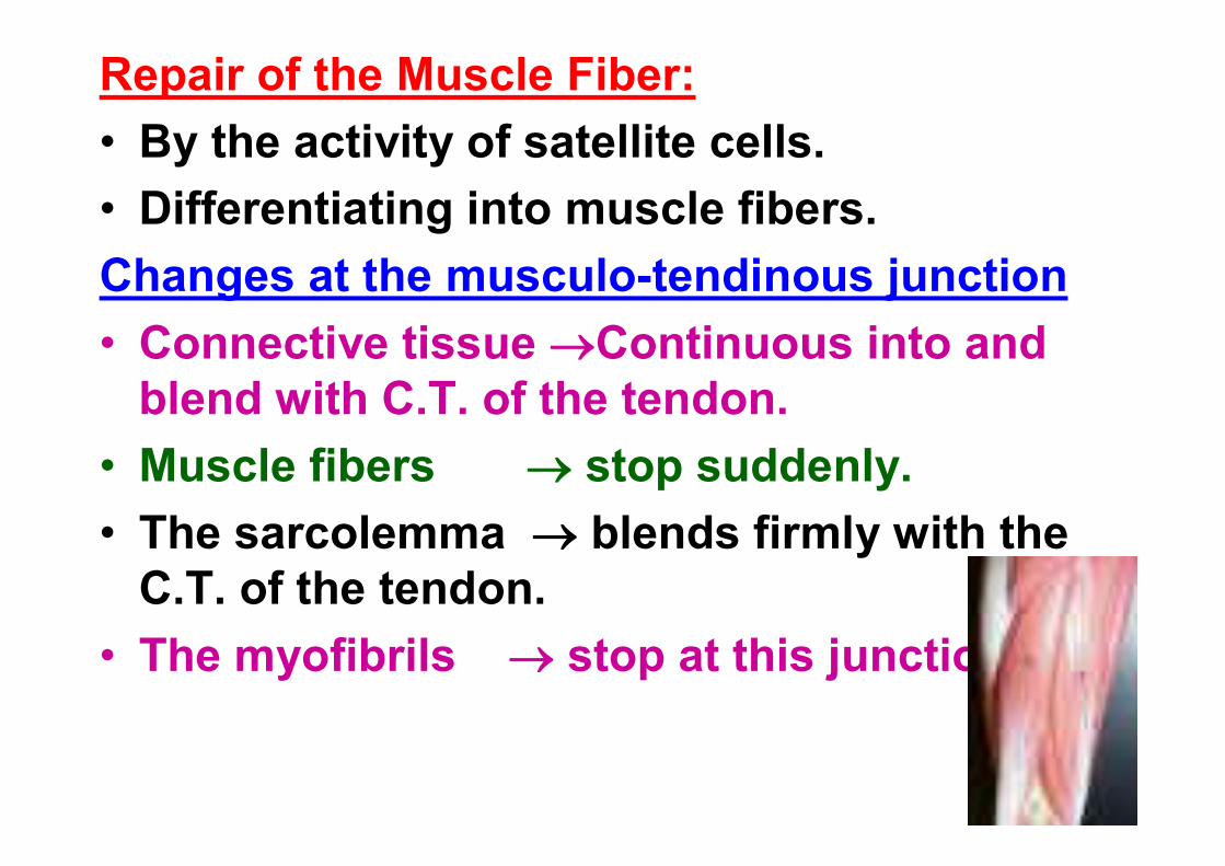
Repair of the Muscle Fiber:• By the activity of satellite cells.• Differentiating into muscle fibers.Changes at the musculo-tendinous junction• Connective tissue Continuous into and
blend with C.T. of the tendon.• Muscle fibers stop suddenly.• The sarcolemma blends firmly with the
C.T. of the tendon.• The myofibrils stop at this junction.