94
SKELETAL MUSCLE PHYSIOLOGY Abraham D. Lee, Ph.D.,P.T. Department of Physical Therapy Office: Collier Building # 4206 Phone #: 419-383-3437 Email: [email protected]
| Date post: | 24-Dec-2015 |
| Category: |
Documents |
| Upload: | grant-moore |
| View: | 217 times |
| Download: | 0 times |
SKELETAL MUSCLE PHYSIOLOGY
Abraham D. Lee, Ph.D.,P.T.
Department of Physical Therapy
Office: Collier Building # 4206
Phone #: 419-383-3437
Email: [email protected]

Contents1. Muscle structure & organization2. Muscle fiber type3. Muscle action 4. Muscle mechanics5. Motor unit and its recruitment6. Local muscle control7. Muscle plasticity8. Summary

Muscle organization• Epimysium: wraps an entire muscle
• Perimysium: wraps a bundle of muscle fibers. This bundle is called fascicle or fasciculus
• Endomysium: wraps an individual muscle fiber
• Sarcolemma: muscle membrane
• Myofibrils: contractile filaments


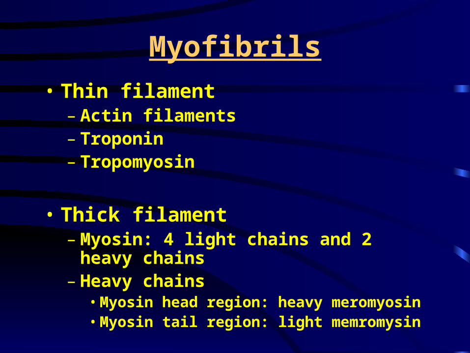
Myofibrils
• Thin filament – Actin filaments– Troponin– Tropomyosin
• Thick filament– Myosin: 4 light chains and 2 heavy chains– Heavy chains
• Myosin head region: heavy meromyosin• Myosin tail region: light memromysin

Muscle pennationMuscle pennation• Longitudinal (non-pennated) architecture: muscle fibers in
parallel to the muscle force generating axis – Example: biceps brachii, sartorius muscle– In these muscles fibers are said to be fusiform or spindle shaped.
• “Pennate” architecture: muscle fibers are oriented at an angle or multiple angles relative to force-generating axis.
1) Unipennate: soleus-25 degree; vastus medialis-5 degree
2) Bipennate: gastrocnemius, rectus femoris3) Multipennate: deltoid


Effect of pennationForce loss
Space saving
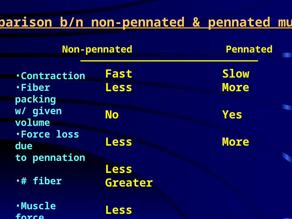
Comparison b/n non-pennated & pennated muscleComparison b/n non-pennated & pennated muscle
•Contraction•Fiber packingw/ given volume•Force loss dueto pennation
•# fiber
•Muscle forceProduction
•CSA
Fast SlowLess More
No Yes
Less More
Less Greater
Less Greater
Non-pennated Pennated

Muscle Fiber Type
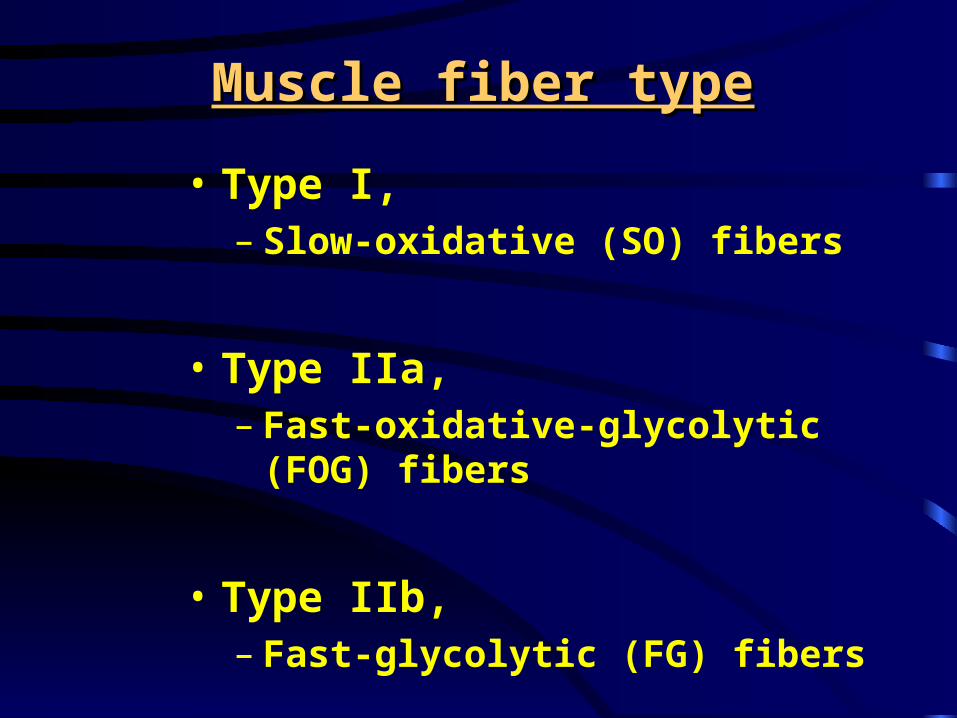
Muscle fiber typeMuscle fiber type
• Type I, – Slow-oxidative (SO) fibers
• Type IIa, – Fast-oxidative-glycolytic (FOG) fibers
• Type IIb, – Fast-glycolytic (FG) fibers
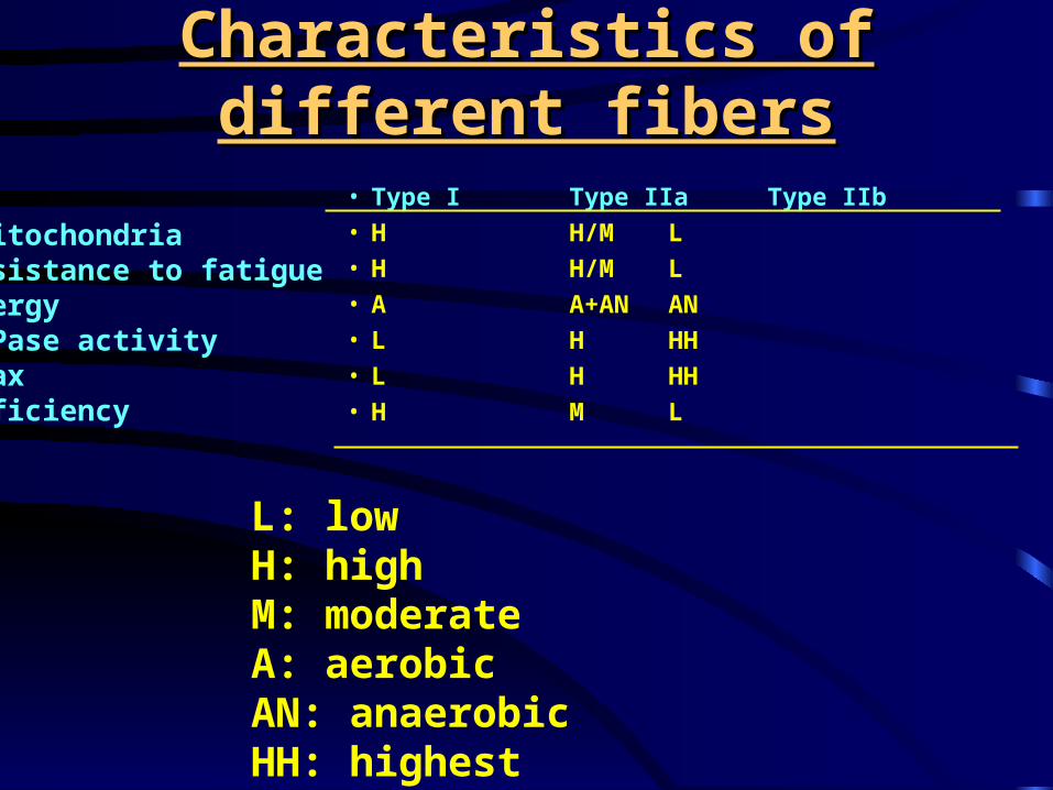
Characteristics of different fibersCharacteristics of different fibers
• Type I Type IIa Type IIb• H H/M L• H H/M L• A A+AN AN• L H HH• L H HH• H M L
#MitochondriaResistance to fatigueEnergyATPase activityVmaxEfficiency
L: lowH: highM: moderateA: aerobicAN: anaerobicHH: highest

Muscle composition in athletesMuscle composition in athletes
% Type I %Type IIa &IIb
70-80 20-30
25-30 70-75
45-55 45-55
47-53 47-53
Distance runnersTrack sprintersWeight liftersNon-athletes
Will fiber type change with training?

Muscle Action
• Excitation-contraction coupling
• Type of muscle action

Excitation-Contraction CouplingExcitation-Contraction Coupling
•Nerve impulse generation and propagation
•Neuromuscular junction transmission•Muscle action potential propagation•Ca2+ release from SR•Ca2+ binding to troponin•Interaction of myosin head and actin•Cross bridge moves: tension development•Ca2+ taken up to SR•Ca2+ removal from troponin•Relaxation


E-C couplingDHPR: dihydropyridine receptorsRyR: ryanodine receptor Other possible mechanism:
Inositol 1,4,5-triphosphate (InsP3)
InsP3 receptor activation
Ca2+ release from SR
May play a role in slow twitch muscle in developmental stage (Talon et al., Am. J. Physiol 282: R1164-R1173, 2002)

E-C coupling

Sliding Filament Theory


Changes during shortening muscle action
• Sarcomere length (distance between two adjacent Z lines): shortens
• A band: no change• I band: shortens• H zone: shortens

Different type of muscle action Different type of muscle action (contraction)(contraction)
• Isometric action
• Isotonic action (dynamic action)– Concentric action
– Eccentric action

Muscle mechanics
It deals with how muscle force is generated and regulated.
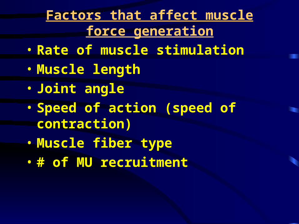
Factors that affect muscle force generation
• Rate of muscle stimulation
• Muscle length
• Joint angle
• Speed of action (speed of contraction)
• Muscle fiber type
• # of MU recruitment

Rate of Muscle Stimulation
• Twitch:
• Tetanus:

Muscle twitchMuscle twitch
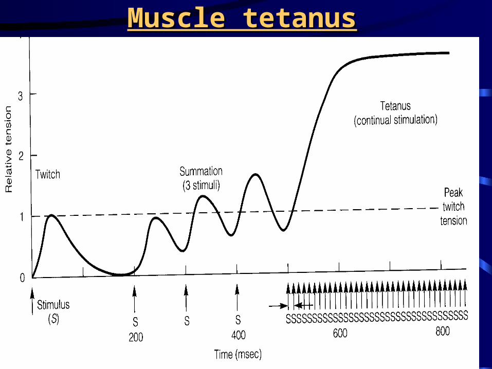
Muscle tetanusMuscle tetanus

Effect of Muscle Length

Force-Length Relationship
• Isolated muscle
• In vivo human muscles

Force-Length Relationship
• Isolated muscle


Force-Length Relationship
• In vivo human muscles– Two things are considered: muscle length
and joint angle– In general, a group of muscles produces
more force (torque) when muscles are lengthened before contraction. But some muscles do not follow this rule.

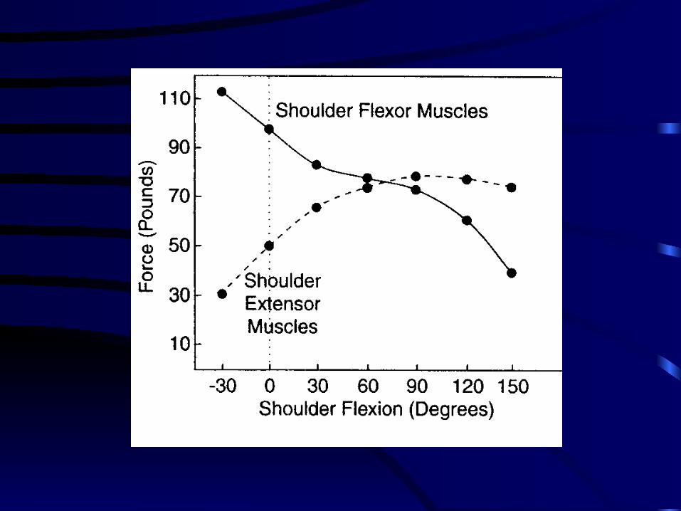
Shoulder muscles
0°
90°
180°
40°
Shoulder flexors (anterior deltoid): causes to flex shoulder joint
Shoulder extensors(posterior deltoid): causes to extend shoulder joint
45°
135°

Knee flexors
• A person is lying on the stomach (prone position)
Knee joint
Thigh Lower leg0°
90° 120°
Hip joint
Trunk
Knee flexors (hamstring muscles): causes to flex knee joint
45°


Hip flexors
• A person is lying on the back (supine position)
hip joint
TrunkThigh0°
90° 120°
Lower leg
Knee joint
Hip flexors (iliopsoas, sartorius): causes to flex hip joint
45°

Knee extensors
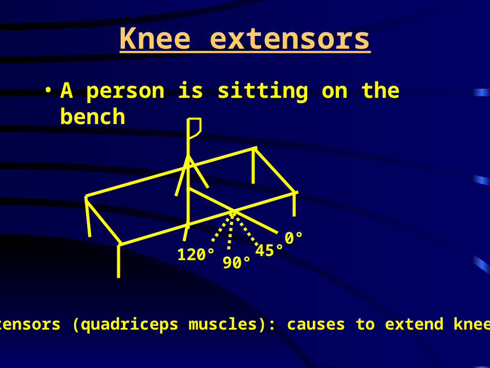
• A person is sitting on the bench
0°
90°120°
Knee extensors (quadriceps muscles): causes to extend knee joint
45°


Elbow flexors
Elbow flexors (biceps brachii): causes to flex elbow joint


Force arm distance

Effect of Velocity (Speed of Action)

Force-velocity curve

Effect of Muscle Fiber

Effect of muscle fiber type on force

Muscle Power
Need to consider two factors:
1. Muscle force
2. Speed of action

Power-Velocity Relationship
Power = work/time = (force x distance)/time = force x speed


Effect of muscle fiber type on power

Factors that affect muscle force/power generation
• Rate of muscle stimulation
• Muscle length
• Joint angle
• Speed of action
• Muscle fiber type
• # of MU recruitment

Motor UnitMotor Unit
How does an individual generate appropriate force for a given task?

Motor Unit (MU)Motor Unit (MU)
Functional unit of movement

Motor Unit (MU)Motor Unit (MU)
•MU consists of •Single -motor neuron•Muscle fibers innervated by
the -motor neuron


Motor Unit (MU)Motor Unit (MU)
•Fast fatigable MU (FF)•High twitch tension•High fatigue index
•Fast fatigue resistant MU (FR)•Intermediate twitch tension•Intermediate fatigue index
•Slow MU (S)•Low twitch tension•Low fatigue index

Reasons for different twitch Reasons for different twitch tension in different MUtension in different MU
•Depends on number of muscle fibers and fiber size
# muscle fiber: FF>FR> SSize of fiber: FF>FR>S
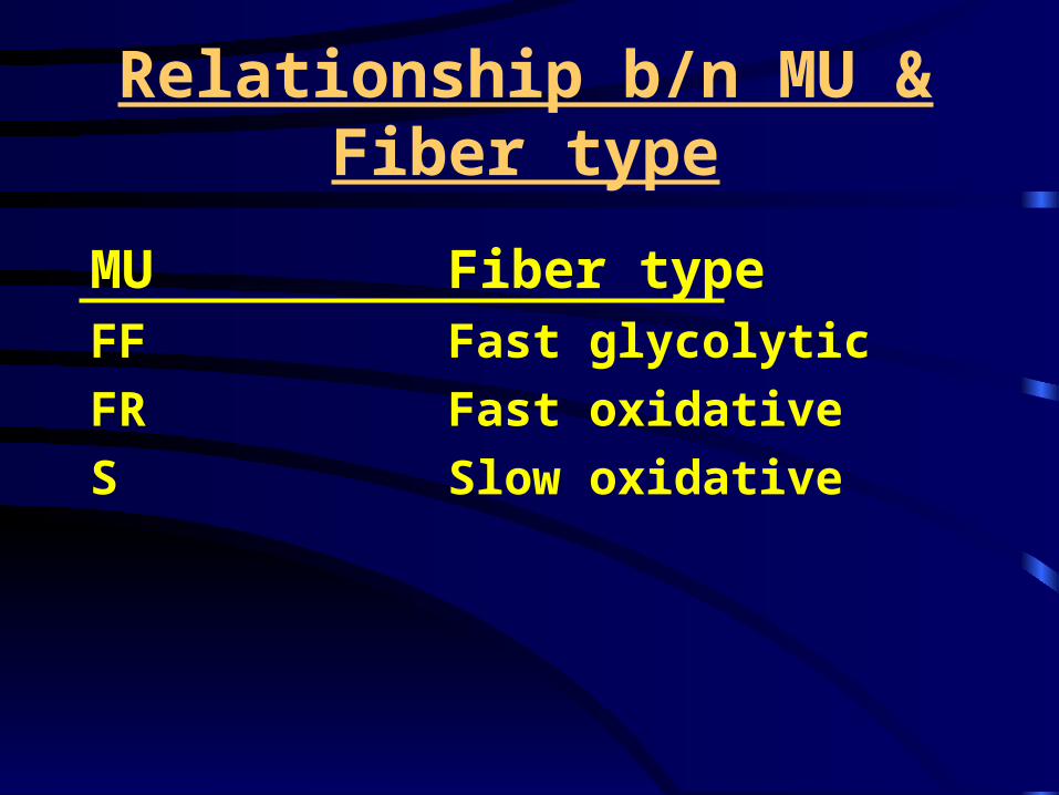
Relationship b/n MU & Fiber type
MU Fiber typeFF Fast glycolytic
FR Fast oxidative
S Slow oxidative

Motor Unit
Muscle # neuron # fibers/MU
• Biceps brachii 774 750
• Gastrocnemius 580 1720
• First lumbrical 98 110
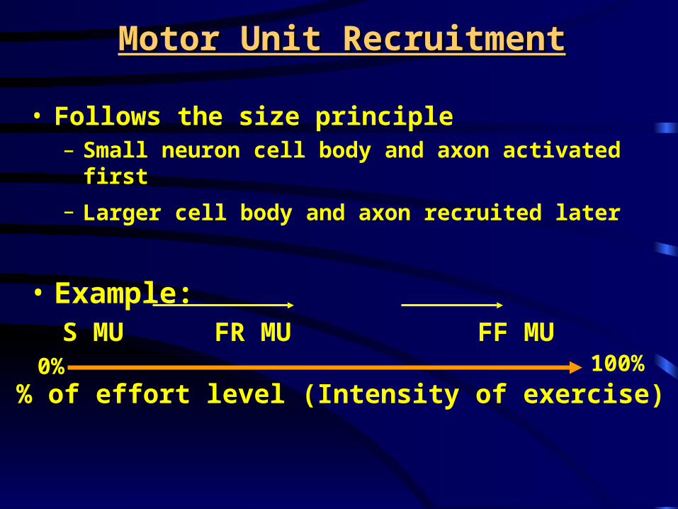
Motor Unit RecruitmentMotor Unit Recruitment
• Follows the size principle– Small neuron cell body and axon activated first
– Larger cell body and axon recruited later
• Example: S MU FR MU FF
MU
% of effort level (Intensity of exercise)0% 100%


Gradation of Muscle Strength
• By increasing # of MU recruited
• By increasing frequency of stimulation

Local Control of Muscle Action
• Muscle spindle: muscle length monitor– Consists of 1) afferent nerves, 2) intrafusal fibers
& 3) γ(gamma)-motor neurons
• Golgi Tendon Organ: muscle tension monitor

Structure of muscle spindle

Action of muscle spindle

Nerve impulse pattern of afferent nerves
Rest Stretch Contraction Return to rest

Speed of stretch on impulse discharge pattern
Clinical implications for individuals with spastic muscle?

Golgi Tendon Organ
•GTO#<spindle # in given muscle
•Composed of network of unmyelinated nerve fibers enclosed by fine capsule
•Activated by either muscle stretch or muscle contraction. More sensitive to muscle contraction.
•Activates inhibitory interneuron in spinalcord, which, in turn, inhibits -motorneuron of contracting muscle (agonists).
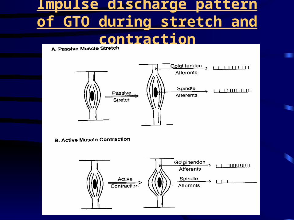
Impulse discharge pattern of GTO during stretch and contraction

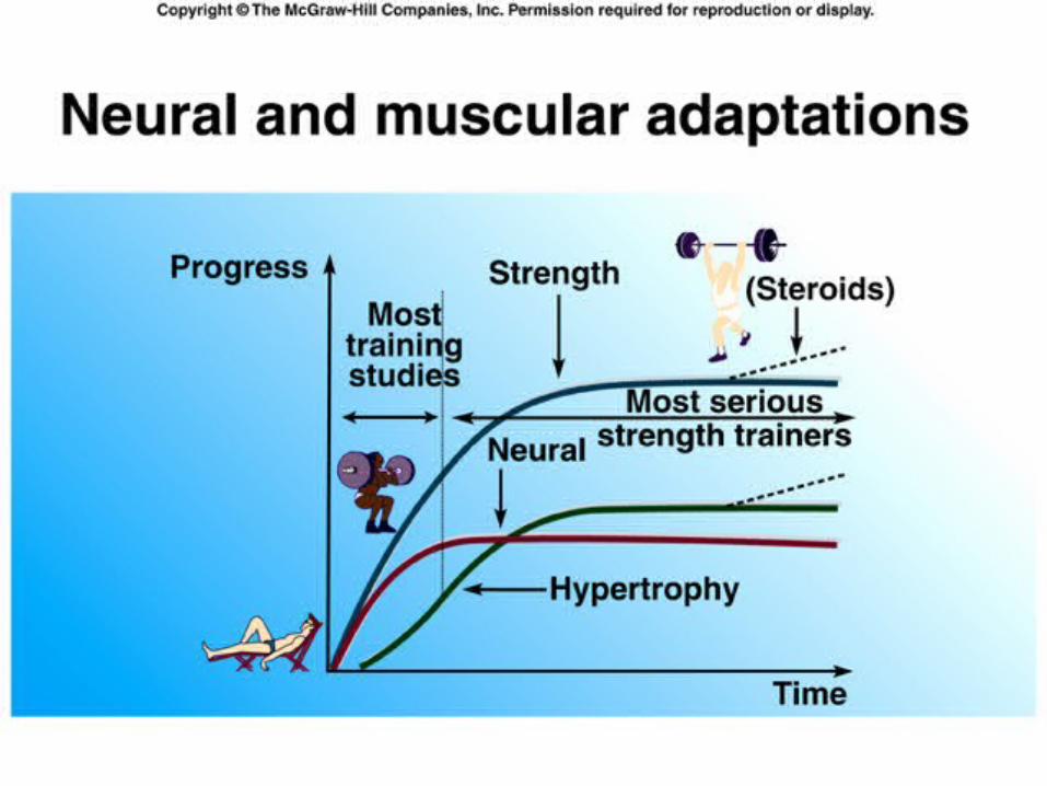
Plasticity of Muscle
• Metabolic and morphological changes to changes in stimulus– Increased stimulus: exercise training– Decreased stimulus: non-weight bearing, bed
rest and aging

Endurance training
• Mode: jogging, running, cycling, swimming, etc
• Adaptations– # of mitochondria– size of mitochondria– Oxidative enzyme activities
• Krebs cycle, beta-oxidation, ETS
– Some glycolytic enzymes – Capillary density


Resistance training
• Strength– Neural factor– Muscle fiber enlargement
(hypertrophy)

33%
27%
38%
31%
6 wk
5-6 month resistance training using triceps brachiiMacDougall et al, EJAP 43:25-34MacDougall et al, EJAP 43:25-34

Resistance TrainingResistance Training

Limb suspensionLimb suspension(Non-weight bearing)(Non-weight bearing)
Berg et al., J. Appl. Physiol. 70:1882-1885, 1991

Limb unloading on muscle strength and X-areaLimb unloading on muscle strength and X-area
Knee extensor Strength
X-area
Dudley et al, in ACSM’s Resource Manual, p.201

Selective muscle atrophy with non-weight bearing
Dudley et al, in ACSM’s Resource Manual, p.201
Bed Rest

Muscle strength change with bed restMuscle strength change with bed rest
Dudley et al, in ACSM’s Resource Manual, p.203
(soleus and gastroc.).)

Changes in skeletal muscles with agingChanges in skeletal muscles with aging
• # of muscle fibers
• Muscle area
• Fiber type distribution
• Muscle strength

McArdle et al, in Exercise Physiology, p639
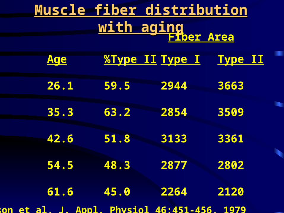
Muscle fiber distribution with agingMuscle fiber distribution with aging
Age %Type II Type I Type II
26.1 59.5 2944 3663
35.3 63.2 2854 3509
42.6 51.8 3133 3361
54.5 48.3 2877 2802
61.6 45.0 2264 2120
Fiber Area
Larson et al, J. Appl. Physiol 46:451-456, 1979

Muscle strength with aging
• A decline in muscle strength is associated with a decrease in muscle mass.
• A decline in lower extremity muscle strength is related to poor functional performance– walking ability, balance, stair-climbing
ability, falls
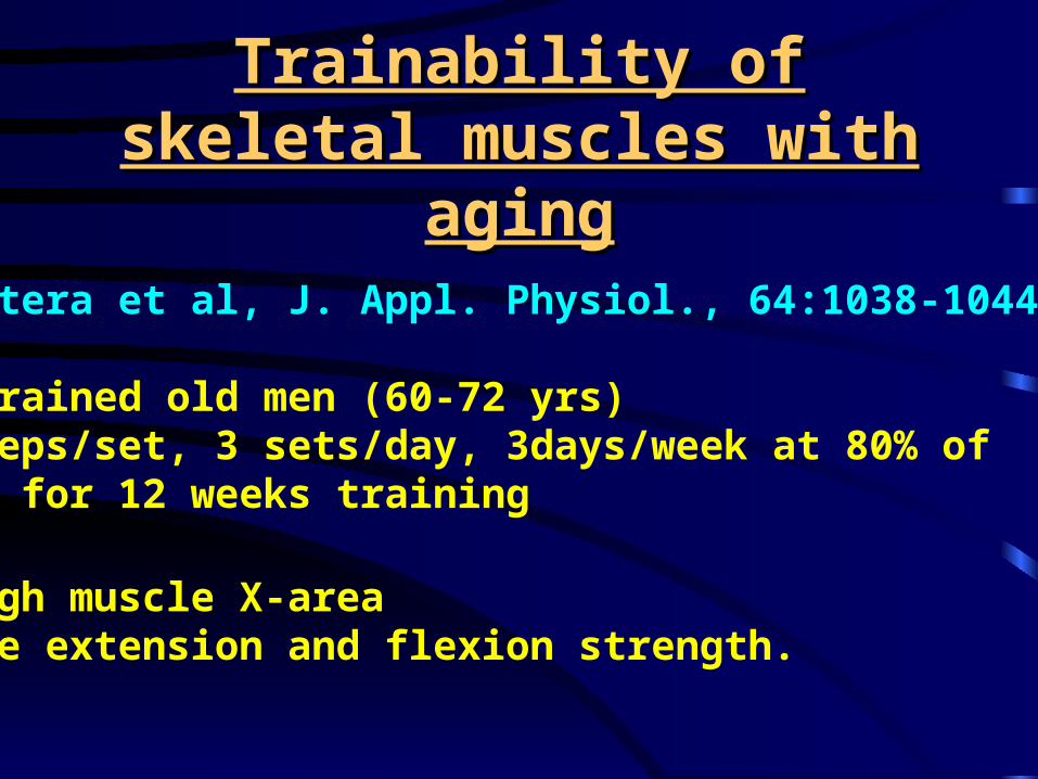
Trainability of skeletal muscles Trainability of skeletal muscles with agingwith aging
Frontera et al, J. Appl. Physiol., 64:1038-1044, 1988
•Untrained old men (60-72 yrs)•8 reps/set, 3 sets/day, 3days/week at 80% of 1 RM for 12 weeks training
•Thigh muscle X-area•Knee extension and flexion strength.

Leg strengthLeg strength
1 RM max
Frontera et al

X-area of quadricepsX-area of quadriceps Frontera et al

Physician’s Role for Physical Activity

Summary
Know followings: names & functions•Muscle structure: connective tissues, pennation, myofibrils •Muscle fiber characteristics•Muscle action: E-C coupling•Sliding filament theory: changes during contraction•Muscle mechanics: force & power-length-velocity •MU: elements, function, characteristics.•Muscle action monitor: muscle spindle and GTO•Changes in muscle in training•Changes in muscle w/ suspension, bed rest aging

The End


