37
Skeletal Muscle Unit Chapter 6
| Date post: | 17-Dec-2015 |
| Category: |
Documents |
| Upload: | cecily-gibson |
| View: | 243 times |
| Download: | 0 times |
Skeletal Muscle UnitChapter 6
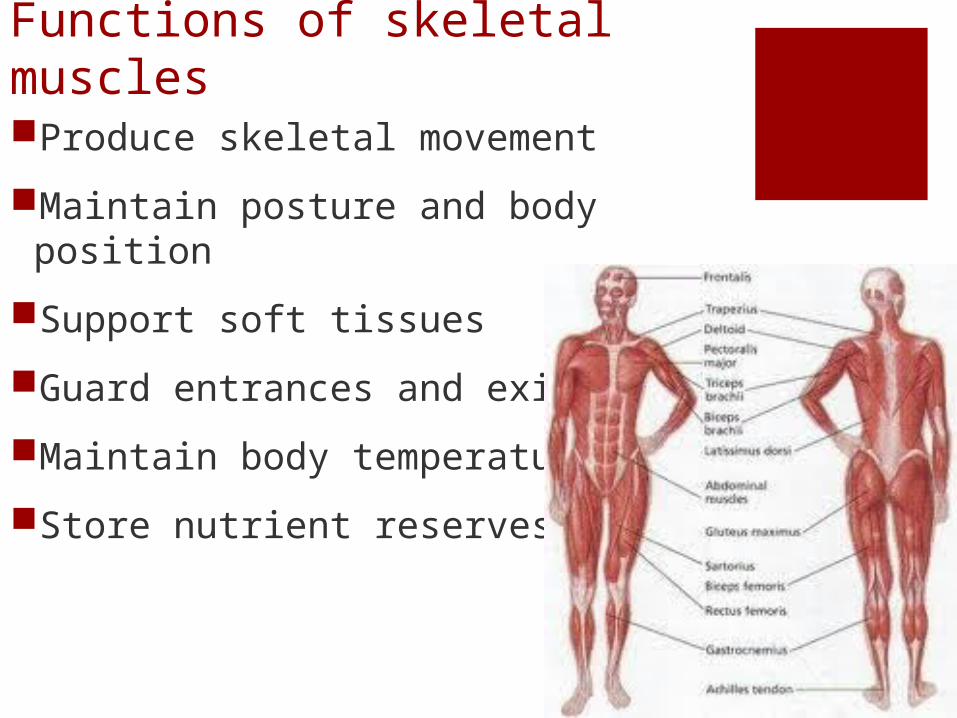
Functions of skeletal musclesProduce skeletal movement
Maintain posture and body position
Support soft tissues
Guard entrances and exits
Maintain body temperature
Store nutrient reserves
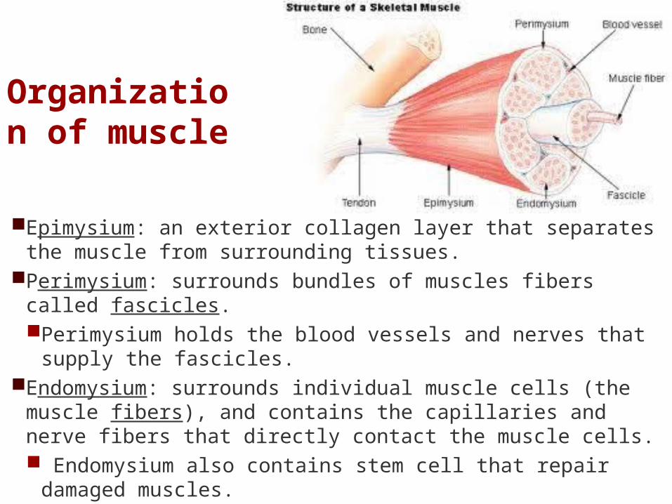
Organization of muscle
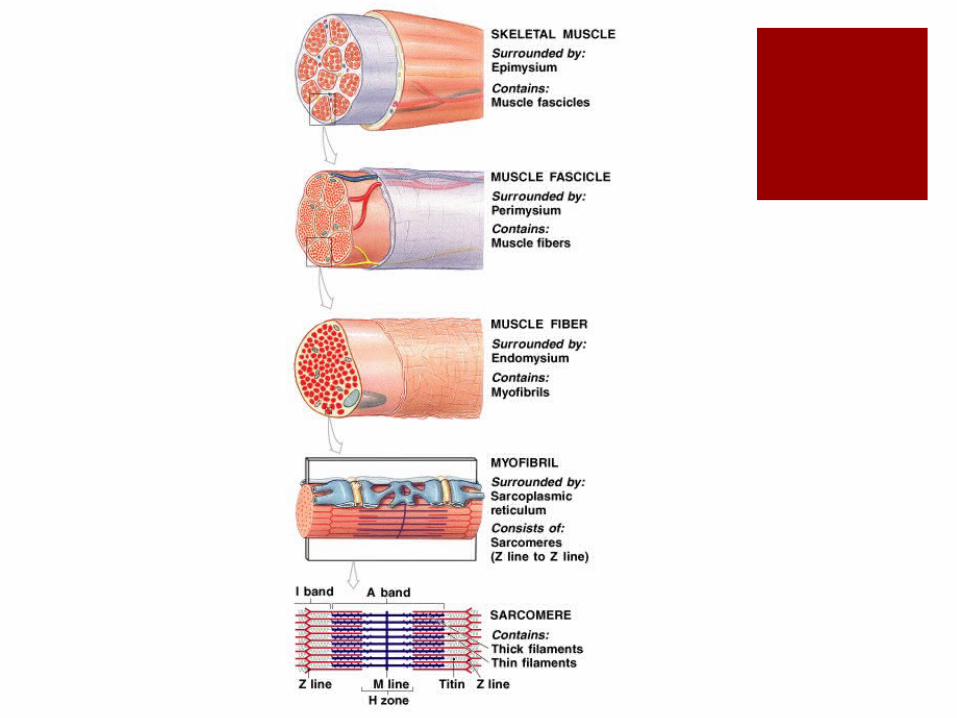
Epimysium: an exterior collagen layer that separates the muscle from surrounding tissues.
Perimysium: surrounds bundles of muscles fibers called fascicles. Perimysium holds the blood vessels and nerves that supply
the fascicles.Endomysium: surrounds individual muscle cells (the muscle
fibers), and contains the capillaries and nerve fibers that directly contact the muscle cells. Endomysium also contains stem cell that repair damaged
muscles.
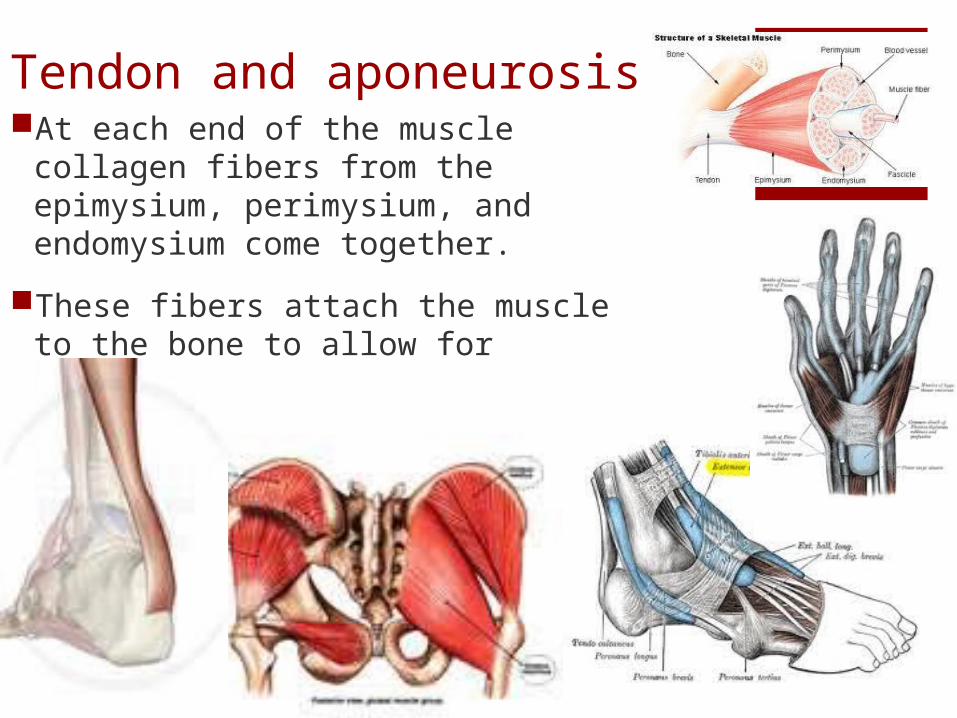
Tendon and aponeurosisAt each end of the muscle collagen
fibers from the epimysium, perimysium, and endomysium come together.
These fibers attach the muscle to the bone to allow for movement
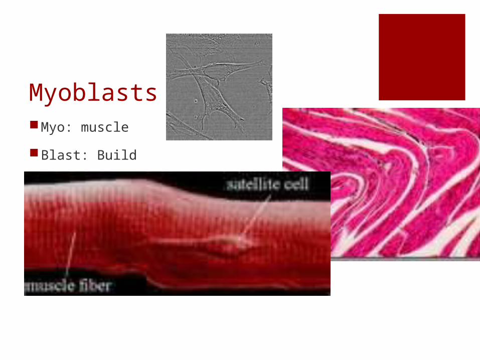
MyoblastsMyo: muscle
Blast: Build
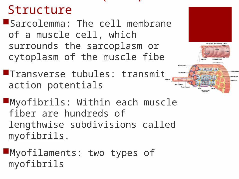
Muscle Fiber (Cell) StructureSarcolemma: The cell membrane of
a muscle cell, which surrounds the sarcoplasm or cytoplasm of the muscle fiber.
Transverse tubules: transmit action potentials
Myofibrils: Within each muscle fiber are hundreds of lengthwise subdivisions called myofibrils.
Myofilaments: two types of myofibrils
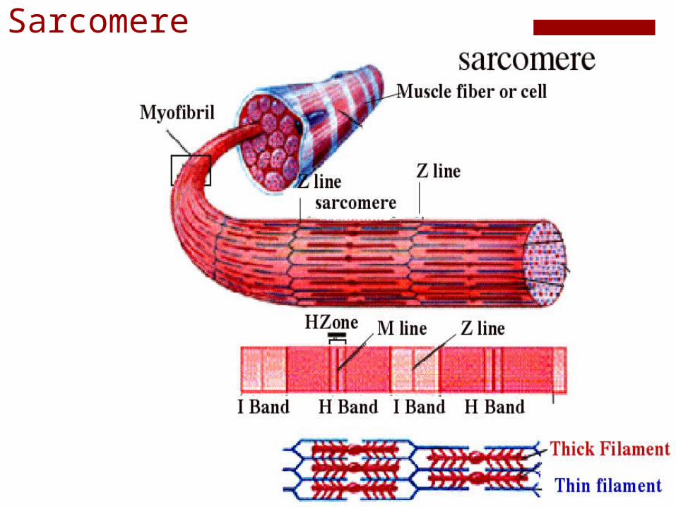
Sarcomere
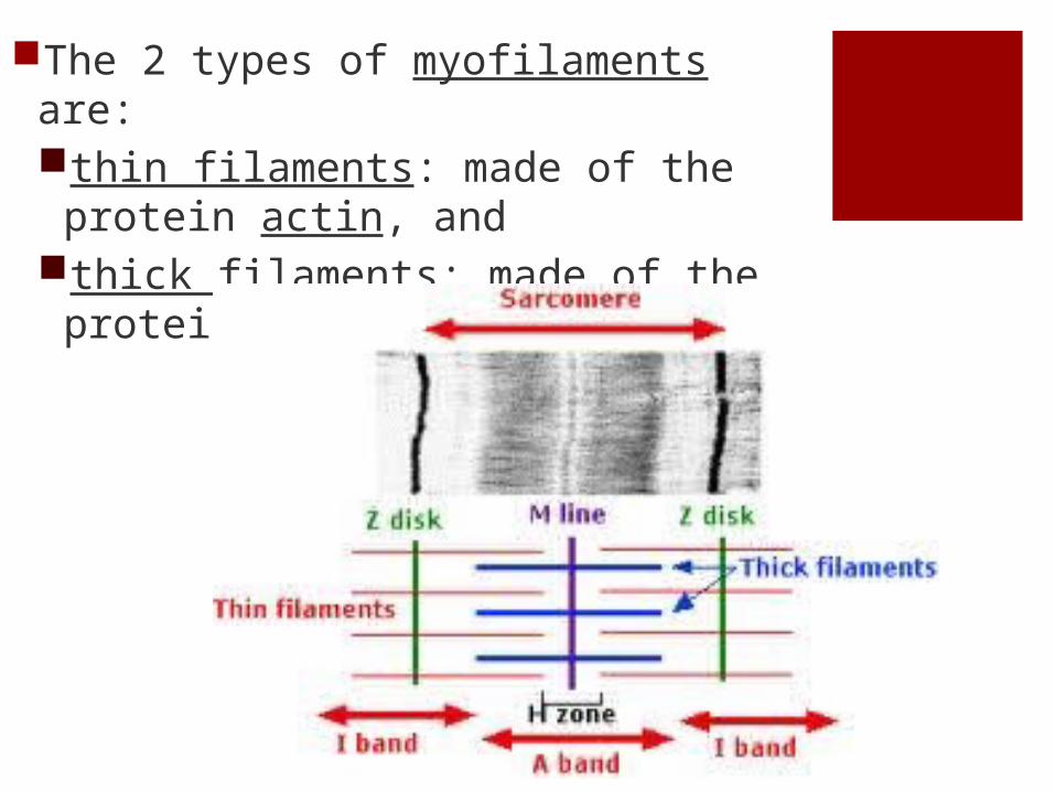
The 2 types of myofilaments are:thin filaments: made of the protein
actin, andthick filaments: made of the protein
myosin.
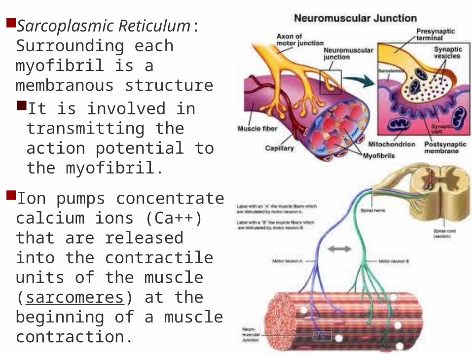
Sarcoplasmic Reticulum: Surrounding each myofibril is a membranous structure It is involved in
transmitting the action potential to the myofibril.
Ion pumps concentrate calcium ions (Ca++) that are released into the contractile units of the muscle (sarcomeres) at the beginning of a muscle contraction.
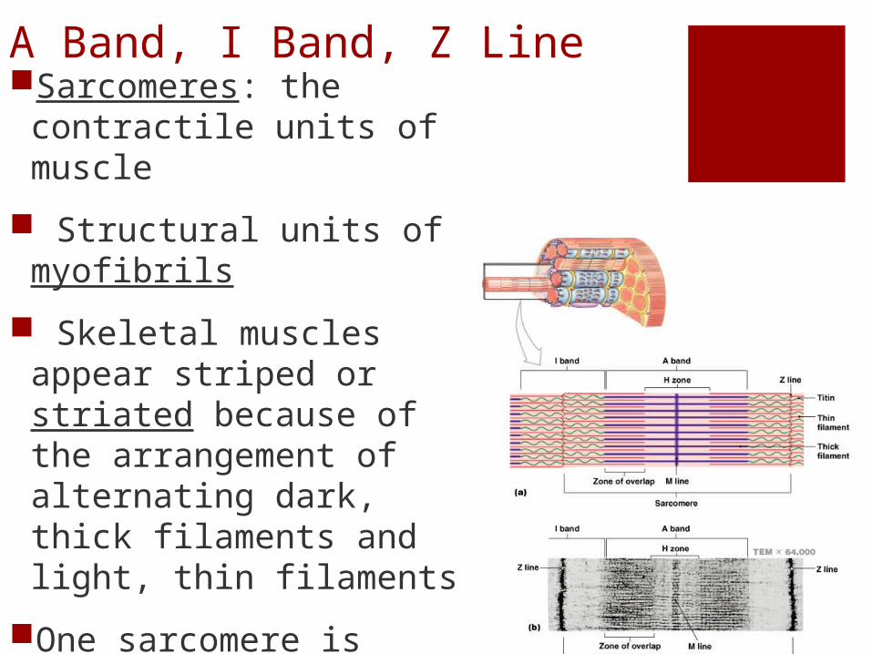
A Band, I Band, Z LineSarcomeres: the
contractile units of muscle
Structural units of myofibrils
Skeletal muscles appear striped or striated because of the arrangement of alternating dark, thick filaments and light, thin filaments
One sarcomere is measured from one Z line to another.
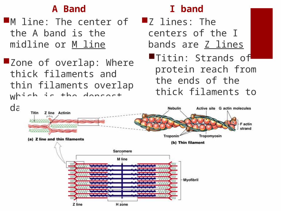
A BandM line: The center of the A
band is the midline or M line
Zone of overlap: Where thick filaments and thin filaments overlap which is the densest, darkest area
I bandZ lines: The centers of
the I bands are Z lines Titin: Strands of
protein reach from the ends of the thick filaments to the Z line and stabilize the filaments
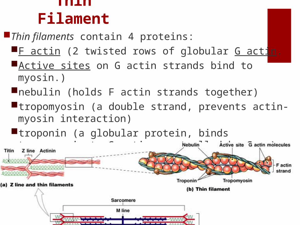
Thin Filament
Thin filaments contain 4 proteins:F actin (2 twisted rows of globular G actin. Active sites on G actin strands bind to myosin.)nebulin (holds F actin strands together)tropomyosin (a double strand, prevents actin-myosin
interaction) troponin (a globular protein, binds tropomyosin to G
actin, controlled by Ca++)
Thin Filament
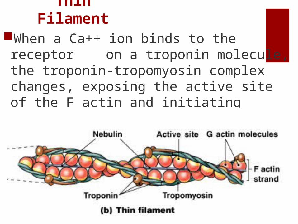
When a Ca++ ion binds to the receptor on a troponin molecule, the troponin-tropomyosin complex changes, exposing the active site of the F actin and initiating contraction.
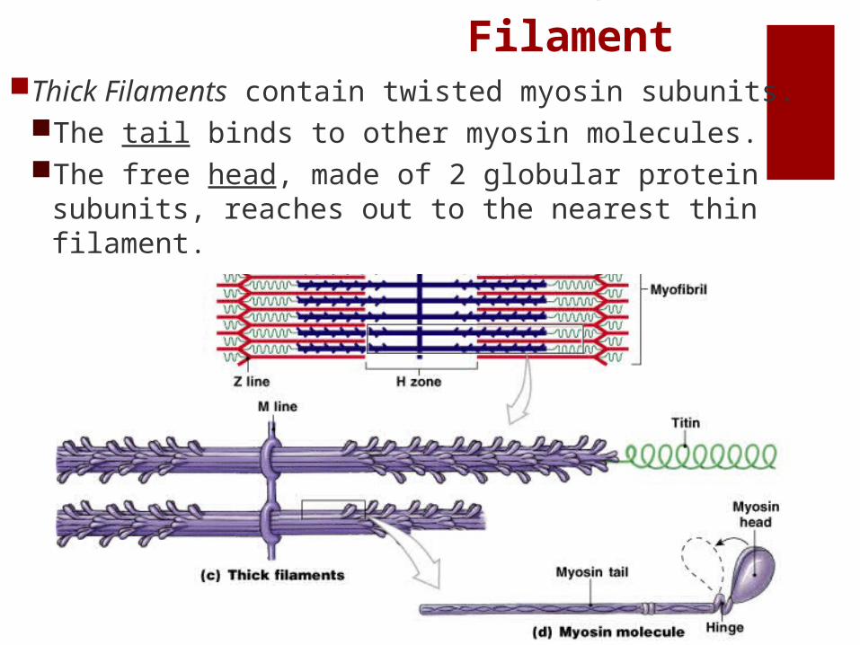
Thick Filament
Thick Filaments contain twisted myosin subunits. The tail binds to other myosin molecules. The free head, made of 2 globular protein subunits,
reaches out to the nearest thin filament.
Thick Filament
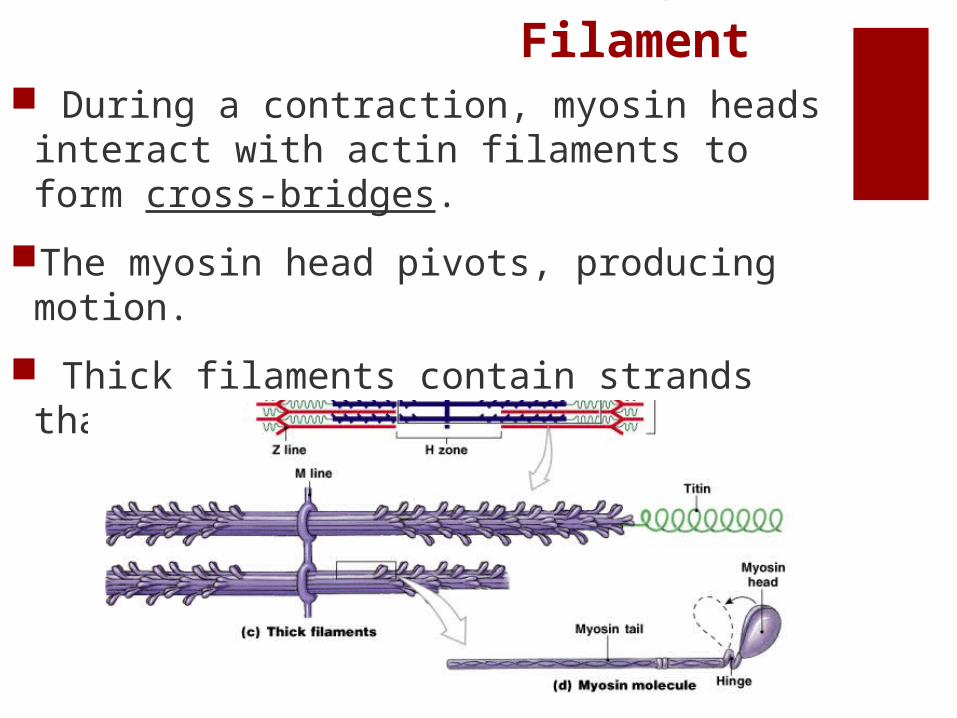
During a contraction, myosin heads interact with actin filaments to form cross-bridges.
The myosin head pivots, producing motion.
Thick filaments contain strands that recoil after stretching.
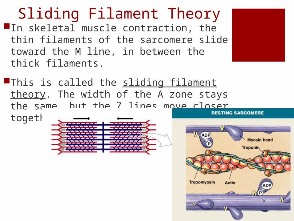
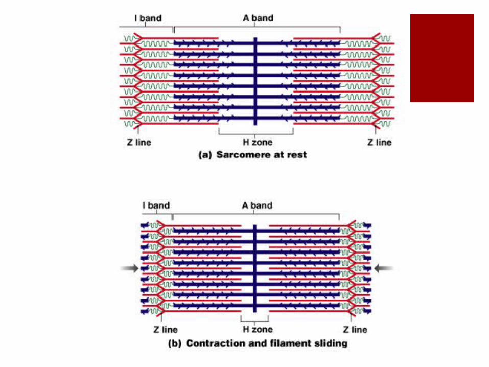
Sliding Filament TheoryIn skeletal muscle contraction, the thin
filaments of the sarcomere slide toward the M line, in between the thick filaments.
This is called the sliding filament theory. The width of the A zone stays the same, but the Z lines move closer together.
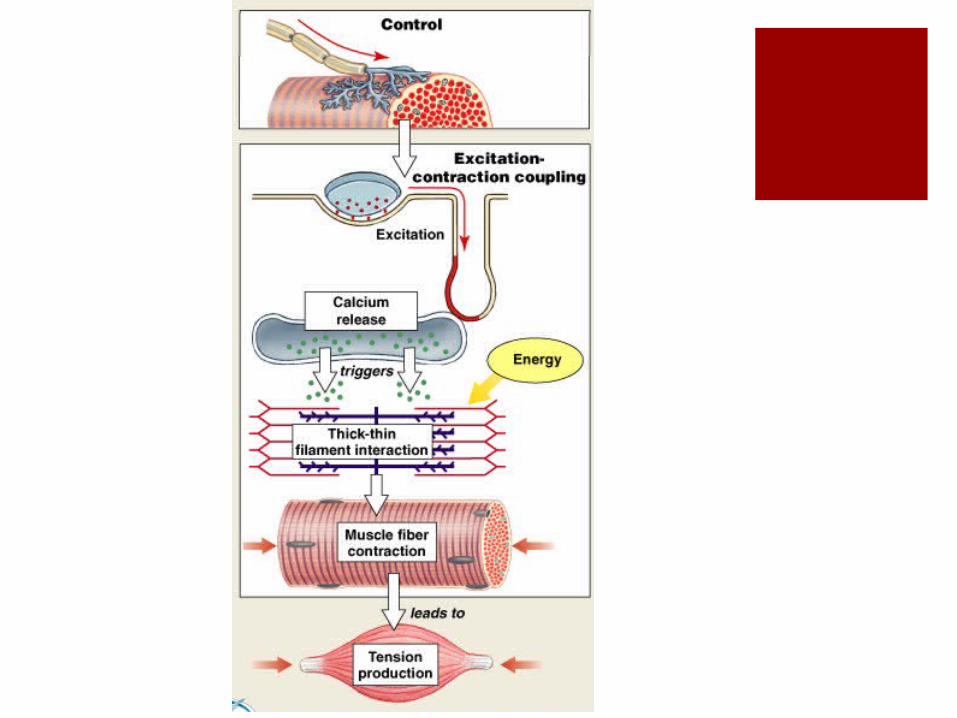
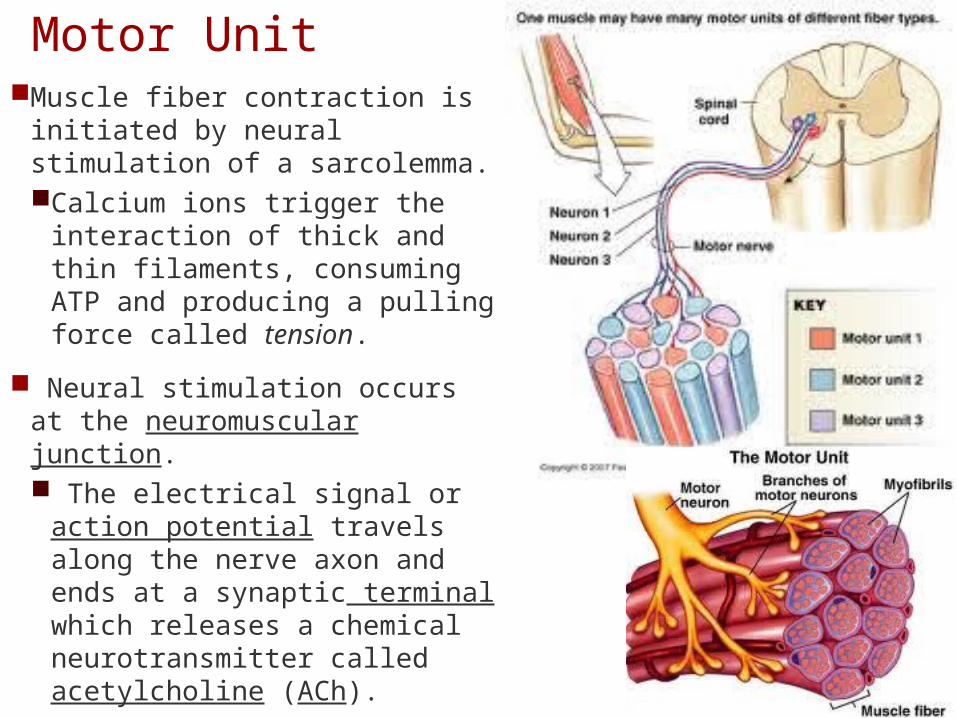
Motor UnitMuscle fiber contraction is
initiated by neural stimulation of a sarcolemma. Calcium ions trigger the
interaction of thick and thin filaments, consuming ATP and producing a pulling force called tension.
Neural stimulation occurs at the neuromuscular junction. The electrical signal or
action potential travels along the nerve axon and ends at a synaptic terminal which releases a chemical neurotransmitter called acetylcholine (ACh).
Action PotentialACh travels across a short gap and binds
to membrane receptors on the sarcolemma, causing sodium ions to rush into the sarcoplasm.
The increase in sodium ions generates an action potential in the sarcolemma which travels along the T tubules.
When the action potential reaches a triad, calcium ions are released, triggering contraction.
This step requires the myosin heads to have previously broken down ATP and stored the potential energy in the “cocked” position.
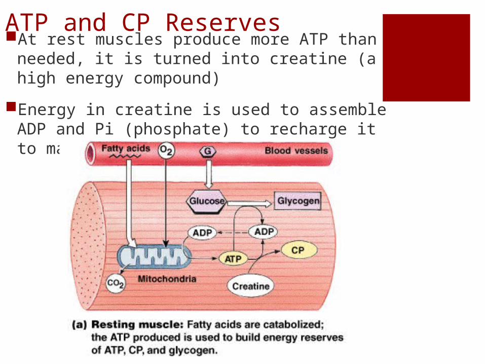
ATP and CP ReservesAt rest muscles produce more ATP than
needed, it is turned into creatine (a high energy compound)
Energy in creatine is used to assemble ADP and Pi (phosphate) to recharge it to make ATP.
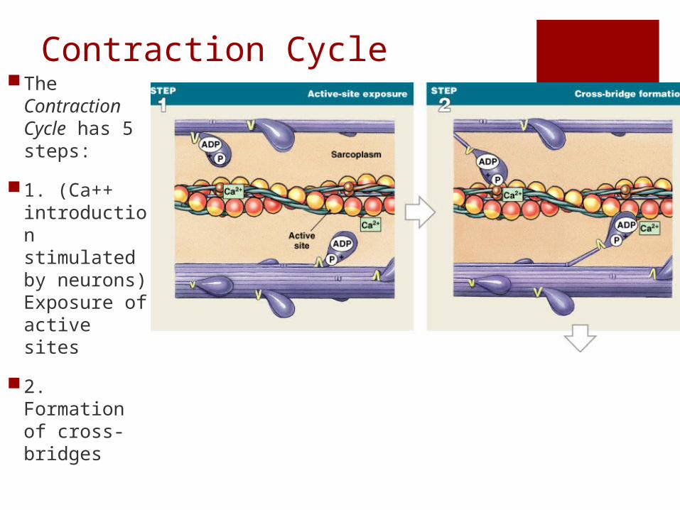
Contraction CycleThe
Contraction Cycle has 5 steps:
1. (Ca++ introduction stimulated by neurons) Exposure of active sites
2. Formation of cross-bridges
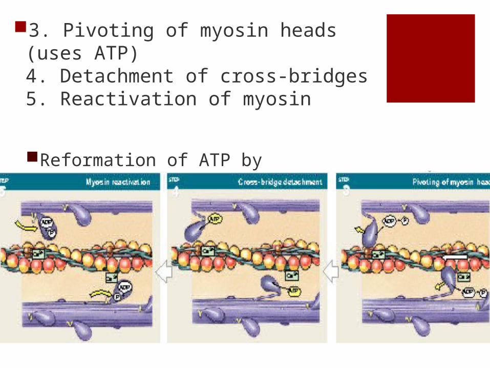
3. Pivoting of myosin heads (uses ATP)4. Detachment of cross-bridges 5. Reactivation of myosin
Reformation of ATP by reassembling ADP with Creatine Phosphate

Aerobic vs AnaerobicAerobic
metabolism uses O2 and provides 95% of ATP for a resting cell the primary
energy source of resting muscles
34 ATP molecules produced per glucose molecule
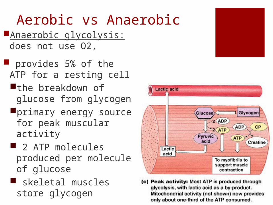
Aerobic vs AnaerobicAnaerobic glycolysis: does
not use O2,
provides 5% of the ATP for a resting cellthe breakdown of glucose
from glycogenprimary energy source
for peak muscular activity
2 ATP molecules produced per molecule of glucose
skeletal muscles store glycogen
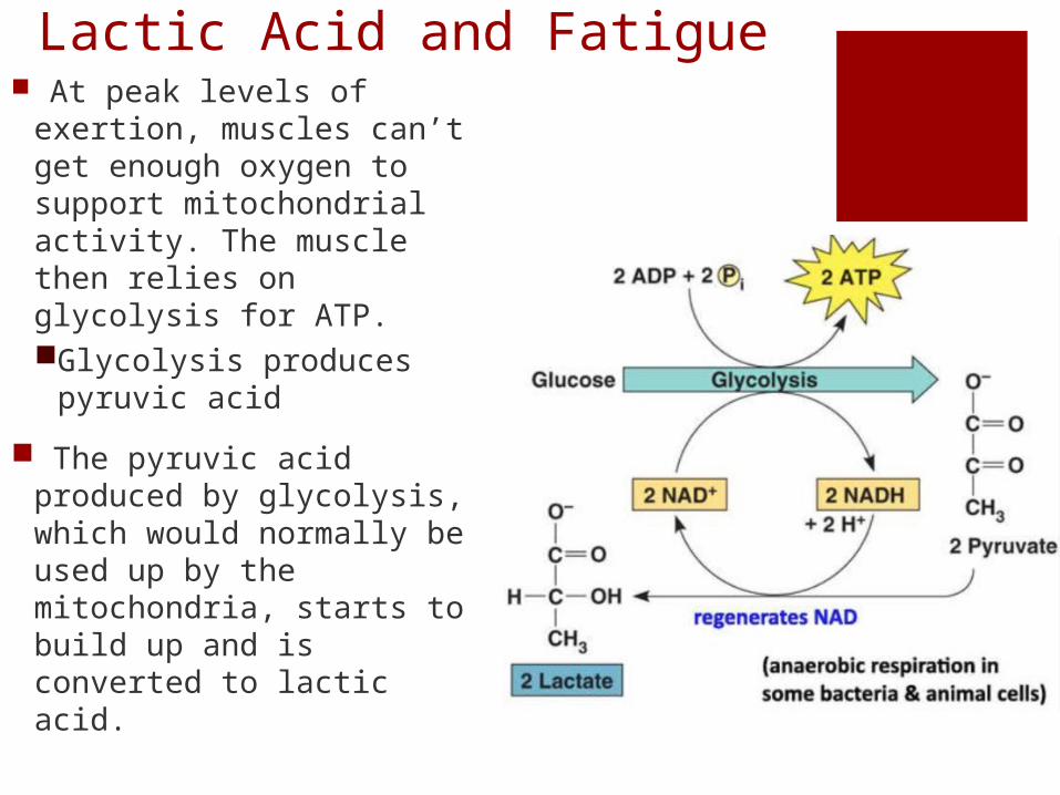
Lactic Acid and Fatigue At peak levels of
exertion, muscles can’t get enough oxygen to support mitochondrial activity. The muscle then relies on glycolysis for ATP.Glycolysis produces
pyruvic acid
The pyruvic acid produced by glycolysis, which would normally be used up by the mitochondria, starts to build up and is converted to lactic acid.
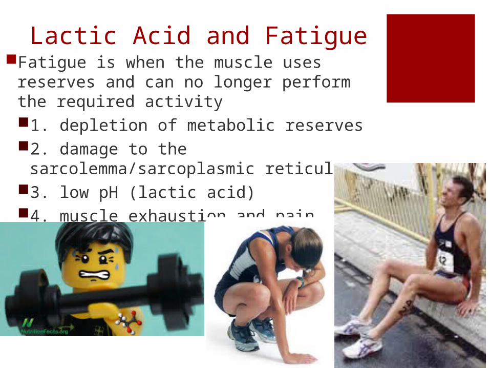
Lactic Acid and FatigueFatigue is when the muscle uses reserves
and can no longer perform the required activity1. depletion of metabolic reserves2. damage to the
sarcolemma/sarcoplasmic reticulum3. low pH (lactic acid)4. muscle exhaustion and pain
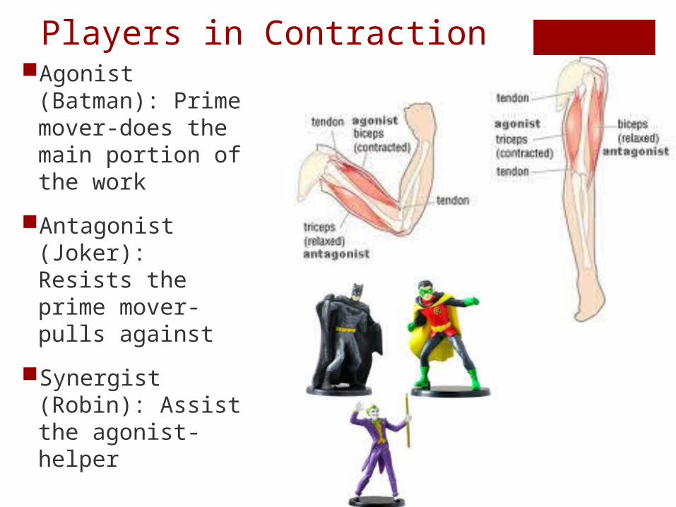
Players in ContractionAgonist (Batman):
Prime mover-does the main portion of the work
Antagonist (Joker): Resists the prime mover-pulls against
Synergist (Robin): Assist the agonist-helper
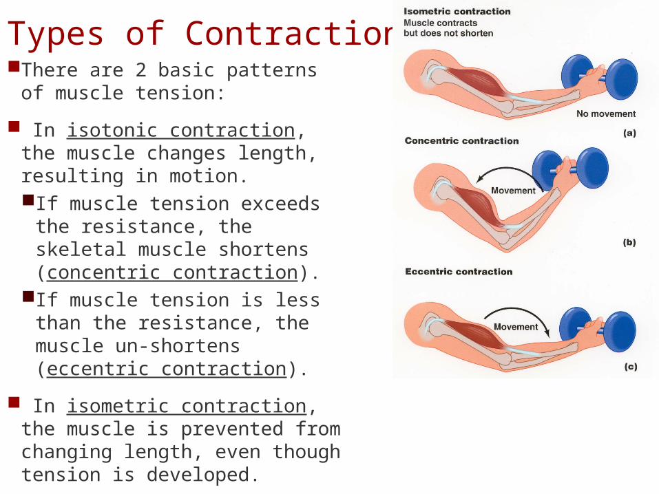
Types of ContractionsThere are 2 basic patterns of
muscle tension:
In isotonic contraction, the muscle changes length, resulting in motion. If muscle tension exceeds the
resistance, the skeletal muscle shortens (concentric contraction).
If muscle tension is less than the resistance, the muscle un-shortens (eccentric contraction).
In isometric contraction, the muscle is prevented from changing length, even though tension is developed.
Recovery Period
After high levels of exertion, it can take hours or days for muscles to return to their normal condition.
During the recovery period, oxygen is once again available and mitochondrial activity resumes. Cori cycle: Lactic acid is carried by the blood stream to
the liver, where it is converted back into pyruvic acid, and glucose is released to recharge the muscles’ glycogen reserves.
To process excess lactic acid and normalize metabolic activities after exercise, the body uses more oxygen than usual. This elevated need for oxygen, called the oxygen debt, is responsible for heavy breathing after exercise.
Skeletal muscles at rest metabolize fatty acids and store glycogen.
During light activity, muscles can generate ATP through the anaerobic breakdown of carbohydrates, lipids or amino acids.
At peak levels of activity, most of the energy is provided by anaerobic reactions that generate lactic acid as a byproduct.
Heat Production and Loss: The more active muscles are, the more heat they produce. During strenuous exercise, up to 70 percent of the energy produced can be lost as heat, raising body temperature.
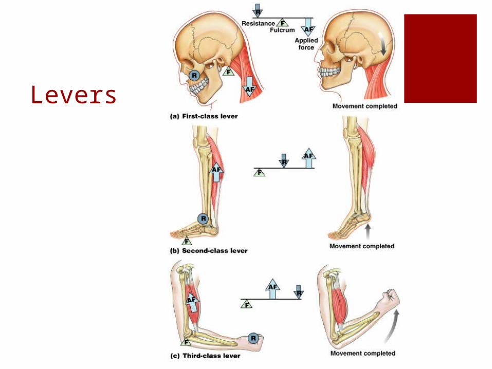
Levers
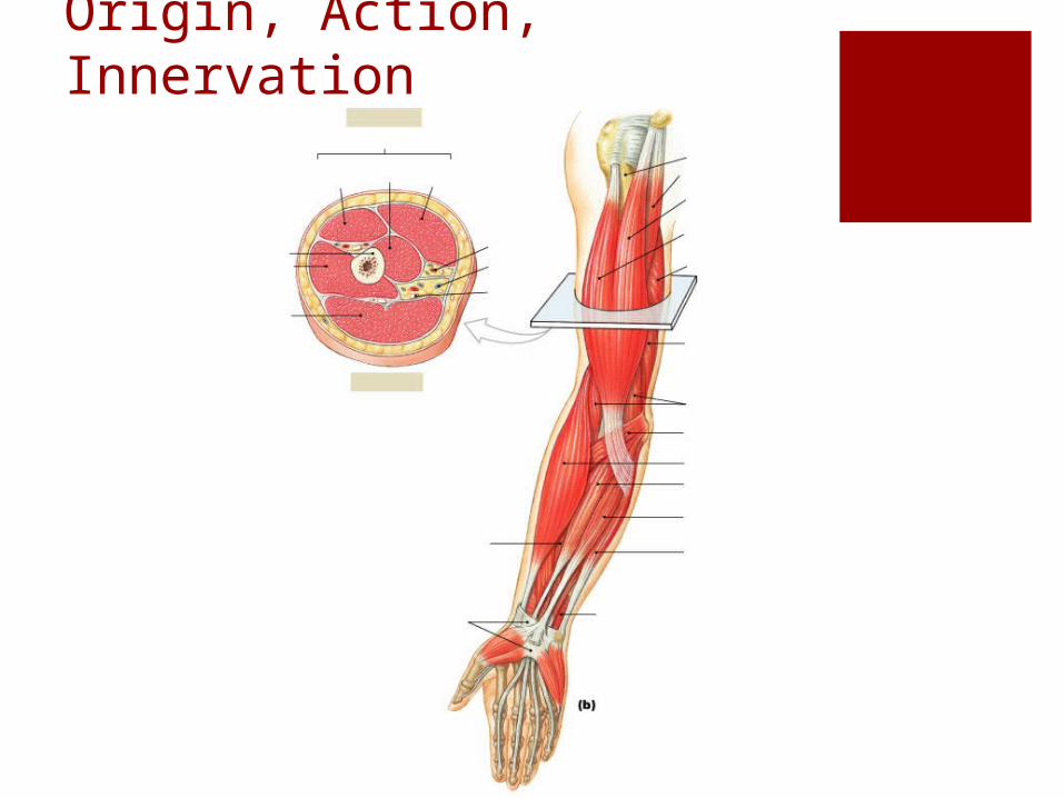
Origin, Action, Innervation
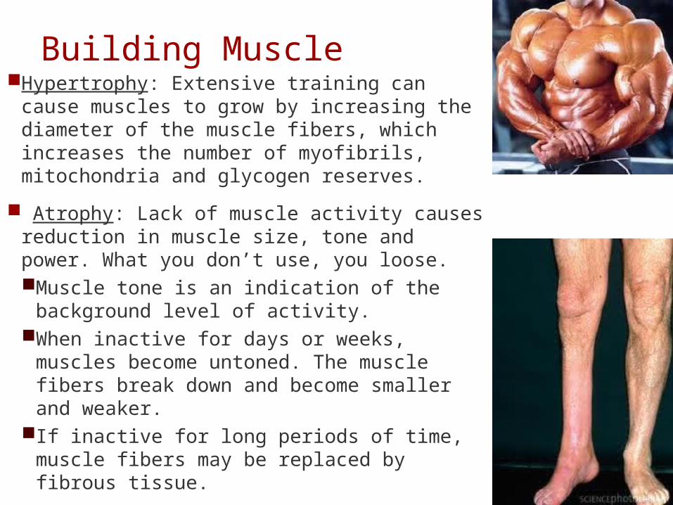
Building MuscleHypertrophy: Extensive training can cause
muscles to grow by increasing the diameter of the muscle fibers, which increases the number of myofibrils, mitochondria and glycogen reserves.
Atrophy: Lack of muscle activity causes reduction in muscle size, tone and power. What you don’t use, you loose. Muscle tone is an indication of the
background level of activity. When inactive for days or weeks, muscles
become untoned. The muscle fibers break down and become smaller and weaker.
If inactive for long periods of time, muscle fibers may be replaced by fibrous tissue.