Skills evaluation in minimally invasive surgery using force/torquesignatures
C. Richards,1 J. Rosen,2 B. Hannaford,2 C. Pellegrini,1 M. Sinanan1
1 Department of Surgery, University of Washington, Box 356410, Seattle, WA 98195, USA2 Department of Electrical Engineering, University of Washington, Box 352500, Seattle, WA 98195, USA
Received: 4 May 1999/Accepted: 1 April 2000/Online publication: 4 August 2000
AbstractBackground:One of the more difficult tasks in surgicaleducation is to teach the optimal application of instrumentforces and torques necessary to facilitate the conduct of anoperation. For laparoscopic surgery, this type of training hastraditionally taken place in the operating room, reducingoperating room efficiency and potentially affecting the safeconduct of the operation. The objective of the current studywas to measure and compare forces and torques (F/T) ap-plied at the tool/hand interface generated during laparoscop-ic surgery by novice (NS) and experienced (ES) surgeonsusing an instrumented laparoscopic grasper and to use thisdata for evaluating the skill level.Methods:Ten surgeons (five-NS, five-ES) performed a cho-lecystectomy and Nissen fundoplication in a porcine model.An instrumented laparoscopic grasper with interchangeablestandard surgical tips equipped with a three-axis F/T sensorlocated at the proximal end of the grasper tube was used tomeasure the F/T at the hand/tool interface. In addition, oneaxis force sensor located at the grasper’s handle was used tomeasure the grasping force. F/T data synchronized withvisual view of the tool operative maneuvers were collectedsimultaneously via a novel graphic user interface incorpo-rated picture-in-picture video technology. Subsequentframe-by-frame video analysis of the operation allowed adefinition of states associated with different tool/tissue in-teractions within each step of the operation. F/T measuredwithin each state were further analyzed using vector quan-tization (VQ). The VQ analysis defines characteristic sets ofF/T in the database that were defined as F/T signature.Results:The magnitude of F/T applied by NS and ES weresignificantly different (p < 0.05) and varied based on thetask being performed. Higher F/T magnitudes were appliedby NS than by ES when performing tissue manipulation,whereas lower F/T magnitudes were applied by NS than byES during tissue dissection. Furthermore, the time to com-plete the surgical procedure was longer for NS by a factor of
1.5–4.8 when compared to the time for ES. State analysissuggests that most of this time is consumed in an [idle] state,in which movements of the surgeon make no tissue contact.Conclusions:Preliminary data suggest that F/T magnitudesassociated with the tool/tissue interactions provide an ob-jective means of distinguishing novices from skilled sur-geons. Clinical F/T analysis using the proposed technologyand methodology may be helpful in training, developingsurgical simulators, and measuring technical proficiencyduring laparoscopic surgery.
Key words: Laparoscopy — Training — Technical Skills— Force-torque signatures — Haptics — Surgical simula-tors — Minimally invasive surgery
One of the most difficult tasks in surgical education is toteach the optimal application of instrument handling neces-sary to conduct an operation. This is especially problematicin the field of minimally invasive surgery (MIS), where theteacher is one step removed from actual tissue contact. Forlaparoscopic surgery, this type of training has traditionallytaken place in the operating room, thereby reducing oper-ating room efficiency and potentially affecting the safe con-duct of the operation.
The use of virtual reality models for teaching these com-plex surgical skills has been a long-term goal of numerousinvestigators [5, 6, 9]. The development of such a systemwould provide a less stressful learning environment for thesurgical novice while eliminating risk to the patient, but itrequires an understanding of the various components thatcomprise a realistic and useful training system [7]. Althoughother studies have focused on the tool-tip/tissue interactionand deformation [1, 3, 4, 8] this research measured theforces and torques (F/T) applied at the surgeon’s hand/toolinterface while performing minimally invasive surgery(MIS).
The objective of this study was to measure the F/T,Correspondence to:M. Sinanan
identifying F/T signatures and analyzing how their signa-tures are distributed while performing laparoscopic surgeryby novice (NS) and experienced (ES) surgeons using aninstrumented endoscopic grasper. F/T signatures are a typi-cal set of force and torque components associated with dif-ferent tool-tip/tissue interactions that characterize clustercenters in a multidimensional database.
Statistical models of the quantitative knowledge gainedin this study can be used to characterize surgical skills fortraining novice surgeons in performing laparoscopic proce-dures. Two areas in which the F/T signature database mightbe used are (a) virtual reality (VR) (developing haptic de-vices for realistic force feedback VR simulations of MISprocedures), and (b) minimally invasive surgical robotics(optimizing mechanisms and actuators).
Materials and methods
Subjects and protocol
Ten surgeons (five NS and five ES) performed a laparoscopic cholecys-tectomy and laparoscopic Nissen fundoplication in a porcine model. Pro-tocols for anesthetic management, euthanasia, and survival procedureswere reviewed and approved by the Animal Care Committee of the Uni-versity of Washington and the Animal Use Review Division of the U.S.Army Veterinary Corps.
Each operation was subdivided into several steps (Table 1). Althoughall the steps were performed in each procedure, data were recorded only
when the instrumented endoscopic tool was used with the following tooltips: atraumatic grasper, curved dissector, or Babcock grasper.
Experimental system setup
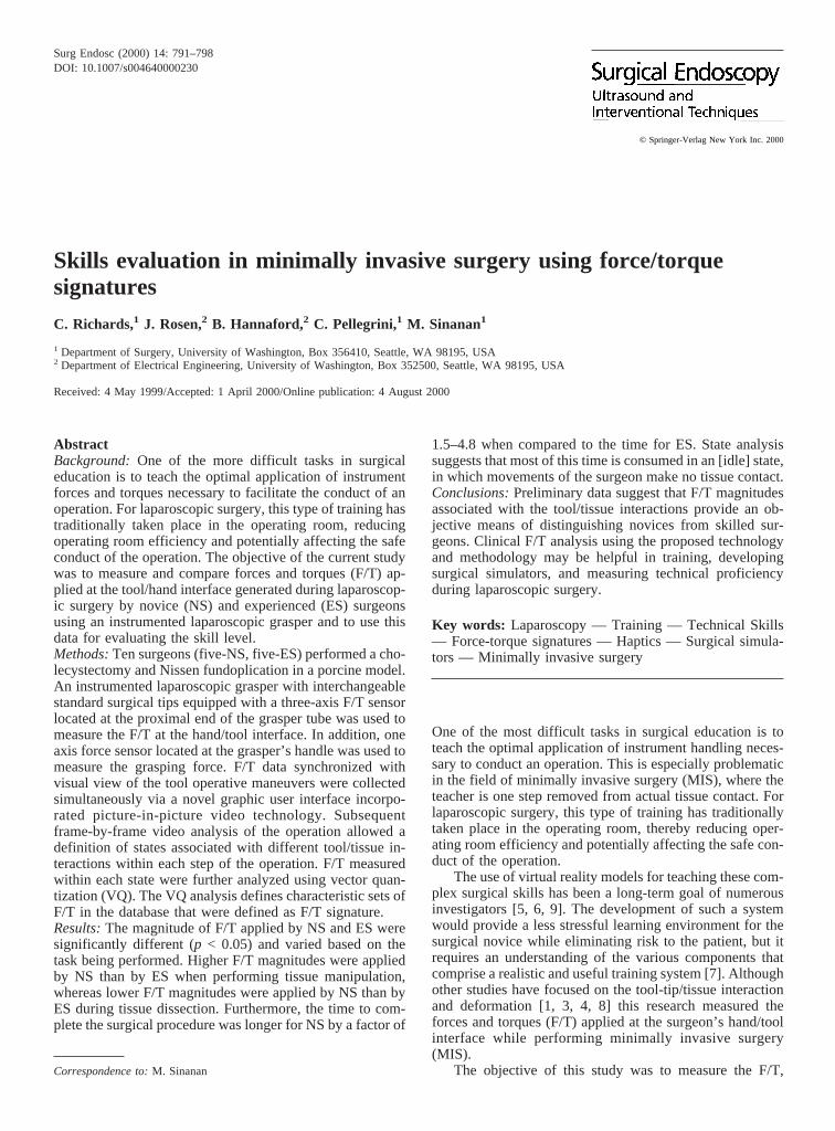
Two types of information were acquired while performing laparoscopicprocedure: (a) F/T data measured at the surgeons’ hand/tool interface, and(b) visual information about the tool tip’s interaction with the tissues. Thetwo sources of information were synchronized in time and recorded simul-taneously for subsequent analysis using picture-in-picture video technol-ogy.
The F/T at the interface between the surgeon’s hand and the endoscopicgrasper handle were measured by two sensors. The first sensor was athree-axis F/T sensor (ATI mini-model), which was mounted into the outertube (proximal end) of a standard reusable 10-mm endoscopic grasper(Karl Storz) (Fig. 1A). The sensor was capable of simultaneous measure-ments of three components of force (Fx ,Fy ,Fz) and three components oftorque (Tx ,Ty ,Tz) in a Cartesian frame (Fig. 1B). The sum of the appliedforces and torques at the tool-tip/tissue interface and the tool/trocar inter-face were transferred through the grasper structure to the surgeon’s hand,as occurs in a normal instrument, and vice versa. The sensor orientationwas such that the x and z axes formed a plane parallel to the tool’s internaljaw contact surface with the jaw closed, and the y and z axes defined aplane perpendicular to that surface (Fig. 1B). A second force sensor wasmounted to the endoscopic grasper handle to permit the measurement ofgrasping force (Fg) applied by the surgeon on the instrument.
The grasper had a reticulating feature that enabled the surgeon tochange the orientation of the tool tip relative to the grasped tissue withoutchanging the handle orientation. The alignment between tool tip originrelative to the sensor remained unchanged since the outer tube and the tooltip are linked mechanically.
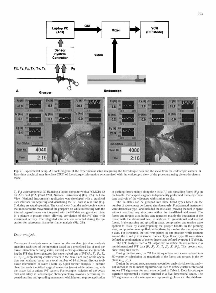
The F/T data were integrated with the laparoscopic camera view ofinstrument activity (Fig. 2). The seven channels F/T data (Fx, Fy, Fz, Tx, Ty,
Fig. 1. The instrumentedendoscopic grasper.A The grasperwith the three-axis force/torquesensor implemented on the outertube and a force sensor located onthe instrument handle.B The tooltip andx,y,zframe aligned with thethree-axis force/torque sensor.
Table 1.Definitions of surgical procedure steps and type of tool tip used (bold steps performed but not recorded)
Procedure Step Description Tool type HandVideoF/T
Laparoscopic LC-1 Positioning gallbladder Atraumatic grasper L +Cholecystectomy LC-2 Exposure of cyctic duct Curved dissector R +
LC-2** Divide of cyctic duct Scissors R –LC-3 Dissection of Gallbladder fossae Curved dissector R +LC-4 Exposure of cyctic artery Curved dissector R –LC-4* Dividing artery Scissors R –
Laparoscopic LNF-1 Dissecting right crus Surgiwand R –Nissen LNF-2 Dissecting left crus Surgiwand R –Fundoplication LNF-3 Dissecting esophagus/blunt Curved dissector R +
LNF-4 Placing a wrap around the esophagus Babcock grasper R +LNF-5 Suture wrap/intracorporeal knot
tying with needle holderCurved dissector R +
LNF-6 Coronal sutures/intracorporeal knot tying Endostitch R –
792
Tz, Fg) were sampled at 30 Hz using a laptop computer with a PCMCIA 12bit A/D card (DAQCard 1200, National Instruments) (Fig. 2A). A Lab-View (National Instruments) application was developed with a graphicaluser interface for acquiring and visualizing the F/T data in real time (Fig.2) during an actual operation. The visual view from the endoscopic camerathat monitored the movement of the grasper’s tip while interacting with theinternal organs/tissues was integrated with the F/T data using a video mixerin a picture-in-picture mode, allowing correlation of the F/T data withinstrument activity. The integrated interface was recorded during the op-eration for subsequent frame-by-frame analysis (Fig. 2B).
Data analysis
Two types of analysis were performed on the raw data: (a) video analysisencoding each step of the operation based on a predefined list of tool-tip/tissue interaction defining states, and (b) vector quantization (VQ) encod-ing the F/T data into signatures that were typical sets of F/T (Fx, Fy, Fz, Tx,Ty, Tz, Fg) representing cluster centers in the data. Each step of the opera-tion was analyzed based on a total number of 14 different discrete tool-tissue interactions or states (Table 2). Upon further analysis, it becameclear that each identified surgical maneuver (state) while interacting withthe tissue had a unique F/T pattern. For example, isolation of the cysticduct and artery in laparoscopic cholecystectomy involves performing re-peated pushing and spreading maneuvers, which in turn require application
of pushing forces mainly along the z axis (Fz) and spreading forces (Fg) onthe handle. Two expert surgeons independently performed frame-by-framestate analysis of the videotape with similar results.
The 14 states can be grouped into three broad types based on thenumber of movements performed simultaneously. Fundamental maneuverswere defined as type I and included the idle state (moving the tool in spacewithout touching any structures within the insufflated abdomen). Theforces and torques used in this state represent mainly the interaction of thetrocar with the abdominal wall in addition to gravitational and inertialforces. In the grasping and spreading states, compression and tension wereapplied to tissue by closing/opening the grasper handle. In the pushingstate, compression was applied on the tissue by moving the tool along thez axis. For sweeping, the tool was placed in one position while rotatingaround the x and y axes (trocar frame). Type II and type III were statesdefined as combinations of two or three states defined by group I (Table 2).
The F/T analysis used a VQ algorithm to define cluster centers in amultidimensional F/T data (Fx ,Fy ,Fz ,Tx ,Ty ,Tz ,Fg). This process wasdone using four steps.
During the first step, the 7D force/torque data vector was reduced to a5D vector by calculating the magnitude of the forces and torques in the xyplane (Fxy ,Txy).
During the second step, a pattern recognition analysis (clustering analy-sis) known as the K-means algorithm was used to define cluster centers andknown F/T signatures for each state defined in Table 2. Each force/torquesignature represented a cluster centered in a five-dimensional space. TheF/T signatures are discrete symbols representing clusters in the database.
Fig. 2. Experimental setup.A Block diagram of the experimental setup integrating the force/torque data and the view from the endoscopic camera.BReal-time graphical user interface (GUI) of force/torque information synchronized with the endoscopic view of the procedure using picture-in-picturemode.
793
During the third step, the entire database was encoded according to thepredefined signatures so that in each time interval the F/T were associatedwith one of the signatures that were predefined based on minimizing theEuclidean distance between them. As a result, a one-dimensional list ofsymbols was generated, replacing the seven-dimensional vector. Each sym-bol represented a F/T magnitude associated with one of the tool/tissueinteraction.
In final step, a statistical analysis of the F/T signatures magnitudedistribution was performed. In order to perform the statistical analysis, theentire F/T database was lumped into two groups—the NS group and the ESgroup. The distribution of the F/T signatures for each tool/tissue interactionwas then calculated for the NS and ES groups performing the differentsteps of the MIS procedures. The distributions of the F/T signatures appliedby the NS and the ES were tested to identified tool/tissue interactions inwhich the F/T magnitude distributions of the two groups were significantlydifferent using the median test (nonparametric method) combined with thefourfold point statistical procedure.
Using the human language as an analogy, the VQ was preformed toidentify the basic “words” of the MIS “language” for creating a “dictio-nary.” Just as a single word is pronounced differently by different people,the same tool/tissue interaction is performed differently by different sur-geons, yet they all share the same meaning, or outcome, as in the realm ofsurgery. The VQ was used to identify the typical F/T associated with eachone of the tool/tissue interactions in the surgery “dictionary.” Or, using thelanguage analogy, it characterized different pronunciations of a word.
Results
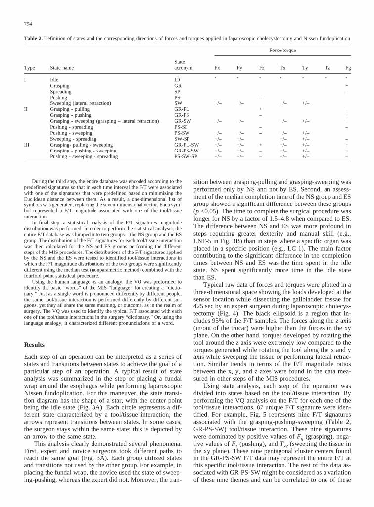
Each step of an operation can be interpreted as a series ofstates and transitions between states to achieve the goal of aparticular step of an operation. A typical result of stateanalysis was summarized in the step of placing a fundalwrap around the esophagus while performing laparoscopicNissen fundoplication. For this maneuver, the state transi-tion diagram has the shape of a star, with the center pointbeing the idle state (Fig. 3A). Each circle represents a dif-ferent state characterized by a tool/tissue interaction; thearrows represent transitions between states. In some cases,the surgeon stays within the same state; this is depicted byan arrow to the same state.
This analysis clearly demonstrated several phenomena.First, expert and novice surgeons took different paths toreach the same goal (Fig. 3A). Each group utilized statesand transitions not used by the other group. For example, inplacing the fundal wrap, the novice used the state of sweep-ing-pushing, whereas the expert did not. Moreover, the tran-
sition between grasping-pulling and grasping-sweeping wasperformed only by NS and not by ES. Second, an assess-ment of the median completion time of the NS group and ESgroup showed a significant difference between these groups(p <0.05). The time to complete the surgical procedure waslonger for NS by a factor of 1.5–4.8 when compared to ES.The difference between NS and ES was more profound insteps requiring greater dexterity and manual skill (e.g.,LNF-5 in Fig. 3B) than in steps where a specific organ wasplaced in a specific position (e.g., LC-1). The main factorcontributing to the significant difference in the completiontimes between NS and ES was the time spent in the idlestate. NS spent significantly more time in the idle statethan ES.
Typical raw data of forces and torques were plotted in athree-dimensional space showing the loads developed at thesensor location while dissecting the gallbladder fossae for425 sec by an expert surgeon during laparoscopic cholecys-tectomy (Fig. 4). The black ellipsoid is a region that in-cludes 95% of the F/T samples. The forces along the z axis(in/out of the trocar) were higher than the forces in the xyplane. On the other hand, torques developed by rotating thetool around the z axis were extremely low compared to thetorques generated while rotating the tool along the x and yaxis while sweeping the tissue or performing lateral retrac-tion. Similar trends in terms of the F/T magnitude ratiosbetween the x, y, and z axes were found in the data mea-sured in other steps of the MIS procedures.
Using state analysis, each step of the operation wasdivided into states based on the tool/tissue interaction. Byperforming the VQ analysis on the F/T for each one of thetool/tissue interactions, 87 unique F/T signature were iden-tified. For example, Fig. 5 represents nine F/T signaturesassociated with the grasping-pushing-sweeping (Table 2,GR-PS-SW) tool/tissue interaction. These nine signatureswere dominated by positive values ofFg (grasping), nega-tive values ofFz (pushing), andTxy (sweeping the tissue inthe xy plane). These nine pentagonal cluster centers foundin the GR-PS-SW F/T data may represent the entire F/T atthis specific tool/tissue interaction. The rest of the data as-sociated with GR-PS-SW might be considered as a variationof these nine themes and can be correlated to one of these
Table 2.Definition of states and the corresponding directions of forces and torques applied in laparoscopic cholecystectomy and Nissen fundoplication
Fig. 3. State analysis of an MISprocedure.A State diagram of placingwrap around the esophagus duringlaparoscopic Nissen fundoplicationshowing differences between novice(dashed line) and expert (solid line)surgeons. The double lines representstates and transitions made by bothgroups.B Time spent at eachtool/tissue interaction by novicesurgeon (NS) and expert surgeon (ES)while performing suture wrap andintracorporeal knot tying with needleholder. The time distribution in eachtool/tissue interaction of the 10subjects is represented by a notch boxplot. The lower and upper lines of thebox are the 25th and 75th percentilesof the sample representing theinterquartile range. The line in themiddle of the box is the samplemedian. The notches in the boxdepicts the 95% confidence intervalabout the median of the sample. Thelines extending above and below thebox define the 95 percentiles of thesampled data.
795
signatures. Moreover, further analysis of this code book (87F/T signatures) showed no overlap between signatures.There was at least one dimension (of the five) that differ-entiated each signature from the others.
Once the code book was defined, the entire database wasencoded into the 87 F/T signatures. This encoding processallowed exploration of a new aspect regarding the differ-ences between NS and ES. This new aspect was related tothe magnitudes of F/T applied by NS and ES during eachstep of the MIS procedures for the different tool/tissue in-teractions (Table 2). The grand median analysis [2] showedthat the F/T magnitudes applied by NS and ES in most of
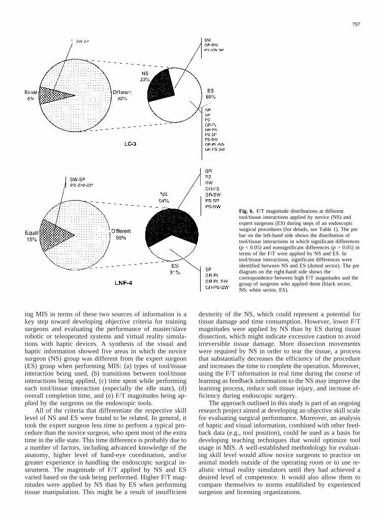
the tool/tissue interactions were significantly different (p<0.05) (Fig. 6). Two steps of two surgical procedures wererepresented in Fig. 6 by two pie diagrams. The pie diagramon the left-hand side defines the tool/tissue interactions inwhich no significant difference was found (gray sector) be-tween NS and ES and cases where a significant differencewas observed (dotted sector). When a significant differencewas identified, the pie diagram on the right hand sideshowed which group (NS, black sector; or ES, white sector)applied higher F/T in each one of the tool/tissue interac-tions. For example, in laparoscopic cholecystectomy step 3(Fig. 6, LC-3), no significant difference in the F/T magni-tude was identified in SW-SP, which is 8% of all the tool/tissue interactions. In the other 92% of tool/tissue interac-tions, a significant difference was identified (left-hand piechart). Of the cases where a significant difference was ob-served, in 23% of the tool/tissue interactions (e.g., SW,GR-SW, PS-SW-SP) NS applied higher F/T magnitudesthan ES; and in 69% of the cases (e.g., GR, SP, PS, GR-PLetc), ES applied higher F/T magnitudes than NS (right-handpie chart) Fig. 6.
In general, a significant difference was identified inmost of the tool/tissue interactions. Moreover, when the sixsurgical steps were divided according to the nature of thetool/tissue interactions—e.g., (a) tissue dissection (LC-2,LC-3, LNF-3), and (b) tissue manipulation (LC-1, LNF-4)—the results showed that higher magnitudes of F/T wereapplied by ES than by NS when dissecting the tissues,whereas lower magnitudes of F/T were applied by ES thanby NS when manipulating the tissues.
Discussion
Minimally invasive surgery is a complex task that requiresa synthesis between visual and haptic information. Analyz-
Fig. 4. Raw data of forces and torques measured at the surgeon’s hand/tool interface while dissecting the gallbladder fossae during laparoscopiccholecystectomy. For the definitions of thex,y,zdirections, see Fig. 1B—A forces andB torques.
Fig. 5. Force/torque signatures of the grasping- pushing-swiping state.
796
ing MIS in terms of these two sources of information is akey step toward developing objective criteria for trainingsurgeons and evaluating the performance of master/slaverobotic or teleoperated systems and virtual reality simula-tions with haptic devices. A synthesis of the visual andhaptic information showed five areas in which the novicesurgeon (NS) group was different from the expert surgeon(ES) group when performing MIS: (a) types of tool/tissueinteraction being used, (b) transitions between tool/tissueinteractions being applied, (c) time spent while performingeach tool/tissue interaction (especially the idle state), (d)overall completion time, and (e) F/T magnitudes being ap-plied by the surgeons on the endoscopic tools.
All of the criteria that differentiate the respective skilllevel of NS and ES were found to be related. In general, ittook the expert surgeon less time to perform a typical pro-cedure than the novice surgeon, who spent most of the extratime in the idle state. This time difference is probably due toa number of factors, including advanced knowledge of theanatomy, higher level of hand-eye coordination, and/orgreater experience in handling the endoscopic surgical in-strument. The magnitude of F/T applied by NS and ESvaried based on the task being performed. Higher F/T mag-nitudes were applied by NS than by ES when performingtissue manipulation. This might be a result of insufficient
dexterity of the NS, which could represent a potential fortissue damage and time consumption. However, lower F/Tmagnitudes were applied by NS than by ES during tissuedissection, which might indicate excessive caution to avoidirreversible tissue damage. More dissection movementswere required by NS in order to tear the tissue, a processthat substantially decreases the efficiency of the procedureand increases the time to complete the operation. Moreover,using the F/T information in real time during the course oflearning as feedback information to the NS may improve thelearning process, reduce soft tissue injury, and increase ef-ficiency during endoscopic surgery.
The approach outlined in this study is part of an ongoingresearch project aimed at developing an objective skill scalefor evaluating surgical performance. Moreover, an analysisof haptic and visual information, combined with other feed-back data (e.g., tool position), could be used as a basis fordeveloping teaching techniques that would optimize toolusage in MIS. A well-established methodology for evaluat-ing skill level would allow novice surgeons to practice onanimal models outside of the operating room or to use re-alistic virtual reality simulators until they had achieved adesired level of competence. It would also allow them tocompare themselves to norms established by experiencedsurgeons and licensing organizations.
Fig. 6. F/T magnitude distributions at differenttool/tissue interactions applied by novice (NS) andexpert surgeons (ES) during steps of an endoscopicsurgical procedures (for details, see Table 1). The piebar on the left-hand side shows the distribution oftool/tissue interactions in which significant differences(p < 0.05) and nonsignificant differences (p > 0.05) interms of the F/T were applied by NS and ES. Intool/tissue interactions, significant differences wereidentified between NS and ES (dotted sector). The piediagram on the right-hand side shows thecorrespondence between high F/T magnitudes and thegroup of surgeons who applied them (black sector,NS; white sector, ES).
797
Acknowledgments.This work was supported by the U.S. Army MedicalResearch and Material Command under DARPA grant DMAD17-97-1-725and by a major grant from U.S. Surgical Corp., a division of Tyco, Inc., tothe University of Washington’s Center for Videoendoscopic Surgery.
References
1. Bicchi A, et al. (1996) A sensorized minimally invasive surgery tool fordetecting tissue elastic properties. Proceedings of the 1996 IEEE In-ternational Conference on Robotics and Automation. pp 884–888
2. Frank H, Althoen SC (1994) Statistics—concepts and application.Cambridge University Press
3. Hannaford B, Trujillo J, Sinanan M, Moreyra M, Rosen J, Brown J,Lueschke R, MacFarlane M (1998) Computerized endoscopic surgicalgrasper, In: Westwood DJ (ed) Proceedings, MMVR-98 (medicinemeets virtual reality). IOS Press, Amsterdam, pp 265–271
4. Morimoto A, Foral R, Kuhlman J, Zucker K, Curet M, Bocklage T,MacFarlane T, Kory L (1997) Force sensor for laparoscopic Babcock.In: Morgan K (ed) Proceedings MMVR-97 (medicine meets virtualreality) IOS Press, Amsterdam, pp 354–361
5. Noar M (1991) Endoscopy simulation: a brave new world? Endoscopy23: 147–149