34
SKIN CANCER WISNUBROTO, dr, SpB(K)Onk Surgical Oncology Consultant
| Date post: | 30-Oct-2014 |
| Category: |
Documents |
| Upload: | firdaus-achmad-jauhar |
| View: | 117 times |
| Download: | 5 times |
SKIN CANCER
WISNUBROTO, dr, SpB(K)OnkSurgical Oncology Consultant

Introduction
900,000 – 1,200,000 new cases per year in US
Non Melanoma skin cancer : High curability Rare metastasis Rare caused of death
Malignant Melanoma : More caused metastasis More caused of death

SKIN CANCER
Skin cancer
Non Melanoma
Malignant Melanoma

Non Melanoma
SQUAMUS CELL CARSINOMABASAL CELL CARSINOMA
INTRA EPITHELIAL Ca (BOWEN’S DISS)KERATOACANTHOMA
MARKEL CELL CaMYCOSIS FUNGOIDESKAPOSI’S SARKOMA
CUTANEUS METASTASIS
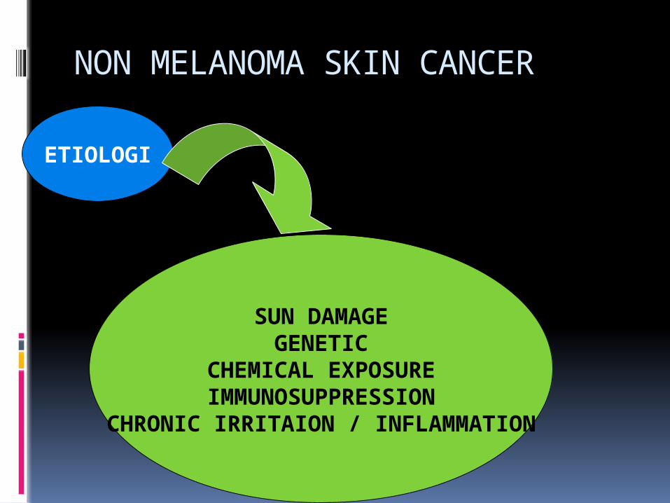
NON MELANOMA SKIN CANCER
ETIOLOGI
SUN DAMAGEGENETIC
CHEMICAL EXPOSUREIMMUNOSUPPRESSION
CHRONIC IRRITAION / INFLAMMATION

SUN DAMAGE Chronic exposure UV light ( 290-320
nm) < ozone Caused DNA damages in epidermis. Changed immune response in tumor. Cumulative exposure in advancing age
RISK FACTORS OF SKIN CANCER

GENETICMany syndrome caused skin cancer : Xeroderma pigmentosum defect DNA repair Basal cell nevus syndrome BCC Albinism melanin exposure UV Epidermodysplasia verruciformis SCC
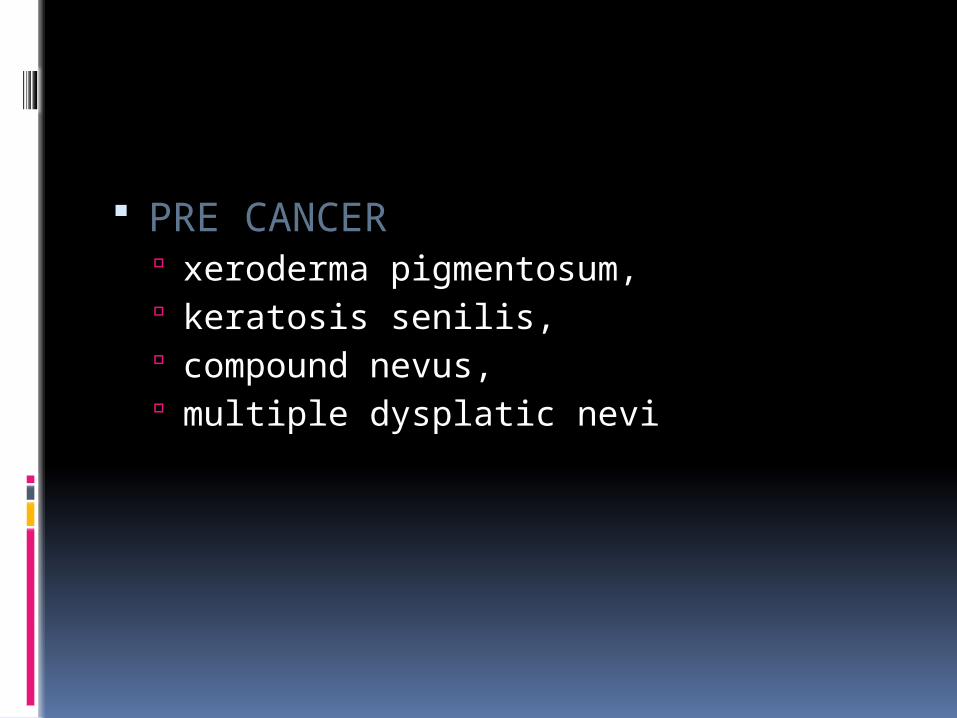
PRE CANCER xeroderma pigmentosum, keratosis senilis, compound nevus, multiple dysplatic nevi
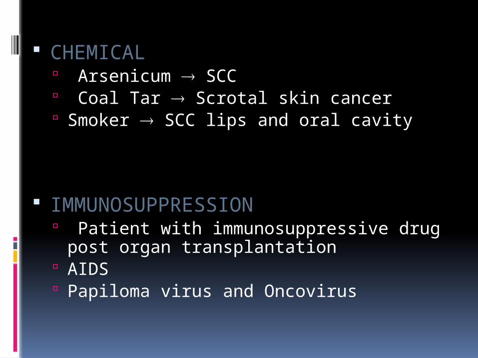
CHEMICAL Arsenicum SCC Coal Tar Scrotal skin cancer Smoker SCC lips and oral cavity
IMMUNOSUPPRESSION Patient with immunosuppressive drug post
organ transplantation AIDS Papiloma virus and Oncovirus
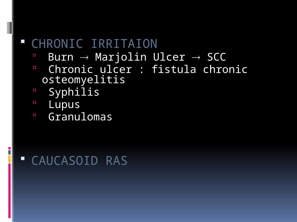
CHRONIC IRRITAION Burn Marjolin Ulcer SCC Chronic ulcer : fistula chronic osteomyelitis Syphilis Lupus Granulomas
CAUCASOID RAS
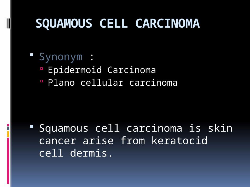
SQUAMOUS CELL CARCINOMA
Synonym : Epidermoid Carcinoma Plano cellular carcinoma
Squamous cell carcinoma is skin cancer arise from keratocid cell dermis.

DIAGNOSIS
High incidence in 50 – 70 year old Dark skin in tropic area Male > Female Normal skin or precancerous lesion
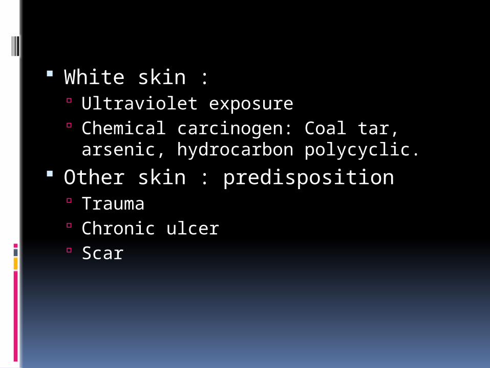
White skin : Ultraviolet exposure Chemical carcinogen: Coal tar, arsenic,
hydrocarbon polycyclic. Other skin : predisposition
Trauma Chronic ulcer Scar
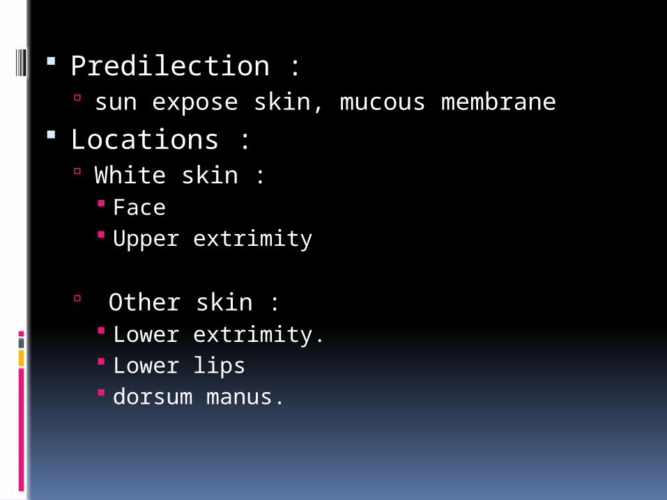
Predilection : sun expose skin, mucous membrane
Locations : White skin :
Face Upper extrimity
Other skin : Lower extrimity. Lower lips dorsum manus.
Tumor : Skin lesion especially skin with
sun exposure or trauma plaque, nodus, papel, solid
tumor or ulcer, fast growing/ progressive: exophytic, endophytic, infiltrative.
de novo or arise from pre cancerous lesion.
Regional node : Lymphadenopathy regionalsingle or multiple, mobile or fixed.
Metastasis : Lung,Liver

Radiology: Chest X-Ray, Bone X-Ray in part of lesion, CT-Scan, MRI
Patologi:a) biopsy: < 2cm excisional biopsy
> 2 cm incisional biopsyb) specimen examination : in primary tumor:
Tumor size Histolopathological fiture, Differential cell grade, Depth infiltration of the tumor, Radicality of excision.

THERAPY
Stage I / II or III (T4 N0 M0) : Wide excision with 1 cm free margin Reconstruction ( if needed).
Stage III (any T N1 M0) : Wide excision and Lymph node dissection.
Stage IV : Palliative therapy.
BASALIOMA
Synonym : Basal cell carcinoma, Ulcus rodent
Basalioma is skin cancer arise from basal cell of the skin.

DIAGNOSIS
> 40 year old Male > Female Predisposition factors:
White skin (type I & II) and albinism with cumulative sun rays exposure
X ray exposure to face acne therapy Basal nevus syndrome (autosomal dominant) Chronic arsenic intoxication Chronic LE Chronic ulcer and fistula.
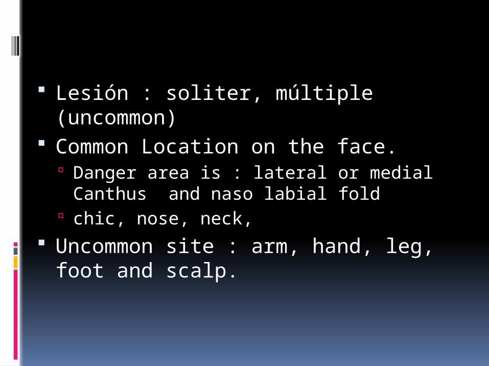
Lesión : soliter, múltiple (uncommon) Common Location on the face.
Danger area is : lateral or medial Canthus and naso labial fold
chic, nose, neck, Uncommon site : arm, hand, leg, foot
and scalp.

Skin Lesion : Type : papule, nodule, translucent like a
pearl, ulcer (rodent ulcer). Color : pink or red, telangiectasi. Pigmented type: brown, dark blue or black. Uncommon regional metastasis or distant
metastasis
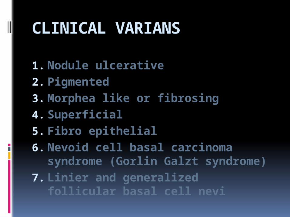
CLINICAL VARIANS
1. Nodule ulcerative2. Pigmented3. Morphea like or fibrosing4. Superficial 5. Fibro epithelial6. Nevoid cell basal carcinoma
syndrome (Gorlin Galzt syndrome)7. Linier and generalized follicular
basal cell nevi

THERAPY
Wide Excision with 0,5 – 1 cm safety margin
Radiation therapy : No radical Non operable Recurrent

MALIGNANT MELANOMA
Arise from melanocyte cell of the skin 1-3 % all malignancy 25 – 40 % grow from nevus pigmentosus
( junctional nevus ), “ Hutchinson’s melanotic freckle “, giant pigmented nevus, “ blue nevus “
Incidence , 35 – 55 year old Male = female Very malignant Rapid metastasis via hematogenic or
lymphogenic

Diagnosis
Anamnesis : Nevus growing fast Itching and ulcer
Tumor : Dark color Plaque or ulcer
Asimetry Border ( undifine) Color (many color) Diameter : more than 6 mm Evolution

Biopsy : < 2 cm excisional biopsy > 2 cm incisional biopsy, with deeper
than base of tumor
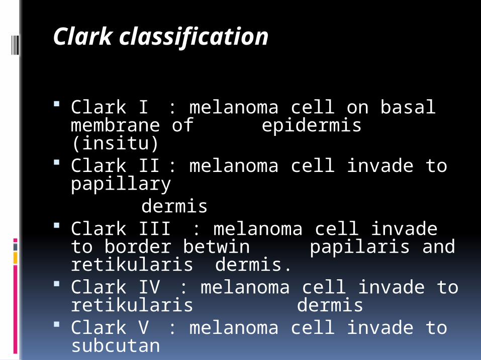
Clark classification
Clark I : melanoma cell on basal membrane of epidermis (insitu)
Clark II : melanoma cell invade to papillary
dermis Clark III : melanoma cell invade to
border betwin papilaris and retikularis dermis.
Clark IV : melanoma cell invade to retikularis dermis
Clark V : melanoma cell invade to subcutan
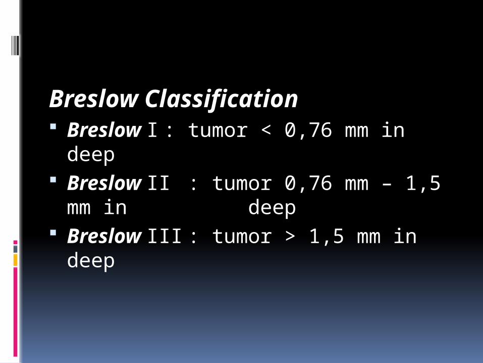
Breslow Classification Breslow I : tumor < 0,76 mm in
deep Breslow II : tumor 0,76 mm – 1,5
mm in deep Breslow III : tumor > 1,5 mm in
deep
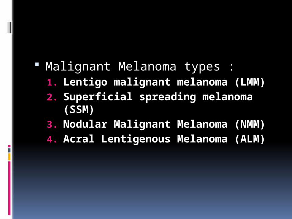
Malignant Melanoma types :1. Lentigo malignant melanoma
(LMM)2. Superficial spreading melanoma
(SSM)3. Nodular Malignant Melanoma
(NMM)4. Acral Lentigenous Melanoma (ALM)
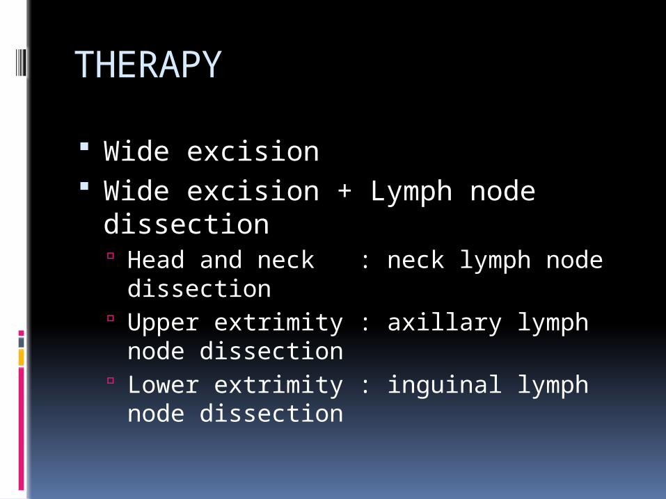
THERAPY
Wide excision Wide excision + Lymph node
dissection Head and neck : neck lymph node
dissection Upper extrimity : axillary lymph node
dissection Lower extrimity : inguinal lymph node
dissection

Other type skin cancer
Adeno carcinoma of the skin : Arise from adnexa of the skin
Merkel Cancer Arise from neuroendocrine cell of the skin
Dermato fibro sarcoma pro tuberans Pre cancer lesion :
Actinic Keratosis Kerato Acantoma Bowen’s Disease Erythroplasia of Queyrat Xeroderma Pigmentosum

Thank you


