Superficial shell abscesses and erosions are common in aquatic chelonia. Ofteninadequate husbandry is an underlying factor. Deep shell abscesses have a similarpathogenesis.
Clinical signs
Deep shell abscesses may penetrate full thickness of the shell, even down to thecoelomic membrane.
Diagnosis and therapy
Diagnosis is by clinical signs. Therapy for superficial lesions includes carefulcleaning and removal of necrotic material from damaged areas of shell. Theanimal’s access to water should be restricted to one hour, twice a day. Topicalantibiotic ointment effective against Gram-negative bacteria such as Pseudomonassp. applied after swimming and feeding (Barten 1996).
When lesions are thought to be deep, a radiograph should be taken for assess-ment and prognosis. In deep lesions debridement under sedation or full anaes-thetic is required and a sample submitted for bacterial culture and sensitivity.Treatment for deep lesions is similar to superficial lesions, except topical treatmentis accompanied by systemic antibiotics for at least four weeks (Barten 1996).
SEPTICAEMIC CUTANEOUS ULCERATIVE DISEASE (SCUD)
Cause and pathogenesis
SCUD is described as a disease syndrome in aquatic turtles and most common insoft shell turtles (Trionychidae). The agent implicated in this specific disease has
Skin Diseases of Exotic Pets Edited by Sue Paterson
been Citrobacter freundii but other Gram-negative bacteria can produce similarsigns. Serratia may be necessary to initiate the infection or as a co-infection. Otherfactors that have been implicated include poor husbandry, poor water quality,abrasions and invertebrate predation.
Clinical signs
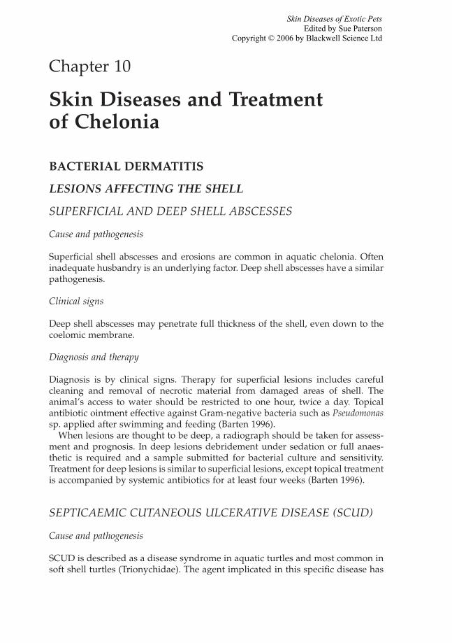
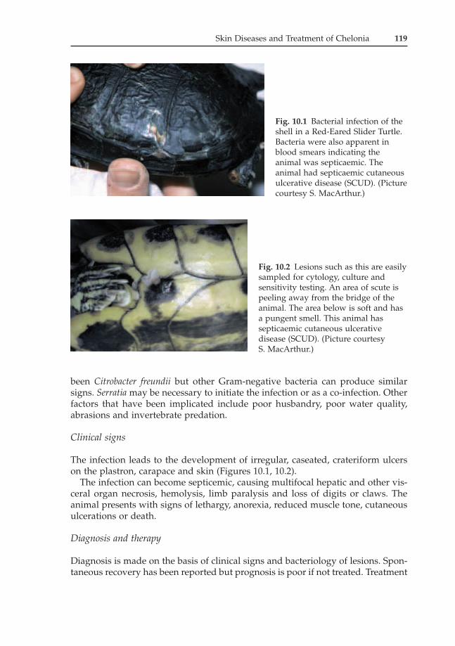
The infection leads to the development of irregular, caseated, crateriform ulcerson the plastron, carapace and skin (Figures 10.1, 10.2).
The infection can become septicemic, causing multifocal hepatic and other vis-ceral organ necrosis, hemolysis, limb paralysis and loss of digits or claws. Theanimal presents with signs of lethargy, anorexia, reduced muscle tone, cutaneousulcerations or death.
Diagnosis and therapy
Diagnosis is made on the basis of clinical signs and bacteriology of lesions. Spon-taneous recovery has been reported but prognosis is poor if not treated. Treatment
Fig. 10.1 Bacterial infection of theshell in a Red-Eared Slider Turtle.Bacteria were also apparent inblood smears indicating theanimal was septicaemic. Theanimal had septicaemic cutaneousulcerative disease (SCUD). (Picturecourtesy S. MacArthur.)
Fig. 10.2 Lesions such as this are easilysampled for cytology, culture andsensitivity testing. An area of scute ispeeling away from the bridge of theanimal. The area below is soft and hasa pungent smell. This animal hassepticaemic cutaneous ulcerativedisease (SCUD). (Picture courtesy S. MacArthur.)
entails debridement of ulcers and abscesses, use of antibiotics and shell supportwith fibreglass and resin if destruction is extensive.
Chloramphenicol has reportedly been effective in one case. Shell lesions cantake one to two years to heal.
OTHER BACTERIAL INFECTIONS
Beneckea chitinovora
Beneckea chitinovora another Gram-negative bacteria is implicated as the cause ofchronic ulcerative shell disease in captive turtles especially soft shell turtlesTrionyx spp. Prior injury to the shell is necessary for the bacteria to cause disease(Wallach 1975). Crustaceans carry these bacteria on their exoskeleton and conta-minate water when used as a food source (Harvey-Clark 1997).
Mycobacterium kansaii
A Chinese soft-shell turtle with a white foci on the carapace was initially treatedwith 20% ammonia (0.5ml/litre water) in the water. The lesions started to respondto therapy but the animal died following anorexia and respiratory distress. Onpost mortem, pulmonary lesions were present as well as papular lesions on skinand carapace. Mycobacterium kansaii was isolated and identified from the lesions.(Oros et al. 2003).
BACTERIAL DERMATITIS OF OTHER AREAS
ULCERATIVE DERMATITIS OF THE FEET
Cause and pathogenesis
A captive breeding program for Western swamp tortoises, Pseudemydura umbrina,were identified with lesions predominantly affecting the feet. There was no evi-dence of trauma, Pseudomonas spp. was consistently cultured and occasionally sec-ondary fungal hyphae demonstrated. The disease outbreaks were associated withenvironmental, husbandry and nutritional factors.
Clinical signs
Ulcerative lesions were identified on the feet. Death was attributed to secondarysepticaemia.
120 Skin Diseases of Exotic Pets
Skin Diseases and Treatment of Chelonia 121
Diagnosis and therapy
Diagnosis was made on the basis of clinical signs and isolation of bacteria andfungi. A review of husbandry led to improvements in the tortoises’ management.Pond temperatures were maintained between 20°C and 27°C. A successful topicalregime was instituted consisting of equal volumes of nystatin, oxytetracycline andwater administered topically with a hydrocolloid gel once daily on affected areasfor a period of two to three weeks. The animals were left out of the water for anhour after application of topical treatment (Ladyman et al. 1998).
OTHER BACTERIAL INFECTIONS
Mycobacterium spp.
Cause and pathogenesis
Mycobacterium is a saprophytic organism probably originating from contami-nated water. Cutaneous lesions are a likely port of entry with haematogenousspread to internal organs. It is zoonotic causing subcutaneous abscesses whendealing with aquatic environment or animals.
Clinical signs
Rhodin and Anver (1977) describe a Side-necked turtle, Phrynops hilari, withchronic nodular ulceration on the mandible and nodules on the webbing of theforefeet. On post mortem, nodules were also present on spleen and liver. Alllesions had the typical staining of Mycobacterium spp. In his review Rhodin alsocites mycobacteriosis causing plastral ulcerations in a Nile soft-shell turtle (Trionyxtriunguis), and a Ganges soft-shell turtle (Aspideretes gangeticus).
FUNGAL DERMATITIS
Cause and pathogenesis
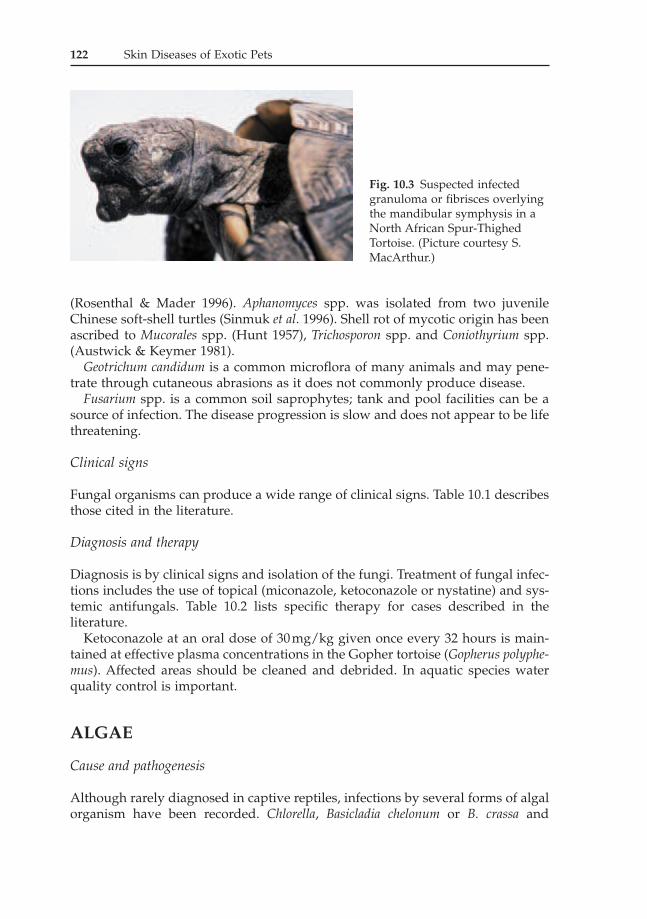
Fungal organisms are opportunists, invading tissue already damaged or infected(Figure 10.3). Predisposing factors include poor water quality, malnutrition andstress. The incidence of mycotic infections in aquatic and semi-aquatic turtles maybe directly or indirectly related to the pH of the water because the growth of mostfungi is inhibited at a pH less than 6.5 (Frye 1991).
In aquatic species, mixed bacterial and fungal infections of the shell are com-monly diagnosed and are often invasive involving bone.
Mycosis in chelonians have included infections by Aspergillus spp., Basidiobolusranarum, Beauveria bassiana, Cladosporium spp., Candida spp., dermatophytes, Fusar-ium spp., Geotrichum spp., Paecilomyces spp., Penicillium spp. and Sporotrichum spp.
(Rosenthal & Mader 1996). Aphanomyces spp. was isolated from two juvenileChinese soft-shell turtles (Sinmuk et al. 1996). Shell rot of mycotic origin has beenascribed to Mucorales spp. (Hunt 1957), Trichosporon spp. and Coniothyrium spp.(Austwick & Keymer 1981).
Geotrichum candidum is a common microflora of many animals and may pene-trate through cutaneous abrasions as it does not commonly produce disease.
Fusarium spp. is a common soil saprophytes; tank and pool facilities can be asource of infection. The disease progression is slow and does not appear to be lifethreatening.
Clinical signs
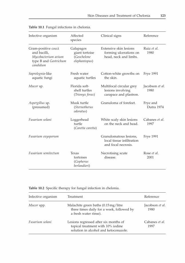
Fungal organisms can produce a wide range of clinical signs. Table 10.1 describesthose cited in the literature.
Diagnosis and therapy
Diagnosis is by clinical signs and isolation of the fungi. Treatment of fungal infec-tions includes the use of topical (miconazole, ketoconazole or nystatine) and sys-temic antifungals. Table 10.2 lists specific therapy for cases described in theliterature.
Ketoconazole at an oral dose of 30mg/kg given once every 32 hours is main-tained at effective plasma concentrations in the Gopher tortoise (Gopherus polyphe-mus). Affected areas should be cleaned and debrided. In aquatic species waterquality control is important.
ALGAE
Cause and pathogenesis
Although rarely diagnosed in captive reptiles, infections by several forms of algalorganism have been recorded. Chlorella, Basicladia chelonum or B. crassa and
122 Skin Diseases of Exotic Pets
Fig. 10.3 Suspected infectedgranuloma or fibrisces overlyingthe mandibular symphysis in aNorth African Spur-ThighedTortoise. (Picture courtesy S.MacArthur.)
Fusarium semitectum Texas Necrotising scute Rose et al.tortoises disease. 2001(Gopherusberlandieri)
Table 10.2 Specific therapy for fungal infection in chelonia.
Infective organism Treatment Reference
Mucor spp. Malachite green baths (0.15mg/litre Jacobson et al.three times daily for a week, followed by 1980a fresh water rinse).
Fusarium solani Lesions regressed after six months of Cabanes et al.topical treatment with 10% iodine 1997solution in alcohol and ketoconazole.
Schizangiella serpentis have been implicated. Whether Basicladial infections areauthentic pathogens is debatable and there is a form of mutual benefit. The algaeprovide camouflage for aquatic turtles. Turtles should shed their scutes regularlyas they grow. Retained scutes are a sign of poor husbandry as the animal has notbeen drying off sufficiently to lose its old scutes. Retained scutes will becomeinfected with algae.
Clinical signs
Algae can cause pitting or discoloration of the shell. Schizangiella serpentis causesgranulomatous lesions and should be removed with wide surgical excision as inci-dence of recurrence is substantial (Frye 1991), followed by topical treatment withketoconazole ointment.
Diagnosis and therapy
Diagnosis should be made on the basis of clinical signs and the identification ofthe algae on culture. The shells of captive turtles should be cleaned weekly. Whereinfection has occurred due to excessive wetting of the scutes then the animals’husbandry should be reviewed and a larger basking area should be included intheir environment to encourage them out of the water to dry off. Treatment forsuperficial algal infections is through the removal of algae with a toothbrush andtopical application of povidone-iodine solutions or a ketoconozole ointment.
VIRAL
GREEN TURTLE FIBROPAPILLOMAS (GTFP)
Cause and pathogenesis
Fibropapillomatosis also termed green turtle fibropapillomas (GTFP) is charac-terised by single to multiple histologically benign fibro-epithelial tumours. Greenturtles, Chelonia mydas and Loggerhead turtles, Caretta caretta, in Florida, Hawaii,Caribbean, Australia and Indonesia are affected by cutaneous and occasional vis-ceral fibropapillomas (Aguirre et al. 1994, Adnyana et al. 1997, Lackovich et al.1999). A viral aetiology remains the main hypothesis. A study in Florida demon-strated that chelonia herpes virus is regularly associated with fibropapillomatosisin sea turtles (Lackovich et al. 1999). At least three herpes viruses have beenreported in marine turtle fibropapilloma (Quackenbush et al. 1998).
GTFP is more prevalent near shore ecosystems and anecdotally more so in areaswith human activity impact. Environmental contaminants such as carcinogensmay induce latent virus infection or immunosuppresion. Another possibility isthat these ecosystems may provide more optimal environment for survival andtransmission of infectious agents and a higher population density of vectors andsusceptible animals (Herbst & Klein 1995, Adnyana et al. 1997).
124 Skin Diseases of Exotic Pets
Fibrous tumours have been observed in Olive ridleys Lepidochelys olivacea, Flat-backs Natator depressus (Herbst & Klein 1995) and loggerheads Caretta caretta,Green turtle Chelonia mydas, Hawksbill Eretmochelys imbricata and Kemp’s ridleyLepidochelys kempii (Harshbarger 2002).
Clinical signs
The tumours are found on areas of soft skin (flippers, neck, chin, inguinal andaxillary regions and tail base) and conjunctivae. Visceral tumours may also bepresent. Some fibropapillomas will decrease in size but others will slowly increasein size, ulcerating due to abrasions. Fibropapillomas can become life threateningif the size of papillomas impairs swimming and if conjunctival fibropapillomasobscure vision.
Diagnosis and therapy
Diagnosis is made on the basis of clinical signs and viral isolation. Therapy is best achieved by surgical removal of fibropapillomas, with wide excisionalmargins.
POXVIRUS
Cause and pathogenesis
A cutaneous pox-like virus infection has been described in a captive Hermann’stortoise Testudo hermanni (Oros et al. 1998). Poxvirus was also attributed but notconfirmed in an amelanotic Californian desert tortoise Xerobates agassizi (Jacobson1991).
Clinical signs
In the case described by Oros white–yellow papular lesions were identified onand around the eyelids. In the second case of Jacobson multiple, raised papularto vesicular lesions were seen on the skin.
Diagnosis and therapy
Diagnosis is by clinical signs and virus isolation. Isolation of infected animals isadvised, to minimise spread and ease treatment and observation, together withtreatment of secondary bacterial and / or fungal infection.
Skin Diseases and Treatment of Chelonia 125
126 Skin Diseases of Exotic Pets
HERPES VIRUS
Cause and pathogenesis
A virus morphologically similar in appearance to herpes virus has been attributedto be the causative agent of grey patch disease of Green sea turtles Chelonia mydas.Epizootics can occur in the summer with high water temperatures, overcrowdingand organic pollution (Jacobson 1991). A disease that appears similar is a super-ficial bacterial dermatitis seen in juvenile Green turtles C. mydas and Loggerheadturtles C. Caretta (Frye 1991). In addition to viral infection, secondary Gram-neg-ative bacteria were thought to be implicated, as well as secondary hypovita-minosis A.
Clinical signs
In Grey Patch Disease young hatchlings in aquaculture developed small papularskin lesions that coalesced into spreading patches. The disease described by Frye(1991) produces a rapidly spreading necrotic dermatitis which is fatal in untreatedanimals.
Diagnosis and therapy
Diagnosis can be made by electron microscopy by the identification of viral inclu-sions. Reported treatment for the disease described by Frye (1991) consisted oftotal immersion three times a week in potassium permanganate (KMnO4 1g/220litres of sea-water) followed three hours later by a fresh sea-water wash. Acyclovirhas been used for treatment of herpes virus in tortoises.
EPIDERMAL SQUAMOUS PAPILLOMAS
Epidermal squamous papillomas have been identified in Mata mata turtle (Chelysfimbriata), Snapping turtle (Chelydra serpentina) and Slider turtle (Trachemys scripta)(Frye 1991). Papilloma-like viral crystalline arrays were observed in skin lesionsof recently imported Bolivian Side-neck turtles (Platemys platcephala) (Jacobson1991).
PARASITIC
Important dermatological parasites of chelonians include:
Evidence of infection with spirorchid flukes (Digenea: Spirorchidae) was identi-fied at necropsy in stranded Green turtles, (Chelonia mydas). Three species ofspirorchid (Hapalotrema mehrai, H. postorchis, and Neospirorchis schistosomatoides)were identified.
Clinical signs
Flukes cause damage to internal organs. Cardiovascular lesions included muralendocarditis, arteritis, and thrombosis, frequently accompanied by aneurysm for-mation. Occlusion of the superficial blood vessels can occur due to the adults orspirorchid fluke eggs. Thrombosis and infarction of the shell may occur.
Diagnosis and therapy
Helminths or ova in shell biopsies are diagnostic. Treatment with praziquantel8–20mg/kg i.m. repeated at 14 days (Harvey-Clark 1997).
MITES
Cause and pathogenesis
Mites (of unknown species) were found under the dermal scutes of an Africanspurred tortoise (Geochelone sulcata).
Diagnosis and therapy
Diagnosis is by clinical signs. Therapy was achieved by debridement of affectedtissue and cleansing with chlorhexidine. This lead to an elimination of the mites(Campillo & Frye 2002).
TICKS
Cause and pathogenesis
Aponomma gervaisi is found on Testudo elegans and the coloration of the tickadapted to the host. Hyalomma aegyptium infests Testudo graeca and H. franchiniitortoises in Libya. Ornithodoros compactus a soft-bodied tick has been found on tor-toises (Frank 1981a).
128 Skin Diseases of Exotic Pets
Reptile ticks such as the African tortoise tick Amblyomma marmoreum, Ambly-omma sparsum on Leopard tortoises and Amblyomma dissimile the Iguana tick haveentered the pet trade through imports and are sustainable in warm climates. Somehave a mammal as their final host and can transmit infectious disease such asspirochaetes and haemofilaria. Heavy burdens can cause anaemia.
Diagnosis and therapy
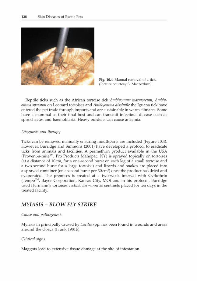
Ticks can be removed manually ensuring mouthparts are included (Figure 10.4).However, Burridge and Simmons (2001) have developed a protocol to eradicateticks from animals and facilities. A permethrin product available in the USA(Provent-a-miteTM, Pro Products Mahopac, NY) is sprayed topically on tortoises(at a distance of 10cm, for a one-second burst on each leg of a small tortoise anda two-second burst for a large tortoise) and lizards and snakes are placed into a sprayed container (one-second burst per 30cm2) once the product has dried andevaporated. The premises is treated at a two-week interval with Cyfluthrin(TempoTM, Bayer Corporation, Kansas City, MO) and in his protocol, Burridgeused Hermann’s tortoises Testudo hermanni as sentinels placed for ten days in thetreated facility.
MYIASIS – BLOW FLY STRIKE
Cause and pathogenesis
Myiasis in principally caused by Lucilia spp. has been found in wounds and areasaround the cloaca (Frank 1981b).
Clinical signs
Maggots lead to extensive tissue damage at the site of infestation.
Fig. 10.4 Manual removal of a tick.(Picture courtesy S. MacArthur.)
Skin Diseases and Treatment of Chelonia 129
Diagnosis and therapy
Maggots are physically removed and the area flushed with a dilute antiseptic solu-tion. Topical or systemic antibiotics may be needed.
LEECHES
Cause and pathogenesis
Leeches can be found attached to wild-caught chelonia.
Diagnosis and therapy
Diagnosis is based on clinical signs and the presence of leeches within the wound.Therapy can be achieved with the use of a saline solution swabbed over thesurface of the leech which facilitates removal (Harkewicz 2001). Secondary infec-tions should be addressed.
NUTRITIONAL
HYPOVITAMINOSIS A
Cause and pathogenesis
A common condition seen in turtles and terrapins fed an all-meat diet. Vitamin Ais needed for maintenance of epithelial integrity.
Clinical signs
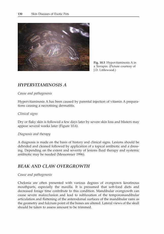
Hypovitaminosis A may present itself with bilateral swollen eyelids due to palpe-bral oedema (Figure 10.5). The hardian glands that secrete sodium chloride losetheir structure with squamous metaplasia of the epithelium (Reichenbach-Klinke& Elkan 1965). Other clinical signs include respiratory infection and more generalsymptoms such as lethargy and anorexia.
Diagnosis and therapy
Diagnosis is based on a history of poorly balanced diet and clinical signs. Oralsupplementation of vitamin A (2000–10000IU/kg feed or 2000 IU/kg p.o. everyseven to fourteen days for two to four treatments) is recommended while address-ing nutrition and secondary infection.
130 Skin Diseases of Exotic Pets
HYPERVITAMINOSIS A
Cause and pathogenesis
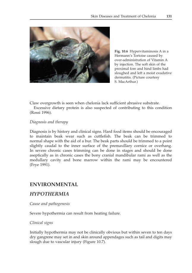
Hypervitaminosis A has been caused by parental injection of vitamin A prepara-tions causing a necrotising dermatitis.
Clinical signs
Dry or flaky skin is followed a few days later by severe skin loss and blisters mayappear several weeks later (Figure 10.6).
Diagnosis and therapy
A diagnosis is made on the basis of history and clinical signs. Lesions should bedebrided and cleaned followed by application of a topical antibiotic and a dress-ing. Depending on the extent and severity of lesions fluid therapy and systemicantibiotic may be needed (Messonnier 1996).
BEAK AND CLAW OVERGROWTH
Cause and pathogenesis
Chelonia are often presented with various degrees of overgrown keratinousmouthparts, especially the maxilla. It is presumed that soft-food diets anddecreased forage time contribute to this condition. Mandibular overgrowth cancause severe malocclusion and lead to subluxation of the temporomandibulararticulation and flattening of the anterodorsal surfaces of the mandibular rami asthe geometry and fulcrum point of the bones are altered. Lateral views of the skullshould be taken to assess amount to be trimmed.
Fig. 10.5 Hypovitaminosis A in a Terrapin. (Picture courtesy ofJ.D. Littlewood.)
Skin Diseases and Treatment of Chelonia 131
Claw overgrowth is seen when chelonia lack sufficient abrasive substrate.Excessive dietary protein is also suspected of contributing to this condition
(Rossi 1996).
Diagnosis and therapy
Diagnosis is by history and clinical signs. Hard food items should be encouragedto maintain beak wear such as cuttlefish. The beak can be trimmed to normal shape with the aid of a bur. The beak parts should be trimmed to a pointslightly caudal to the inner surface of the premaxillary cornice or overhang. In severe chronic cases trimming can be done in stages and should be done aseptically as in chronic cases the bony cranial mandibular rami as well as themedullary cavity and bone marrow within the rami may be encountered (Frye 1991).
ENVIRONMENTAL
HYPOTHERMIA
Cause and pathogenesis
Severe hypothermia can result from heating failure.
Clinical signs
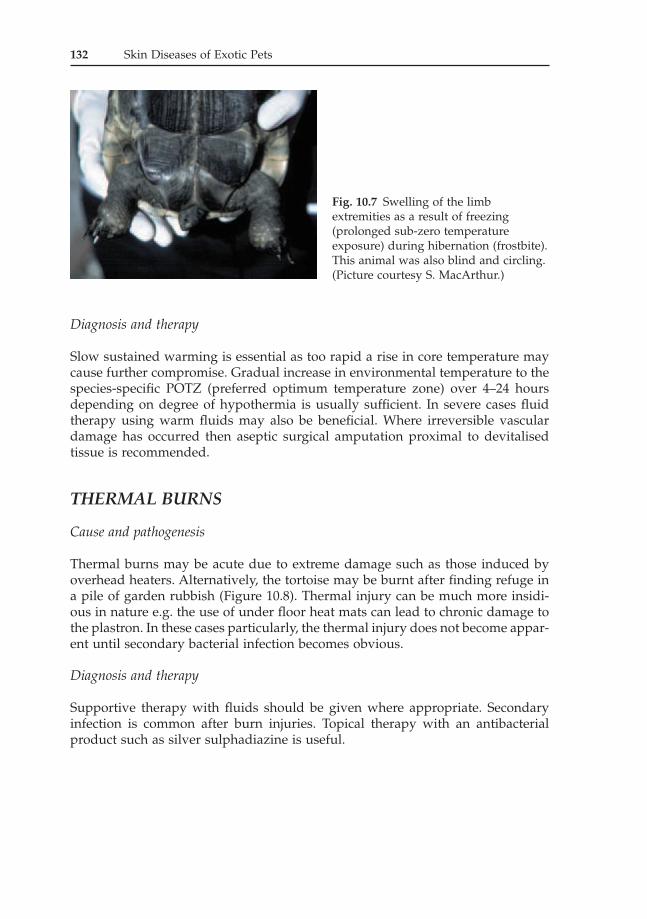
Initially hypothermia may not be clinically obvious but within seven to ten daysdry gangrene may set in and skin around appendages such as tail and digits mayslough due to vascular injury (Figure 10.7).
Fig. 10.6 Hypervitaminosis A in aHermann’s Tortoise caused byover-administration of Vitamin Aby injection. The soft skin of theproximal fore and hind limbs hadsloughed and left a moist exudativedermatitis. (Picture courtesy S. MacArthur.)
132 Skin Diseases of Exotic Pets
Diagnosis and therapy
Slow sustained warming is essential as too rapid a rise in core temperature maycause further compromise. Gradual increase in environmental temperature to thespecies-specific POTZ (preferred optimum temperature zone) over 4–24 hoursdepending on degree of hypothermia is usually sufficient. In severe cases fluidtherapy using warm fluids may also be beneficial. Where irreversible vasculardamage has occurred then aseptic surgical amputation proximal to devitalisedtissue is recommended.
THERMAL BURNS
Cause and pathogenesis
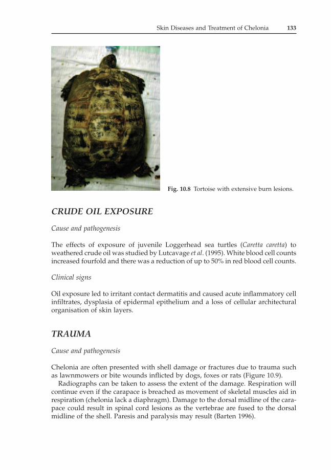
Thermal burns may be acute due to extreme damage such as those induced byoverhead heaters. Alternatively, the tortoise may be burnt after finding refuge ina pile of garden rubbish (Figure 10.8). Thermal injury can be much more insidi-ous in nature e.g. the use of under floor heat mats can lead to chronic damage tothe plastron. In these cases particularly, the thermal injury does not become appar-ent until secondary bacterial infection becomes obvious.
Diagnosis and therapy
Supportive therapy with fluids should be given where appropriate. Secondaryinfection is common after burn injuries. Topical therapy with an antibacterialproduct such as silver sulphadiazine is useful.
Fig. 10.7 Swelling of the limbextremities as a result of freezing(prolonged sub-zero temperatureexposure) during hibernation (frostbite).This animal was also blind and circling.(Picture courtesy S. MacArthur.)
Skin Diseases and Treatment of Chelonia 133
CRUDE OIL EXPOSURE
Cause and pathogenesis
The effects of exposure of juvenile Loggerhead sea turtles (Caretta caretta) toweathered crude oil was studied by Lutcavage et al. (1995). White blood cell countsincreased fourfold and there was a reduction of up to 50% in red blood cell counts.
Clinical signs
Oil exposure led to irritant contact dermatitis and caused acute inflammatory cellinfiltrates, dysplasia of epidermal epithelium and a loss of cellular architecturalorganisation of skin layers.
TRAUMA
Cause and pathogenesis
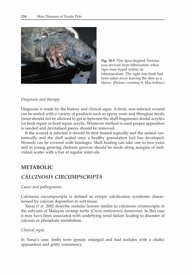
Chelonia are often presented with shell damage or fractures due to trauma suchas lawnmowers or bite wounds inflicted by dogs, foxes or rats (Figure 10.9).
Radiographs can be taken to assess the extent of the damage. Respiration willcontinue even if the carapace is breached as movement of skeletal muscles aid inrespiration (chelonia lack a diaphragm). Damage to the dorsal midline of the cara-pace could result in spinal cord lesions as the vertebrae are fused to the dorsalmidline of the shell. Paresis and paralysis may result (Barten 1996).
Fig. 10.8 Tortoise with extensive burn lesions.
Diagnosis and therapy
Diagnosis is made by the history and clinical signs. A fresh, non-infected woundcan be sealed with a variety of products such as epoxy resin and fibreglass mesh,(resin should not be allowed to get in between the shell fragments) dental acrylicsfor beak repair or hoof repair acrylic. Whatever method is used proper appositionis needed and devitalised pieces should be removed.
If the wound is infected it should be first treated topically and the animal sys-temically and the shell sealed once a healthy granulation bed has developed.Wounds can be covered with bandages. Shell healing can take one to two yearsand in young growing chelonia grooves should be made along margins of indi-vidual scutes with a bur at regular intervals.
METABOLIC
CALCINOSIS CIRCUMPSCRIPTA
Cause and pathogenesis
Calcinosis circumpscripta is defined as ectopic calcification syndrome charac-terised by calcium deposition in soft tissue.
Yanai et al. 2002 describe nodular lesions similar to calcinosis cirumscripta inthe subcutis of Malayan swamp turtle (Cuora ambionensis kamaroma). In this caseit may have been associated with underlying renal failure leading to disorder ofcalcium or phosphate metabolism.
Clinical signs
In Yanai’s case, limbs were grossly enlarged and had nodules with a chalkyappearance and gritty consistency.
134 Skin Diseases of Exotic Pets
Fig. 10.9 This Spur-thighed Tortoisewas revived from hibernation whenraps were heard within itshibernaculum. The right fore-limb hadbeen eaten away leaving the skin as asleeve. (Picture courtesy S. MacArthur.)
HEREDITARY AND CONGENITAL
Minor abnormalities of chelonian shell pattern have been noted (Reichenbach-Klinke & Elkan 1965, Bellairs 1981). These are possibly related to poorly controlledegg-incubation periods.
Skin Diseases and Treatment of Chelonia 135
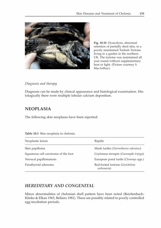
Fig. 10.10 Dysecdysis, abnormalretention of partially shed skin, in apoorly maintained Turkish Tortoiseliving in a garden in the northernUK. The tortoise was maintained allyear round without supplementaryheat or light. (Picture courtesy S.MacArthur.)
Diagnosis can be made by clinical appearance and histological examination. His-tologically there were multiple lobular calcium deposition.
NEOPLASIA
The following skin neoplasia have been reported:
MISCELLANEOUS
DYSECDYSIS
Cause and pathogenesis
Sloughing of scutes has been seen with underlying renal disease and with animalshoused in an environment with very moist substrate (Figure 10.10).
Diagnosis and therapy
To aid in the assessment of renal function uric acid and calcium:phosphorous ratioin plasma should be measured. Where infection has occurred due to excessivewetting of the scutes, then the animals’ husbandry should be reviewed and alarger basking area should be included in their environment to encourage themout of the water to dry off.
Environmental causes are easier to rectify accompanied by systemic antibiotics.Renal disease carries a poor prognosis (Rossi 1996).
REFERENCES
Adnyana, W., Ladds, P.W. and Blair, D. (1997) ‘Observations of fibropapillomatosis in greenturtles, Chelonia mydas in Indonesia’. Australian Veterinary Journal, 75:10, pp. 737–742.
Aguirre, A.A., Balazs, G.H., Zimmerman, B. and Sparker, T.R. (1994) ‘Evaluation of Hawaiian green turtles, Chelonia mydas, for potential pathogens associated with fibro-papillomas’. Journal of Wildlife Diseases, 30:1, pp. 8–15.
Austwick P.K.C. and Keymer I.F. (1981) ‘Fungi and actinomycetes.’ In: Diseases of the Reptilia, Vol. I. Cooper, J.E. and Jackson O.F. (eds). Academic Press, London, pp. 193–231.
Barten, S.L. (1996) ‘Shell damage’. In: Reptile Medicine and Surgery. W.B. Saunders Company,Philadelphia, pp. 413–417.
Bellairs, A.D.A. (1981) ‘Congenital and developmental diseases’. In: Diseases of the Reptilia, Vol. II. Cooper, J.E. and Jackson, O.F. (eds). Academic Press, London, pp. 469–486.
Burridge, M.J. and Simmons, L. (2001) ‘Control and eradication of exotic tick infestationson reptiles’. Proceedings of the Association of Reptilian and Amphibian Veterinarians. Orlando,Florida, pp. 21–23.
Cabanes, F.J., Alonso, J.M., Castella, G., et al. (1997) ‘Cutaneous Hyalohyphomycosis causedby Fusarium solani in a Loggerhead sea turtle, Caretta caretta’. Journal of Clinical Microbi-ology, 35:12, pp. 3343–3345.
Campillo, N.J. and Frye, F.L. (2002) ‘Preliminary report of subepidermal mite infestation inan African Spurred tortoise, Geochelone sulcata’. Proceedings of the Association of Reptilianand Amphibian Veterinarians. Reno, Nevada, p. 17.
Frank, W. (1981a) ‘Endoparasites’. In: Diseases of the Reptilia, Vol. I. Cooper, J.E. and Jackson,O.F. (eds). Academic Press, London, pp. 291–358.
Frank, W. (1981b) ‘Ectoparasites’. In: Diseases of the Reptilia, Vol. I. Cooper, J.E. and Jackson,O.F. (eds). Academic Press, London, pp. 359–383.
Frye, F.L. and Dutra, F.R. (1974, Dec.) ‘Mycotic granuloma involving the forefeet of a turtle’.Veterinary Medicine / Small Animal Clinician, pp. 1554–1556.
Frye, F.L. (1991) Biomedical and Surgical Aspects of Captive Reptile Husbandry, Vols I and II.Krieger Publishing Company, Malabar.
136 Skin Diseases of Exotic Pets
Harshbarger, J.C. (2002) ‘Marine turtle fibroma and fibropapilloma cases in the registry oftumours in lower animals’. Proceedings of the Sixth International Symposium on the Path-ology of Reptiles and Amphibians, Saint Paul, MN, pp. 129–145.
Harkewicz, K.A. (2001) ‘Dermatology of reptiles: A clinical approach to diagnosis and treat-ment’. In: Veterinary Clinics of North America – Exotic Animal Practice, 4:2, pp. 441–462.
Harvey-Clark, C.J. (1997) ‘Dermatologic (skin) disorders’. In: The Biology, Husbandry andHealthcare of Reptiles, Vol. III, Ackerman, L. (ed.). T.F.H Publications Inc., Neptune City,NJ, pp. 654–680.
Herbst, L.H. and Klein, P.A. (1995) ‘Green turtle fibropapillomatosis: Challenges to assess-ing the role of environmental cofactors’. Environmental Health Perspectives, 103:4, pp.27–30.
Hunt, T.J. (1957) ‘Notes on disease and mortality in testudines’. Herpetologica 13, p. 19.Jacobson, E.R., Calderwood, M.B. and Clubb, S.L. (1980) ‘Mucormycosis in hatchling Soft-
shell turtles’. Journal of the American Veterinary Medical Association, 177:9, pp. 835–837.Jacobson, E.R., Mansell, J.L., Sundberg, J.P., et al. (1989) ‘Cutaneous fibropapillomas of
Green turtles, Chelonia mydas’. Journal of Comparative Pathology, 101, pp. 39–52.Jacobson, E.R. (1991) ‘Disease of the integumentary system of reptiles’. In: Dermatology for
Small Animal Practitioner, Nesbitt, G.H. and Ackerman, L.L. (eds). Veterinary LearningSystems Co. Inc., Trenton, NJ, pp. 225–239.
Lackovich, J.K., Brown, D.R., Homer, B.L., et al. (1999) ‘Association of herpes virus withfibropapillomatosis of the Green turtle, Chelonia mydas and the Loggerhead turtle, Carettacareta in Florida’. Diseases of Aquatic Organisms, 37, pp. 89–97.
Ladyman, J.M., Kuchling, G., Burford, D., et al. (1998) ‘Skin disease affecting the conserva-tion of the Western swamp tortoise, Pseudemydura umbrina’. Australian Veterinary Journal,76:11, pp. 743–745.
Lutcavage, M.E., Lutz, P.L., Bossart, G.D. and Hudson, D.M. (1995) ‘Physiological and clin-icopathological effects of crude oil on Loggerhead sea turtles’. Archives of EnvironmentalContamination and Toxicology, 28, pp. 417–422.
Messonnier, S.P. (1996) Common Reptile Diseases and Treatment. Blackwell Science, Oxford.Oros, J., Rodriguez, J.L., Deniz, S., et al. (1998) ‘Cutaneous poxvirus-like infection in a
captive Hermann’s tortoise, Testudo hermanni’. Veterinary Record 31, pp. 508–509.Oros, J., Acosta, B., Gaskin, J.M., et al. (2003) ‘Mycobacterium kansaii infection in a Chinese
soft-shell turtle, Pelodiscus sinensis’. Veterinary Record, April 12, pp. 474–476.Quackenbush, S.L., Work, T.M., Balazs, G.H., et al. (1998) ‘Three closely related herpes
viruses are associated with fibropapillomatosis in marine turtles’. Virology, 246:2, pp.392–399.
Reichenbach-Klinke, H. and Elkan, E. (1965) Principal Diseases of Lower Vertebrates, Part 3,Diseases of Reptiles. T.H.F Publications Inc., Neptune, NJ.
Rhodin, A.G.J. and Anver, M.R. (1977) ‘Mycobacteriosis in turtles: Cutaneous andhepatosplenic involvement in a Phrynops hilari’. Journal of Wildlife Diseases, 13, pp. 180–183.
Rose, F.L., Koke, J. Koehn, R. and Smith, D. (2001) ‘Identification of the etiological agent fornecrotizing scute disease in the Texas tortoise’. Journal of Wildlife Diseases, 37:2, pp. 223–228.
Rosenthal, K.L. and Mader, D.R. (1996) Reptile Medicine and Surgery. W.B. SaundersCompany, Philadelphia, pp. 117–124.
Rossi, J.V. (1996) ‘Dermatology’. In: Reptile Medicine and Surgery. W.B. Saunders Company,Philadelphia, pp. 104–117.
Ruiz, J.M., Arteaga, E., Martinez, J., et al. (1980) ‘Cutaneous and renal geotrichiosis in aGiant tortoise Geochelone elephantopus’. Sabouraudia 18, pp. 51–59.
Sinmuk, S., Suda, H. and Hatai, K. (1996) ‘Aphanomyces infection in juvenile Soft-shelledturtle, Pelodiscus sinensis, imported from Singapore’. Mycoscience, 37, pp. 249–254.
Wallach, J.D. (1975) ‘The pathogenesis and aetiology of ulcerative shell disease in turtles’.Journal of Zoo Animal Medicine, 6, pp. 11–13.
Yanai, T., Noda, A., Hasegawa, K., et al. (2002) ‘Calcinosis circumscripta in a Malayan boxturtle, Cuora amboinensis kamaroma’. Proceedings of the Sixth International Symposium on thePathology of Reptiles and Amphibian. Saint Paul, MN, pp. 111–115.