SLS Symposium on Chemistry and Physics Tuesday, December 4, 2012 10:00 to 12:15, WBGB/019 10:00 Interface Fermi states of LaAlO3/SrTiO3 and related heterostructures C. Cancellieri , M.L. Reinle-Schmitt, M. Kobayashi, V. N. Strocov, T. Schmitt, S. Gariglio, J.-M. Triscone, P.R. Willmott 10:30 Structure of a functioning catalyst for methane steam reforming Renata Bessa Duarte , José M. Corrêa Bueno, Maarten Nachtegaal, Olga Safonova, and Jeroen van Bokhoven 11:00 Coffee 11:15 The cold stage: A new sample environment for TOMCAT Annabelle Medebach , Bernd R. Pinzer, Cedric Dubois, Hans Joerg Limbach, Martin Schneebeli and Marco Stampanoni 11:45 High Repetition Rate Laser Pump / X-Ray Probe Studies on Biological Systems, Metal Oxide Nanoparticles and Metal Complexes J. Rittmann , Ch. Milne, M. Reinhardt, M.H. Rittmann-Frank, F. Santomauro, M.Silatani, R. Abela, and M. Chergui
Transcript
SLS Symposium on
Chemistry and Physics
Tuesday, December 4, 2012
10:00 to 12:15, WBGB/019
10:00 Interface Fermi states of LaAlO3/SrTiO3 and related heterostructures C. Cancellieri, M.L. Reinle-Schmitt, M. Kobayashi, V. N. Strocov, T. Schmitt, S. Gariglio, J.-M. Triscone, P.R. Willmott 10:30 Structure of a functioning catalyst for methane steam reforming Renata Bessa Duarte, José M. Corrêa Bueno, Maarten Nachtegaal, Olga Safonova, and Jeroen van Bokhoven 11:00 Coffee 11:15 The cold stage: A new sample environment for TOMCAT Annabelle Medebach, Bernd R. Pinzer, Cedric Dubois, Hans Joerg Limbach, Martin Schneebeli and Marco Stampanoni 11:45 High Repetition Rate Laser Pump / X-Ray Probe Studies on Biological Systems, Metal Oxide Nanoparticles and Metal Complexes J. Rittmann, Ch. Milne, M. Reinhardt, M.H. Rittmann-Frank, F. Santomauro, M.Silatani, R. Abela, and M. Chergui
Interface Fermi states of LaAlO3/SrTiO3 and related heterostructures
C. Cancellieri1, M.L. Reinle-Schmitt1, M. Kobayashi1, V. N. Strocov1, T. Schmitt1, S. Gariglio2, J.-M. Triscone2, P.R. Willmott1
1 Swiss Light Source, Paul Scherrer Institute, 5232 Villigen, Switzerland
2DPMC, University of Geneva, 24 Quai Ernest-Ansermet, 1211 Geneva4, Switzerland
At the interface between complex oxides, unexpected electronic properties different from those of the constituent bulk materials can arise. A particularly interesting example is the appearance of 2-dimensional conductivity at the interface of the band insulators LaAlO3
(LAO) and SrTiO3 (STO) above a critical LAO thickness of 4 unit cells (u.c.) [1]. A very recent related heterostructure is the diluted system of (LaAlO3)x(SrTiO3)1−x/SrTiO3 (LASTO:x/STO) which also shows interfacial conductivity above a certain critical LASTO thickness which scales inversely to the LAO content [2]. The interfaces of LAO/STO and LASTO:0.5/STO heterostructures have been investigated by soft x-ray photoelectron spectroscopy for different layer thicknesses across the insulator-to-metal interface transition. The valence band and Fermi edge were probed using resonant photoemission across the Ti L2,3 absorption edge. The presence of a Fermi-edge signal originating from the partially filled Ti 3d orbitals is only found in the conducting samples. No Fermi-edge signal could be detected for insulating samples below the critical thickness. Furthermore, the angular dependence of the Fermi intensity allows the spatial localization perpendicular to the interface of the conducting electron density.
Fig.1 (a) (Color) Photoemission spectra at different photon energies for conducting (a) 4.5 u.c. LAO/STO and (b) 6 u.c. LASTO/STO and for insulating (c) 2.5 u.c. LAO/STO and (d) 4 u.c. LASTO/STO. The color map
corresponds to different emission angles , indicated on the right of each panel, from 0° (normal emission, black lines) to 80◦ (grazing emission, pink lines) in 5° steps. References: [1] S. Thiel et al., Science 313, 1942 (2006). [2] M. L. Reinle-Schmitt et al., Nat. Comm. 3, 932 (2012).
Structure of a functioning catalyst for methane steam reforming
Renata Bessa Duartea,b
, José M. Corrêa Buenob, Maarten Nachtegaal
c, Olga Safonova
c, and Jeroen van Bokhoven
a,c
a Institute for Chemical and Bioengineering, Swiss Federal Institute for Technology, Wolfgang-Pauli-Strasse 10, 8093
Zürich, Switzerland,b Universidade Federal de São Carlos, Rod. Washington Luis Km 235, 13565-905 São Carlos-SP,
Brazil, c Paul Scherrer Institute (PSI), 5232 Villigen, Switzerland
Conversion of methane into higher value products is an important process to reduce our
dependence on petroleum. Rh is one of the most active metals for methane steam reforming which
produces hydrogen.[1] The most common support for methane reforming catalysts is γ-Al2O3. CeO2
attracts attention mainly due to its oxygen-storage properties and chemical interaction with noble
metals. To further enhance the temperature stability of supported CeO2 it can be promoted with
Sm2O3.[2] To understand the influence of cerium and samarium oxides on the structure of rhodium
nanoparticles and its relation to catalytic performance during methane steam reforming (SR) it is
important to determine the structure under reaction conditions. Giving this, Rh catalysts supported
on mixed oxides of Sm2O3-CeO2-Al2O3 were investi-
gated in situ by X-ray absorption spectroscopy (XAS)
in combination with on-line mass-spectrometry.
The values of CH4 conversion obtained for the cata-
lysts during methane steam reforming were higher for
the samples supported on alumina promoted by CeO2
and Sm2O3. All catalysts showed deactivation with
time on stream; however Rh/Al2O3 showed the
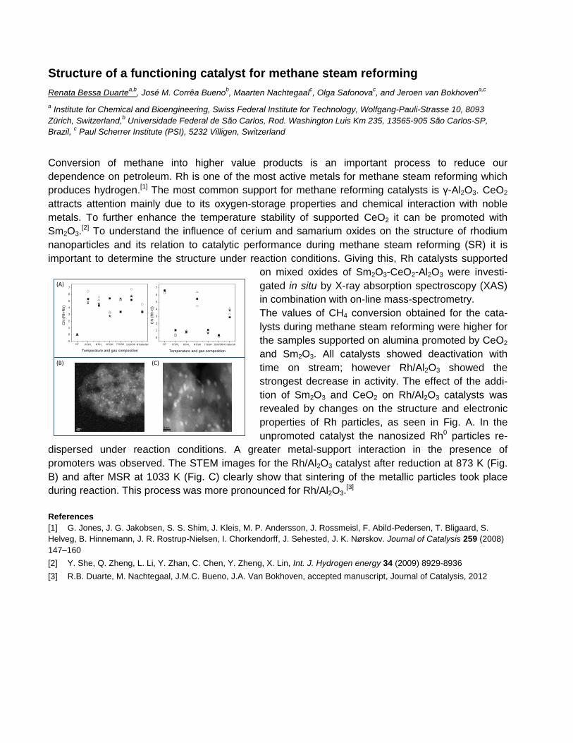
strongest decrease in activity. The effect of the addi-
tion of Sm2O3 and CeO2 on Rh/Al2O3 catalysts was
revealed by changes on the structure and electronic
properties of Rh particles, as seen in Fig. A. In the
unpromoted catalyst the nanosized Rh0 particles re-
dispersed under reaction conditions. A greater metal-support interaction in the presence of
promoters was observed. The STEM images for the Rh/Al2O3 catalyst after reduction at 873 K (Fig.
B) and after MSR at 1033 K (Fig. C) clearly show that sintering of the metallic particles took place
during reaction. This process was more pronounced for Rh/Al2O3.[3]
References
[1] G. Jones, J. G. Jakobsen, S. S. Shim, J. Kleis, M. P. Andersson, J. Rossmeisl, F. Abild-Pedersen, T. Bligaard, S.
Helveg, B. Hinnemann, J. R. Rostrup-Nielsen, I. Chorkendorff, J. Sehested, J. K. Nørskov. Journal of Catalysis 259 (2008)
147–160
[2] Y. She, Q. Zheng, L. Li, Y. Zhan, C. Chen, Y. Zheng, X. Lin, Int. J. Hydrogen energy 34 (2009) 8929-8936
[3] R.B. Duarte, M. Nachtegaal, J.M.C. Bueno, J.A. Van Bokhoven, accepted manuscript, Journal of Catalysis, 2012
-1
0
1
2
3
4
5
6
7
RT
RT/afterSR1033/SR773/SRRT/SRRT/H2
873/H2
CN
(R
h-R
h)
Temperature and gas composition
0
1
2
3
4
5
6
7
RT RT/afterSR1033/SR773/SRRT/SRRT/H2
873/H2
CN
(R
h-O
)
Temperature and gas composition
(A)
(C)(B)
The cold stage: A new sample environment forTOMCAT
Annabelle Medebach1, Bernd R. Pinzer1, Cedric Dubois2, Hans JoergLimbach2, Martin Schneebeli3 and Marco Stampanoni1,4
1Paul Scherrer Institut, Swiss Light Source, 5232 Villigen PSI2Nestle Research Center, Department of Food Science & Technology, 1000 Lausanne 26
3WSL Institute for Snow and Avalanche Research SLF, Snow and Permafrost, 7260 Davos Dorf4Institute of Biomedical Engineering, ETH Zurich, 8092 Zurich, Switzerland
Over the past years the need for special thermal boundary conditions during a scan atthe TOMCAT beamline has been increasing. After commissioning a laser furnace in 2011for high temperature scans we are now commissioning a new dedicated setup for the tem-perature range between −45◦C and +20◦C. This temperature range has been chosen asmany materials, for example snow and ice cream, show interesting structural changes inthis range. This research complements the material science research done in our group athigh temperatures by investigating ternary systems that are highly variable.
In order to study samples at temperatures below room temperature at the TOMCATBeamline so far a CryoJet has been used. The CryoJet can only cool a very small area.This can lead to highly fluctuating temperatures and undesired high temperature gradientsinside the sample. To overcome these problems a new sample environment has been takeninto operation. The cold stage provides temporally and spatially stable thermal boundaryconditions for the samples throughout the measurement including movement into and outof the beam. Additionally, the temperature boundaries during the sample transfer are alsocontrolled. We present the cold stage and its characterization.
The cold stage has been used for first experiments on frozen sucrose in water solution.Using propagation based phase contrast imaging (PCI) we can differentiate the similarlyabsorbing sugar solution from the ice crystals without the addition of contrast agents. Weshow the results of a first time lapse study at constant temperature.
(a) Temperature stability at the sample over10 hours with scans every 30 minutes
(b) Ice crystals in sucrose solutiongrown at −30◦C
High Repetition Rate Laser Pump / X-Ray Probe Studies on Biological Systems, Metal Oxide Nanoparticles and Metal Complexes
J. Rittmann1,3, Ch. Milne2, M. Reinhardt3, M.H. Rittmann-Frank1,3, F. Santomauro1,3, M.Silatani1,3, R. Abela2, and M. Chergui3
1 Swiss Light Source, Paul Scherrer Institut, CH 5232 Villigen, Switzerland 2 SwissFEL, Paul Scherrer Institut, CH 5232 Villigen, Switzerland 3 École Polytechnique Fédérale de Lausanne, Laboratoire de Spectroscopie Ultrarapide, Facultè des Sciences de Base, CH-1015 Lausanne, Switzerland [email protected]
The talk will provide an overview of our recent time-resolved studies on various systems in solution using our recently implemented high repetition rate laser pump / x-ray probe setup [1]. We will present the first picosecond X-ray absorption studies of a protein, myoglobin, in physiological solutions (pH7, room temperature and pressure) where we probed the photo-induced ligand detachment and recombination in solution. By recording the Fe K-edge absorption spectra, we are able to specifically monitor the dynamics at the active site, i.e. the Fe atom in the porphyrin. We performed studies on bare and dye coated metal oxide nanoparticles (NPs), such as TiO2 and ZnO NP as used in dye sensitized solar cells, in order to follow the charge carrier dynamics. The ruthenium dye sensitizer coated TiO2 NPs were probed at the Ti K-edge and the Ru L-edges. We were able to monitor the relaxation dynamics of NPs excited via direct band gap excitation and electron injection via the dye, allowing us to obtain a detailed picture of the electron injection induced electronic and structure changes in the conduction band of the NP. Finally, in studies on metal complexes such as Fe(CN)6 and Cu-diimine complexes, we aim at exploring the role of solvent effects on the relaxation processes of these complexes. The static and transient x-ray absorption spectra allow an unambiguous assignment of electronic and geometric structure in both, the ground and the excited state (cf. fig 1) and provide information about chemical processes such as photoaquation in the case of Fe(CN)6.
Figure 1 left: Transient X-Ray absorption (excited state – ground state) signal at the Fe K edge (blue). The transient can be
explained by a solvation of CN in the excited state and replacement with an H2O molecule (black, calculated). Right:
Structure of the ground state (a) and the excited state (b) of the complex.