21
Small Angle Neutron Scattering Studies on Polymeric Micelles and Lipids System Name: Md. Nasir Uddin Phy-6900, December 03, 2008
| Date post: | 15-Mar-2018 |
| Category: |
Documents |
| Upload: | truongtuyen |
| View: | 216 times |
| Download: | 1 times |
Small Angle Neutron Scattering Studies on
Polymeric Micelles and Lipids System
Name: Md. Nasir Uddin
Phy-6900, December 03, 2008
OutlineOutline
Why Neutron?
Instrumentation
Scattering basics
Contrast variation
Time resolved SANS
Experimental results
Conclusion
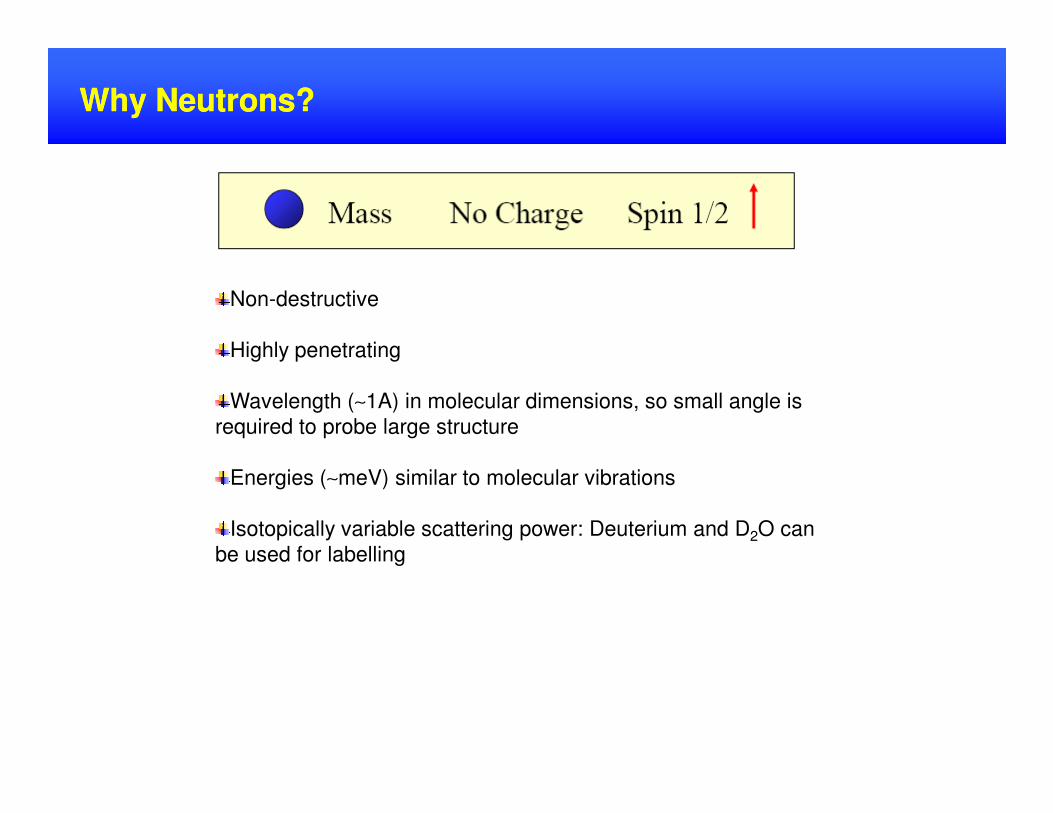
Why Neutrons? Why Neutrons?
Non-destructive
Highly penetrating
Wavelength (∼1A) in molecular dimensions, so small angle is Wavelength (∼1A) in molecular dimensions, so small angle is
required to probe large structure
Energies (∼meV) similar to molecular vibrations
Isotopically variable scattering power: Deuterium and D2O can
be used for labelling
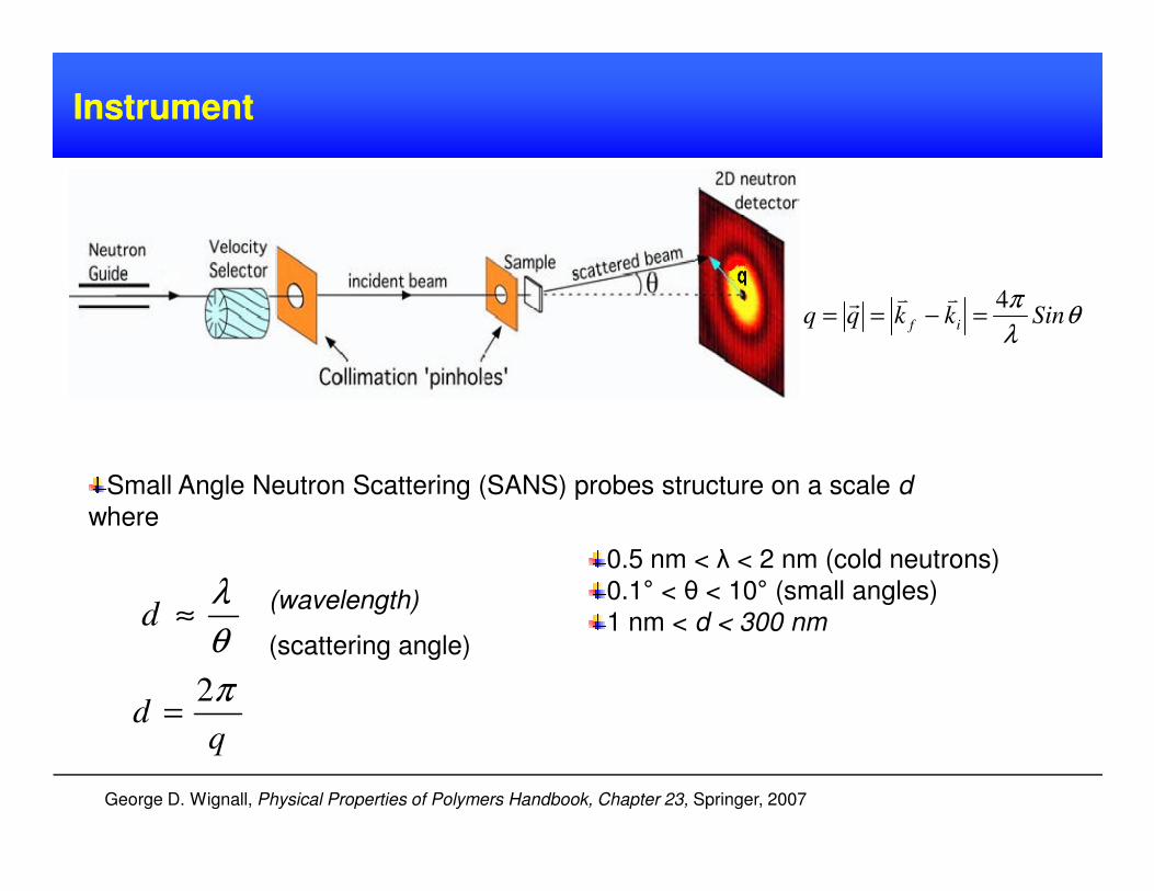
InstrumentInstrument
θλ
πSinkkqq if
4=−==
vvr
George D. Wignall, Physical Properties of Polymers Handbook, Chapter 23, Springer, 2007
0.5 nm < λ < 2 nm (cold neutrons)
0.1° < θ < 10° (small angles)
1 nm < d < 300 nm(wavelength)
(scattering angle)
Small Angle Neutron Scattering (SANS) probes structure on a scale d
where
θ
λ≈d
qd
π2=
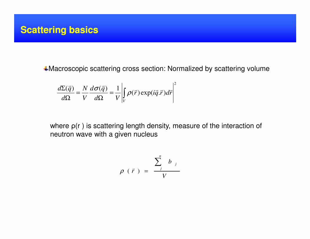
Macroscopic scattering cross section: Normalized by scattering volume
Scattering basicsScattering basics
2
).exp()(1)()(∫=
Ω=
Ω
Σ
V
rdrqirVd
qd
V
N
d
qd vvvvvv
ρσ
V
b
r
n
j
j∑=)(
vρ
where ρ(r ) is scattering length density, measure of the interaction of
neutron wave with a given nucleus
Scattering basicsScattering basics
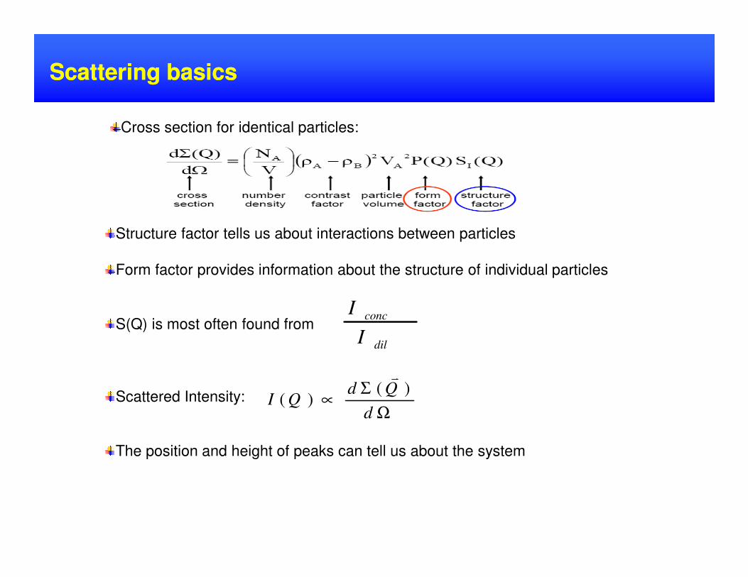
Structure factor tells us about interactions between particles
Form factor provides information about the structure of individual particles
Cross section for identical particles:
S(Q) is most often found from
Scattered Intensity:
The position and height of peaks can tell us about the system
dil
conc
I
I
Ω
Σ∝
d
QdQI
)()(
v
Types of ScatteringTypes of Scattering
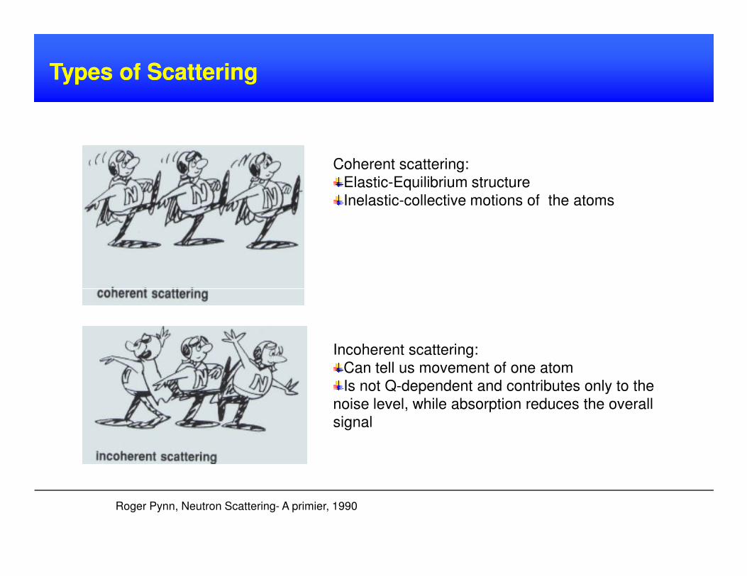
Coherent scattering:
Elastic-Equilibrium structure
Inelastic-collective motions of the atoms
Incoherent scattering:
Can tell us movement of one atom
Is not Q-dependent and contributes only to the
noise level, while absorption reduces the overall
signal
Roger Pynn, Neutron Scattering- A primier, 1990
Contrast VariationContrast Variation
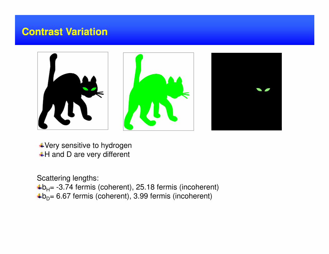
Very sensitive to hydrogen
H and D are very different
Scattering lengths:
bH= -3.74 fermis (coherent), 25.18 fermis (incoherent)
bD= 6.67 fermis (coherent), 3.99 fermis (incoherent)
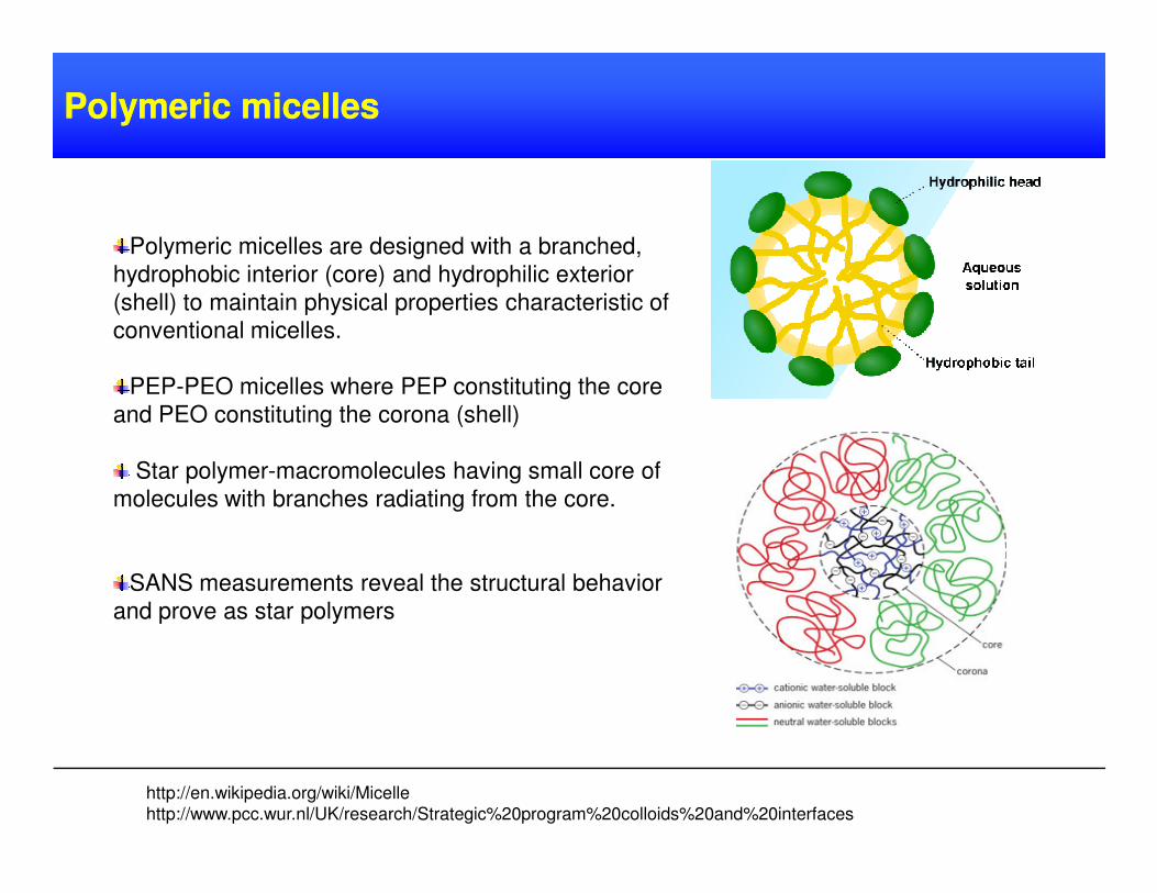
Polymeric micelles are designed with a branched,
hydrophobic interior (core) and hydrophilic exterior
(shell) to maintain physical properties characteristic of
conventional micelles.
PEP-PEO micelles where PEP constituting the core
and PEO constituting the corona (shell)
Polymeric micelles Polymeric micelles
Star polymer-macromolecules having small core of
molecules with branches radiating from the core.
SANS measurements reveal the structural behavior
and prove as star polymers
http://en.wikipedia.org/wiki/Micelle
http://www.pcc.wur.nl/UK/research/Strategic%20program%20colloids%20and%20interfaces
ExperimentalExperimental ResultsResults:: polymericpolymeric micellesmicelles II
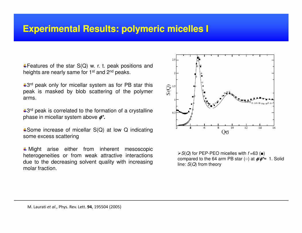
Features of the star S(Q) w. r. t. peak positions andheights are nearly same for 1st and 2nd peaks.
3rd peak only for micellar system as for PB star thispeak is masked by blob scattering of the polymerarms.
3rd peak is correlated to the formation of a crystalline
phase in micellar system above φφφφ*.
M. Laurati et al., Phys. Rev. Lett. 94, 195504 (2005)
phase in micellar system above φφφφ*.
Some increase of micellar S(Q) at low Q indicatingsome excess scattering
Might arise either from inherent mesoscopicheterogeneities or from weak attractive interactionsdue to the decreasing solvent quality with increasingmolar fraction.
S(Q) for PEP-PEO micelles with f =63 ()
compared to the 64 arm PB star () at φφφφ/φφφφ*≈ 1. Solid
line: S(Q) from theory
ExperimentalExperimental ResultsResults :: polymericpolymeric micellesmicelles IIII
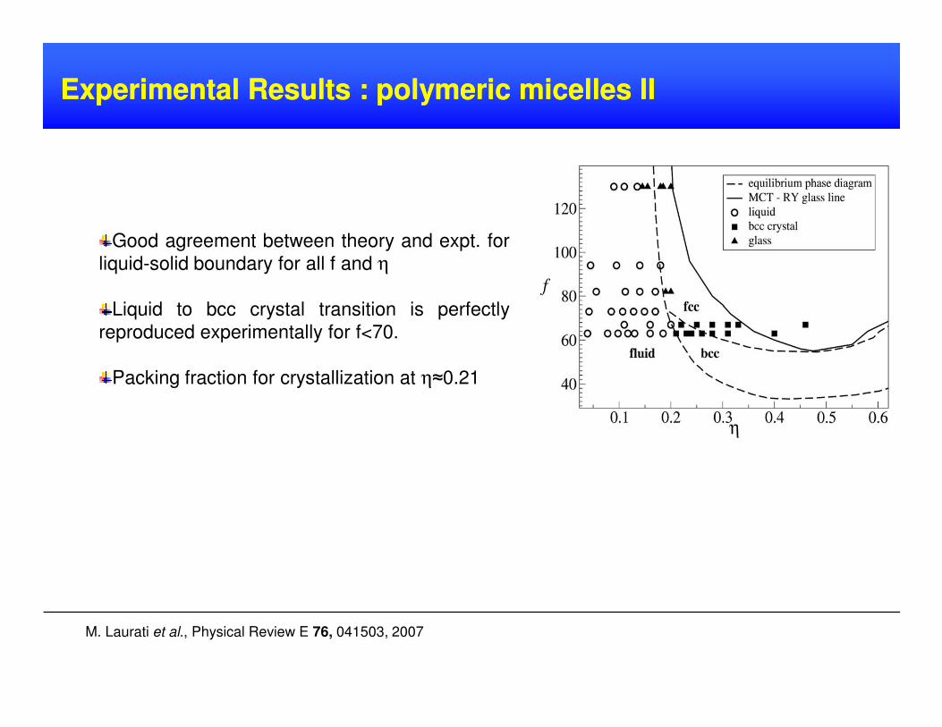
Good agreement between theory and expt. for
liquid-solid boundary for all f and η
Liquid to bcc crystal transition is perfectly
reproduced experimentally for f<70.
Packing fraction for crystallization at η≈0.21
M. Laurati et al., Physical Review E 76, 041503, 2007
ExperimentalExperimental ResultsResults :: polymericpolymeric micellesmicelles IIIIII
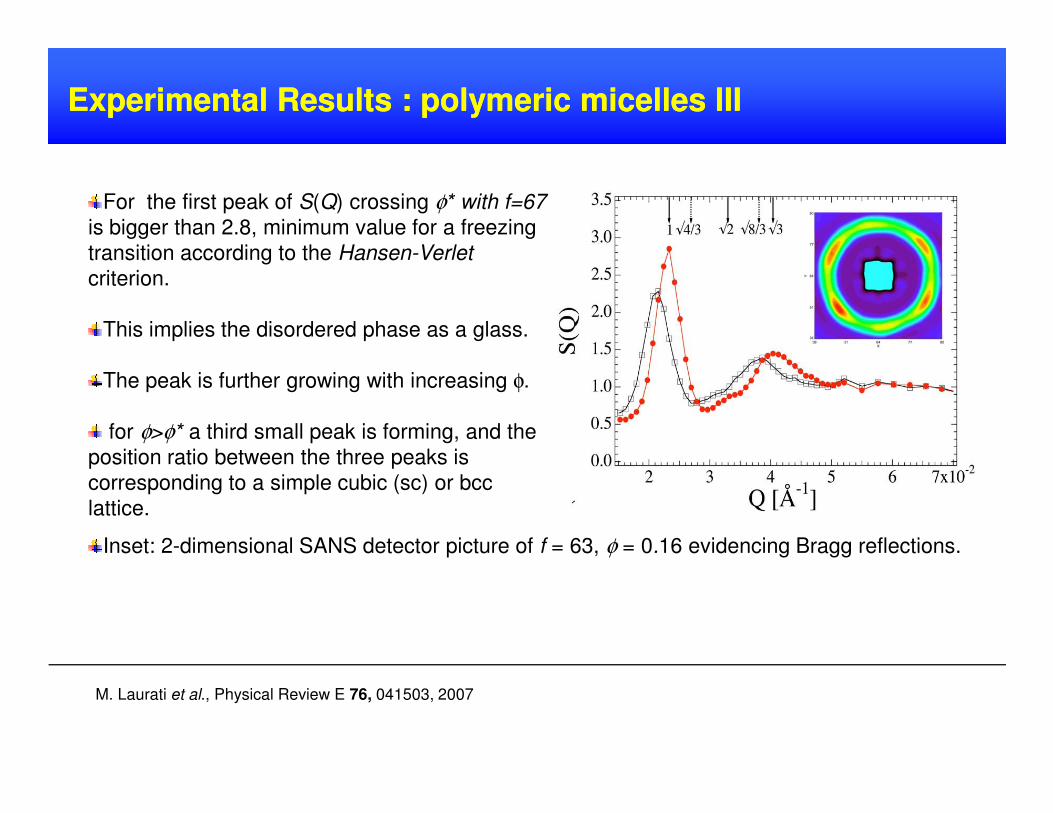
For the first peak of S(Q) crossing φ* with f=67
is bigger than 2.8, minimum value for a freezing
transition according to the Hansen-Verlet
criterion.
This implies the disordered phase as a glass.
The peak is further growing with increasing φ.
Inset: 2-dimensional SANS detector picture of f = 63, φ = 0.16 evidencing Bragg reflections.
for φ>φ* a third small peak is forming, and the
position ratio between the three peaks is
corresponding to a simple cubic (sc) or bcc
lattice.
M. Laurati et al., Physical Review E 76, 041503, 2007
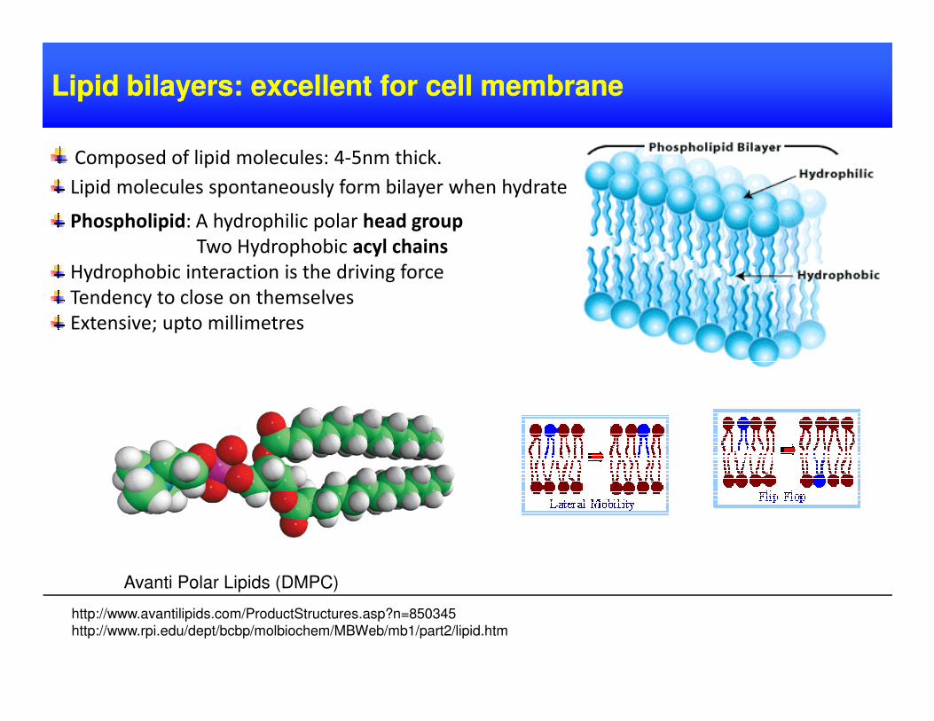
Lipid bilayers: excellent for cell membrane Lipid bilayers: excellent for cell membrane
Composed of lipid molecules: 4-5nm thick.
Lipid molecules spontaneously form bilayer when hydrated
Phospholipid: A hydrophilic polar head group
Two Hydrophobic acyl chains
Hydrophobic interaction is the driving force
Tendency to close on themselves
Extensive; upto millimetres
Avanti Polar Lipids (DMPC)
http://www.avantilipids.com/ProductStructures.asp?n=850345
http://www.rpi.edu/dept/bcbp/molbiochem/MBWeb/mb1/part2/lipid.htm
Describe as sending the neutron beam into a sample and watching how it is temporarily
broaden when reaching the detector after passing through the sample
Useful for kinetics studies because perturbations are inevitable in most experimental
techniques
Time resolved SANS Time resolved SANS
If time scale is shorter - time slicing required
With chopper at the source it is possible to get 50µs - 100ms time resolution.
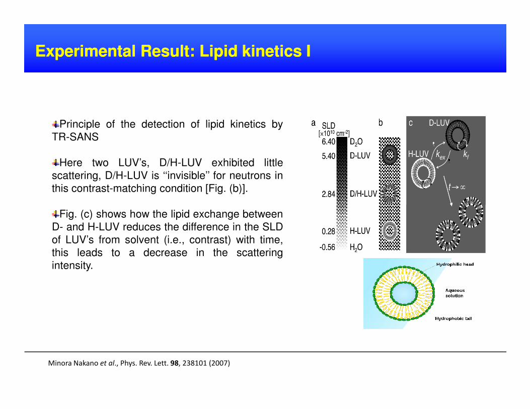
Experimental Result: Lipid kinetics IExperimental Result: Lipid kinetics I
Principle of the detection of lipid kinetics by
TR-SANS
Here two LUV’s, D/H-LUV exhibited little
scattering, D/H-LUV is ‘‘invisible’’ for neutrons in
this contrast-matching condition [Fig. (b)].
Fig. (c) shows how the lipid exchange between
D- and H-LUV reduces the difference in the SLD
of LUV’s from solvent (i.e., contrast) with time,
this leads to a decrease in the scattering
intensity.
Minora Nakano et al., Phys. Rev. Lett. 98, 238101 (2007)
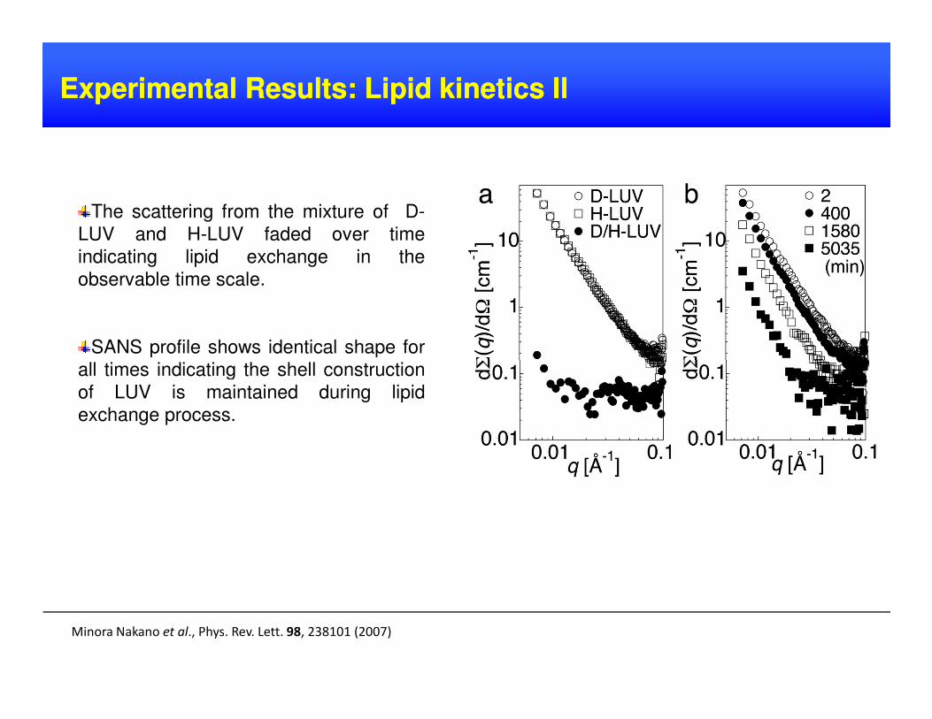
ExperimentalExperimental ResultsResults:: LipidLipid kineticskinetics IIII
The scattering from the mixture of D-
LUV and H-LUV faded over time
indicating lipid exchange in the
observable time scale.
SANS profile shows identical shape for
all times indicating the shell construction
Minora Nakano et al., Phys. Rev. Lett. 98, 238101 (2007)
all times indicating the shell construction
of LUV is maintained during lipid
exchange process.
ExperimentalExperimental ResultsResults :: LipidLipid kineticskinetics IIIIII
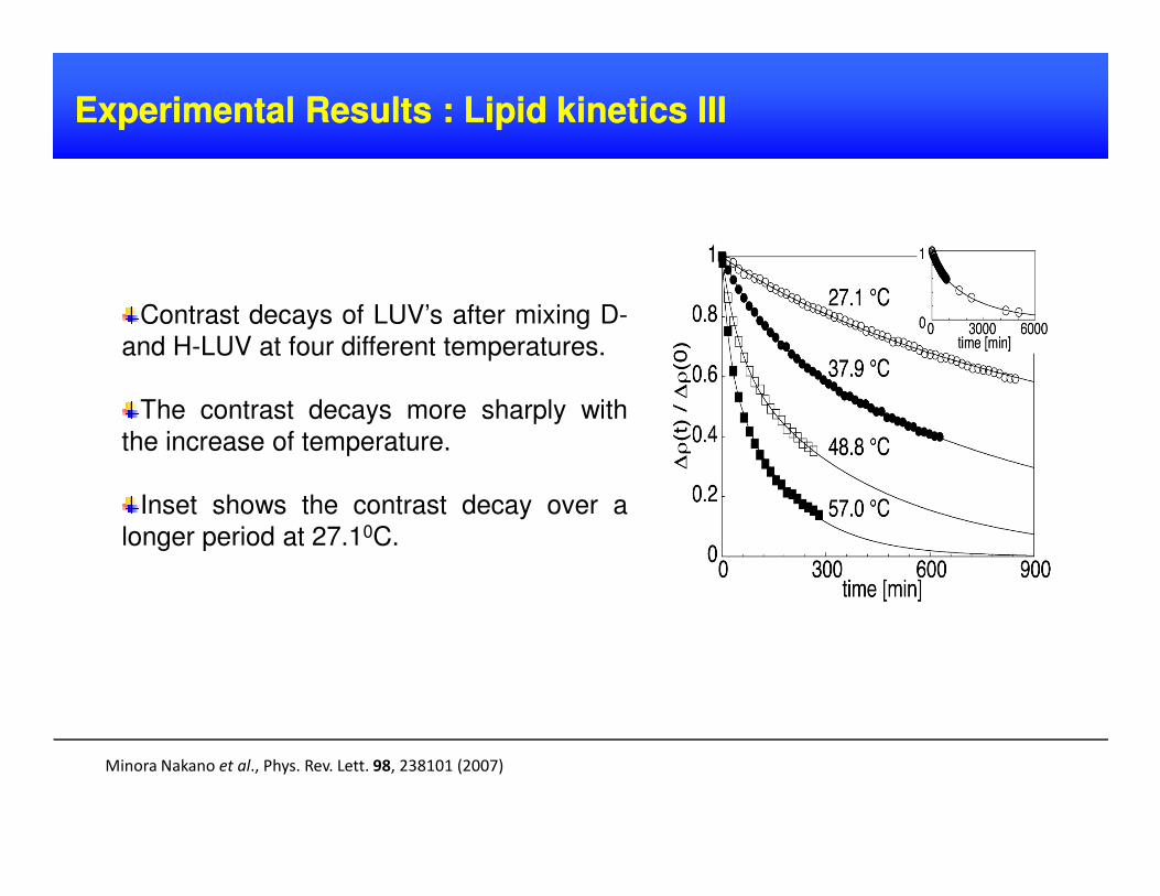
Contrast decays of LUV’s after mixing D-
and H-LUV at four different temperatures.
The contrast decays more sharply with
the increase of temperature.
Minora Nakano et al., Phys. Rev. Lett. 98, 238101 (2007)
the increase of temperature.
Inset shows the contrast decay over a
longer period at 27.10C.
ExperimentalExperimental ResultsResults :: LipidLipid kineticskinetics IVIV
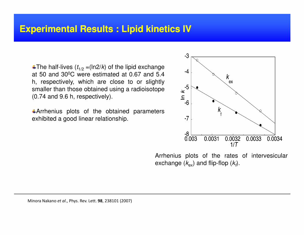
The half-lives (t1/2 =(ln2/k) of the lipid exchange
at 50 and 300C were estimated at 0.67 and 5.4
h, respectively, which are close to or slightly
smaller than those obtained using a radioisotope
(0.74 and 9.6 h, respectively).
Arrhenius plots of the obtained parameters
exhibited a good linear relationship.
Minora Nakano et al., Phys. Rev. Lett. 98, 238101 (2007)
exhibited a good linear relationship.
Arrhenius plots of the rates of intervesicular
exchange (kex) and flip-flop (kf).
SANS is a routine technique available at neutron-scattering facilities associated withresearch nuclear reactors.
No home version of this technique.
Neutron sources are very expensive to build and to maintain.
DrawbacksDrawbacks ofof SANSSANS
Another problem with this technique is that neutron flux is very low.
The interaction of neutrons with matter is weak.
Small-angle neutron scattering (SANS) is a very popular method used by
physicists, material scientists, chemists and biologists.
SANS can determine structures, phase transitions, and morphology.
Possible to do experiments in bulk systems
ConclusionConclusion
TR-SANS is a new method to determine kinetics studies of lipid and
polymeric micelles precisely.

Thank you
Questions?


