NORIKO TANAKA, TAKUHITO TADA, MASAHIRO TOKUNAGA et al. Small-bowel Obstruction due to Pouch-type Internal Hernia through a Defect in the Broad Ligament of the Uterus Diagnosed with Multidetector Computed Tomography: A Case Report. Osaka City Medical Journal. 2017, 63, 111-115
Small-bowel Obstruction due to Pouch-type Internal Hernia through a Defect in the Broad Ligament of
the Uterus Diagnosed with Multidetector Computed Tomography: A Case Report
TAKESHI SUNAMI2), SATOSHI DOISHITA3), and TOHRU TAKESHITA4)
Departments of Radiology1) and Surgery2), Izumi Municipal Hospital; Department of Diagnostic and Interventional Radiology3), Osaka City University Graduate School of Medicine; and Department of Radiology4),
Osaka Prefectural Medical Center for Respiratory and Allergic Diseases
Abstract Small-bowel obstruction due to internal hernia through a defect in the broad ligament of the uterus
is rare, and its clinical diagnosis is usually difficult because of the lack of specific symptoms. This
report presents a case of small-bowel obstruction due to pouch-type internal-hernia through a defect
in the broad ligament, for which multidetector computed tomography proved useful for preoperative
diagnosis. A herniated small-bowel loop appearing as a “sac-like mass” was considered diagnostic for
distinguishing the pouch-type internal hernia from the fenestra-type internal hernia.
enhanced CT of the abdomen and pelvis was immediately performed using a 16-row MDCT scanner
(Aquilion 16, TOSHIBA, Tokyo, Japan). MDCT showed multiple dilated fluid-filled loops of the
proximal small-bowel and collapsed distal ileum, which was consistent with mechanical SBO.
In the pelvic cavity, encapsulated small-bowel with air-fluid was observed as a “sac-like mass” on
the left side of the uterus (Figs. 1, 2, and 3). Within this sac-like mass, crowded mesenteric fat tissue
and vessels were seen as a radial form converging toward the ventral side. Proximal and distal
transition points were identified on the ventral side of the sac-like mass, adjacent to one another,
suggestive of closed-loop SBO (Figs. 1 and 2). At the apex of the sac-like mass, a band-like structure
between the uterus and the left ovary corresponding to the left fallopian tube was stretched and
displaced ventrally (Figs. 1 and 2). Widening of the distance between the uterus and the left ovary
was also noted (Fig. 2). Based on these MDCT findings, a diagnosis of SBO due to IH through a
defect in the left broad ligament, and in particular the pouch-type, was made.
Subseqently, emergency surgery was performed. During surgery, the adhesion was not identified
in abdominal cavity. A moderate quantity of yellow serous ascites was identified. Dilatation of
Figure 1. Contrast-enhanced axial MDCT image shows a sac-like cluster of dilated small-bowel with air-fluid level (white arrows) on the left side of the uterus. The left fallopian tube (black arrows) is observed as a band-like structure. MDCT, multidetector computed tomography.
- 112 -
Tanaka et al
the proximal small-bowel was observed. A 10-cm long ileal loop, which was 30-cm proximal from
the terminal ileum, had herniated through an 8-mm aperture in the anterior leaf of the left broad
ligament (Fig. 4), and was entrapped between the anterior and posterior leaves of the broad ligament
located in the parametrial tissue. Because the entrapped bowel loop could not be liberated, the hernial
orifice was enlarged by a diameter of 7-mm. Then, traction and repositioning of the entrapped bowel
loop were performed. The herniated bowel loop had initially appeared congested, but its color and
peristalsis improved rapidly. No bowel resection was required and the defect was closed to prevent
relapse. The postoperative course was uneventful, and the patient was discharged on postoperative
day 10.
Figure 2. Contrast-enhanced coronal MDCT images are presented from the ventral side a) to the dorsal side c).a) The small-bowel loops with mesenteric fat tissue and vessels (open arrow) have herniated into a defect in the left
broad ligament.b) The herniated small-bowel loops (white arrows) are observed as a “sac-like mass” on the left side of the uterus (U).
The left fallopian tube (black arrows) is stretched and displaced superiorly.c) Widening of the distance between the uterus (U) and the left ovary (white arrowhead) is also noted.MDCT, multidetector computed tomography.
Figure 3. Contrast-enhanced reformatted sagittal MDCT image clearly depicts the encapsulation of the herniated small-bowel loop, as a “sac-like mass” (white arrows). MDCT, multidetector computed tomography.
- 113 -
Small-bowel Obstruction due to Pouch-type Internal Hernia through a Defect in the Broad Ligament of the Uterus
Discussion IH through a defect in the broad ligament is rare, accounting for only 4%-7% of all cases of
IH7-10). According to a report by Baron12), the first case of this type of IH was reported in 1861 during
an autopsy by Quain. Since then, approximately 200 cases have been reported in the English and
Japanese medical literature including both the fenestra-type and the pouch-type13,14). The fenestra-
type is defined as herniation of an abdominal viscus through a full-thickness defect in the broad
ligament. This type involves both the anterior and posterior leaves of the broad ligament, with
no hernial sac. Thus, the herniated viscus is located outside the parametrial tissue, in the pelvic
peritoneum. On the other hand, the pouch-type is defined as herniation of an abdominal viscus
through a defect in only one leaf in the broad ligament. With this type, either the anterior or the
posterior leaf is involved. The hernia sac is the broad ligament itself. The herniated viscus enters
the parametrial tissue and gets trapped in it.
MDCT has proven to be useful in the preoperative diagnosis of various types of IH3-10). Its findings
of fenestra-type IH through a defect in the broad ligament have been reported as follows7-10): 1)
mechanical SBO with a double transition zone (closed-loop SBO) located lateral to the uterus; 2)
a cluster of dilated fluid-filled small-bowel loops in the pelvic cavity; 3) displacement of the uterus
to the contralateral side, displacement of the ipsilateral fallopian tube ventrally, and displacement
of the rectosigmoid colon dorsolaterally by the herniated small-bowel loops; and 4) widening of the
distance between the uterus and ipsilateral ovary. These findings were also observed in the present
pouch-type case, with the exception that no displacement of the uterus or rectosigmoid colon by the
herniated small-bowel loops was recognized.
In this case, the most impressive finding was that the herniated small-bowel loop appeared as
a sac-like mass. Such an appearance is only seen when the herniated small-bowel loops lie within
a small enclosed space6). In the present case, this finding indicated that the hernial sac was lying
within the broad ligament itself and that the contents were trapped in the parametrial tissue. This
finding was considered diagnostic for distinguishing pouch-type IH from fenestra-type IH.
Differential diagnoses for IH through a defect in the broad ligament may indicate sigmoid
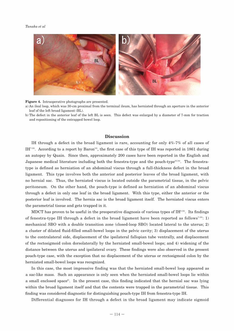
Figure 4. Intraoperative photographs are presented.a) An ileal loop, which was 30-cm proximal from the terminal ileum, has herniated through an aperture in the anterior
leaf of the left broad ligament (BL).b) The defect in the anterior leaf of the left BL is seen. This defect was enlarged by a diameter of 7-mm for traction
and repositioning of the entrapped bowel loop.
- 114 -
Tanaka et al
mesocolon hernia, IH through a peritoneal defect in the pouch of Douglas, and internal supravesical
hernia15-16). The key to differentiating these hernias is to identify the hernial orifice: the orifice of
sigmoid mesocolon hernia is located in the sigmoid mesocolon itself or near the root of the sigmoid
mesocolon, whereas that of IH through a peritoneal defect in the pouch of Douglas is located in the
pelvic floor, and that of supravesical hernia is the supravesical fossa. Furthermore, when a patient
has a history of abdominal or pelvic surgery, SBO due to a fibrous band around the uterus should be
considered among the differential diagnoses.
In conclusion, we encountered a case of SBO due to pouch-type IH through a defect in the broad
ligament. During preoperative diagnosis, MDCT provided several useful findings suggesting this
extremely rare disease. A herniated small-bowel loop appearing as a “sac-like mass” was considered
the diagnostic key for distinguishing pouch-type IH from fenestra-type IH. Therefore when clinical
manifestations and abdominal radiography suggest SBO, MDCT is recommended.
References 1.Martin LC, Merkle EM Thompson WM. Review of internal hernias: radiographic and clinical findings. AJR
Am J Roentgenol 2006;186:703-717. 2. Hunt AB. Fenestrate and pouches in the broad ligament as an actual and potential cause of strangulated
intraabdominal hernia. Surgery, Gynecology and Obstetrics 1934;58:906-913. 3. Coulier B, Broze B, Mailleux P, Maldague P. Small-bowel internal herniation through the falciform ligament:
64-row MDCT diagnosis. Emerg Radiol 2010;17:73-78. 4. Azar AR, Abraham C, Coulier B, Broze B. Ileocecal herniation through the foramen of Winslow: MDCT
diagnosis. Abdom Imaging 2010;35:574-577. 5. Liao YH, Lin CH, Lin WC. Right paraduodenal hernia: characteristic MDCT findings. Abdom Imaging 2011;
36:130-133. 6. Hongo N, Mori H, Matsumoto S, Okino Y, Takaji R, Komatsu E. Internal hernias after abdominal surgeries:
MDCT features. Abdom Imaging 2011;36:349-362. 7. Mailleux P, Ramboux A. Small bowel obstruction due to an internal herniation through a defect of the broad
ligament. JBR- BTR 2010;93:201-203. 8. Kosaka N, Uematsu H, Kimura H, Yamamori S, Hirano K, Itoh H. Utility of multi-detector CT for pre-operative
diagnosis of internal hernia through a defect in the broad ligament (2007: 1b). Eur Radiol 2007;17:1130-1133. 9. Barbier Brion B, Daragon C, Idelcadi O, Mantion G, Kastler B, Delabrousse E. Small bowel obstruction due to
broad ligament hernia: computed tomography findings. Hernia 2011;15:353-355.10. Quiroga S, Sarrias M, Sánchez JL, Rivero J. Small bowel obstruction secondary to internal hernia through a
defect of the broad ligament: preoperative multi-detector CT diagnosis. Abdom Imaging 2012;37:1089-1091.11. Aufort S, Charra L, Lesnik A, Bruel JM, Taourel P. Multidetector CT of bowel obstruction: value of post-
processing. Eur Radiol 2005;15:2323-2329.12. Baron A. Defect in the broad ligament and its association with intestinal strangulation. Br J Surg 1948;36:91-
94.13. Haku T, Daidouji K, Kawamura H, Matsuzaki M. Internal herniation through a defect of the broad ligament
of the uterus. Abdom Imaging 2004;29:161-163.14. Tanioka Y, Hirano A, Okita K, Kimura T, Kato A, Yamashita S, et al. A case report of internal hernia through
an abnormal defect in the broad ligament of the uterus. Nihon Shokakibyo Gakkai Zasshi 2010;107:620-624. (In Japanese).
15. Takeyama N, Gokan T, Ohgiya Y, Satoh S, Hashizume T, Hataya K, et al. CT of internal hernias. Radiographics 2005;25:997-1015.
16. Doishita S, Takeshita T, Uchima Y, Kawasaki M, Shimono T, Yamashita A, et al. Internal hernias in the era of multidetector CT: correlation of imaging and surgical findings. Radiographics 2016;36:88-106.
- 115 -
Small-bowel Obstruction due to Pouch-type Internal Hernia through a Defect in the Broad Ligament of the Uterus


![SMALL BOWEL OBSTRUCTION IN TROCAR SITE HERNIA. CASE …€¦ · hernia repair at the moment of diagnosis and if the incarcerated loop is vital. [4] Trocar site hernias are one of](https://static.documents.pub/doc/80x56/5f0cf7db7e708231d43805c7/small-bowel-obstruction-in-trocar-site-hernia-case-hernia-repair-at-the-moment.jpg)