Anna Tramontano,§ Claudia Bonaccini,§,+ Patrick Trojer,| Ingo Bauer,⊥ Gerald Brosch,*,⊥ and Gianluca Sbardella*,‡
Istituto Pasteur - Fondazione Cenci Bolognetti, Dipartimento di Studi Farmaceutici, UniVersita degli Studi di Roma “La Sapienza”, P.le AldoMoro 5, I-00185 Roma, Dipartimento di Scienze Farmaceutiche, UniVersita degli Studi di Salerno, Via Ponte Don Melillo, I-84084 Fisciano(SA), Dipartimento di Scienze Biochimiche, A. Rossi Fanelli, UniVersita degli Studi di Roma “La Sapienza”, P.le Aldo Moro 5, 00185 Roma,Howard Hughes Medical Institute, Department of Biochemistry, DiVision of Nucleic Acids Enzymology, Robert Wood Johnson Medical School,UniVersity of Medicine and Dentistry of New Jersey, 683 Hoes Lane, Piscataway, New Jersey 08854, and DiVision of Molecular Biology,Biocenter-Innsbruck Medical UniVersity, Fritz-Preglstrasse 3, 6020 Innsbruck, Austria
ReceiVed October 17, 2006
The screening of the inhibition capabilities of dye-like small molecules from a focused library against bothhuman PRMT1 andAspergillus nidulansRmtA is reported as well as molecular modeling studies (homologymodeling, molecular docking, and 3-D QSAR) of the catalytic domain of the PRMT1 fungal homologueRmtA. The good correlation between computational and biological results makes RmtA a reliable tool forscreening arginine methyltransferase inhibitors. In addition, the binding mode analyses of tested derivativesreveal the crucial role of two regions, the pocket formed by Ile12, His13, Met16, and Thr49 and the SAMcisteinic binding site subsite. These regions should be taken into account in the design of novel PRMTinhibitors.
Introduction
Similar to other posttranslational covalent modifications, suchas phosphorylation, acetylation, and ubiquitination, histonemethylation regulates a broad range of DNA and chromatin-templated nuclear events, including transcription.1,2 Histones canbe methylated on lysine as well as arginine residues, preferen-tially on the amino-terminal tails of histones H3 and H4, andthis methylation is a stable epigenetic mark that does not alterthe overall charge of the histone tails. Nevertheless, the recentidentification and structural characterization of “histonedemethylases”3-12 led us to consider that histone methylationmight also play a dynamic role in the epigenetic code.3-5
However, with increasing methylation13 comes an increase inbasicity, hydrophobicity, and an influence on the affinity foranionic molecules such as DNA.14,15 As a matter of fact,similarly to histone acetylation, histone methylation can modu-late histone interaction with DNA and chromatin associatedproteins, resulting in an alteration of nucleosomal structures andfunctions and ultimately contributing to different biologicalprocesses.16 Histone methyltransferases display remarkablespecificity in the level of methylation they catalyze, and thelatest findings suggest that this could have functional signifi-cance in transcription.17,18The nine mammalian protein argininemethyltransferases (PRMTs) thus far identified19,20 share a
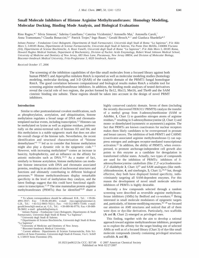
highly conserved catalytic domain. Seven of them (includingthe recently discovered FBXO11/ PRMT9) catalyze the transferof a methyl group fromS-adenosylmethionine (SAM orAdoMet, 1, Chart 1) to guanidino nitrogen atoms of arginineresidues,21 resulting inS-adenosylhomocysteine (2, Chart 1) andmono- or dimethylated (symmetric or asymmetric) arginine. Thefact that PRMTs are known coactivators for nuclear receptorsmakes them likely candidates to be overexpressed in prostateand breast cancers. The inhibition of both PRMT1 and CARM1(coactivator-associated arginine methyltransferase-1) can sup-press estrogen and androgen receptor-mediated transcriptionalactivation.22 In addition, the ability of PRMT5, when overex-pressed, to promote anchorage-independent cell growth alsopoints to this enzyme as a candidate for deregulation intransformed cellular states. Actually, two types of compoundsare used for the inhibition of PRMTs: inhibitors ofS-adenosylhomocysteine catabolism (like 2′,3′-acycloadenosine-2′,3′-dialdehyde3, Chart 1)23 and SAM analogues (like meth-ylthioadenosine,4, and sinefungin,5, Chart 1),24,25but, thougheffective, they both have displayed limited specificity, indis-criminately targeting all SAM-dependent enzymes. For thisreason the development of novel small molecule selectiveinhibitors of PRMTs is highly desirable.
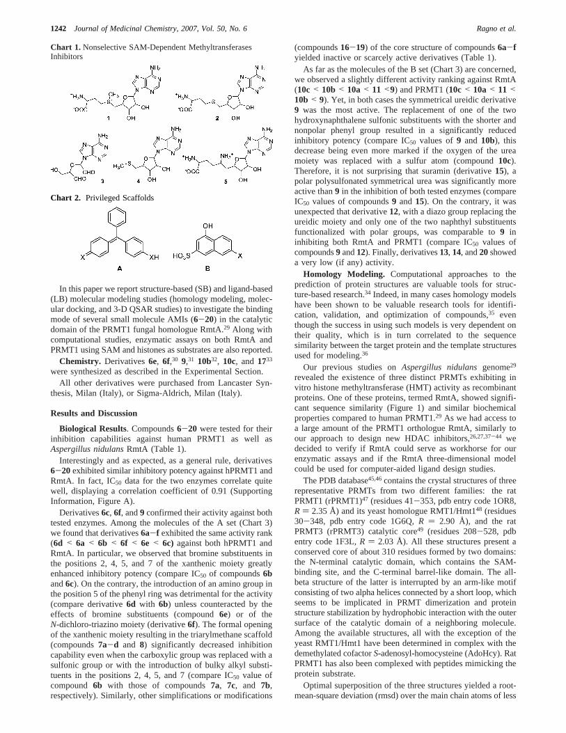
Recently a few compounds selected through a randomscreening were described as reversible arginine methyltrans-ferase inhibitors (AMIs) by Bedford and co-workers.22 Beinginterested in small molecule modulators of epigenetic targetsand, particularly, of histone-modifying enzymes,26-28 we focusedour attention on AMI structures and noticed that all of themwere dyes or dye-like derivatives. Particularly, two scaffolds(A andB, Chart 2) emerged as privileged ones.
This finding, together with the aim to develop a rationalapproach toward arginine methyltransferase inhibitors, promptedus to explore the affinity for the target binding site of selectedAMIs as well as of a focused library (Chart 3) of dye-like smallmolecule compounds (mostly containing privileged structuresscaffoldsA andB).
* To whom correspondence should be addressed. R.R., Tel.:+39-06-4991-3937; Fax: +39-06-491491; e-mail: [email protected];G.B., Tel.: +43-512-9003-70211; Fax:+43-512-9003-73100; e-mail:[email protected]; G.S., Tel.:+39-089-96-9770; Fax:+39-089-96-9602; e-mail: [email protected].
† Istituto Pasteur - Fondazione Cenci Bolognetti, Dipartimento di StudiFarmaceutici, Universita` degli Studi di Roma “La Sapienza”.
‡ Universitadegli Studi di Salerno.§ Dipartimento di Scienze Biochimiche, Universita` degli Studi di Roma
“La Sapienza”.| University of Medicine and Dentistry of New Jersey.⊥ Biocenter-Innsbruck Medical University.+ Current address: Dipartimento di Scienze Farmaceutiche, Polo Sci-
entifico di Sesto Fiorentino, Universita` degli Studi di Firenze, Via U. Schiff6, I-50019 Sesto Fiorentino (FI).
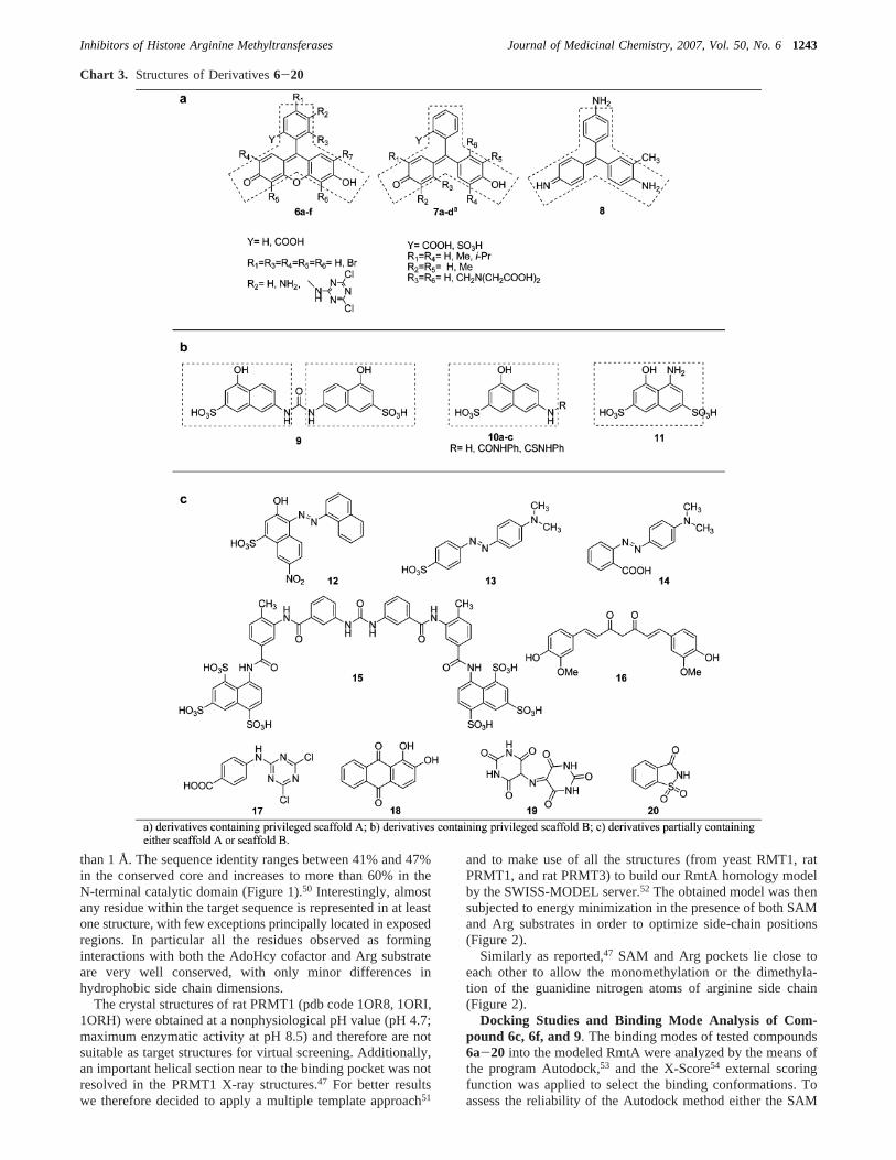
In this paper we report structure-based (SB) and ligand-based(LB) molecular modeling studies (homology modeling, molec-ular docking, and 3-D QSAR studies) to investigate the bindingmode of several small molecule AMIs (6-20) in the catalyticdomain of the PRMT1 fungal homologue RmtA.29 Along withcomputational studies, enzymatic assays on both RmtA andPRMT1 using SAM and histones as substrates are also reported.
Chemistry. Derivatives6e, 6f,30 9,31 10b32, 10c, and 1733
were synthesized as described in the Experimental Section.All other derivatives were purchased from Lancaster Syn-
thesis, Milan (Italy), or Sigma-Aldrich, Milan (Italy).
Results and Discussion
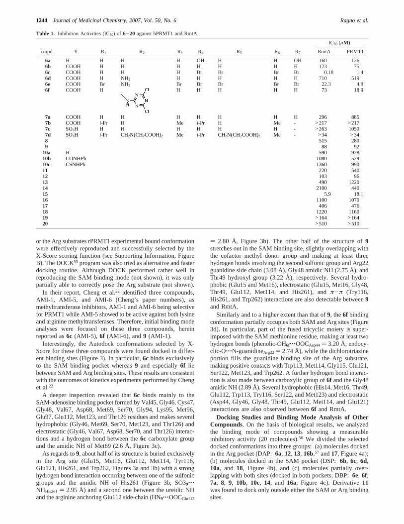
Biological Results. Compounds6-20 were tested for theirinhibition capabilities against human PRMT1 as well asAspergillus nidulansRmtA (Table 1).
Interestingly and as expected, as a general rule, derivatives6-20exhibited similar inhibitory potency against hPRMT1 andRmtA. In fact, IC50 data for the two enzymes correlate quitewell, displaying a correlation coefficient of 0.91 (SupportingInformation, Figure A).
Derivatives6c, 6f, and9 confirmed their activity against bothtested enzymes. Among the molecules of the A set (Chart 3)we found that derivatives6a-f exhibited the same activity rank(6d < 6a < 6b < 6f < 6e < 6c) against both hPRMT1 andRmtA. In particular, we observed that bromine substituents inthe positions 2, 4, 5, and 7 of the xanthenic moiety greatlyenhanced inhibitory potency (compare IC50 of compounds6band6c). On the contrary, the introduction of an amino group inthe position 5 of the phenyl ring was detrimental for the activity(compare derivative6d with 6b) unless counteracted by theeffects of bromine substituents (compound6e) or of theN-dichloro-triazino moiety (derivative6f). The formal openingof the xanthenic moiety resulting in the triarylmethane scaffold(compounds7a-d and 8) significantly decreased inhibitioncapability even when the carboxylic group was replaced with asulfonic group or with the introduction of bulky alkyl substi-tuents in the positions 2, 4, 5, and 7 (compare IC50 value ofcompound 6b with those of compounds7a, 7c, and 7b,respectively). Similarly, other simplifications or modifications
(compounds16-19) of the core structure of compounds6a-fyielded inactive or scarcely active derivatives (Table 1).
As far as the molecules of the B set (Chart 3) are concerned,we observed a slightly different activity ranking against RmtA(10c< 10b < 10a< 11 <9) and PRMT1 (10c< 10a< 11 <10b < 9). Yet, in both cases the symmetrical ureidic derivative9 was the most active. The replacement of one of the twohydroxynaphthalene sulfonic substituents with the shorter andnonpolar phenyl group resulted in a significantly reducedinhibitory potency (compare IC50 values of9 and 10b), thisdecrease being even more marked if the oxygen of the ureamoiety was replaced with a sulfur atom (compound10c).Therefore, it is not surprising that suramin (derivative15), apolar polysulfonated symmetrical urea was significantly moreactive than9 in the inhibition of both tested enzymes (compareIC50 values of compounds9 and15). On the contrary, it wasunexpected that derivative12, with a diazo group replacing theureidic moiety and only one of the two naphthyl substituentsfunctionalized with polar groups, was comparable to9 ininhibiting both RmtA and PRMT1 (compare IC50 values ofcompounds9 and12). Finally, derivatives13, 14, and20showeda very low (if any) activity.
Homology Modeling. Computational approaches to theprediction of protein structures are valuable tools for struc-ture-based research.34 Indeed, in many cases homology modelshave been shown to be valuable research tools for identifi-cation, validation, and optimization of compounds,35 eventhough the success in using such models is very dependent ontheir quality, which is in turn correlated to the sequencesimilarity between the target protein and the template structuresused for modeling.36
revealed the existence of three distinct PRMTs exhibiting invitro histone methyltransferase (HMT) activity as recombinantproteins. One of these proteins, termed RmtA, showed signifi-cant sequence similarity (Figure 1) and similar biochemicalproperties compared to human PRMT1.29 As we had access toa large amount of the PRMT1 orthologue RmtA, similarly toour approach to design new HDAC inhibitors,26,27,37-44 wedecided to verify if RmtA could serve as workhorse for ourenzymatic assays and if the RmtA three-dimensional modelcould be used for computer-aided ligand design studies.
The PDB database45,46contains the crystal structures of threerepresentative PRMTs from two different families: the ratPRMT1 (rPRMT1)47 (residues 41-353, pdb entry code 1OR8,R ) 2.35 Å) and its yeast homologue RMT1/Hmt148 (residues30-348, pdb entry code 1G6Q,R ) 2.90 Å), and the ratPRMT3 (rPRMT3) catalytic core49 (residues 208-528, pdbentry code 1F3L,R ) 2.03 Å). All these structures present aconserved core of about 310 residues formed by two domains:the N-terminal catalytic domain, which contains the SAM-binding site, and the C-terminal barrel-like domain. The all-beta structure of the latter is interrupted by an arm-like motifconsisting of two alpha helices connected by a short loop, whichseems to be implicated in PRMT dimerization and proteinstructure stabilization by hydrophobic interaction with the outersurface of the catalytic domain of a neighboring molecule.Among the available structures, all with the exception of theyeast RMT1/Hmt1 have been determined in complex with thedemethylated cofactorS-adenosyl-homocysteine (AdoHcy). RatPRMT1 has also been complexed with peptides mimicking theprotein substrate.
Optimal superposition of the three structures yielded a root-mean-square deviation (rmsd) over the main chain atoms of less
1242 Journal of Medicinal Chemistry, 2007, Vol. 50, No. 6 Ragno et al.
than 1 Å. The sequence identity ranges between 41% and 47%in the conserved core and increases to more than 60% in theN-terminal catalytic domain (Figure 1).50 Interestingly, almostany residue within the target sequence is represented in at leastone structure, with few exceptions principally located in exposedregions. In particular all the residues observed as forminginteractions with both the AdoHcy cofactor and Arg substrateare very well conserved, with only minor differences inhydrophobic side chain dimensions.
The crystal structures of rat PRMT1 (pdb code 1OR8, 1ORI,1ORH) were obtained at a nonphysiological pH value (pH 4.7;maximum enzymatic activity at pH 8.5) and therefore are notsuitable as target structures for virtual screening. Additionally,an important helical section near to the binding pocket was notresolved in the PRMT1 X-ray structures.47 For better resultswe therefore decided to apply a multiple template approach51
and to make use of all the structures (from yeast RMT1, ratPRMT1, and rat PRMT3) to build our RmtA homology modelby the SWISS-MODEL server.52 The obtained model was thensubjected to energy minimization in the presence of both SAMand Arg substrates in order to optimize side-chain positions(Figure 2).
Similarly as reported,47 SAM and Arg pockets lie close toeach other to allow the monomethylation or the dimethyla-tion of the guanidine nitrogen atoms of arginine side chain(Figure 2).
Docking Studies and Binding Mode Analysis of Com-pound 6c, 6f, and 9. The binding modes of tested compounds6a-20 into the modeled RmtA were analyzed by the means ofthe program Autodock,53 and the X-Score54 external scoringfunction was applied to select the binding conformations. Toassess the reliability of the Autodock method either the SAM
Chart 3. Structures of Derivatives6-20
Inhibitors of Histone Arginine Methyltransferases Journal of Medicinal Chemistry, 2007, Vol. 50, No. 61243
or the Arg substrates rPRMT1 experimental bound conformationwere effectively reproduced and successfully selected by theX-Score scoring function (see Supporting Information, FigureB). The DOCK55 program was also tried as alternative and fasterdocking routine. Although DOCK performed rather well inreproducing the SAM binding mode (not shown), it was onlypartially able to correctly pose the Arg substrate (not shown).
In their report, Cheng et al.22 identified three compounds,AMI-1, AMI-5, and AMI-6 (Cheng’s paper numbers), asmethyltransferase inhibitors, AMI-1 and AMI-6 being selectivefor PRMT1 while AMI-5 showed to be active against both lysineand arginine methyltransferases. Therefore, initial binding modeanalyses were focused on these three compounds, hereinreported as6c (AMI-5), 6f (AMI-6), and 9 (AMI-1).
Interestingly, the Autodock conformations selected by X-Score for these three compounds were found docked in differ-ent binding sites (Figure 3). In particular,6c binds exclusivelyto the SAM binding pocket whereas9 and especially6f liebetween SAM and Arg binding sites. These results are consistentwith the outcomes of kinetics experiments performed by Chenget al.22
A deeper inspection revealed that6c binds mainly to theSAM-adenosine binding pocket formed by Val45, Gly46, Cys47,Gly48, Val67, Asp68, Met69, Ser70, Gly94, Lys95, Met96,Glu97, Glu112, Met123, and Thr126 residues and makes severalhydrophobic (Gly46, Met69, Ser70, Met123, and Thr126) andelectrostatic (Gly46, Val67, Asp68, Ser70, and Thr126) interac-tions and a hydrogen bond between the6c carboxylate groupand the amidic NH of Met69 (2.6 Å, Figure 3c).
As regards to9, about half of its structure is buried exclusivelyin the Arg site (Glu15, Met16, Glu112, Met114, Tyr116,Glu121, His261, and Trp262, Figures 3a and 3b) with a stronghydrogen bond interaction occurring between one of the sulfonicgroups and the amidic NH of His261 (Figure 3b, SO39‚‚‚NHHis261 ) 2.95 Å) and a second one between the ureidic NHand the arginine anchoring Glu112 side-chain (HN9‚‚‚OOCGlu112
) 2.80 Å, Figure 3b). The other half of the structure of9stretches out in the SAM binding site, slightly overlapping withthe cofactor methyl donor group and making at least threehydrogen bonds involving the second sulfonic group and Arg22guanidine side chain (3.08 Å), Gly48 amidic NH (2.75 Å), andThr49 hydroxyl group (3.22 Å), respectively. Several hydro-phobic (Glu15 and Met16), electrostatic (Glu15, Met16, Gly48,Thr49, Glu112, Met114, and His261), andπ-π (Try116,His261, and Trp262) interactions are also detectable between9and RmtA.
Similarly and to a higher extent than that of9, the6f bindingconformation partially occupies both SAM and Arg sites (Figure3d). In particular, part of the fused tricyclic moiety is super-imposed with the SAM methionine residue, making at least twohydrogen bonds (phenolic-OH6f‚‚‚OOCAsp44 ) 3.20 Å; endocy-clic-O‚‚‚N-guanidineArg22 ) 2.74 Å), while the dichlorotriazineportion fills the guanidine binding site of the Arg substrate,making positive contacts with Trp113, Met114, Gly115, Glu121,Ser122, Met123, and Trp262. A further hydrogen bond interac-tion is also made between carboxylic group of6f and the Gly48amidic NH (2.89 Å). Several hydrophobic (His14, Met16, Thr49,Glu112, Trp113, Try116, Ser122, and Met123) and electrostatic(Asp44, Gly46, Gly48, Thr49, Glu112, Met114, and Glu121)interactions are also observed between6f and RmtA.
Docking Studies and Binding Mode Analysis of OtherCompounds. On the basis of biological results, we analyzedthe binding mode of compounds showing a measurableinhibitory activity (20 molecules).56 We divided the selecteddocked conformations into three groups: (a) molecules dockedin the Arg pocket (DAP:6a, 12, 13, 16b,57 and17, Figure 4a);(b) molecules docked in the SAM pocket (DSP:6b, 6c, 6d,10a, and 18, Figure 4b), and (c) molecules partially over-lapping with both sites (docked in both pockets, DBP:6e, 6f,7a, 8, 9, 10b, 10c, 14, and 16a, Figure 4c). Derivative11was found to dock only outside either the SAM or Arg bindingsites.
Table 1. Inhibition Activities (IC50) of 6-20 against hPRMT1 and RmtA
IC50 (µM)
cmpd Y R1 R2 R3 R4 R5 R6 R7 RmtA PRMT1
6a H H H H OH H H OH 160 1266b COOH H H H H H H H 123 756c COOH H H H Br Br Br Br 0.18 1.46d COOH H NH2 H H H H H 710 5196e COOH Br NH2 Br Br Br Br Br 22.3 4.86f COOH H H H H H H 73 18.9
7a COOH H H H H H H H 296 8857b COOH i-Pr H Me i-Pr H Me - >217 >2177c SO3H H H H H H H - >263 10507d SO3H i-Pr CH2N(CH2COOH)2 Me i-Pr CH2N(CH2COOH)2 Me - >34 >348 515 2809 88 92
1244 Journal of Medicinal Chemistry, 2007, Vol. 50, No. 6 Ragno et al.
As illustrated in Figure 4a, the molecules of the DAP groupoccupy the Arg pocket plus a portion of the area surroundingthe entry site of the substrate side chain (not shown). Theformation of a hydrogen bond with the Glu15 side-chaincarboxyl is the most common interaction in this group, notice-able for three compounds (6a, 16b, and17) out of five. Amongthem,6a, the second most active molecule of the set, also makespositive interactions with the hydroxyl of Tyr116 and amidicnitrogen of His261. Moreover, the phenyl group seems to makea π-π interaction with the indole ring of Trp262, while the
tricycle portion is lying between Met16 and His261 side chains(Supporting Information, Figure C).
On the other hand, derivative12, the most active of the DAPgroup, makes important hydrogen bonding interactions betweenits sulfonic group and the guanidine moiety of Arg295. Inaddition the unsubstituted naphthyl ring stacks between theTrp262 indole and the Tyr116 phenol rings makingπ-πinteractions. (Supporting Information, Figure D). As regards tothe less active derivatives13, 16b, and 17, although a fewrelevant interactions (i.e., hydrogen bond with Glu15) arepresent, as in the case of6a and 12, major hydrophobic andelectrostatic interactions are lacking (Supporting Information,Figure E). Additional docking studies performed in the presenceof the SAM cofactor revealed similar results with differencesonly for compounds12 and17. (See Supporting Information,Figure F): derivative12displays a different binding pose, while17 is shifted toward SAM and the negatively charged carboxy-late seems to make electrostatic interaction with the positivelycharged SAM methyl group. No relevant interactions are madebetween6a, 13, 12, and16b with SAM.
As regards to compounds of DSP group, it is noteworthy thatthe two similar derivatives6b and6d display a different bindingscenario from the one above-described for6c, as they are shiftedtoward the Arg site and while losing the interactions with amidicnitrogen Asp68, they gain some interactions with key residuesat the interface between the SAM and Arg sites (Glu112 orAsp68 carboxylate for6b or 6d, respectively; see SupportingInformation, Figure G). Compounds10a and 18 occupy thesame space but, though making a few important interactionssuch as an hydrogen bond with the Thr49 hydroxyl, most
Figure 1. Sequence alignment of the targetA. nidulans(A.N.) RmtA and human PRMT1to the structural alignment of yeast RMT1, rat PRMT1,and rat PRMT3. Cylinders and arrows indicate the common secondary structural elements (helices and strands, respectively) in the template structures.Red boxes point out the catalytic domain regions. Residue numbering is indicated on the left of each sequence row. Small letters indicate residueswhich are not structurally aligned. Identical residues between target and templates are bold. Residues highlighted in yellow are identical in all thesequences. Red asterisks indicate residues having a role in cofactor binding and/or catalytic mechanism; blue asterisks indicate residues importantfor the dimerization process.
Figure 2. Superimposition of RmtA homology model (blue) andrPRMT1 X-rays structure (green). The SAM binding site, the Argbinding site, and the backbone of histone H4 are highlighted in yellow,cyan, and purple, respectively.
Inhibitors of Histone Arginine Methyltransferases Journal of Medicinal Chemistry, 2007, Vol. 50, No. 61245
hydrophobic interactions are lacking due to their smaller size(Supporting Information, Figure H).
The docking area of the third group of molecules (DBP),partially overlapping both Arg and SAM sites, indicates adifferent interaction profile. Taken together, the molecules ofthis group make contacts within 3 Å that define a binding sitecomprising at least 18 residues (Ile12, His13, Glu15, Leu17,Arg22, Gly46, Cys47, Gly48, Thr49, Leu52, Asp68, Glu112,Met114, Gly115, Tyr116, Glu121, His261, and Trp262, Sup-
porting Information, Figure I). Among these residues, Arg22(6f, 10b and14), Gly46 (6f, 7a, 10, and14), Glu112 (6f, 7a, 8,9, 10b, 10c, 14, and16a), Met114 (6f, 7a, 10b, and10c), andHis261 (9, 10c, and 16a) are the most involved in ligandbinding. The most active compound6eshows peculiar hydrogenbonding interactions: a strong one with the hydroxyl group ofTyr116 (3.07 Å) and a weak one with His13 (3.73 Å) (notshown). However, the strong point of6eseems to be the numberof positive contacts that the six bromine atoms make in mostly
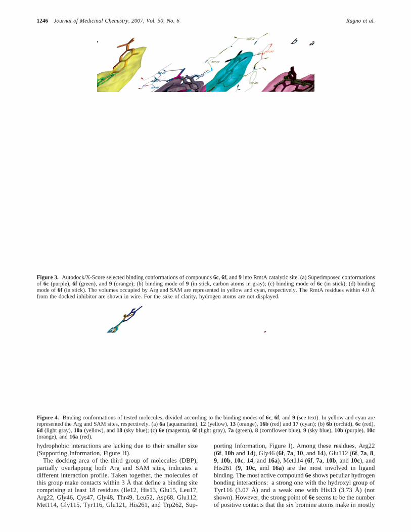
Figure 3. Autodock/X-Score selected binding conformations of compounds6c, 6f, and9 into RmtA catalytic site. (a) Superimposed conformationsof 6c (purple),6f (green), and9 (orange); (b) binding mode of9 (in stick, carbon atoms in gray); (c) binding mode of6c (in stick); (d) bindingmode of6f (in stick). The volumes occupied by Arg and SAM are represented in yellow and cyan, respectively. The RmtA residues within 4.0 Åfrom the docked inhibitor are shown in wire. For the sake of clarity, hydrogen atoms are not displayed.
Figure 4. Binding conformations of tested molecules, divided according to the binding modes of6c, 6f, and9 (see text). In yellow and cyan arerepresented the Arg and SAM sites, respectively. (a)6a (aquamarine),12 (yellow), 13 (orange),16b (red) and17 (cyan); (b)6b (orchid),6c (red),6d (light gray),10a (yellow), and18 (sky blue); (c)6e (magenta),6f (light gray),7a (green),8 (cornflower blue),9 (sky blue),10b (purple),10c(orange), and16a (red).
1246 Journal of Medicinal Chemistry, 2007, Vol. 50, No. 6 Ragno et al.
filling the Arg binding area and part of SAM binding area(Supporting Information, Figure J). The less active derivatives7a, 8, 10b, 10c, 14, and16a though showing docking positionssimilar to that of6e are smaller in volume (data not shown)and make less positive contacts.
In addition to the above-mentioned docking studies, we alsogenerated the human PRMT1 (hPRMT1) using the sameprotocol described for RmtA homology modeling. Subsequently,we performed a 3-D comparison of the RmtA andhPRMT1models and we found that only five (13.5%) of the 37 residuesthat are in contact with the SAM and arginine substrates haddifferent side-chains, and more interestingly, four out of fiveshowed some similarities (Cys47RmtA f Ser39hPRMT1; Val67RmtA
f Ile59hPRMT1; Asp68RmtA f Val60hPRMT1; Met69RmtA fCys61hPRMT1; Met96RmtA f Val88hPRMT1; see Supporting In-formation, Figure K). Once the hPRMT1/SAM/H4 ternary
complex was geometry optimized we applied the Autodock/X-SCORE protocol to inspect the binding site of derivatives6-20(data not shown). As expected the binding modes of thecompounds in the h-PRMT1 were similar to those describedfor RmtA with only marginal differences in the structureconformations, consistent with the aforementioned good cor-relation between pIC50 values.
3-D QSAR Studies. As learned from the foregoing dockingstudies, the three different binding modes adopted by molecules6-20 could represent structure-based aligned training sets tobuild 3-D QSAR models useful as validation tools for thedocking studies themselves as well as tools to design new PRMTinhibitors. With this aim, three 3-D QSAR models were derivedusing the GRID/GOLPE procedure.58 Obviously, it has to bestressed that due to the limited number of molecules in eachgroup the 3-D QSAR models are of limited validity and wereonly built to gain qualitative support to the above-describedbinding modes and to get some insights to design new PRMTinhibitors.
In building the 3-D QSAR models, different GRID probesalone (C3, N1, O, OH, OH2, and DRY) and combinations ofthem (C3+N1, DRY+N1, DRY+O, O+C3, O+N1, OH2+C3,OH2+DRY, OH+C3, OH+DRY, and OH+N1) were tried, andonly the preliminary GOLPE/GRID models showing the higherq2 values were refined by sequential fractional factorial design(FFD) selections. The combination of the water (OH2) and themethyl (C3) probes gave the best preliminary models for theDAP and DBP groups while only the carbonyl oxygen (O) probesupplied the best model for the DSP group alignment. Afterhaving imported the molecular interaction fields (MIF) intoGOLPE, a series of fractional factorial design (FFD) selectionsallowed the definition of the final 3-D QSAR models whosestatistical parameters are listed in Table 2. As can be seen, in
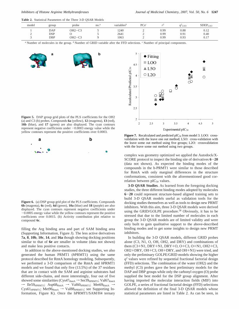
Table 2. Statistical Parameters of the Three 3-D QSAR Models
model group probe no.a variablesb PCsc r2 q2LOO SDEPLOO
a Number of molecules in the group.b Number of GRID variable after the FFD selections.c Number of principal components.
Figure 5. DAP group grid plots of the PLS coefficients for the OH2(a) and C3 (b) probes. Compounds6a (yellow), 12 (magenta),13 (red),16b (blue), and17 (green) are also displayed. The cyan contoursrepresent negative coefficients under-0.0003 energy value while theyellow contours represent the positive coefficients over 0.0003.
Figure 6. (a) DSP group grid plot of the PLS coefficients. Compounds6b (magenta),6c (red),6d (green),10a(blue) and18 (purple) are alsodisplayed. The cyan contours represent negative coefficients under-0.0005 energy value while the yellow contours represent the positivecoefficients over 0.0015. (b) Activity contribution plot relative tocompound6c.
Figure 7. Recalculated and predicted pIC50 from model 3. LOO: cross-validation with the leave one out method; L5O: cross-validation withthe leave some out method using five groups; L2O: crossvalidationwith the leave some out method using two groups.
Inhibitors of Histone Arginine Methyltransferases Journal of Medicinal Chemistry, 2007, Vol. 50, No. 61247
term of cross-validation data (q2 and SDEP values), allthree models, although developed with a limited number ofcompounds, were statistically robust withq2 and SDEP valuesranging from 0.88 to 0.93 and from 0.12 to 0.40, respectively.Although in models 1 and 2 the molecules seem not to beoptimally aligned, the 3-D QSAR models seem to supportthe above-described binding modes obtained by dockingstudies.
As already outlined in the binding mode analysis section, onlyderivative12, the most active out of the DAP group, is fullyburied in the arginine substrate site, which is correctly recog-nized by the PLS analysis of the OH2 and C3 GRID fields. Inparticular in the OH2 PLS coefficient plot (Figure 5a) a largepositive yellow polyhedron runs over the12diaza group whichis close to the guanidine group of the arginine substrate.Although in the above binding mode analysis the12diaza groupdid not seem to make any significant interaction, the PLSanalysis of both the actual field and activity contribution plots(Supporting Information, Figure L) indicates that aπ-rich groupshould be retained in designing new compounds competitivefor the arginine site. This consideration is also supported bythe observation that less active molecules such as13 and16bpossessed hydrogen bonding acceptor groups in the same12diaza moiety area. To prevent the model from being guided bythe misalignment of12, we also built a 3-D QSAR model withonly four compounds, and, taking into account the statistical
limits of such a model, the final 3-D QSAR model was charac-terized withr2 andq2 values of 0.99 and 0.84, respectively.
Regarding the 3D-QSAR model 2, it was built using onlyfive molecules which were docked exclusively in the SAMbinding site; derivative6c, the most active among all testedmolecules, belongs to this group. As in the case of compound12 in model 1,6c seems misaligned (Figures 4b and 6), yet a3-D QSAR model endowed with highr2 (0.99) andq2 (0.68)values without compound6cwas also obtained, confirming thatmodel 2 reflects a structure-activity correlation belonging toall five molecules in the set.
In Figure 6a is reported the PLS coefficients plot of the 3-DQSAR model for the DSP group in which one large yellowpolyhedron (positive values) and a few small cyan polyhedrons(negative values) are present. To properly understand the roleof the yellow polyhedron, the activity contribution plot forcompound6c is also reported (Figure 6b). As shown, the mostimportant contribution to the activity seems due to the benzoategroup while no polyhedron can be associated to the bromineatoms, the importance of which still emerged from biologicalassays. This disagreement could be attributed to the fact thatthe SAM binding site is not fully represented by the alignedmolecules, and thus the 3-D QSAR model here only supportsthe hypothesis that there is some correlation between theinhibitory activity with both the bound conformations and theirspatial disposition (alignment).
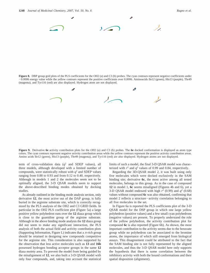
Figure 8. DBP group grid plots of the PLS coefficients for the OH2 (a) and C3 (b) probes. The cyan contours represent negative coefficients under-0.0006 energy value while the yellow contours represent the positive coefficients over 0.0006. Aminoacids Ile12 (green), His13 (purple), Thr49(magenta), and Tyr116 (red) are also displayed. Hydrogen atom are not displayed.
Figure 9. Derivative 6e activity contribution plots for the OH2 (a) and C3 (b) probes. The6e docked conformation is displayed as atom typecolors. The cyan contours represent negative activity contribution areas while the yellow contours represent the positive activity contribution areas.Amino acids Ile12 (green), His13 (purple), Thr49 (magenta), and Tyr116 (red) are also displayed. Hydrogen atoms are not displayed.
1248 Journal of Medicinal Chemistry, 2007, Vol. 50, No. 6 Ragno et al.
The 3-D QSAR model 3 was developed on moleculespartially occupying both the SAM and Arg binding sites (DBP,Figure 4c). Differently from the above analyzed models, all ninemolecules of this group were found superimposed to each other,leading to a robust model (Figure 4c and Supporting Informa-tion, Figure M), as also confirmed by highq2 values obtainedby cross-validations using the group method (leave some out,LSO): with five (L5O,q2 ) 0.90) and two (L2O,q2 ) 0.61)groups the DBP molecules were highly self-predictive(Figure 7).
Analysis of the PLS coefficient plots (Figure 8) revealed thatsome areas around the structure-based aligned molecules shouldbe further explored by the inclusion of additional ligands. Inparticular, in both the OH2 and the C3 PLS coefficients plot(Figure 8a and 8b, respectively) at 0.0006 energy level a largeyellow polyhedron is noticeable close to the tricyclic moiety of6e(magenta circle). This polyhedron is not correlated with anyresidue of RmtA but lies in an open space, maybe the entranceof SAM. A second yellow polyhedron (blue circle) could beassociated to a space close to the dibromoaminobenzoate moietyof 6ecorresponding to a pocket formed by Ile12, His13, Met16,and Thr49 residues. It is noteworthy that this area is also filledby 6f and9, that together with6eare the most potent compoundsof the DBP group. Two other cyan-colored polyhedrons(negative PLS coefficients) are visible in Figure 8a (OH2 probe)and almost absent in Figure 8b (C3 probe): the polyhedronshighlighted by green circles, mainly associated either withelectrostatic (7a, 8, 10b, and16a) or hydrogen bonding (6e, 6f,and9) interactions with Arg22 (not shown) and the polyhedronscircled in red, mainly associated either with electrostatic (6f,10c, and14, not shown) or hydrogen bonding (7a, 8, 9 10b,and10c, not shown) interactions with either Glu112 side-chaincarboxyl (6f, 9, 10b, 10c, and 14, not shown) or Met114carbonilic oxygen (6f, 7a, 8, 9, 10b, and10c, not shown). Thenegative PLS coefficients generated by a methyl probe (smallcyan polyhedrons in Figure 8b) indicate areas that have to befilled to maintain or increase the activity. One of these areascoincides with the binding site of the cysteinic portion of SAM,a site delimited by Arg22, Asp44, Gly46, Cys47, Ile51, Leu52,and Glu112 (not shown).
The aforementioned important role of the interactions ofcompounds of the DBP group with the pocket composed ofIle12, His13, Met16, and Thr49 residues is more clearlyexplained in Figure 9, reporting the activity contribution plotsrelative to derivative6e, the most active in this group. In thisfigure, yellow polyhedrons, associated with positive activitycontribution areas, are closely related to this pocket. Again thesmall cyan polyhedrons, associated with negative activitycontribution areas, highlight regions lacking in positive stericalinteractions (green and red circles in Figure 9b).
Conclusions
In this report we describe the binding mode analysis of afocused library of small molecule compounds into the catalyticdomain of the PRMT1 fungal homologue RmtA as well asenzymatic assays performed on recombinant RmtA and PRMT1proteins using SAM and histones as substrates. Computationaland biological results were in good agreement. Moreover, thegood correlation between pIC50 values against the two enzymes(R ) 0.91) supported our hypothesis of using RmtA as a modelto screen for arginine methyltransferase inhibitors.
Moreover, the agreement between molecular modeling studies(molecular docking and 3-D QSAR) and biological resultsallowed us to shed more light on the molecular mechanism of
inhibition of small molecule inhibitors of histone argininemethyltransferases. In particular the structure-based 3-D QSARhigh statistical coefficients seem to support the docking experi-ments. If considered together, the docking and the 3-D QSARstudies allowed a deep binding mode analysis of testedmolecules. In addition, these analyses hinted that two regions,the pocket formed by Ile12, His13, Met16, and Thr49 and theSAM cysteinic portion binding site, should be taken into accountto design novel active inhibitors.
Further molecular modeling, synthetic and biological effortsare ongoing to develop more robust 3-D QSAR models.
Materials and MethodsMelting points were determined on a Gallenkamp melting point
apparatus and are uncorrected.1H NMR spectra were recorded at300 MHz on a Bruker Avance 300 spectrometer; chemical shiftsare reported inδ (ppm) units relative to the internal referencetetramethylsilane (Me4Si). Electronic impact mass spectrometry (EI-MS) was performed on a Finnigan LCQ DECA TermoQuest (SanJose, CA) mass spectrometer. All compounds were routinelychecked by TLC and1H NMR. TLC was performed on aluminum-backed silica gel plates (Merck DC-Alufolien Kieselgel 60 F254)with spots visualized by UV light or using a KMnO4 alkalinesolution. All solvents were reagent grade and, when necessary, werepurified and dried by standard methods. Concentration of solutionsafter reactions and extractions involved the use of a rotaryevaporator operating at a reduced pressure of ca. 20 Torr. Organicsolutions were dried over anhydrous sodium sulfate. Analyticalresults are within(0.40% of the theoretical values. Reagents werepurchased from Lancaster Synthesis, Milan (Italy), or Sigma-Aldrich, Milan (Italy). As a rule, samples prepared for physicaland biological studies were dried in high vacuum over P2O5 for 20h at temperatures ranging from 25 to 110°C, depending on thesample melting point.
Preparation of 4-Amino-3,5-dibromo-2-(2,4,5,7-tetrabromo-6-hydroxy-3-oxo-3H-xanthen-9-yl )benzoic Acid (6e).A stirredmixture of 6-aminofluorescein (0.576 mmol, 0.2 g) in glacial aceticacid (7 mL) was cooled at 0°C. A solution of bromine (4.606 mmol,0.74 g) in acetic acid (3 mL) was then added dropwise, and stirringwas continued at room temperature for additional 15 min. Aftercompletion (TLC monitoring: CHCl3:AcOEt 1:1, on silica gelplate), the reaction mixture was diluted with water (10 mL) andfiltered by vacuum filtration. The precipitate was washed with water(3 × 5 mL) and air-dried in a vacuum desiccator to give TLC pure6e in 91% yield (0.82 g; mp: 291-292 °C), MS (EI, 70 eV)m/z821; 1H NMR (DMSO-d6): δ 6.53 (s, 1H), 7.10 (s, 1H), 8.07 (s,1H), 10.79 (s, 1H).
Preparation of 4-(4,6-Dichloro-1,3,5-triazin-2-ylamino)-2-(6-hydroxy-3-oxo-3H-xanthen-9-yl)benzoic Acid (6f).30 A solutionof 6-aminofluorescein (0.288 mmol, 0.1 g) in acetone (2 mL) wasadded dropwise to a solution of cyanuric chloride (0.2851 mmol,0.053 g) in acetone (3 mL) magnetically stirred at 5°C. Theresulting mixture was kept stirring for 3 h at 3-5 °C (TLCmonitoring: AcOEt on silica gel plate) and then filtered to give6fas a TLC pure yellow precipitate.(0.13 g, yield, 92%; mp:> 350°C); MS (EI, 70 eV)m/z 494;1H NMR (DMSO-d6): δ 6.79-6.88(m, 4H), 6.95 (s, 2H), 7.73 (s, 1H), 8.18-8.28 (m, 2H), 11.72(s, 1H).
Preparation of 7,7′-Carbonylbis(azanediyl)bis(4-hydroxynaph-thalene-2-sulfonic acid) (9).31 J-acid (2.00 g, 3.964 mmol) wasadded to 50 mL of water, with dropwise addition of aqueous NaOH10% to facilitate dissolution; the pH of the final solution was about7. The solution was then stirred and heated to 60°C and triphosgenepowder (0.40 g, 1.348 mmol) rapidly added. The resulting mixturewas magnetically stirred for 6 h while keeping temperature (60°C) and pH (7-8) constant. When the reaction was complete, J-acidurea9 was obtained by salting-out. (1.80 g, Yield 90%). MS (EI,70 ev)m/z: 504; 1H NMR (DMSO-d6): δ 7.04 (m, 1H), 7.51 (m,1H), 7.55 (m, 1H), 8.00 (m, 1H), 8.02 (m, 1H), 9.27 (s, 1H), 9.61(s, 1H).
Inhibitors of Histone Arginine Methyltransferases Journal of Medicinal Chemistry, 2007, Vol. 50, No. 61249
Preparation of 4-Hydroxy-7-(3-phenylureido)naphthalene-2-sulfonic Acid (10b).32 A stirred solution of J acid (1.943 mmol,0.5 g) in 8 mL of alkaline water solution containing NaOH (1.943mmol, 0.077 g), at room temperature, was treated with a solutionof phenyl isocianate (1.943 mmol, 0.23 g) in tetrahydrofuran (5mL). The resulting mixture was magnetically stirred at roomtemperature for 24 h (TLC monitoring: CHCl3 on silica gel plate),acidified with a water solution of HCl 2 N (3 mL), evaporated underreduced pressure, and purified by column chromatography (silicagel/ethyl acetate:isopropanol: water 12:3:1.). (0.49 g, Yield, 70%;mp: > 350 °C); MS (EI, 70 eV)m/z 358; 1H NMR (DMSO-d6):δ 6.92-6.98 (m, 2H), 7.24-7.29 (t, 2H), 7.42-7.48 (m, 4H), 7.85(s, 1H), 7.95-7.98 (d, 1H), 8.72 (s, 1H), 8.93 (s, 1H), 10.02 (s,1H).
Preparation of 4-Hydroxy-7-(3-phenylthioureido)naphtha-lene-2-sulfonic Acid (10c).A stirred solution of J-acid (1.943mmol, 0.5 g) in 8 mL of alkaline water solution containing NaOH(1.943 mmol, 0.077 g), at room temperature, was treated with asolution of phenyl isothiocyanate (1.943 mmol, 0.278 g) intetrahydrofuran (5 mL). The resulting mixture was magneticallystirred at room temperature for 12 h (TLC monitoring: CHCl3 onsilica gel plate), acidified with a water solution of HCl 2 N (3 mL),evaporated under reduced pressure, and purified by crystallization(CH3CN) to yield TLC pure10c (0.55 g, Yield: 75%; mp:>350°C); MS (EI, 70 eV)m/z 374;1H NMR (DMSO-d6): δ 7.05-7.10(m, 2H), 7.31-7.34 (t, 2H), 7.47-7.57 (m, 4H), 7.83-7.84 (s, 1H),7.97-8.00 (d, 1H), 9.82 (s, 1H), 9.95 (s, 1H), 10.08 (s, 1H).
Preparation of 4-(4,6-Dichloro-1,3,5-triazin-2-ylamino)ben-zoic Acid (17).33 A solution of 4-aminobenzoic acid (3.656 mmol,0.5 g) in acetone (10 mL) was added to a solution of cyanuricchloride (3.61 mmol, 0.67 g) in acetone (12 mL) magneticallystirred at 0°C. Aqueous sodium hydrogenocarbonate 0.3 M (12mL) was then added to the mixture over 30 min while maintaininga temperature of 0-5 °C. The solution was stirred for 2 h (TLCmonitoring: CHCl3:ethyl acetate 1:1, on silica gel plate), and thenaqueous HCl 12% was added until a precipitate formed which wasfiltered and washed with acetone. (0.99 g, Yield: 95%; mp:>350°C); MS (EI, 70 eV)m/z 284;1H NMR (DMSO-d6): δ 7.67-7.70(d, 2H), 7.78-7.95 (d, 2H), 11.37 (s, 1H).
Cloning of Human PRMT1 and Aspergillus nidulansRmtA.Human PRMT1 (isoform 2) was amplified from a HeLa cDNAlibrary (Invitrogen) using the following primer set: PRMT1fwd5′-GGATCCGAGAATTTTGTAGCCACCTTGGCTAA-3′ andPRMT1rev 5′-CTCGAGTCAGCGCATCCGGTAGTCGGTGGA-3′. The PCR fragment was subcloned into a pGEM-Teasy vector(Promega), released by digestion with the restriction endonucleasesBamHI and XhoI (New England Biolabs), and inserted into theBamHI/XhoI sites of the bacterial expression plasmid pGEX6P1(Amersham) using T4 Ligase (Roche).
Total Aspergillus RNA was used to prepare cDNA usingSuperscript reverse transcriptase (Life Technologies, Inc.) accordingto the manufacturer’s instructions employing an oligo-dT17
standard primer. For amplification of fragments ofrmtA from thecDNA of A. nidulans, degenerate oligodeoxynucleotide primerswere used. Forward primer was 5′-GGNATHCAYGARGARATG-3′ based on the amino acid sequence GIHEEM, and reverse primerwas 5′-ACNCAYTGGAARCARAC-3′ based on THWKQT. Aproduct of 750 bp was isolated and cloned into pGEM-T vector(Promega). A full-length genomic copy ofrmtAwas obtained usingcDNA PCR products to screen a genomic library ofAspergillusnidulansDNA. For amplification of the 3′ end ofrmtA, the 3′ rapidamplification of cDNA ends (RACE) protocol59 was performed.The first PCR was performed with forward primer 5′-ATACCGTC-GAGCTCAAG-3′ and an adapter primer of sequence 5′-GACTC-GAGTCGACATCGA-3′. For “nested PCR”, primer 5′-GACTA-GAGTACACGATGGA-3′ (forward) and dT4 adapter primer 5′-GACTCGAGTCGACATCGATTTT-3′ (reverse) were used. Ampli-fied products were cloned into pGEM-T vector (Promega).
Preparation of GST-RmtA and GST-PRMT1 Fusion Pro-teins.Expression of the GST-PRMT1 fusion protein was performedin BL21 cells. At OD600 nm ) 0.6 expression was induced with 1
mM isopropylâ-D-thiogalactopyranoside (IPTG), and the cultureswere grown for 3 h upon induction. Bacterial cells were harvestedby centrifugation (3000g for 20 min), resuspended in sonicationbuffer (PBS, 0.01% TritonX100, 0.2 mM PMSF, 1µg/mL aprotinin,1µg/mL leupeptin, 1µg/mL pepstatin), and sonicated three timesfor 30 s with a sonic dismembrator 550 (Fisher Scientific; amplitude40%). The sonicated suspension was centrifuged for 20 min at10000g, and the supernatant was incubated with preequilibratedglutathione-sepharose-4-fast-flow overnight at 4°C on a rotatingwheel. The resin was transferred to a gravity-flow column andwashed with five column volumes (CV) of PBS+ 0.1% Tri-tonX100. Bound GST-PRMT1 was released using five times oneCV of elution buffer (50 mM Tris pH 8.0, 10 mM glutathione).Purified GST-PRMT1 was dialyzed against BC100 buffer (20 mMTris pH 7.9, 10% glycerol, 100 mM KCl, 0.2 mM EDTA, 10 mM2-ME, 0.2 mM PMSF), and protein concentration was estimatedby SDS-PAGE and Bradford assay.
The coding sequence of RmtA was cloned into a pGEX-5X-1expression vector (Amersham Pharmacia Biotech). RmtA-Proteinwas expressed in BL21 cells in LB-medium. Cultures (250 mL)with an A600 of 0.4 were induced with a final concentration of 1mM IPTG and grown for 4 h at 37°C. After centrifugation of cellsat 4000g, the pellet was resuspended in 6 mL of GST-binding buffer(140 mM NaCl, 2.7 mM KCl, 10 mM Na2HPO4, 1.8 mM KH2-PO4, pH 7.3) containing one protease inhibitor tablet (Complete,Roche, Mannheim, Germany) per 50 mL of buffer. For cell lysis,lysozyme was added at a final concentration of 5 mg/mL bindingbuffer, and cells were passed through a french press with a pressuresetting of 1000 psi. The resulting lysate was centrifuged at 20000gfor 10 min at 4°C. GST fusion protein was purified from solubleextracts by binding to a GST-HiTrap column (Amersham PharmaciaBiotech). Proteins were eluted with 50 mM Tris-HCl, 10 mMreduced glutathione, pH 8.0, and assayed for histone methyltrans-ferase activity.
Histone Methyltransferase Assay (HMT).For inhibition assays,affinity purified GST-RmtA and GST-PRMT1 fusion proteins wereused as the enzyme source. HMT activities were assayed asdescribed29 using chicken erythrocyte core histones as substrate.A 500 ng amount of GST-RmtA and GST-PRMT1 fusion proteinswas incubated with different concentrations of compounds for 15min at room temperature, and 20µg of chicken core histones and0.55µCi of [3H]-S-adenosyl-L-methionine (SAM) were added. Thismixture was incubated for 30 min at 30°C. Reaction was stoppedby TCA precipitation (25% final concentration), and samples werekept on ice for 20 min. Whole sample volumes were collected ontoa glass fiber filter (Whatman GF/F) preincubated with 25% TCA.Filters were washed three times with 3 mL of 25% TCA and thenthree times with 1 mL of ethanol. After the filters were dried for10 min at 70°C, radioactivity was measured by liquid scintillationspectrophotometry (3 mL of scintillation cocktail).
Preparation of the RmtA-SAM-H4 Ternary Complex. TheRmtA model from the homology model experiment was mergedwith the S-adenosyl-L-homocysteine (AdoHcy) and the 19mersubstrate peptide taken from the experimental PRMT1 structure(pdb entry code 1or8).47 The AdoHcy was modified to SAM usingan experimental SAM structure extracted from the FTSJ RNAmethyltransferase (pdb entry code 1ej0)60 which showed a SAMconformation similar to the AdoHcy found in the 1or8 complex.The experimental 19mer peptide lacked of most of the side-chainatoms, which were added using the xLeap module of the AMBERsuite.61 The RmtA-SAM-H4 complex was solvated (SOLVA-TEOCT command) in a box extending 10 Å with 16658 watermolecules (TIP3 model) and neutralized with 10 Na+ ions. Thesolvated complex was then refined by minimization using theSANDER module of AMBER. The parameters for the SAM werecalculated using the antechamber module of AMBER, and theatomic charges were calculated using the AM1 Hamiltonian.
Molecular Docking. All the tested molecules were built, startingfrom ASCII text, using the stand alone version of PRODRG,62 inconjunction with the GROMACS suite.63 Docking studies wereperformed by means of the Autodock 3.0.5 program using a grid
1250 Journal of Medicinal Chemistry, 2007, Vol. 50, No. 6 Ragno et al.
spacing of 0.375 Å and 39× 50 × 56 number of points thatembraced both the SAM and Arg binding sites. The grid wascentered on the mass center of the experimental bound AdoMetand Arg substrates. The GA-LS method was adopted using thedefault setting except for the maximum number of energy evalu-ations increased from 250000 to 2500000. Autodock generated 100possible binding conformations for each molecule that wereclustered using a tolerance of 2.0 Å. The AutoDockTool (ADT)graphical interface64 was used to prepare the enzyme PDBQS file.The protein atom charges as calculated during the complexminimization were retained for the docking calculations.
The SAM andN-acetyl,O-methyl-capped Arg PRODRG-gener-ated conformations were docked into the RmtA to assess thedocking protocol. The SAM and Arg were docked back into theirbinding sites, and the Xscore54-selected conformations showed root-mean-square deviations (rmsd) of 0.60 and 1.47, respectively. TheAutodock scoring function did not select conformations with lowerrmsd values. Trials with the DOCK program did not successfullydock back the substrates into the RmtA; therefore, we continuedto use Autodock. To analyze the docking results, the ADT wasused and the Chimera 1.2176 program65 was used to produce theimages.
Docking studies were not performed on nonactive compounds(7b, 7c, 7d, and 20) and on compound15 due to its largedimensions.
3-D QSAR Studies. GRID Calculations. The interactionenergies were calculated by using the program GRID66 (version22) with a grid spacing of 1 Å and the grid dimensions (Å):Xmin/Xmax, 58.25/20.25;Ymin/Ymax, 69.25/31.25;Zmin/Zmax, 75.25/37.25.
GOLPE Analyses. PLS models were calculated with GOLPE4.5.1267 running on a SGI O2 R10000 equipped with the IRIXoperating system 6.5.11. To measure the goodness of the modelthe statistical indicesr2, q2, and SDEP were employed.
Probe Selection.In this study we started with five probes (Csp3,DRY, OH2, NHamide,Osp2) and combinations of them, according tothe nature of the RmtA active site. Preliminary PLS analyses wererun on the DSP, DAP, and DBP groups, and the probe combinationdisplaying higher values ofr2 andq2 was selected to develop thefinal model for each group.
Variable Preselection. The resulting probe-target interactionenergies for each compound were unfolded to produce the corre-sponding one-dimensional vector variables, which were assembledin the so-called X matrix. This matrix was pretreated first by usinga cutoff of 5 kcal/mol to produce a more symmetrical distributionof energy values and then zeroing small variable values andremoving variables with small standard deviation, using appropriatecutoffs. In addition, variables taking only two and three distributionwere also removed.
Smart Region Definition (SRD). A number of seeds (3000)were selected using a D-optimal design criterion in the weight space.Structural differences between different molecules in the series willbe reflected in groups of variables, and therefore groups weregenerated around each seed in the 3-D space. Variables with adistance of no more than 1 Å to theseeds were included in thegroups. If two neighboring groups (with a distance smaller than 2Å) contained the same information the groups were collapsed. Thegroups were used in the variable selection procedure replacing theoriginal variables. The effect of the groups on the predictivity wasevaluated, and groups instead of individual variables were removedfrom the data file.
Region Selection.The effect of the grouped variables on thepredictivity was evaluated using a fractional factorial design (FFD)procedure. A number of reduced models (twice the number ofvariables) were built removing some of the variables according tothe FFD design. The effect of dummy variables (20%) on thepredictivity was calculated, and only if a variable had a positiveeffect on the predictivity larger than the effect of the average dummyvariable was included in the final model. The FFD selection wasrepeated until ther2 andq2 value did not increase significantly. In
the FFD selection the cross-validation was conducted using fiverandom groups for 20 times and a maximum of two principalcomponents.
Cross-Validation. The models were validated using the leaveone out (LOO) method. Molecules were assigned in a random wayto five groups of equal size. Reduced models were built keepingout one group at a time.
Acknowledgment. Many thanks are due to Prof. GabrieleCruciani and Prof. Sergio Clementi (Molecular Discovery andMIA srl) for the use the GOLPE program in their chemometriclaboratory (University of Perugia, Italy) and for having providedthe GRID program. This work was partially supported by PRIN2004 (A.M.), AIRC 2005 (A.M.), and Regione Campania 2003,LR 5/02 (G.S.) grants.
Supporting Information Available: Characterization data forcompounds6e, 6f, 9, 10b, 10c, and17, sequence homology, andalignment and additional binding mode informations. This materialis available free of charge via the Internet at http://pubs.acs.org.
References(1) Strahl, B. D.; Allis, C. D. The language of covalent histone
modifications.Nature2000, 403, 41-45.(2) Jenuwein, T.; Allis, C. D. Translating the histone code.Science2001,
293, 1074-1080.(3) Shi, Y.; Lan, F.; Matson, C.; Mulligan, P.; Whetstine, J. R.; Cole, P.
A.; Casero, R. A.; Shi, Y. Histone demethylation mediated by thenuclear amine oxidase homolog LSD1.Cell 2004, 119, 941-953.
(5) Forneris, F.; Binda, C.; Vanoni, M. A.; Mattevi, A.; Battaglioli, E.Histone demethylation catalysed by LSD1 is a flavin-dependentoxidative process.FEBS Lett.2005, 579, 2203-2207.
(6) Chen, Y.; Yang, Y.; Wang, F.; Wan, K.; Yamane, K.; Zhang, Y.;Lei, M. Crystal structure of human histone lysine-specific demethy-lase 1 (LSD1).Proc. Natl. Acad. Sci. U.S.A.2006, 103, 13956-13961.
(7) Stavropoulos, P.; Blobel, G.; Hoelz, A. Crystal structure andmechanism of human lysine-specific demethylase-1.Nat. Struct. Mol.Biol. 2006, 13, 626-632.
(8) Yamane, K.; Toumazou, C.; Tsukada, Y.; Erdjument-Bromage, H.;Tempst, P.; Wong, J.; Zhang, Y. JHDM2A, a JmjC-containing H3K9demethylase, facilitates transcription activation by androgen receptor.Cell 2006, 125, 483-495.
(9) Whetstine, J. R.; Nottke, A.; Lan, F.; Huarte, M.; Smolikov, S.; Chen,Z.; Spooner, E.; Li, E.; Zhang, G.; Colaiacovo, M.; Shi, Y. Reversalof histone lysine trimethylation by the JMJD2 family of histonedemethylases.Cell 2006, 125, 467-481.
(10) Tsukada, Y.; Fang, J.; Erdjument-Bromage, H.; Warren, M. E.;Borchers, C. H.; Tempst, P.; Zhang, Y. Histone demethylation by afamily of JmjC domain-containing proteins.Nature2006, 439, 811-816.
(11) Klose, R. J.; Yamane, K.; Bae, Y.; Zhang, D.; Erdjument-Bromage,H.; Tempst, P.; Wong, J.; Zhang, Y. The transcriptional repressorJHDM3A demethylates trimethyl histone H3 lysine 9 and lysine 36.Nature2006, 442, 312-316.
(12) Cloos, P. A.; Christensen, J.; Agger, K.; Maiolica, A.; Rappsilber,J.; Antal, T.; Hansen, K. H.; Helin, K. The putative oncogene GASC1demethylates tri- and dimethylated lysine 9 on histone H3.Nature2006, 442, 307-311.
(13) Zhang, Y.; Reinberg, D. Transcription regulation by histone methy-lation: interplay between different covalent modifications of the corehistone tails.Genes DeV. 2001, 15, 2343-2360.
(14) Byvoet, P.; Shepherd, G. R.; Hardin, J. M.; Noland, B. J. Thedistribution and turnover of labeled methyl groups in histone fractionsof cultured mammalian cells.Arch. Biochem. Biophys.1972, 148,558-567.
(15) Baxter, C. S.; Byvoet, P. Intercalating agents as probes of the spatialrelationship between chromatin components.Biochem. Biophys. Res.Commun.1975, 63, 286-291.
(16) Rice, J. C.; Allis, C. D. Histone methylation versus histone acety-lation: new insights into epigenetic regulation.Curr. Opin. Cell Biol.2001, 13, 263-273.
(17) Turner, B. M. Memorable transcription.Nat. Cell Biol.2003, 5, 390-393.
(18) Lee, D. Y.; Teyssier, C.; Strahl, B. D.; Stallcup, M. R. Role of proteinmethylation in regulation of transcription.Endocr. ReV. 2005, 26,147-170.
Inhibitors of Histone Arginine Methyltransferases Journal of Medicinal Chemistry, 2007, Vol. 50, No. 61251
(19) Bedford, M. T.; Richard, S. Arginine methylation an emergingregulator of protein function.Mol. Cell. 2005, 18, 263-272.
(20) Cook, J. R.; Lee, J. H.; Yang, Z. H.; Krause, C. D.; Herth, N.;Hoffmann, R.; Pestka, S. FBXO11/PRMT9, a new protein argininemethyltransferase, symmetrically dimethylates arginine residues.Biochem. Biophys. Res. Commun.2006, 342, 472-481.
(21) Gary, J. D.; Clarke, S. RNA and protein interactions modulated byprotein arginine methylation.Prog. Nucleic Acid Res. Mol. Biol.1998,61, 65-131.
(22) Cheng, D.; Yadav, N.; King, R. W.; Swanson, M. S.; Weinstein, E.J.; Bedford, M. T. Small molecule regulators of protein argininemethyltransferases.J. Biol. Chem.2004, 279, 23892-23899.
(23) a) Bartel, R. L.; Borchardt, R. T. Effects of adenosine dialdehydeon S-adenosylhomocysteine hydrolase and S-adenosylmethionine-dependent transmethylations in mouse L929 cells.Mol. Pharmacol.,1984, 25, 418-424.; b) Schwerk, C.; Schulze-Osthoff, K. Methyl-transferase inhibition induces p53-dependent apoptosis and a novelform of cell death.Oncogene2005, 24, 7002-7011.
(24) Casellas, P.; Jeanteur, P. Protein methylation in animal cells. II.Inhibition of S-adenosyl-L-methionine:protein( arginine ) N- meth-yltransferase by analogs of S-adenosyl-L-homocysteine.Biochim.Biophys. Acta, Nucleic Acids Protein Synth.1978, 519, 255-268.
(25) Barbes, C.; Sanchez, J.; Yebra, M. J.; Robert-Gero, M.; Hardisson,C. Effects of sinefungin and S-adenosylhomocysteine on DNA andprotein methyltransferases from Streptomyces and other bacteria.FEMS Microbiol. Lett.1990, 69, 239-243.
(26) Massa, S.; Mai, A.; Sbardella, G.; Esposito, M.; Ragno, R.; Loidl,P.; Brosch, G. 3-(4-aroyl-1H-pyrrol-2-yl)-N-hydroxy-2-propenamides,a new class of synthetic histone deacetylase inhibitors.J. Med. Chem.2001, 44, 2069-2072.
(27) Mai, A.; Massa, S.; Ragno, R.; Esposito, M.; Sbardella, G.; Nocca,G.; Scatena, R.; Jesacher, F.; Loidl, P.; Brosch, G. Binding modeanalysis of 3-(4-benzoyl-1-methyl-1H-2-pyrrolyl)-N-hydroxy-2-pro-penamide: a new synthetic histone deacetylase inhibitor inducinghistone hyperacetylation, growth inhibition, and terminal cell dif-ferentiation.J. Med. Chem.2002, 45, 1778-1784.
(28) Ornaghi, P.; Rotili, D.; Sbardella, G.; Mai, A.; Filetici, P. A novelGcn5p inhibitor represses cell growth, gene transcription and histoneacetylation in budding yeast.Biochem. Pharmacol.2005, 70, 911-917.
(29) Trojer, P.; Dangl, M.; Bauer, I.; Graessle, S.; Loidl, P.; Brosch, G.Histone methyltransferases in Aspergillus nidulans: evidence for anovel enzyme with a unique substrate specificity.Biochemistry2004,43, 10834-10843.
(30) Barskii, V. E.; Ivanov, V. B.; Skliar Iu, E.; Mikhailov, G. I.[Dichlorotriazineylaminofluorescein-a new fluorochrome for cy-tochemical and histochemical detection of proteins].IzV. Akad. Nauk.SSSR Biol.1968, 5, 744-747.
(31) Peng, X.; Yu, H.; Hang, Y.; Wang, J. N,N′;-phosgenation withtriphosgene in the synthesis of direct dyes containing the ureylenegroup.Dyes Pigm.1996, 32, 193-198.
(32) Adam, J. M.; Kaser, A. Anionic disazo dyes. EP0290384, 1988.(33) Birkett, H. E.; Cherryman, J. C.; Chippendale, A. M.; Evans, J. S.
O.; Harris, R. K.; James, M.; King, I. J.; McPherson, G. J. Structuralinvestigations of three triazines: Solution-state NMR studies ofinternal rotation and structural information from solid-state NMR,plus a full structure determination from powder X-ray diffraction inone case.Magn. Reson. Chem.2003, 41, 324-336.
(34) Kuntz, I. D. Structure-based strategies for drug design and discovery.Science1992, 257, 1078-1082.
(35) Hillisch, A.; Pineda, L. F.; Hilgenfeld, R. Utility of homology modelsin the drug discovery process.Drug DiscoVery Today2004, 9, 659-669.
(36) Tramontano, A.; Morea, V. Assessment of homology-based predic-tions in CASP5.Proteins2003, 53 Suppl. 6, 352-368.
(37) Mai, A.; Massa, S.; Ragno, R.; Cerbara, I.; Jesacher, F.; Loidl, P.;Brosch, G. 3-(4-Aroyl-1-methyl-1H-2-pyrrolyl)-N-hydroxy-2-alky-lamides as a new class of synthetic histone deacetylase inhibitors. 1.Design, synthesis, biological evaluation, and binding mode studiesperformed through three different docking procedures.J. Med. Chem.2003, 46, 512-524.
(38) Rosato, R. R.; Grant, S. Histone deacetylase inhibitors in cancertherapy.Cancer Biol. Ther.2003, 2, 30-37.
(39) Mai, A.; Massa, S.; Cerbara, I.; Valente, S.; Ragno, R.; Bottoni, P.;Scatena, R.; Loidl, P.; Brosch, G. 3-(4-Aroyl-1-methyl-1H-2-pyrro-lyl)-N-hydroxy-2-propenamides as a new class of synthetic histonedeacetylase inhibitors. 2. Effect of pyrrole-C2 and/or -C4 substitutionson biological activity.J. Med. Chem.2004, 47, 1098-1109.
(40) Ragno, R.; Mai, A.; Massa, S.; Cerbara, I.; Valente, S.; Bottoni, P.;Scatena, R.; Jesacher, F.; Loidl, P.; Brosch, G. 3-(4-Aroyl-1-methyl-1H-pyrrol-2-yl)-N-hydroxy-2-propenamides as a new class of syn-thetic histone deacetylase inhibitors. 3. Discovery of novel leadcompounds through structure-based drug design and docking studies.J. Med. Chem.2004, 47, 1351-1359.
(41) Mai, A.; Massa, S.; Rotili, D.; Cerbara, I.; Valente, S.; Pezzi, R.;Simeoni, S.; Ragno, R. Histone deacetylation in epigenetics: anattractive target for anticancer therapy.Med. Res. ReV. 2005, 25, 261-309.
(42) Mai, A.; Massa, S.; Rotili, D.; Simeoni, S.; Ragno, R.; Botta, G.;Nebbioso, A.; Miceli, M.; Altucci, L.; Brosch, G. Synthesis andBiological Properties of Novel, Uracil-Containing Histone Deacety-lase Inhibitors.J. Med. Chem.2006, 49, 6046-6056.
(43) Mai, A.; Massa, S.; Valente, S.; Simeoni, S.; Ragno, R.; Bottoni, P.;Scatena, R.; Brosch, G. Aroyl-Pyrrolyl Hydroxyamides: Influenceof Pyrrole C4-Phenylacetyl Substitution on Histone DeacetylaseInhibition. ChemMedChem2006, 1, 225-237.
(44) Ragno, R.; Simeoni, S.; Valente, S.; Massa, S.; Mai, A. 3-D QSARstudies on histone deacetylase inhibitors. A GOLPE/GRID approachon different series of compounds.J. Chem. Inf. Model.2006, 46,1420-1430.
(45) Kouranov, A.; Xie, L.; de la Cruz, J.; Chen, L.; Westbrook, J.; Bourne,P. E.; Berman, H. M. The RCSB PDB information portal forstructural genomics.Nucleic Acids Res.2006, 34, 302-305.
(46) Berman, H. M.; Westbrook, J.; Feng, Z.; Gilliland, G.; Bhat, T. N.;Weissig, H.; Shindyalov, I. N.; Bourne, P. E. The Protein Data Bank.Nucleic Acids Res.2000, 28, 235-242.
(47) Zhang, X.; Cheng, X. Structure of the predominant protein argininemethyltransferase PRMT1 and analysis of its binding to substratepeptides.Structure2003, 11, 509-520.
(48) Weiss, V. H.; McBride, A. E.; Soriano, M. A.; Filman, D. J.; Silver,P. A.; Hogle, J. M. The structure and oligomerization of the yeastarginine methyltransferase, Hmt1.Nat. Struct. Biol.2000, 7, 1165-1171.
(49) Zhang, X.; Zhou, L.; Cheng, X. Crystal structure of the conservedcore of protein arginine methyltransferase PRMT3.EMBO J.2000,19, 3509-3519.
(50) Multiple sequence alignment was accomplished by the use of BioEditsoftware v. 7.0.1 and then manually adjusted. For reference see: Hall,T. A. BioEdit: a user-friendly biological sequence alignment editorand analysis program for Windows 95/98/NT.Nucl. Acids Symp. Ser.1999, 41, 95-98.
(51) Venclovas, C. Comparative modeling in CASP5: progress is evident,but alignment errors remain a significant hindrance.Proteins2003,53 Suppl. 6, 380-388.
(52) Schwede, T.; Kopp, J.; Guex, N.; Peitsch, M. C. SWISS-MODEL:An automated protein homology-modeling server.Nucleic Acids Res.2003, 31, 3381-3385.
(53) Goodsell, D. S.; Morris, G. M.; Olson, A. J. Automated docking offlexible ligands: applications of AutoDock.J. Mol. Recognit.1996,9, 1-5.
(54) Wang, R.; Lai, L.; Wang, S. Further development and validation ofempirical scoring functions for structure-based binding affinityprediction.J. Comput.-Aided Mol. Des.2002, 16, 11-26.
(55) Shoichet, B. K.; Stroud, R. M.; Santi, D. V.; Kuntz, I. D.; Perry, K.M. Structure-based discovery of inhibitors of thymidylate synthase.Science1993, 259, 1445-1450.
(56) Docking studies were not performed on non-active compounds (7b,7c, 7d, and20) and on compound15 due to its large dimensions.
(57) The two tautomeric forms of compound16 (keto and enol, indicatedas16a and16b, respectively) docked in different sites.
(58) Cruciani, G.; Watson, K. A. Comparative molecular field analysisusing GRID force-field and GOLPE variable selection methods in astudy of inhibitors of glycogen phosphorylase b.J. Med. Chem.1994,37, 2589-2601.
(59) Frohman, M. A.; Dush, M. K.; Martin, G. R. Rapid production offull-length cDNAs from rare transcripts: amplification using a singlegene-specific oligonucleotide primer.Proc. Natl. Acad. Sci. U.S.A.1988, 85, 8998-9002.
(61) Case, D. A.; Cheatham, T. E., 3rd; Darden, T.; Gohlke, H.; Luo, R.;Merz, K. M., Jr.; Onufriev, A.; Simmerling, C.; Wang, B.; Woods,R. J. The Amber biomolecular simulation programs.J. Comput.Chem.2005, 26, 1668-1688.
(62) Schuttelkopf, A. W.; van Aalten, D. M. PRODRG: a tool for high-throughput crystallography of protein-ligand complexes.Acta Crys-tallogr., Sect. D: Biol. Crystallogr.2004, 60, 1355-1363.
(63) Van Der Spoel, D.; Lindahl, E.; Hess, B.; Groenhof, G.; Mark, A.E.; Berendsen, H. J. GROMACS: fast, flexible, and free.J. Comput.Chem.2005, 26, 1701-1718.
1252 Journal of Medicinal Chemistry, 2007, Vol. 50, No. 6 Ragno et al.
(64) Gillet, A.; Sanner, M.; Stoffler, D.; Olson, A. Tangible interfacesfor structural molecular biology.Structure2005, 13, 483-491.
(65) Pettersen, E. F.; Goddard, T. D.; Huang, C. C.; Couch, G. S.;Greenblatt, D. M.; Meng, E. C.; Ferrin, T. E. UCSF Chimera-avisualization system for exploratory research and analysis.J. Comput.Chem.2004, 25, 1605-1612.
(66) Goodford, P. J., A computational procedure for determining energeti-
cally favorable binding sites on biologically important macromol-ecules.J. Med. Chem.1985, 28, 849-857.