Small RNA Deep Sequencing Identifies MicroRNAs and Other SmallNoncoding RNAs from Human Herpesvirus 6B
Lee Tuddenham,a Jette S. Jung,b Béatrice Chane-Woon-Ming,a Lars Dölken,b,c and Sébastien Pfeffera
Architecture et Réactivité de l’ARN, Université de Strasbourg, Institut de Biologie Moléculaire et Cellulaire du CNRS, Strasbourg, Francea; Max von Pettenkofer Institute,Ludwig Maximilians University Munich, Munich, Germanyb; and University of Cambridge, Department of Medicine, Cambridge, United Kingdomc
Roseolovirus, or human herpesvirus 6 (HHV-6), is a ubiquitous human pathogen infecting over 95% of the population by theage of 2 years. As with other herpesviruses, reactivation of HHV-6 can present with severe complications in immunocompro-mised individuals. Recent studies have highlighted the importance of herpesvirus-derived microRNAs (miRNAs) in modulatingboth cellular and viral gene expression. An initial report which computed the likelihood of various viruses to encode miRNAsdid not predict HHV-6 miRNAs. To experimentally screen for small HHV-6-encoded RNAs, we conducted large-scale sequenc-ing of Sup-T-1 cells lytically infected with a laboratory strain of HHV-6B. This revealed an abundant, 60- to 65-nucleotide RNAof unknown function derived from the lytic origin of replication (OriLyt) that gave rise to smaller RNA species of 18 or 19 nucle-otides. In addition, we identified four pre-miRNAs whose mature forms accumulated in Argonaute 2. In contrast to the case forother betaherpesviruses, HHV-6B miRNAs are expressed from direct repeat regions (DRL and DRR) located at either side of thegenome. All miRNAs are conserved in the closely related HHV-6A variant, and one of them is a seed ortholog of the humanmiRNA miR-582-5p. Similar to alphaherpesvirus miRNAs, they are expressed in antisense orientation relative to immediate-early open reading frames (ORFs) and thus have the potential to regulate key viral genes.
Human herpesvirus 6 (HHV-6) is one of eight herpesvirusesknown to infect humans and was first isolated in 1986 from
immunocompromised patients suffering from lymphoprolifera-tive disorders (43). HHV-6 is a ubiquitous human pathogen withseroprevalence rates exceeding 95% in industrialized nations. To-gether with HHV-7, it belongs to the Roseolovirus genus within thebetaherpesvirus subfamily. It exists as two variants, HHV-6A andHHV-6B, which differ with respect to their biological properties,tropisms, and clinical manifestations (14). In 1988, HHV-6B wasidentified as the causative agent of the childhood disease exan-them subitum (roseola or 3-day fever) (57). Primary infectiontypically presents as an acute febrile illness, sometimes followed bya red rash. Additional symptoms may include otitis, meningitis,and seizures (42). While severe complications resulting from pri-mary infection, such as encephalitis and encephalopathy, are rare,reactivation of HHV-6 in transplant patients, particularly thosereceiving hematopoietic stem cells, can present with life-threatening complications, most notably due to encephalitis (31,58). In addition, HHV-6 may facilitate the progression of otherviral infections, trans-activating HHV-7, human cytomegalovirus(HCMV), and HIV-1, which generally results in a poorer progno-sis (reviewed in reference 14). Finally, HHV-6 has been implicatedin a range of neurological diseases, most prominently multiplesclerosis (MS), although a causative role of HHV-6 in MS pathol-ogy remains controversial (36).
The HHV-6 genome consists of a linear, double-strandedDNA molecule of 160 to 162 kb, comprising a unique region (U)of 145 kb flanked by terminal direct repeats (DRL and DRR) of 8 to9 kb. HHV-6 has a broad cellular tropism, as its receptor (CD46)is expressed on all nucleated cells. The sites of latency are notprecisely defined yet but could consist of CD4� T cells, mono-cytes, and early bone marrow progenitor cells (14, 32, 49).
HHV-6, like HCMV (although it is less well studied), uses amultitude of approaches to modulate the host-pathogen environ-ment and establish latency. Previous studies have focused on the
role of viral proteins that modulate major histocompatibilitycomplex (MHC) class I gene expression and of viral chemokinesand receptors that interact with the host (34). Recently, manymembers of the herpesvirus family have been shown to expresstheir own microRNAs (miRNAs) (11, 19, 40, 41). Since the sem-inal discovery of miRNAs in Epstein-Barr virus (EBV) (41), morethan 235 miRNAs of viral origin have been identified and listed inthe miRNA repository miRBase (v.17) (25, 26). Conservedthroughout metazoan evolution, miRNAs derive from a large pri-mary transcript (pri-miRNA) which is sequentially processed bythe nuclear and cytoplasmic RNase III enzymes Drosha and Dicerto generate the mature, �22-nucleotide (nt) miRNA. ThismiRNA becomes functional only after its incorporation into anArgonaute (Ago)-containing RNA-induced silencing complex(RISC), where it will mediate the regulation of target messengerRNAs via binding to partially complementary sites, usually lo-cated in the 3=-untranslated regions (3=-UTRs) (3).
The vast majority of virus-encoded miRNAs derive from her-pesviruses, and so far HHV-3, HHV-6, and HHV-7 are the onlyherpesviruses for which miRNAs have not been characterized. ForHHV-3, deep sequencing of latently infected trigeminal gangliafailed to identify any miRNAs (55). To resolve the question ofwhether HHV-6 encodes miRNAs, we performed small RNA
cloning followed by massively parallel sequencing to identify thesmall RNAs deriving from the virus during lytic infection.
We cloned small RNAs both from total RNA isolated from aCD4� T lymphocyte cell line (Sup-T-1) infected with a laboratorystrain of HHV-6B and from RNAs coimmunoprecipitated withAgo2. Here we report that HHV-6B encodes at least four pre-miRNAs. The respective miRNAs are all conserved in the closelyrelated virus HHV-6A, although they display intervariant and in-terstrain sequence variations. These viral miRNAs are encoded attwo positions within the genome (in both DRL and DRR) and areencoded in antisense orientation relative to predicted HHV-6B-specific genes. One HHV-6 miRNA is a seed ortholog of the hu-man miRNA miR-582-5p, which is not expressed in Sup-T-1 Tcells. Interestingly, 5= RNA ligase-mediated rapid amplification ofcDNA ends (5= RLM-RACE) to detect viral miRNA-inducedcleavage products enabled us to identify a locus expressingmiRNAs from both strands of the genome. In addition, we iden-tified a novel viral transcript of 60 to 65 nt deriving from theminimal efficient origin of lytic replication (OriLyt) that gives riseto abundant RNA species of 18 and 19 nt that may represent a newclass of origin-derived viral small RNAs.
MATERIALS AND METHODSCell culture and virus infection. Sup-T-1 cells (CRL-1942; ATCC) weremaintained in RPMI 1640 medium containing 7% fetal bovine serum(FBS). A total of 3 � 105 cells/ml were infected with HHV-6B (using astrain that is routinely used for immunofluorescence-based antibody de-tection in the laboratory) by cocultivation of uninfected Sup-T-1 cellswith infected cells at a ratio of 1:1,000. Samples were harvested at 0, 24, 48,72, and 96 h postinfection (hpi), and total RNA was isolated using TRIzol(Invitrogen, Carlsbad, CA) according to the manufacturer’s recommen-dations.
Immunofluorescence staining. For immunofluorescence analysis todetermine the rate of HHV-6 infection, 2 � 105 cells in 150 �l phosphate-buffered saline (PBS) per slide were pelleted on a glass slide (by cytospincentrifugation) at 800 rpm for 10 min. Slides were air dried, fixed, andpermeabilized with fixative solution for a CINAkit (Argene, Sherley, NY)and permeabilization solution for a CINAkit (Argene, Sherley, NY). Theprimary antibody (sc-65447; Santa Cruz) for fluorescence staining wasapplied at a concentration of 0.5% for 1 h, followed by 1 h of incubationwith the Alexa Fluor 488-conjugated donkey anti-mouse secondary anti-body (Invitrogen, Darmstadt, Germany). Evans blue (1%) was used forcounterstaining. Approximately 80% of the cells used for deep sequencingwere positive for HHV-6B p41 staining.
IP of hAgo2 complexes. For RISC immunoprecipitation (IP), 1 � 108
cells were used for each replicate and were washed twice in PBS before lysisin 10 ml of lysis buffer containing 25 mM Tris-HCl, pH 7.5, 150 mM KCl,2 mM EDTA, 0.5% NP-40, 0.5 mM dithiothreitol (DTT), and Completeprotease inhibitor (Roche). DTT and protease inhibitors were always pre-pared freshly and added immediately before use. Lysates were incubatedfor 30 min at 4°C and cleared by centrifugation at 20,000 � g for 30 min at4°C. Total RNA was prepared from 100 �l of cell lysate by using an miR-Neasy kit (Qiagen) following the manufacturer’s instructions. Ago2 im-munoprecipitation was performed as previously described (16). Briefly, 6�g of purified monoclonal hAgo2 antibody (anti-hAgo2; 11A9) or controlmonoclonal bromodeoxyuridine (BrdU) antibody (Abcam) was added to5 ml of RPMI medium and incubated with 30 �l of protein G-Sepharosebeads (GE Healthcare) in Pierce centrifuge columns (Thermo Scientific)with constant rotation at 4°C overnight. Columns were drained by gravityflow and washed once with lysis buffer. Beads were subsequently incu-bated with 5 ml of cell lysate for 2.5 h with constant rotation at 4°C. Afterincubation, the beads were washed four times with IP wash buffer (300mM NaCl, 50 mM Tris-HCl, pH 7.5, 5 mM MgCl2, 0.1% NP-40, 1 mM
NaF) and once with PBS to remove residual detergents. RNA was recov-ered from the beads by adding 700 �l of Qiazol to the columns. After 5min, the Qiazol lysates were collected from the columns. This step wasrepeated once, and the Qiazol lysates were combined. RNA was preparedusing an miRNeasy kit (Qiagen) according to the manufacturer’s instruc-tions. RNA samples were eluted in 30 �l H2O and stored at �80°C untilsubjected to further analyses.
cDNA synthesis and quantitative PCR on a LightCycler instrument.To monitor the efficiency of Ago2 IP and to check for selective enrichmentof HHV-6B miRNA candidates, cDNAs were prepared from 2.5 �l Ago2IP, BrdU IP, and total RNAs (1:10) in a single-step reaction mixture by useof a miScript reverse transcription (RT) kit (Qiagen). LightCycler quan-titative RT-PCR (qRT-PCR) was performed for the cellular miRNA let-7a, using the forward primer let-7a for (5=-TGAGGTAGTAGGTTGTATAGTT), or with the respective sequences of the mature HHV-6 miRNAcandidates by using a miScript SYBR green PCR kit (Qiagen) followingthe manufacturer’s instructions.
Small RNA cloning and sequencing. RNAs were extracted from non-infected and HHV-6B-infected (7 days postinfection [dpi]) Sup-T-1 cellsby using TRI reagent (MRC, Inc.) per the manufacturer’s instructions.Small RNA cloning was conducted with 30 �g of total RNA as previouslydescribed (40), except that PCR products were not concatenated and in-stead were sent directly for large-scale sequencing. Small RNA sequencingwas performed at the Institut de Génétique et de Biologie Moléculaire etCellulaire (IGBMC), Illkirch, France, using an Illumina Genome AnalyzerIIx instrument with a read length of 36 bp. Similarly, for deep sequencingof small RNAs incorporated into Ago2-containing RISCs, a small RNAlibrary was created from hAgo2 IP samples from Sup-T-1 cells infectedwith HHV-6B at 4 days postinfection, using a read length of 54 bases.
Processing and annotation of small RNA sequences. An in-housePerl analysis pipeline was used to analyze the vast amount of data pro-duced during next-generation small RNA sequencing. After 3= adaptorremoval and size selection (exclusion of trimmed reads shorter than 15nt), nonredundant sequences were mapped to the genomes from whichthey may have been derived and to other already annotated RNAs by usingNexalign (http://genome.gsc.riken.jp/osc/english/software/src/nexalign-1.3.5.tgz), permitting up to 2 mismatches. The Homo sapiens andHHV-6B genome sequences were downloaded from the UCSC repository(assembly version hg19) and the RefSeq or GenBank database (accessionnumber NC_000898.1 for the Z29 strain and AB021506.1 for the HSTstrain), respectively. The following sources of annotated transcripts wereused: miRBase v.16 for miRNAs, GenBank v.180 for Homo sapiens rRNA,tRNA, small nuclear-small nucleolar RNA (sn-snoRNA), small cytoplas-mic RNA (scRNA), and Piwi-interacting RNA (piRNA) and Repbasev.16.01 for Homo sapiens and common ancestral repeats. By doing so,small RNAs that mapped unambiguously to sequences from one singlefunctional category were easily classified, while the others were identifiedby applying the following annotation rule based on the abundances ofvarious types of sequences in the cell: rRNA � tRNA � sn-snoRNA �miRNA � piRNA � repeat � pathogen genome � host genome � un-known. Sequences annotated as HHV-6B sequences were representedgraphically on the HHV-6B (strain Z29) genome by using the R/Biocon-ductor package girafe (52).
Northern blotting. Total RNA was extracted from Sup-T-1 cells in-fected with HHV-6B (laboratory strain) by using TRI reagent (MRC, Inc.)following 1, 2, 3, and 4 days of infection. Total RNA (10 �g) or 5 �l ofBrdU IP or Ago2 IP sample was mixed with an equal volume of formam-ide loading dye and heated at 95°C for 30 s prior to separation in a 17.5%urea-acrylamide gel. RNA was transferred to a HybondNX membrane(Amersham Biosciences) in Milli-Q water and chemically cross-linked at60°C by using 1-ethyl-3-[3-dimethylaminopropyl]carbodiimide hydro-chloride (EDC) (Sigma) for 1 h 30 min, followed by extensive washing inMilli-Q water prior to prehybridization. Membranes were prehybridizedfor 2 h in PerfectHyb Plus solution (Sigma) at 42°C. For each of the 8candidate miRNAs, antisense DNA oligonucleotides containing 3 locked
nucleic acid (LNA) bases (Eurogentec) or standard antisense DNA oligo-nucleotides (for hhv6b-miR-Ro6-3-5p, hhv6b-miR-Ro6-4, and se-quences antisense to positions 69121 to 69141 and 69141 to 69161 ofHHV-6B [strain Z29]) were 5= end labeled with 25 �Ci of [�-32P]dATP byusing T4 polynucleotide kinase (Fermentas). The labeled probes werehybridized to the blot overnight at 42°C. The blot was then washed at 55°Ctwice for 10 min with low-stringency buffer (5� SSC [1� SSC is 0.15 MNaCl plus 0.015 M sodium citrate]– 0.1% SDS), followed by a single washin a higher-stringency buffer (1� SSC– 0.1% SDS) for 5 min. Northernblots were exposed overnight to phosphorimager plates (Fuji) andscanned using an FLA-5000 series phosphorimager (Fuji).
5= RACE. To determine whether hhv6b-miR-Ro6-3 could directcleavage of the viral transcript (B1) expressed antisense to the pre-miRNA, we performed 5= RACE by using non-tobacco alkaline phospha-tase (TAP)-, non-calf intestine phosphatase (CIP)-treated RNA following3 days of infection with HHV-6B. RNA from Sup-T-1 cells served as aninternal control for the specificity of the RACE product. RNA (5 �g) wastreated with DNase twice according to the manufacturer’s instructions(Fermentas) and was purified by phenol-chloroform extraction. The RNApellet was precipitated and resuspended in water. An RNA oligonucleo-tide (200 pmol; 5=-GUUCAGAGUUCUACAGUCCGACGAUC) wasadded and denatured at 90°C for 2 min. Ligation was performed for 1.5 hat 37°C by using T4 RNA ligase 1 (NEB). RNA was purified as previouslydescribed, and one quarter of the product was used for the RT step. Anoligonucleotide specific to B1 (5=-GCCCAGGGAGGACGGATAGCAGGAGTCG) was used for RT at 55°C, using Superscript III polymerase (In-vitrogen) following the manufacturer’s instructions. No-RT controlsserved as negative controls. The resulting product(s) was purified andamplified in an initial PCR using Phusion high-fidelity DNA polymerase(Finnzymes), using the adaptor-specific 5= oligonucleotide 30.300 (5=-CAGCCAACAGGCACCGTTCAGAGTTCTACA) and the RT primer withthe following PCR program: 98°C for 30 s followed by 40 cycles of 98°C for10 s and 72°C for 25 s, with a final step of 72°C for 2 min. A second roundof PCR was used to amplify B1-specific products, using an internal primerfor B1 (5=-CGCAGACTGGCGTGTGGACCGT) and the 5= oligonucleo-tide (30.300) under the same PCR conditions. The PCR products werepurified, an A residue was added by performing 20 cycles of two-step PCRwith DreamTaq polymerase (Fermentas), and the product was repurifiedand cloned into pDRIVE following the manufacturer’s recommenda-tions. Eleven positive clones were obtained and sequenced using theM13Rev primer; sequences were mapped to the HHV-6B genome by us-ing BLAST.
Microarray data accession number. The data discussed in this paperhave been deposited in NCBI’s Gene Expression Omnibus (GEO) and areaccessible through GEO Series accession number GSE34196.
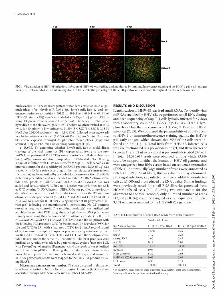
RESULTS AND DISCUSSIONIdentification of HHV-6B-derived small RNAs. To identify viralmiRNAs encoded by HHV-6B, we performed small RNA cloningand deep sequencing of Sup-T-1 cells lytically infected for 7 dayswith a laboratory strain of HHV-6B. Sup-T-1 is a CD4� T lym-phocyte cell line that is permissive to HHV-6, HHV-7, and HIV-1infection (7, 13). We confirmed the permissibility of Sup-T-1 cellsto HHV-6 by immunofluorescence staining against the HHV-6p41 early antigen, which showed that 80% of the cells were in-fected at 3 dpi (Fig. 1). Total RNA from HHV-6B-infected cellswas size fractionated in a polyacrylamide gel, and RNA species ofbetween 19 and 24 nt were cloned as previously described (39, 40).In total, 24,380,617 reads were obtained, among which 93.9%could be mapped to either the human or HHV-6B genome, andwere categorized into RNA classes based on sequence annotation(Table 1). An unusually large number of reads were annotated asrRNA (71.58%). Most likely, this was due to nonsynchronized,prolonged infection, i.e., infected cells were added to uninfectedcells at 1:1,000 and thus reduced the RNA quality. Similar findingswere previously noted for small RNA libraries generated fromHCMV-infected cells (40). Allowing two mismatches for thealignment to the viral genome, only a limited number of reads(12,350 [0.05%]) could be assigned as viral sequences. Of these,9,138 sequences mapped to the HHV-6B Z29 genome.
FIG 1 Visualization of HHV-6B infection. Infection of HHV-6B was verified and monitored by immunofluorescence staining of the HHV-6 p41 early antigenin Sup-T-1 cells infected with a laboratory strain of HHV-6B. The percentage of HHV-6B-positive cells increased throughout the 3-day time course.
TABLE 1 Distribution of small RNA reads from both librariesa
RNA classification
% of total clones
HHV-6B total RNA HHV-6B Ago2 IP RNA
rRNA 71.58 0.29tRNA 1.12 0.25sn-snoRNA 1.00 0.45scRNA 0.14 0.09miRNA 11.97 94.96Repeats 0.93 0.13Host genome 6.82 1.03HHV-6B (Z29) genome 0.05 0.69Unknown 6.38 2.11Total 100.00 100.00a sn-snoRNA, small nuclear-small nucleolar RNA; scRNA, small cytoplasmic RNA.Shading indicates the species examined in this study.
To enhance the sensitivity and boost the degree of confidencein our analysis, we performed small RNA cloning and deep se-quencing analysis of RNAs obtained after Ago2 immunoprecipi-tation (Ago2 IP) of Sup-T-1 cells lytically infected with HHV-6B.In total, 30,225,650 reads were obtained, of which 97.93% weremappable. We observed that �95% of all reads were annotated asmiRNAs. Due to the prevalence of miRNAs within the Ago2 sam-ple, we could be confident that the majority of sequences derivingfrom the virus (207,394 reads [0.69%]) were likely to representgenuine viral miRNAs (Table 1). Such a limited number of viralsequences is intriguing compared to results for other herpesvi-ruses, particularly those for the betaherpesvirus family (17, 40),where viral miRNAs often constitute 10 to 60% of the cellularmiRNA pool late in infection. However, it was previously notedfor mouse CMV (MCMV) that the proportion of viral sequencespresent in a given library is dependent on the efficiency of viralinfection, multiplicity of infection (MOI), and cell type. For in-stance, in a small-scale library created from bone marrow macro-phages infected with MCMV, only �0.5% of sequences repre-sented viral sequences, compared to �30% for mouse embryonicfibroblasts (MEFs) (8). Similarly, a recent report showed that lessthan 2% of small RNA sequences cloned from marmoset T cellsinfected with herpesvirus saimiri mapped to the viral genome(12). Similar to the case for HHV-6B, miRNAs characterized dur-ing lytic infection of the integrating, oncogenic Marek’s diseasevirus (MDV) contributed only �1% of cloned sequences (9).
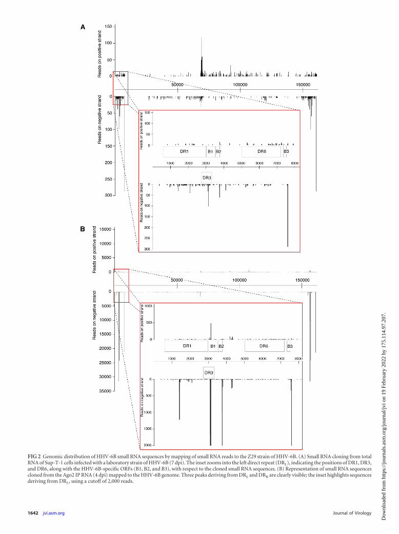
Distribution of HHV-6B small RNA sequences across the ge-nome. To simplify the analysis and to reduce noise, only se-quences with zero mismatches to HHV-6B (strain Z29) and be-tween 19 and 24 nt (with no corresponding human genomic hits)were used to search for putative miRNA candidates. Sequenceswere mapped to the HHV-6B Z29 reference genome (GenBankaccession no. NC_000898.1) to look for hot spots across the ge-nome. The HHV-6B genome consists of a unique long region (U)of 145 kb flanked by two identical repetitive regions of 8 to 9 kbthat contain direct repeats (DRL and DRR). The distribution ofHHV-6B small RNA sequences across the genome was quite strik-ing. Mapping of HHV-6B small RNA sequences revealed that themajority of viral sequences derived from either the direct repeatregions or the region between open reading frames (ORFs) U41and U42 that contains the OriLyt (15). Entire genomic represen-tations of cloned sequences from both total RNA (Fig. 2A) andRNA obtained from Ago2 IP (Fig. 2B) are given with reference tothe Z29 strain. Comparing whole genome reads between totalRNA and Ago2 IP RNA, we observed a distinct enrichment incertain sequences deriving from the direct repeat regions (Fig.2B). Reads mapped from small RNA cloning of total RNA clearlydefined a central peak which correlated with the OriLyt (Fig. 2A).Sequences deriving from in or around the OriLyt of both rat andmouse cytomegaloviruses (RCMV and MCMV, respectively), aswell as some other herpesviruses, such as herpes simplex virus 1(HSV-1) and HSV-2, have previously been reported to encodemiRNAs or postulated to act as RNA primers for the replication ofthe viral genome (8, 17, 30, 35).
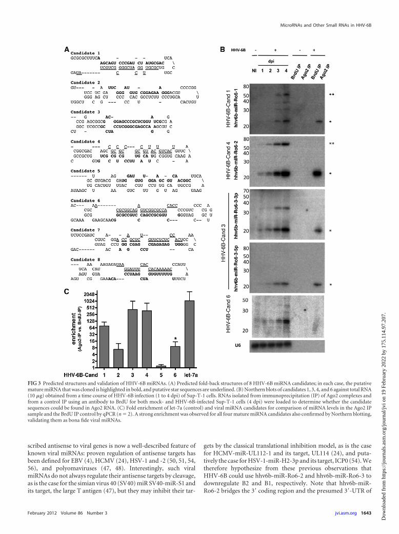
Identification of HHV-6 miRNAs. Those sequences that werecloned multiple times and had characteristic 5= homogeneity wereassessed manually for the ability to fold into typical stem-loopprecursor structures by using Mfold (59). Eight sequences (namedcandidates 1 to 8) deriving from eight putative classical pre-miRNA stem-loop structures that matched perfectly to the
HHV-6B Z29 genome were initially selected for validation (Fig.3A). To validate these candidates, we performed Northern blotanalysis of RNA taken from a time course of HHV-6B infectionfrom 1 to 4 dpi; noninfected Sup-T-1 cells were used as a control.In addition, to verify if these were bona fide miRNAs, we loadedRNA samples from Ago2 IPs (and from BrdU IPs as controls)from both noninfected and infected cells (4 dpi). Three of thesesequences (candidates 1, 3, and 4) were validated as novelHHV-6B miRNAs by their detection in both total RNA and Ago2IP RNA by Northern blotting; both the 3p and 5p arms of candi-date 3 were detected by Northern blotting of total RNA and RNAisolated from Ago2 IP samples (Fig. 3B).
Although sequences representing candidates 2, 6, 7, and 8 werecloned from RNA isolated after Ago2 IP (237, 289, 1,214, and1,639 times, respectively), we were unable to formally validatethem as miRNAs. While candidates 2, 7, and 8 remained undetect-able in both total RNA and Ago2 IP RNA by Northern blotting,candidate 6 strongly accumulated during the time course of infec-tion, as two distinct RNA species, but interestingly, it remainedundetectable in the Ago2 IP samples. In addition, a small nonspe-cific signal was detected in the control (Fig. 3B). These resultsindicate that this small RNA most probably represents a degrada-tion product or as yet unclassified small RNA species.
We further verified these results by using quantitative real-timePCR to identify enrichment of the small viral RNAs in Ago2 versusBrdU control samples; consistent with the Northern blot data,qPCR revealed significant enrichment of candidates 1, 3, and 4 inthe Ago2 IP samples (Fig. 3C). Candidates 2 and 6 were signifi-cantly enriched in the Ago2 IP samples compared to BrdU IPcontrols, although the signal for candidate 6 was weak and non-specific, reflecting the Northern blot data (Fig. 3A and C). Candi-date 5 was cloned from total RNA and was not present in the Ago2IP RNA sequence data, but it was selected as a candidate because itderived from a nonrepetitive genomic region and folded into ahairpin structure. No enrichment in Ago2 IP RNA was observedfor candidate 5 by qPCR, confirming the sequencing data; no sig-nal was observable by Northern blot analysis (Fig. 3C and data notshown).
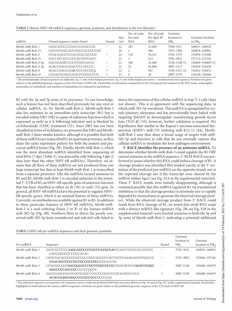
To distinguish HHV-6 from the related HHV-7, HHV-6BmiRNAs are named hhv6b-miR-Ro6-1 (candidate 1), hhv6b-miR-Ro6-2 (candidate 4), and hhv6b-miR-Ro6-3-3p and hhv6b-miR-Ro6-3-5p (candidates 3-3p and 3-5p, respectively). We referto these miRNAs by their official names hereinafter. The se-quences of the mature miRNAs and their star sequences are givenin Table 2, along with the numbers of times they were cloned fromboth total RNA and Ago2 IP RNA libraries. The sequences of thepredicted pre-miRNAs shown in Fig. 3A are given in Table 3, withreference to the Z29 strain of HHV-6B.
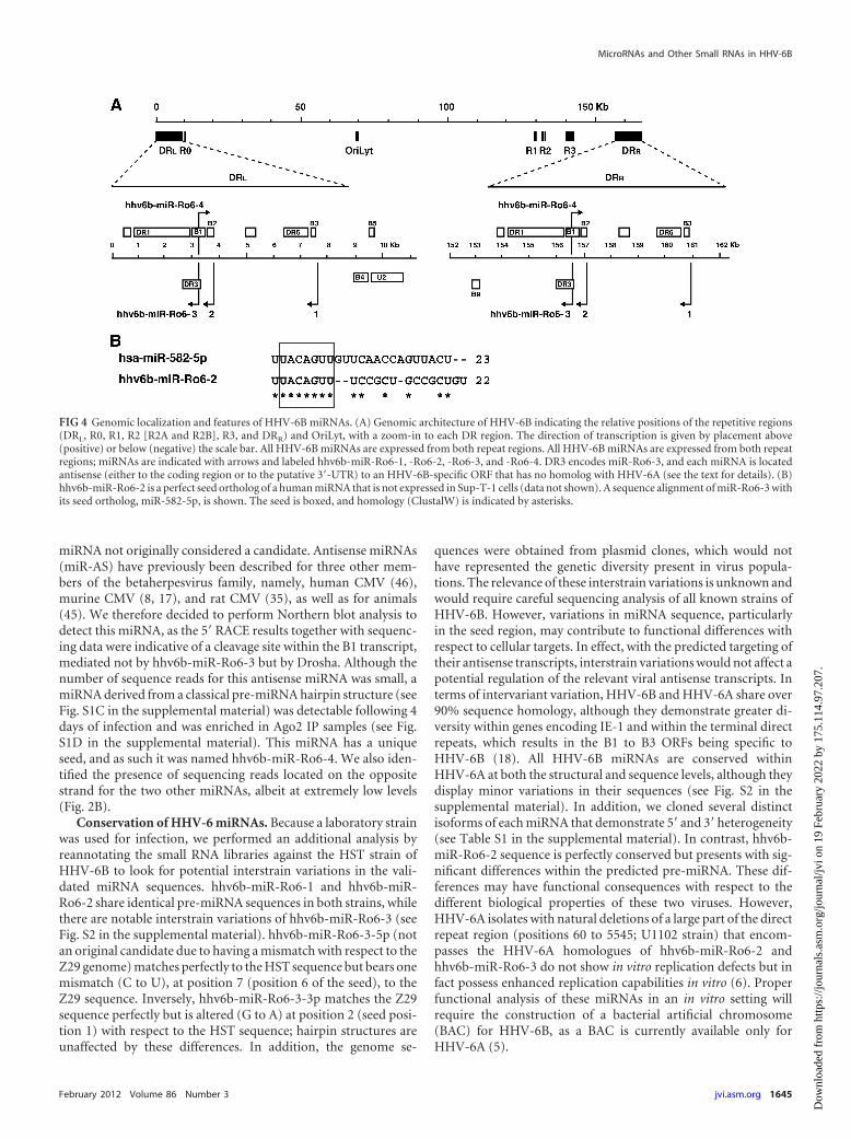
Characteristics of HHV-6 miRNAs. A schematic representa-tion of HHV-6B miRNAs with respect to their genomic localiza-tion in the direct repeats is given in Fig. 4A. Note that hhv6b-miR-Ro6-3 lies antisense to the predicted B1 ORF and is encodedwithin DR3, while hhv6b-miR-Ro6-2 lies antisense to the pre-dicted B2 ORF. B1 and B2 are unique to HHV-6B among all her-pesviruses and were recently identified as 2 of the 8 immediate-early (IE) genes expressed by HHV-6B via transcriptome analyses(53). The functions of B1 and B2 are unknown, but it is thoughtthat due to their classification as IE genes and their specificity toHHV-6B, they may play important roles in HHV-6B-specific in-fection (53). The identification of viral miRNAs that are tran-
FIG 2 Genomic distribution of HHV-6B small RNA sequences by mapping of small RNA reads to the Z29 strain of HHV-6B. (A) Small RNA cloning from totalRNA of Sup-T-1 cells infected with a laboratory strain of HHV-6B (7 dpi). The inset zooms into the left direct repeat (DRL), indicating the positions of DR1, DR3,and DR6, along with the HHV-6B-specific ORFs (B1, B2, and B3), with respect to the cloned small RNA sequences. (B) Representation of small RNA sequencescloned from the Ago2 IP RNA (4 dpi) mapped to the HHV-6B genome. Three peaks deriving from DRL and DRR are clearly visible; the inset highlights sequencesderiving from DRL, using a cutoff of 2,000 reads.
scribed antisense to viral genes is now a well-described feature ofknown viral miRNAs: proven regulation of antisense targets hasbeen defined for EBV (4), HCMV (24), HSV-1 and -2 (50, 51, 54,56), and polyomaviruses (47, 48). Interestingly, such viralmiRNAs do not always regulate their antisense targets by cleavage,as is the case for the simian virus 40 (SV40) miR SV40-miR-S1 andits target, the large T antigen (47), but they may inhibit their tar-
gets by the classical translational inhibition model, as is the casefor HCMV-miR-UL112-1 and its target, UL114 (24), and puta-tively the case for HSV-1-miR-H2-3p and its target, ICP0 (54). Wetherefore hypothesize from these previous observations thatHHV-6B could use hhv6b-miR-Ro6-2 and hhv6b-miR-Ro6-3 todownregulate B2 and B1, respectively. Note that hhv6b-miR-Ro6-2 bridges the 3= coding region and the presumed 3=-UTR of
FIG 3 Predicted structures and validation of HHV-6B miRNAs. (A) Predicted fold-back structures of 8 HHV-6B miRNA candidates; in each case, the putativemature miRNA that was cloned is highlighted in bold, and putative star sequences are underlined. (B) Northern blots of candidates 1, 3, 4, and 6 against total RNA(10 �g) obtained from a time course of HHV-6B infection (1 to 4 dpi) of Sup-T-1 cells. RNAs isolated from immunoprecipitation (IP) of Ago2 complexes andfrom a control IP using an antibody to BrdU for both mock- and HHV-6B-infected Sup-T-1 cells (4 dpi) were loaded to determine whether the candidatesequences could be found in Ago2 RNA. (C) Fold enrichment of let-7a (control) and viral miRNA candidates for comparison of miRNA levels in the Ago2 IPsample and the BrdU IP control by qPCR (n � 2). A strong enrichment was observed for all four mature miRNA candidates also confirmed by Northern blotting,validating them as bona fide viral miRNAs.
B2 with the 3p and 5p arms of its precursor. To our knowledge,such a feature has not been described previously for any viral orcellular miRNA. As for hhv6b-miR-Ro6-2, hhv6b-miR-Ro6-3also lies antisense to an immediate-early transcript (B1) but isencoded within DR3. DR3 is a gene of unknown function which isexpressed as early as 6 h following infection and is blocked bycycloheximide (CHX) treatment (53). Since DR3 has not beenclassified in terms of its kinetics, we presume that DR3 and hhv6b-miR-Ro6-3 share similar kinetics, although it is possible that bothof these miRNAs are transcribed by an upstream promoter, as theyshare the same expression pattern for both the mature and pre-cursor miRNA forms (Fig. 3B). Finally, hhv6b-miR-Ro6-1, whichwas the most abundant miRNA identified from sequencing oftotal RNA (7 dpi) (Table 2), was detectable only following 3 dpi (2days later than the other HHV-6B miRNAs). Therefore, we as-sume that all three of these miRNAs are not produced from onelarge transcript but that at least hhv6b-miR-Ro6-1 is transcribedfrom a separate promoter. Like the miRNAs located antisense toB1 and B2, hhv6b-miR-Ro6-1 is encoded antisense to the poten-tial 3=-UTR of B3, an HHV-6B-specific gene of unknown functionthat has been classified as either an IE (38) or early (53) gene. Ingeneral, all HHV-6B miRNAs have the potential to regulate HHV-6B-specific genes, which is an unusual feature of these miRNAs.Currently, no antibodies are available against B1 to B3. In additionto these particular features of HHV-6B miRNAs, hhv6b-miR-Ro6-2 is a seed ortholog (bases 2 to 8) of the human miRNAmiR-582-5p (Fig. 4B). Northern blots to detect the poorly con-served miR-582-5p from noninfected and infected cells failed to
detect the expression of this cellular miRNA in Sup-T-1 cells (datanot shown). This is in agreement with the sequencing data, inwhich miR-582-5p was absent. This miRNA is upregulated in cer-tain pituitary adenomas and has provisionally been linked withtargeting SMAD3 to downregulate transforming growth factorbeta (TGF-�) (10); however, further validation is required. Wehypothesize that similar to the Kaposi’s sarcoma-associated her-pesvirus (KSHV) miR-155 ortholog miR-K12-11 (44), hhv6b-miR-Ro6-2 may thus share a broad range of targets with miR-582-5p and function in cells that do not normally express thiscellular miRNA to modulate the host-pathogen environment.
5= RACE identifies the presence of an antisense miRNA. Todetermine whether hhv6b-miR-Ro6-3 could regulate the B1 ORFcarried antisense to the miRNA sequence, 5= RLM-RACE was per-formed to assess whether this RNA could induce cleavage of B1. Acleavage product was identified that resided exactly at the 5= ter-minus of the predicted pre-miRNA on the opposite strand, not atthe expected cleavage site if the transcript were cleaved by themiRNA within Ago2 (see Fig. S1A in the supplemental material).These 5= RACE results were initially disappointing, although itremained possible that this miRNA regulated B1 via translationalinhibition or that the cleavage product is extremely rare or rapidlydegraded by exonucleases to generate the observed cleavage prod-uct. While the observed cleavage product from 5= RACE couldresult from RNA cleavage of B1, we noted that small RNA readswith a distinct miRNA-like signature (Fig. 2B; see Fig. S1B in thesupplemental material) were located antisense to both the 5p and3p arms of hhv6b-miR-Ro6-3, indicating a potential additional
TABLE 2 Mature HHV-6B miRNA sequences, genomic positions, and distribution in the two librariesa
miRNA Cloned sequence (major form) StrandSize(nt)
No. of readsfor totalRNA
No. of readsfor Ago2 IPRNA
Genomiclocation inDRL
Genomic locationin DRR
hhv6b-miR-Ro6-1 AAGCAGUCCCGAUCUAUGCGAC � 22 287 25,449 7590–7611 160911–160932hhv6b-miR-Ro6-1� CGCGUUGGCAUCGGGCGCUGCUAU 24 1 946 7557–7580 160878–160901hhv6b-miR-Ro6-2 UUACAGUUUCCGCUGCCGCUGU � 22 110 55,321 3754–3775 157076–157096hhv6b-miR-Ro6-2� CGCCGCCGCCGUUACUGUCACU 22 2 612 3794–3815 157115–157136hhv6b-miR-Ro6-3-5p GACGGAGUCCGCUCGGUAUCG � 21 100 21,460 3128–3148 (1) 156449–156469 (1)hhv6b-miR-Ro6-3-3p AGACCGAGCGGGCUCCAUCCG � 21 32 61,900 3097–3117 156418–156438hhv6b-miR-Ro6-4 AUACCGAGCGGACUCCGUCGCU � 22 2 947 3130–3151 (2) 156451–156472hhv6b-miR-Ro6-4� CGGAUGGAGCCCGCUCGGUCUCG � 23 0 20 3097–3119 156418–156440a The predominantly cloned sequences are indicated. 5p, 5= arm of the hairpin precursor; 3p, 3= arm of the hairpin precursor; *, nonfunctional star sequence. Positions are givenrelative to the published genomic sequence of the Z29 strain of HHV-6B. Nucleotides in bold denote mismatches to the Z29 genome but matches to the HST strain, nontemplatenucleotides are underlined, and numbers of mismatches are indicated in parentheses.
TABLE 3 HHV-6B pre-miRNA sequences and their genomic positions
a The indicated sequences correspond to the sequences used to make the predicted fold-back structures shown in Fig. 3A and in Fig. S1C in the supplemental material. Nucleotideshighlighted in bold indicate the mature miRNA sequences. Positions are given relative to the published genomic sequence of the Z29 strain of HHV-6B.
miRNA not originally considered a candidate. Antisense miRNAs(miR-AS) have previously been described for three other mem-bers of the betaherpesvirus family, namely, human CMV (46),murine CMV (8, 17), and rat CMV (35), as well as for animals(45). We therefore decided to perform Northern blot analysis todetect this miRNA, as the 5= RACE results together with sequenc-ing data were indicative of a cleavage site within the B1 transcript,mediated not by hhv6b-miR-Ro6-3 but by Drosha. Although thenumber of sequence reads for this antisense miRNA was small, amiRNA derived from a classical pre-miRNA hairpin structure (seeFig. S1C in the supplemental material) was detectable following 4days of infection and was enriched in Ago2 IP samples (see Fig.S1D in the supplemental material). This miRNA has a uniqueseed, and as such it was named hhv6b-miR-Ro6-4. We also iden-tified the presence of sequencing reads located on the oppositestrand for the two other miRNAs, albeit at extremely low levels(Fig. 2B).
Conservation of HHV-6 miRNAs. Because a laboratory strainwas used for infection, we performed an additional analysis byreannotating the small RNA libraries against the HST strain ofHHV-6B to look for potential interstrain variations in the vali-dated miRNA sequences. hhv6b-miR-Ro6-1 and hhv6b-miR-Ro6-2 share identical pre-miRNA sequences in both strains, whilethere are notable interstrain variations of hhv6b-miR-Ro6-3 (seeFig. S2 in the supplemental material). hhv6b-miR-Ro6-3-5p (notan original candidate due to having a mismatch with respect to theZ29 genome) matches perfectly to the HST sequence but bears onemismatch (C to U), at position 7 (position 6 of the seed), to theZ29 sequence. Inversely, hhv6b-miR-Ro6-3-3p matches the Z29sequence perfectly but is altered (G to A) at position 2 (seed posi-tion 1) with respect to the HST sequence; hairpin structures areunaffected by these differences. In addition, the genome se-
quences were obtained from plasmid clones, which would nothave represented the genetic diversity present in virus popula-tions. The relevance of these interstrain variations is unknown andwould require careful sequencing analysis of all known strains ofHHV-6B. However, variations in miRNA sequence, particularlyin the seed region, may contribute to functional differences withrespect to cellular targets. In effect, with the predicted targeting oftheir antisense transcripts, interstrain variations would not affect apotential regulation of the relevant viral antisense transcripts. Interms of intervariant variation, HHV-6B and HHV-6A share over90% sequence homology, although they demonstrate greater di-versity within genes encoding IE-1 and within the terminal directrepeats, which results in the B1 to B3 ORFs being specific toHHV-6B (18). All HHV-6B miRNAs are conserved withinHHV-6A at both the structural and sequence levels, although theydisplay minor variations in their sequences (see Fig. S2 in thesupplemental material). In addition, we cloned several distinctisoforms of each miRNA that demonstrate 5= and 3= heterogeneity(see Table S1 in the supplemental material). In contrast, hhv6b-miR-Ro6-2 sequence is perfectly conserved but presents with sig-nificant differences within the predicted pre-miRNA. These dif-ferences may have functional consequences with respect to thedifferent biological properties of these two viruses. However,HHV-6A isolates with natural deletions of a large part of the directrepeat region (positions 60 to 5545; U1102 strain) that encom-passes the HHV-6A homologues of hhv6b-miR-Ro6-2 andhhv6b-miR-Ro6-3 do not show in vitro replication defects but infact possess enhanced replication capabilities in vitro (6). Properfunctional analysis of these miRNAs in an in vitro setting willrequire the construction of a bacterial artificial chromosome(BAC) for HHV-6B, as a BAC is currently available only forHHV-6A (5).
FIG 4 Genomic localization and features of HHV-6B miRNAs. (A) Genomic architecture of HHV-6B indicating the relative positions of the repetitive regions(DRL, R0, R1, R2 [R2A and R2B], R3, and DRR) and OriLyt, with a zoom-in to each DR region. The direction of transcription is given by placement above(positive) or below (negative) the scale bar. All HHV-6B miRNAs are expressed from both repeat regions. All HHV-6B miRNAs are expressed from both repeatregions; miRNAs are indicated with arrows and labeled hhv6b-miR-Ro6-1, -Ro6-2, -Ro6-3, and -Ro6-4. DR3 encodes miR-Ro6-3, and each miRNA is locatedantisense (either to the coding region or to the putative 3=-UTR) to an HHV-6B-specific ORF that has no homolog with HHV-6A (see the text for details). (B)hhv6b-miR-Ro6-2 is a perfect seed ortholog of a human miRNA that is not expressed in Sup-T-1 cells (data not shown). A sequence alignment of miR-Ro6-3 withits seed ortholog, miR-582-5p, is shown. The seed is boxed, and homology (ClustalW) is indicated by asterisks.
Other small RNAs identified in HHV-6. An overall analysis ofsmall viral RNAs in the Ago2 RNA sample indicated that the fourmiRNA hairpins verified to give rise to HHV-6B miRNAs com-prise �80% of all viral sequences. Note that candidates 7 and 8form fold-back hairpins (Fig. 3A) with characteristic 5= homoge-neity and were enriched in libraries generated from Ago2 RNAcompared to total RNA (Fig. 2A and B and 5A and B). Thesecandidates were tested by both qPCR and Northern blotting; how-ever, we were unable to verify these as miRNAs, likely owing totheir low abundance. Considering that �95% of all cellular RNAsfound in the Ago2 IP samples were classifiable as miRNAs, it isinteresting to speculate that these sequences may simply derivefrom rare HHV-6B miRNAs not detectable under the conditionsused. Experiments to assess the role of HHV-6B miRNAs in la-tency and in vivo will assess whether these sequences representmiRNAs. Sequences and genomic positions of all the candidatescan be found within Table S2 in the supplemental material. Abreakdown of the distribution of sequence reads across the ge-nome identifies �85% as deriving from the direct repeats (DR),�7.5% as deriving from the OriLyt, and �7.5% as deriving fromelsewhere. As mentioned, miRNAs have been verified from in oraround the OriLyt regions for other herpesviruses, namely,MCMV (8, 17), RCMV (35), and HSV-1 (30). As such, Northernblots were performed for the three most abundant sequences de-riving from the origin: candidate 8 (not defined as a miRNA), onthe negative strand (positions c68744 to c68765), and two contig-uous sequences that do not form a typical hairpin, located on thepositive strand (positions 69121 to 69141 and 69141 to 69161)(Fig. 5A and B). Interestingly, probes against the contiguous se-quences identified a novel viral transcript of �62 nt spanning thisregion, located near sites for the origin binding protein (OBP)(29) (Fig. 5C). Note that sequencing data from both libraries in-dicate multiple RNA species deriving from this locus (sequencesin Ago2 RNA ranging from 17 to 32 nt), and this is clearly visibleas a broad set of vertical bars in the graphical representation ofOriLyt sequences and by a smear on the Northern blot (Fig. 5A, B,and C). Notably, abundant RNA species of 18 and 19 nt wereidentified from only one arm of the precursor; the Northern blotsignal indicates that these were not present in Ago2 RNA at com-parable levels to those in total RNA samples (as seen for viralmiRNAs in Fig. 3B). As such, their presence in Ago2 cloning al-most certainly represents a nonspecific binding to Ago2 due totheir abundance within the cell, highlighting the benefit of includ-ing RNA from Ago2 IP on Northern blots to distinguish RNAspecies within the Ago2 sequence from other small RNAs orbreakdown products. Interestingly, Tsao et al. recently character-ized the expression of lytic genes in HHV-6B and identified anumber of intergenic noncoding transcripts (53). Among thesetranscripts, they identified a 316-nt RNA located such that it couldcontain the �60-nt RNA we detected. It appears reasonable to
hypothesize that these small RNAs derive from this novel viralRNA being degraded specifically to generate small RNA speciesthat may represent RNA primers to assist in viral replication. Al-ternatively, these small RNAs could represent a new class of smallRNAs that may be incorporated into another member of theArgonaute family. Performing immunoprecipitations with thethree remaining Argonaute proteins will resolve this interestinghypothesis. Further work is required to elucidate their processingand potential functions in HHV-6 biology.
Conclusions. Here we showed by using a deep sequencing ap-proach that HHV-6B encodes at least four pre-miRNAs, two ofwhich lie antisense to each other at the same genomic locus. ThesemiRNAs are expressed during lytic infection and are all conservedin HHV-6A. The use of small RNA cloning from Ago2 IP samplesgreatly simplified the analysis by removing the majority of RNAbreakdown products, providing a clean data set to aid in identify-ing novel viral miRNAs. HHV-6B miRNAs accumulate duringHHV-6B infection and, similar to HSV-1/2 miRNAs, are encodedantisense to IE HHV-6B-specific ORFs (B1, B2, B3, and DR3), inline with the paradigm that viral miRNAs can regulate IE genes tomodulate their own transcription. Like the case for KSHV-miR-K12-11 and miR-155, hhv6b-miR-Ro6-2 is a seed ortholog of acellular miRNA (miR-582-5p) that is not expressed in the maintarget cell of the virus, in this case CD4� T cells. These data pro-vide good starting points for identifying the cellular and viral tar-gets of these miRNAs. Similar to HCMV, HHV-6 is a betaherpes-virus, and its miRNAs were identified in cells undergoing activelytic infection (19, 23, 40).
This work opens up questions regarding the cloning bias ob-served in deep sequencing data sets for certain miRNAs; the viralmiRNAs reported in this study were present at low abundances inthe sequencing data sets. Nevertheless, the miRNA candidates allconform to standard rules governing miRNA classification (1).This was confirmed by their Northern blot detection in both totalRNA and Ago2 IP RNA. The inclusion of Ago2 IP RNA in North-ern blots aided in our discrimination of miRNAs from non-miRNA-like small RNAs or breakdown products, such as candi-date 6.
In terms of the use of bioinformatics to predict a given miRNA,caution is warranted. In the original study by Pfeffer et al., nopre-miRNA hairpins were predicted for HHV-6, although thelikelihood that HHV-6 encodes miRNAs was stated at 84% (40).More recently, an open access large database (Vir-miR) was pub-lished that predicted viral miRNAs in over 2,000 viral genomes(33), but none of our experimentally validated miRNAs were pre-dicted in that study. These reports open up speculation as to theefficacy of bioinformatic predictions when miRNAs derive fromcomplex genomic loci such as direct repeats. An improved ap-proach relies on the combination of prediction tools and deepsequencing data sets to scan for putative pre-miRNA hairpins, as
FIG 5 Identification of an abundant small RNA mapping to the OriLyt. (A) Representation of small RNA reads mapping to the region encompassing the originof lytic replication from total RNA of Sup-T-1 cells infected with HHV-6B (7 dpi). Sites for OBP are given, along with the positions of the indirect repeat regions(IDRs) and the neighboring genes U41 and U42. The horizontal gray bar indicates the position of the sequence that was analyzed by Northern blotting. (B)Sequences mapping to the OriLyt obtained following sequencing of RNAs within the Ago2 IP sample. A clear enrichment of two sets of antisense sequences wasvisible following IP. The leftmost sequence (candidate 8, indicated by an arrow) was enriched �50 times (compared to that in panel A). The horizontal gray barindicates the position of the sequence that was analyzed by Northern blotting. (C) Northern blots for two contiguous sequences enriched in Ago2 RNA andderiving from a GC-rich region between OBP1 and the IDR. A novel viral transcript encompassing positions 69121 to 69161 was clearly detectable and increasedin abundance throughout the time course of infection. One arm of this transcript generates a range of abundant small RNAs which are not detectable by Northernblotting of Ago2 RNA and may represent a novel class of origin-derived small RNAs.
in miRCat (37), miRDeep (20, 21), and miRAnalyzer (27, 28)software. At least one strand of each HHV-6B pre-miRNA waspredicted by one or more of these programs for either the totalRNA library, the Ago2 IP library, or both (see Table S3 in thesupplemental material), which provides further support regard-ing their miRNA status.
Unlike all other human herpesviruses, HHV-6A has beenshown to integrate into host telomeres as a method of latency inindividuals with chromosomally integrated HHV-6 (CIHHV-6),rather than residing as an episome. Importantly, the virus can bereactivated from this state and passed on through generations ofsuch individuals (2). It will be interesting to see if these miRNAsare expressed in latently infected cells as well as in CIHHV-6 pa-tients. The distribution of HHV-6 miRNAs is somewhat differentfrom what has been found to date in other betaherpesviruses butresembles the genomic organization of the miRNAs of the chickenMarek’s disease virus, another integrating herpesvirus (9).
The identification of an abundant transcript of 60 to 65 ntexpressed from the OriLyt could be reminiscent of other viralpre-miRNAs recently identified to be expressed from the OriLytregions of MCMV (8, 17), RCMV (35), bovine herpesvirus(BoHV) (22), and HSV-1/2 (30). However, similar to our results,none of the miRNAs reported in these papers were reported con-clusively to be in Ago2 RNA; hence, herpesvirus origin-derivedsmall RNAs represent an area of research that clearly deservesfurther investigation.
As with all viral miRNAs, a thorough decoding of cellular andviral targets is essential to understand the potential therapeuticuses of targeting the viral miRNAs of a given virus in the context ofhuman disease. The identification of miRNAs from HHV-6 willprovide researchers with new avenues of research concerning thehost-pathogen relationship during HHV-6 infection.
ACKNOWLEDGMENTS
We thank members of our laboratories for critically reviewing the manu-script prior to publication. We thank Timo Lassmann for providing uswith an early version of Nexalign.
This work was supported by a grant from the German Bundesminis-terium für Bildung und Forschung (NGFN-Plus 01GS0801) to L.D. andby the European Research Council (ERC starting grant ncRNAVIR260767) and a starting grant from the Centre National de la RechercheScientifique (ATIP) to S.P.
REFERENCES1. Ambros V, et al. 2003. A uniform system for microRNA annotation. RNA
9:277–279.2. Arbuckle JH, et al. 2010. The latent human herpesvirus-6A genome
specifically integrates in telomeres of human chromosomes in vivo and invitro. Proc. Natl. Acad. Sci. U. S. A. 107:5563–5568.
4. Barth S, et al. 2008. Epstein-Barr virus-encoded microRNA miR-BART2down-regulates the viral DNA polymerase BALF5. Nucleic Acids Res. 36:666 – 675.
5. Borenstein R, Frenkel N. 2009. Cloning human herpes virus 6A genomeinto bacterial artificial chromosomes and study of DNA replication inter-mediates. Proc. Natl. Acad. Sci. U. S. A. 106:19138 –19143.
6. Borenstein R, Zeigerman H, Frenkel N. 2010. The DR1 and DR6 firstexons of human herpesvirus 6A are not required for virus replication inculture and are deleted in virus stocks that replicate well in T-cell lines. J.Virol. 84:2648 –2656.
7. Boyd MT, Simpson GR, Cann AJ, Johnson MA, Weiss RA. 1993. Asingle amino acid substitution in the V1 loop of human immunodefi-ciency virus type 1 gp120 alters cellular tropism. J. Virol. 67:3649 –3652.
8. Buck AH, et al. 2007. Discrete clusters of virus-encoded microRNAs areassociated with complementary strands of the genome and the 7.2-kilobase stable intron in murine cytomegalovirus. J. Virol. 81:13761–13770.
9. Burnside J, et al. 2006. Marek’s disease virus encodes microRNAs thatmap to meq and the latency-associated transcript. J. Virol. 80:8778 – 8786.
10. Butz H, et al. 2011. MicroRNA profile indicates downregulation of theTGFbeta pathway in sporadic non-functioning pituitary adenomas. Pitu-itary 14:112–124.
11. Cai X, et al. 2005. Kaposi’s sarcoma-associated herpesvirus expresses anarray of viral microRNAs in latently infected cells. Proc. Natl. Acad. Sci.U. S. A. 102:5570 –5575.
12. Cazalla D, Xie M, Steitz JA. 2011. A primate herpesvirus uses the inte-grator complex to generate viral microRNAs. Mol. Cell 43:982–992.
13. Cermelli C, et al. 1997. SupT-1: a cell system suitable for an efficientpropagation of both HHV-7 and HHV-6 variants A and B. New Micro-biol. 20:187–196.
14. De Bolle L, Naesens L, De Clercq E. 2005. Update on human herpesvirus6 biology, clinical features, and therapy. Clin. Microbiol. Rev. 18:217–245.
15. Dewhurst S, Dollard SC, Pellett PE, Dambaugh TR. 1993. Identificationof a lytic-phase origin of DNA replication in human herpesvirus 6B strainZ29. J. Virol. 67:7680 –7683.
16. Dolken L, et al. 2010. Systematic analysis of viral and cellular microRNAtargets in cells latently infected with human gamma-herpesviruses byRISC immunoprecipitation assay. Cell Host Microbe 7:324 –334.
17. Dolken L, et al. 2007. Mouse cytomegalovirus microRNAs dominate thecellular small RNA profile during lytic infection and show features ofposttranscriptional regulation. J. Virol. 81:13771–13782.
18. Dominguez G, et al. 1999. Human herpesvirus 6B genome sequence:coding content and comparison with human herpesvirus 6A. J. Virol.73:8040 – 8052.
19. Dunn W, et al. 2005. Human cytomegalovirus expresses novel micro-RNAs during productive viral infection. Cell. Microbiol. 7:1684 –1695.
20. Friedlander MR, et al. 2008. Discovering microRNAs from deep sequenc-ing data using miRDeep. Nat. Biotechnol. 26:407– 415.
21. Friedlander MR, Mackowiak SD, Li N, Chen W, Rajewsky N. miRDeep2accurately identifies known and hundreds of novel microRNA genes inseven animal clades. Nucleic Acids Res., in press.
22. Glazov EA, et al. 2010. Characterization of microRNAs encoded by thebovine herpesvirus 1 genome. J. Gen. Virol. 91:32– 41.
23. Grey F, et al. 2005. Identification and characterization of humancytomegalovirus-encoded microRNAs. J. Virol. 79:12095–12099.
24. Grey F, Nelson J. 2008. Identification and function of human cytomeg-alovirus microRNAs. J. Clin. Virol. 41:186 –191.
25. Griffiths-Jones S. 2006. miRBase: the microRNA sequence database.Methods Mol. Biol. 342:129 –138.
26. Griffiths-Jones S, Saini HK, van Dongen S, Enright AJ. 2008. miRBase:tools for microRNA genomics. Nucleic Acids Res. 36:D154 –D158.
27. Hackenberg M, Rodriguez-Ezpeleta N, Aransay AM. 2011. miRanalyzer:an update on the detection and analysis of microRNAs in high-throughput sequencing experiments. Nucleic Acids Res. 39:W132–W138.
28. Hackenberg M, Sturm M, Langenberger D, Falcon-Perez JM, AransayAM. 2009. miRanalyzer: a microRNA detection and analysis tool for next-generation sequencing experiments. Nucleic Acids Res. 37:W68 –W76.
29. Inoue N, Dambaugh TR, Rapp JC, Pellett PE. 1994. Alphaherpesvirusorigin-binding protein homolog encoded by human herpesvirus 6B, abetaherpesvirus, binds to nucleotide sequences that are similar to ori re-gions of alphaherpesviruses. J. Virol. 68:4126 – 4136.
30. Jurak I, et al. 2010. Numerous conserved and divergent microRNAsexpressed by herpes simplex viruses 1 and 2. J. Virol. 84:4659 – 4672.
31. Kim YJ, et al. 2002. Human herpesvirus-6 as a possible cause of enceph-alitis and hemorrhagic cystitis after allogeneic hematopoietic stem celltransplantation. Leukemia 16:958 –959.
32. Kondo K, et al. 2002. Strong interaction between human herpesvirus 6and peripheral blood monocytes/macrophages during acute infection. J.Med. Virol. 67:364 –369.
33. Li SC, Shiau CK, Lin WC. 2008. Vir-Mir db: prediction of viral mi-croRNA candidate hairpins. Nucleic Acids Res. 36:D184 –D189.
34. Lusso P. 2006. HHV-6 and the immune system: mechanisms of immu-nomodulation and viral escape. J. Clin. Virol. 37(Suppl 1):S4 –S10.
35. Meyer C, et al. 2011. Cytomegalovirus microRNA expression is tissuespecific and is associated with persistence. J. Virol. 85:378 –389.
36. Moore FG, Wolfson C. 2002. Human herpes virus 6 and multiple sclero-sis. Acta Neurol. Scand. 106:63– 83.
37. Moxon S, et al. 2008. A toolkit for analysing large-scale plant small RNAdatasets. Bioinformatics 24:2252–2253.
38. Oster B, Hollsberg P. 2002. Viral gene expression patterns in humanherpesvirus 6B-infected T cells. J. Virol. 76:7578 –7586.
39. Pfeffer S. 2007. Identification of virally encoded microRNAs. MethodsEnzymol. 427:51– 63.
40. Pfeffer S, et al. 2005. Identification of microRNAs of the herpesvirusfamily. Nat. Methods 2:269 –276.
41. Pfeffer S, et al. 2004. Identification of virus-encoded microRNAs. Science304:734 –736.
42. Pruksananonda P, et al. 1992. Primary human herpesvirus 6 infection inyoung children. N. Engl. J. Med. 326:1445–1450.
43. Salahuddin SZ, et al. 1986. Isolation of a new virus, HBLV, in patientswith lymphoproliferative disorders. Science 234:596 – 601.
44. Skalsky RL, et al. 2007. Kaposi’s sarcoma-associated herpesvirus encodesan ortholog of miR-155. J. Virol. 81:12836 –12845.
45. Stark A, et al. 2008. A single Hox locus in Drosophila produces functionalmicroRNAs from opposite DNA strands. Genes Dev. 22:8 –13.
46. Stark TJ, Arnold JD, Spector DH, Yeo GW. 2012. High-resolutionprofiling and analysis of viral and host small RNAs during human cyto-megalovirus infection. J. Virol. 86:226 –235.
47. Sullivan CS, Grundhoff AT, Tevethia S, Pipas JM, Ganem D. 2005.SV40-encoded microRNAs regulate viral gene expression and reduce sus-ceptibility to cytotoxic T cells. Nature 435:682– 686.
48. Sullivan CS, et al. 2009. Murine polyomavirus encodes a microRNA thatcleaves early RNA transcripts but is not essential for experimental infec-tion. Virology 387:157–167.
49. Takahashi K, et al. 1989. Predominant CD4 T-lymphocyte tropism ofhuman herpesvirus 6-related virus. J. Virol. 63:3161–3163.
50. Tang S, et al. 2008. An acutely and latently expressed herpes simplex virus2 viral microRNA inhibits expression of ICP34.5, a viral neurovirulencefactor. Proc. Natl. Acad. Sci. U. S. A. 105:10931–10936.
51. Tang S, Patel A, Krause PR. 2009. Novel less-abundant viral microRNAsencoded by herpes simplex virus 2 latency-associated transcript and theirroles in regulating ICP34.5 and ICP0 mRNAs. J. Virol. 83:1433–1442.
52. Toedling J, Ciaudo C, Voinnet O, Heard E, Barillot E. 2010. Girafe—anR/Bioconductor package for functional exploration of aligned next-generation sequencing reads. Bioinformatics 26:2902–2903.
53. Tsao EH, et al. 2009. Microarray-based determination of the lytic cascadeof human herpesvirus 6B. J. Gen. Virol. 90:2581–2591.