SmeA, a small membrane protein with multiplefunctions in Streptomyces sporulation includingtargeting of a SpoIIIE/FtsK-like protein to celldivision septa
Nora Ausmees,1** Helene Wahlstedt,1
Sonchita Bagchi,1 Marie A. Elliot,2 Mark J. Buttner3
and Klas Flärdh4*1Department of Cell and Molecular Biology, UppsalaUniversity, BMC Box 596, 75124, Uppsala, Sweden.2Department of Biology, McMaster University, 1280 MainSt. West, Hamilton, Ontario, L8S 4K1, Canada.3Department of Molecular Microbiology, John InnesCentre, Norwich NR4 7UH, UK.4Department of Cell and Organism Biology, LundUniversity, Sölvegatan 35, 223 62 Lund, Sweden.
Summary
Sporulation in aerial hyphae of Streptomyces coeli-color involves profound changes in regulation of fun-damental morphogenetic and cell cycle processes toconvert the filamentous and multinucleoid cells tosmall unigenomic spores. Here, a novel sporulationlocus consisting of smeA (encoding a small putativemembrane protein) and sffA (encoding a SpoIIIE/FtsK-family protein) is characterized. Deletion ofsmeA-sffA gave rise to pleiotropic effects on sporematuration, and influenced the segregation of chro-mosomes and placement of septa during sporulation.Both smeA and sffA were expressed specifically inapical cells of sporogenic aerial hyphae simulta-neously with or slightly after Z-ring assembly. Thepresence of smeA-like genes in streptomycete chro-mosomes, plasmids and transposons, often pairedwith a gene for a SpoIIIE/FtsK- or Tra-like protein,indicates that SmeA and SffA functions might berelated to DNA transfer. During spore developmentSffA accumulated specifically at sporulation septawhere it colocalized with FtsK. However, sffA didnot show redundancy with ftsK, and SffA functionappeared distinct from the DNA translocase activitydisplayed by FtsK during closure of sporulationsepta. The septal localization of SffA was dependent
on SmeA, suggesting that SmeA may act as anassembly factor for SffA and possibly other proteinsrequired during spore maturation.
Introduction
The life cycle of streptomycetes is one of the mostcomplex developmental processes among prokaryotes. Agerminating spore grows out to form a vegetative myce-lium of branched hyphae. Nutrient limitation and possiblyother stimuli then trigger a complex multicellular develop-ment, which includes the emergence of a specializedaerial mycelium on the surface of the colony and theproduction of antibiotics and other secondary metabolites(Chater and Losick, 1997; Elliot et al., 2007). Sub-sequently, a profound reprogramming of regulation andmodification of fundamental morphogenetic and cell cycleprocesses take place in the aerial hyphae, leading to thedifferentiation of these filamentous, multinucleoid cellsinto chains of unigenomic spores that have thick cell wallsand low metabolic activity, permitting dispersal and long-term survival.
A key morphogenetic event in the sporulation of aerialhyphae is the switch in the mode of cell division from thewidely spaced thin crosswalls, or vegetative septa, thatdelimit the multinucleoid hyphal cells in the substratemycelium to sporulation septation, which partitions thesporogenic aerial hyphal cell into prespores (Chaterand Losick, 1997; Chater, 2000). Sporulation septa arethicker, regularly spaced between every nucleoid, andlead to deep cell wall constriction and eventually separa-tion of daughter cells (Wildermuth and Hopwood, 1970;Flärdh and Van Wezel, 2003). Sporulation requires adevelopmentally induced upregulation of ftsZ (Flärdhet al., 2000), encoding the tubulin homologue that directsbacterial cell division (Margolin, 2005). FtsZ then polymer-izes into helical filaments that are remodelled to form aseries of regularly spaced FtsZ rings along the sporulatinghyphal cell, and subsequently organize the formation ofsporulation septa (Schwedock et al., 1997; Grantcharovaet al., 2005). Simultaneously, mechanisms for segrega-tion of chromosomes have to be activated in order toensure that each spore inherits one copy of the genome.
The ParA and ParB proteins, which are similar to proteinsinvolved in chromosome and plasmid partitioning in manyother bacteria, have a role in developmentally controllednucleoid partitioning in Streptomyces coelicolor (Kimet al., 2000; Jakimowicz et al., 2006). However, the rela-tively mild phenotype caused by the lack of ParA andParB (only about 13% of the spores contained aberrantamounts of DNA) implies that additional proteins areinvolved (Kim et al., 2000). It has been previouslyreported that the constriction of sporulation septa in strep-tomycetes initiates before nucleoid partitioning is com-plete (Schwedock et al., 1997; Miguelez et al., 1998;Flärdh, 2003; Noens et al., 2005), and thus, some mecha-nism for transport or movement of DNA through theclosing septa must exist. One candidate likely to beinvolved in this process is the S. coelicolor FtsK protein(SCO5750), which is homologous to the SpoIIIE/FtsKDNA translocases (Errington et al., 2001). Indeed, FtsKwas recently reported to localize at sporulation septa andto affect the integrity of chromosomes after sporulationin S. coelicolor, suggesting that it may be involved insporulation-specific genome segregation (Wang et al.,2007). However, as the majority of spores of the ftsK nullmutant contained intact chromosomes, other mecha-nisms are likely to be involved as well.
Some of the key regulators of the development of aerialhyphae into spores in S. coelicolor are known, but ouroverall understanding of the pathways and mechanisms isstill fragmentary. Commitment to sporulation requires theRNA polymerase sigma factor WhiG (Chater et al., 1989).In the absence of whiG, the aerial hyphae remain straightand form widely spaced vegetative-type hyphal cross-walls (Flärdh et al., 1999). Two target genes under thecontrol of whiG have been characterized, whiH encodinga GntR-family repressor and whiI encoding a responseregulator (Ryding et al., 1998; Ainsa et al., 1999). whiHand whiI are both required for efficient sporulation septa-tion, but the respective mutants have different phenotypes(Ainsa et al., 1999; Flärdh et al., 1999; Tian et al., 2007).The genes whiA and whiB constitute a second convergingpathway in the control of sporulation in aerial hyphae andare both required for sporulation septation in the aerialhyphae (Chater, 1972; Flärdh et al., 1999; Ainsa et al.,2000). Orthologues of WhiA are ubiquitous among Gram-positive bacterial genomes, and WhiB is the foundingmember of a large family of Actinobacteria-specific pro-teins (Soliveri et al., 2000; Gao et al., 2006). Both whiAand whiB are upregulated during sporulation in a whiG-independent fashion (Soliveri et al., 1992; Ainsa et al.,1999), but their exact functions are unclear. These fiveearly whi genes are all required for full sporulation septa-tion, most spore maturation processes, and the develop-mental upregulation of a number of genes including ftsZ,parAB and sigF (encoding a s factor that affects spore
maturation), as well as ssgB (which encodes a member ofthe SsgA-like family of proteins that appear to modifypeptidoglycan/cell wall during sporulation) and the genesof the whiE cluster (which encode the biosyntheticgenes for the grey spore pigment) (Kelemen et al., 1996;1998; Flärdh et al., 2000; Chater, 2001; Sevcikova andKormanec, 2003; Noens et al., 2005; Jakimowicz et al.,2006). Apart from some sWhiG regulon members, no directtargets for any of the sporulation regulatory proteins havebeen identified, and it is still an open question as to howthese early regulatory genes can trigger the profoundmorphogenetic changes taking place during the differen-tiation of aerial hyphae into spores.
In this article, we characterize a novel locus whoseexpression depends on both whiA and the whiG/whiI/whiH pathway, and is constrained to the sporogenic aerialhyphae at a time coinciding with the assembly of multipleFtsZ rings. The locus consists of two genes with new rolesin sporulation: one encodes a small putative membraneprotein of unknown function (SmeA) and the other amember of the SpoIIIE/FtsK family (SffA). We show thatdeletion of these genes has a pleiotropic effect on sporedevelopment, and that smeA is required for the specifictargeting of SffA to the sporulation septa. SffA influencesthe accuracy of nucleoid partitioning, but has a differentfunction from that of FtsK.
Results
smeA-sffA (SCO1415-16) constitutes a novelsporulation locus
In order to identify new genes involved in spore develop-ment, and to further elucidate the regulatory networkscontrolling spore formation in S. coelicolor, we haveanalysed gene expression profiles of the wild-type strainM145 and sporulation-deficient mutant strains throughoutdevelopment using a whole-genome microarray approach(comprehensive results will be published elsewhere, P.Salerno et al., in preparation). In a pilot microarray experi-ment we compared the developmental gene expressionprofile of the wild-type strain with those of its congenicmutants lacking whiG, whiA, whiH and whiI to identifygenes that were upregulated during development in theparent strain, but failed to be upregulated in at least one ofthe mutants. This initial analysis detected activation ofseveral known sporulation genes (Fig. S1), includingopen reading frames (ORFs) in the whiE cluster and ssgB(Kelemen et al., 1998; Kormanec and Sevcikova, 2002).In addition, it revealed a number of previously unrecog-nized genes which appeared to have sporulation-specificexpression patterns (to be described elsewhere), includ-ing a putative operon consisting of genes SCO1415 andSCO1416, whose expression was upregulated in the later
stages of development in the wild-type strain but not inany of the whi mutant strains examined (Fig. S1).SCO1415 encoded a small protein (63 amino acids) andwas designated smeA (small membrane protein). In silicoanalysis of SmeA revealed a centrally located putativetransmembrane domain and indicated a putativeN-terminal signal peptide, but did not reveal any motifs toinfer a possible cellular function (see Discussion andFig. 6) (Cserzo et al., 1997; Nielsen et al., 1997). Never-theless, BLAST and PSI-BLAST searches revealed thatsimilar proteins are widespread among Streptomyces andFrankia (see Discussion). SCO1416 encoded a proteinwith an N-terminal transmembrane region and aC-terminal domain with similarity to ATPase domains ofSpoIIIE/FtsK-family proteins, and as such, was termedsffA (Figs S2 and S3).
To confirm the expression pattern revealed by themicroarray experiment, transcription of smeA was inves-tigated using S1 nuclease protection assays on indepen-
dent RNA preparations. The largest protected fragmentscorresponded to a transcription start site ~40–50 bpupstream of the smeA start codon, and transcripts wereseen only in samples from time points at which visiblesigns of sporulation were detected (Fig. 1A), confirmingthat transcription of smeA is activated during sporulationin S. coelicolor. No smeA transcripts were detected insamples from the whiG, whiA and whiI mutants, and onlyvery faint bands were seen in the whiH mutant (Fig. 1Aand data not shown), indicating that smeA expressionwas completely dependent on the products of whiG, whiAand whiI, and strongly affected by the whiH gene product.The smeA mRNA 5′ ends were also amplified and clonedusing the rapid amplification of 5′ cDNA ends (5′RACE)method, from RNA prepared independently of a sporulat-ing wild-type culture. Of 10 sequenced inserts, six showedan mRNA start point at 28 or 29 bp upstream of thepredicted start codon for smeA, two started at 38 bp andtwo at 46 bp from the start codon (Fig. 1A, bottom). This
Fig. 1. A. S1 nuclease protection analysis oftranscription of smeA during development onSM agar plates in the wild-type strain M145and in the non-sporulating whiA and whiImutants. Numbers above indicate time inhours of development. M designates a lanecontaining a DNA size marker, and tRNAindicates a control reaction with an equalamount of yeast tRNA. Below is shown thebeginning of smeA open reading frame withupstream region. Capital letters indicate thepredicted smeA start codon, bold lettersindicate the first 5′ nucleotides of the mRNAtranscripts identified by 5′RACE, and theprimer sequence used for S1 assays isunderlined. The start point of the shortesttranscript could not be exactly assigned to theG or A as polyC tailing of the cDNA was used.A terminal G on the mRNA could therefore notbe distinguished from the added stretch of Con the complementary strand.B. Schematic presentation of the smeA-sffAlocus, deletion mutants and clones used inphenotypic studies and for complementation.LoxP indicates in-frame deletion of smeA orsmeA-sffA, and aac(3)IV indicatesreplacement of sffA by an apramycinresistance cassette. Plasmids pSmeA-SffA,pSffA and pSmeA correspond to plasmidspNA276, pNA277 and pNA485 respectively.C. Left: complementation of the light greyspore phenotype of the DsmeA and theDsmeA-sffA strains by smeA, sffA andsmeA-sffA in trans. Triple plus symbol (+++)designates restoration of spore colour to thewild-type level; single plus (+) and double plus(++) symbols indicate spore colour that isdarker than that of the mutant but lighter thanwild-type colour. Right: photograph of a6-day-old MS plate containing the followingstrains: wild-type M145, DsmeA-sffA,DsmeA-sffA with smeA-sffA in trans andDsmeA-sffA with only sffA in trans.
corresponded very well with the sizes of the fragmentsdetected in the S1 nuclease protection assay. However, itis unclear whether the multiple 5′ ends represent multipletranscription start sites or could be generated by mRNAprocessing.
To further study the putative sporulation-related roles ofsmeA and sffA, we deleted both genes from the genomeof the wild-type strain M145, which normally producesdark grey spores on MS agar plates. A DsmeA-sffA strain(K151, Table 1) produced uniformly light grey colonies,indicating that indeed, smeA and sffA were important fornormal sporulation (Fig. 1B and C). Introduction of smeA-sffA (pNA276, Table 1) in trans restored the spore pig-mentation of the mutant to the wild-type level, confirming
that the observed developmental defect was indeedcaused by smeA-sffA deletion (Fig. 1B and C). In order toclarify the individual contributions of smeA and sffA to thedefective sporulation phenotype, we constructed anin-frame DsmeA deletion mutant (K150, Table 1; Fig. 1B)and a DsffA mutant (K152, Table 1; Fig. 1B). The smeAdeletion strain had a light grey spore phenotype, similar tothat of the DsmeA-sffA strain, while deletion of sffA aloneresulted in a very slight reduction in spore pigmentation,suggesting that the lack of SmeA was primarily respon-sible for the light grey phenotypes of both the DsmeA andDsmeA-sffA mutants. However, smeA alone in trans wasonly able to partially complement the DsmeA strain (wheresffA was still present) and not the DsmeA-sffA strain,
Table 1. Strains and plasmids.
Strain/plasmid Description Source
StrainS. coelicolor
J1984 M145 sigF::tsr Kelemen et al. (1998)J2400 M145 whiG::hyg Flärdh et al. (1999)J2401 M145 whiA::hyg Flärdh et al. (1999)J2408 M145 DwhiH::ermE Flärdh et al. (1999)J2450 M145 whiI::hyg Ainsa et al. (1999)K102 M145 Dglk-esp119 ftsZ17(Spo) Grantcharova et al.
(2003)K127 M145 ftsZ::pKF40[F(ftsZ–egfp)Hyb], merodiploid strain, contains both
ftsZ and ftsZ–egfpK. Flärdh Grantcharova
et al. (2005)K150 M145 DsmeA::loxP This studyK151 M145 DsmeA-sffA::loxP This studyK152 M145 DsffA::[aac(3)IV oriT] This studyK158 M145 sffA::pNA303[F(sffA–egfp)Hyb], sffA is replaced by sffA–egfp This studyK159 M145 DsmeA::loxP sffA::pNA303[f(sffA–egfp)Hyb] This studyK161 M145 DftsK::[aac(3)IV oriT] This studyK169 M145 DsmeA-sffA::loxP DftsK::[aac(3)IV oriT] This studyK170 M145 ftsK::pNA585 [F(ftsK–mCherry)Hyb] This studyK171 M145 DsmeA::loxP ftsK::pNA585 [F(ftsK–mCherry)Hyb] This studyM145 Plasmid-free prototroph Kieser et al. (2000)
E. coliDH5a Cloning strainDY380 D(mrr-hsdRMS-mcrBC) mcrA recA1 l cl857 D(cro-bio)<>tet, for
PlasmidpIJ82 Hygromycin-resistant derivative of pSET152 Helen Kieser, JIC,
Norwich, UKpNA276 (pSmeA-SffA) pSET152 containing smeA and sffA with upstream region This studypNA277 (pSffA) pSET152 containing sffA with upstream region where smeA is replaced by loxP This studypNA303 (pSffA–GFP) Plasmid pEGFP-N2 encoding the C-terminal part of SffA fused to EGFP This studypNA473 (pPsmeA–mCh) pSET152 containing the promoter region of smeA translationally fused to mCherry This studypNA485 (pSmeA) pIJ82 containing smeA with upstream region and aac(3)IV which replaces sffA This studypNA540 (pPsigF–mCh) pSET152 containing the promoter region of sigF translationally fused to mCherry This studypNA547 pSET152 containing smeA with internally inserted mCherry and sffA This studypNA585 Plasmid pEGFP-N2 encoding the C-terminal part of FtsK fused to mCherry and
with egfp deletedThis study
pSET152 E. coli–S. coelicolor shuttle vector, apramycin resistance Bierman et al. (1992)
indicating that both SmeA and SffA are needed for optimalsporulation (Fig. 1C). Full complementation was onlyobtained when smeA and sffA were expressed in cis,suggesting that coexpression was necessary for theoptimal function of both SmeA and SffA. Together, theseresults strongly suggest that smeA-sffA constitutes anovel sporulation locus in S. coelicolor.
SmeA and SffA influence multiple developmentalprocesses during later stages of sporulation
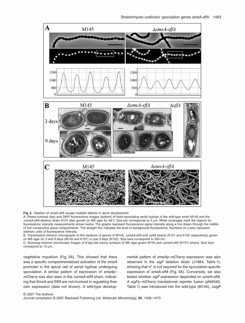
Because reduced spore pigmentation is often indicative ofdefects in underlying developmental processes leading tospore maturation, we studied the DsmeA-sffA strain byphase-contrast, fluorescence and electron microscopy togain better understanding of the developmental roles ofSmeA and SffA. Growth of the vegetative mycelium wasnot affected by deletion of smeA-sffA; however, severaldifferences between the wild-type and the DsmeA-sffAstrains were observed in sporulating aerial hyphae. First,fluorescence microscopy of fixed and DAPI-stainedsporulating hyphae revealed that the chromosomes inDsmeA-sff spores displayed a more diffuse stainingpattern than chromosomes in wild-type spores (Fig. 2A).The lower panel in Fig. 2A shows the distribution of fluo-rescence signal intensity along a line through the middleof five consecutive spore compartments. Peaks repre-senting wild-type chromosomes were sharp and declinedalmost to a background level at positions correspondingto constrictions between spore compartments, whereaspeaks of the mutant strain were wider and flatter, indicat-ing the inability of the DsmeA-sffA strain to condenseits chromosomal DNA to the wild-type level duringsporulation. Furthermore, DAPI staining also revealedthat smeA-sffA deletion caused a slight defect in chromo-some segregation. Anucleate spore compartments withinspore chains occurred with a frequency of 0.6% in theDsmeA-sffA strain, compared with the wild-type frequencyof < 0.1% (> 10 000 spore compartments were countedfor both wild-type and DsmeA-sffA strains).
Transmission electron microscopy (TEM) revealedarchitectural defects in the spore envelope of the DsmeA-sffA strain (Fig. 2B). The cell walls of the mutant sporeswere thinner and showed less defined layers than those ofthe wild-type spores, which had clearly discernible layersof light and dense material, as described previously (McVit-tie, 1974). In particular, the pronounced electron-denseouter layer of the wild-type spores appeared considerablythinner and ‘paler’ in the mutant (Fig. 2B). Reducedspore wall thickness is often correlated with decreasedspore resistance to detergents and high temperatures(Potuckova et al., 1995; Molle et al., 2000; Mazza et al.,2006), and correspondingly, we observed that mutantspores were more susceptible to heat than wild-type
spores. For example, 10.3% of the wild-type spores sur-vived treatment at 65°C for 20 min, whereas the corre-sponding figure for mutant spores was 2.4% (after 30 minthe corresponding values were 3.3% and 0.4% respec-tively). However, mutant and wild-type spores appeared tobe equally resistant to detergents (up to 5% SDS, data notshown). Intriguingly, SmeA and Sff also appeared to berequired for spore separation, as scanning electronmicroscopy (SEM) images of 8-day-old colonies grown onMS agar revealed that, while free spores were readilyapparent on the surface of the wild-type strain, spores fromthe DsmeA-sff strain appeared almost exclusively in chains(Fig. 2C). Furthermore, the uneven compartment size ofdeveloping spore chains of the DsmeA-sffA strain, seenclearly in Fig. 2C, was also observed by phase-contrastmicroscopy and by TEM (data not shown).
The phenotype of the DsmeA strain was indistinguish-able from that of the DsmeA-sffA double mutant strain atthe level of microscopic observation. In contrast, the sffAdeletion strain showed apparently normal spore matura-tion and spore wall structure, except that phase-contrastand electron microscopy revealed an uneven septationdefect similar to what is characteristic of the DsmeA-sffAstrain (example in a young DsffA spore chain is shown inFig. 2B). Taken together, these observations show thatthe products of the smeA-sffA operon influence severalprocesses of normal formation of prespore compartmentsand spore maturation, including division septum place-ment, chromosome segregation and condensation, sporeseparation, spore pigmentation, maturation of the sporeenvelope and development of heat resistance.
The smeAp promoter is induced specifically insporogenic aerial hyphal cells independently of thesporulation sigma factor gene sigF
Interestingly, most features of the pleiotropic smeA-sffAphenotype overlap with the phenotype associated with thedeletion of the RNA polymerase sigma factor gene sigF,which is also known to affect late stages of sporulation(Potuckova et al., 1995). sigF is specifically induced insporulating hyphae (Sun et al., 1999) and like smeA-sffA,its transcription depends on the early whi genes (whiG,whiA, whiB, whiH, whiI and whiJ) (Kelemen et al., 1996).Thus it was possible that smeA-sffA transcription wassF-dependent. To test this hypothesis, we fused the pro-moter region, ribosome binding site and start codon ofsmeA to the gene for the red fluorescent protein mCherry(smeAp–mCherry, pNA473, Table 1), and used this trans-lational fusion to monitor smeA expression. In the wild-type, a strong fluorescence signal was observed insporulating aerial hyphae with constrictions or sporulationsepta, and a weaker signal was seen in some uncon-stricted aerial hyphae, but no signal was detected in the
vegetative mycelium (Fig. 3A). This showed that therewas a specific compartmentalized activation of the smeApromoter in the apical cell of aerial hyphae undergoingsporulation. A similar pattern of expression of smeAp–mCherry was also seen in the DsmeA-sffA strain, indicat-ing that SmeA and SffA are not involved in regulating theirown expression (data not shown). A wild-type develop-
mental pattern of smeAp–mCherry expression was alsoobserved in the sigF deletion strain (J1984, Table 1),showing that sF is not required for the sporulation-specificexpression of smeA-sffA (Fig. 3A). Conversely, we alsotested whether sigF expression depended on smeA-sffA.A sigFp–mCherry translational reporter fusion (pNA540,Table 1) was introduced into the wild-type (M145), DsigF
Fig. 2. Deletion of smeA-sffA causes multiple defects in spore development.A. Phase-contrast (top) and DAPI fluorescence images (bottom) of fixed sporulating aerial hyphae of the wild-type strain M145 and theDsmeA-sffA deletion strain K151 after growth on MS agar for 48 h. Size bar corresponds to 5 mm. White rectangles mark the regions forfluorescence intensity measurements shown below. The graphs represent fluorescence signal intensity along a line drawn through the middleof five consecutive spore compartments. The straight line indicates the level of background fluorescence. Numbers on y-axis representarbitrary units of fluorescence intensity.B. Transmission electron micrographs of thin sections of spores of M145, DsmeA-sffA and DsffA strains (K151 and K152 respectively) grownon MS agar for 3 and 9 days (M145 and K151) or just 3 days (K152). Size bars correspond to 200 nm.C. Scanning electron microscopic images of 9-day-old colony surfaces of MS agar-grown M145 and DsmeA-sffA (K151) strains. Size barscorrespond to 10 mm.
and DsmeA-sffA strains. Strong sporulation-specificexpression of mCherry fluorescence was observed in allthree strains, showing that sigF expression did notdepend on SmeA-SffA or sF itself (Fig. 3A and data notshown). Thus, sigF and smeA-sffA are expressed inde-pendently of each other in sporulating aerial hyphae.
Activation of smeA-sffA expression occurs after Z-ringformation, but is not dependent on regular septation
Expression of both sigF and smeA-sffA depends on theearly whi genes. As the early whi genes are also neededfor the formation of sporulation septa, and ftsZ mutantsthat are specifically defective in the formation of sporula-tion septa have pleiotropic sporulation defects including astrong reduction in the production of the grey sporepigment (Flärdh et al., 2000; Grantcharova et al., 2003), ithas been speculated that septum formation might repre-sent a morphological checkpoint that triggers the expres-sion of late sporulation genes whose products are needed
after compartmentalization of the aerial hyphae (Flärdhet al., 2000). To test whether smeA-sffA was subject tothis putative cell-division checkpoint, we first needed todetermine the timing of smeA-sffA expression in relationto Z-ring formation. For this purpose, the smeAp–mCherryreporter was introduced into strain K127, which producesboth wild-type FtsZ and FtsZ–EGFP. Most sporulatinghyphae displayed both FtsZ–EGFP and smeAp–mCherryfluorescence. However, some unconstricted aerialhyphae had clearly visible Z-rings but no detectablemCherry fluorescence (Fig. 3B), but we never detectedsmeAp–mCherry expression without an FtsZ–EGFPsignal. These results suggested that smeA expressionoccurred no earlier than, and possibly after, FtsZ-ringformation. The same experiment was conducted with thesigFp–mCherry reporter, which allowed comparison withthis previously known late sporulation gene (Kelemenet al., 1996). The timing of sigF expression was indistin-guishable from that of smeA-sffA (Fig. 3C), indicating thatboth smeA-sffA and sigF gene expression could in prin-
Fig. 3. Developmental timing of smeA and sigF expression.A. Translational fusions of the smeA and sigF promoters to mCherry (smeAp–mCh and sigFp–mCh) were monitored in the wild-type M145,the DsigF strain J1984 and the DsmeA-sffA strain K151. Overlays of phase-contrast and fluorescence images (red) show compartmentalizedpromoter activity only in sporulating aerial hyphae.B and C. Phase-contrast images and fluorescence overlay images of FtsZ–EGFP (green) and mCherry (red) of live aerial hyphae in differentdevelopmental stages of the KF127 strain containing the smeAp–mCherry fusion (B) and the KF127 strain containing the sigFp–mCherryfusion (C) are shown. Hyphae in an earlier developmental stage display only FtsZ–EGFP structures, while those in later stages show bothFtsZ–EGFP and smeAp–mCherry (B) or sigFp–mCherry (C) fluorescence, showing similar timing of the firing of both promoters.D. Phase-contrast image of aerial hyphae of the ftsZ17(Spo) strain carrying the smeAp–mCherry fusion (top) and the same image overlaidwith the mCherry fluorescence image (red, bottom) are shown. Insets in both panels depict separated phase-dark hyphae showing a high levelof smeAp-driven mCherry production.
ciple be subject to septum checkpoint regulation. We nextintroduced the smeAp–mCherry promoter probe into astrain carrying the mutant allele ftsZ17(Spo) (K102,Table 1), which is severely impaired in the formation ofregular Z-rings and sporulation septa in the aerial myce-lium (Grantcharova et al., 2003). If expression of smeA-sffA required normal FtsZ-ring formation and septation,we would expect to see reduced smeAp–mCherry fluo-rescence in the aerial hyphae in this mutant background.Interestingly, many aerial hyphae showed smeAp–mCherry fluorescence with intensity comparable to thewild-type situation (Fig. 3D). Furthermore, it has beenshown that sporulation septa form with low frequency inthe ftsZ17(Spo) strain, causing separation of some aerialhyphal fragments (Grantcharova et al., 2003). Such sepa-rated fragments (inset in Fig. 3D) showed not only intensemCherry fluorescence, but were also thick and phase-dark, reminiscent of the spore-like aerial hyphal fragmentsformed by whiH and ftsZD2p mutants (Flärdh et al., 1999;2000; Grantcharova et al., 2003). Thus, there does notseem to be a quantitative dependence of smeA expres-sion on the formation of multiple Z-rings and sporulationsepta in aerial hyphae. However, it is still possible that thedependence was qualitative, and formation of one, or afew, sporulation septa per aerial hypha suffices to triggersmeA-sffA expression.
SffA localizes to late sporulation septa in aSmeA-dependent manner
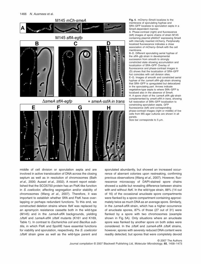
The homology of SffA to FtsK-like DNA translocases andthe increased frequency of anucleate spores in the DsmeA-sffA mutant suggested a possible involvement of this locusin segregation of chromosomes. It was therefore importantto determine whether SmeA and SffA were targeted to thesporulation septa or to other specific sites. Unfortunately,all attempts to create a fully functional tagged derivative ofSmeA were unsuccessful. Both N-terminal and C-terminalfusions to mCherry or EGFP, respectively, were non-functional, as was SmeA carrying a short C-terminal FLAGtag. However, insertion of mCherry internally, immediatelyfollowing a putative signal sequence cleavage site, pro-duced a partially functional fusion protein as indicated by aslight restoration of spore pigmentation on MS agar plates(pNA547, Table 1). This fusion protein (mCh–SmeA) dis-played a developmental expression pattern similar to thesmeAp–mCherry reporter (Fig. 3A) and the SffA–EGFPprotein (see below and Fig. 4) and was evenly distributedin the cell periphery (Fig. 4A), thus supporting the pre-dicted membrane localization of SmeA. However, as thefusion protein was only weakly active, we cannot excludethat the native SmeA protein might have a more distinctcellular localization pattern, such as, for example, colocal-ization with SffA to the sporulation septa.
Replacement of sffA by sffA–egfp in the chromosome ofthe wild-type strain M145 (K158, Table 1) did not cause thecharacteristic uneven septation phenotype of the DsffAstrain, indicating that the SffA–EGFP fusion protein was atleast partially functional. In agreement with the resultsseen with the smeAp–mCherry reporter, no SffA–EGFPfluorescence was detected during vegetative growth.Weak SffA–EGFP fluorescence appeared in some earlynon-constricted sporogenic hyphae (Fig. 4B). In laterstages, when visible constrictions had appeared betweenspore compartments, SffA–EGFP had re-localized andaccumulated into strongly fluorescent foci in the middle ofthe constricting septa (Fig. 4C and D). Thus, SffAhas a celltype-specific expression and septal localization in S. coeli-color, which would be consistent with a hypothetical role asa translocase protein suggested by its homology to FtsK/SpoIIIE proteins. In addition, SffA–EGFP was also seen atsome vegetative-type septa in the subapical segment ofaerial hyphae (data not shown), which otherwise showedno SffA–GFP upregulation.
To investigate the cellular role of SmeA and the putativefunctional relationship with SffA, we studied the localiza-tion of SffA–EGFP in a DsmeA background. sffA wasreplaced by sffA–egfp in the chromosome of the DsmeAstrain (K159, Table 1), and as expected, the DsmeA sffA–egfp strain was phenotypically similar to the DsmeA strainand showed normal activation of sffA–egfp expression insporogenic hyphae (Fig. 4E). Strikingly, however, SffA–EGFP did not relocalize to the constricting sporulationsepta of the DsmeA deletion strain (Fig. 4F and G). Incontrast, SffA–EGFP localization to the vegetative-typesepta (arrows in Fig. 4E and F) in the non-sporulating partof the hyphae occurred both in the smeA and smeA +
strains. Introduction of smeA-sffA in trans fully restored thedynamic localization pattern of SffA–EGFP (Fig. 4H) andthe dark grey spore colour to the smeA sffA–egfp strain.Thus, SmeA was specifically required for SffA accumula-tion at the constricting sporulation septa, but the localiza-tion of SffA–EGFP to vegetative-type septa in basal partsof sporulating aerial hyphae was SmeA-independent. It isimportant to note that while both the DsmeA (SffA mislo-calized) and the DsmeA-sffA (SffA missing) strains hadvery similar phenotypes, the DsffA strain had a less severesporulation defect, suggesting that the mislocalization ofSffA was not solely responsible for the developmentalphenotype of the smeA strains. Consequently, SmeA mustfulfil other functions in addition to its role as a localizationdeterminant for the DNA-translocase-domain protein SffA.
Sporulation septa contain two SpoIIIE/FtsK-familyproteins with distinct functions
The septal localization of SffA resembled that of FtsK andSpoIIIE proteins from other bacteria, which localize to the
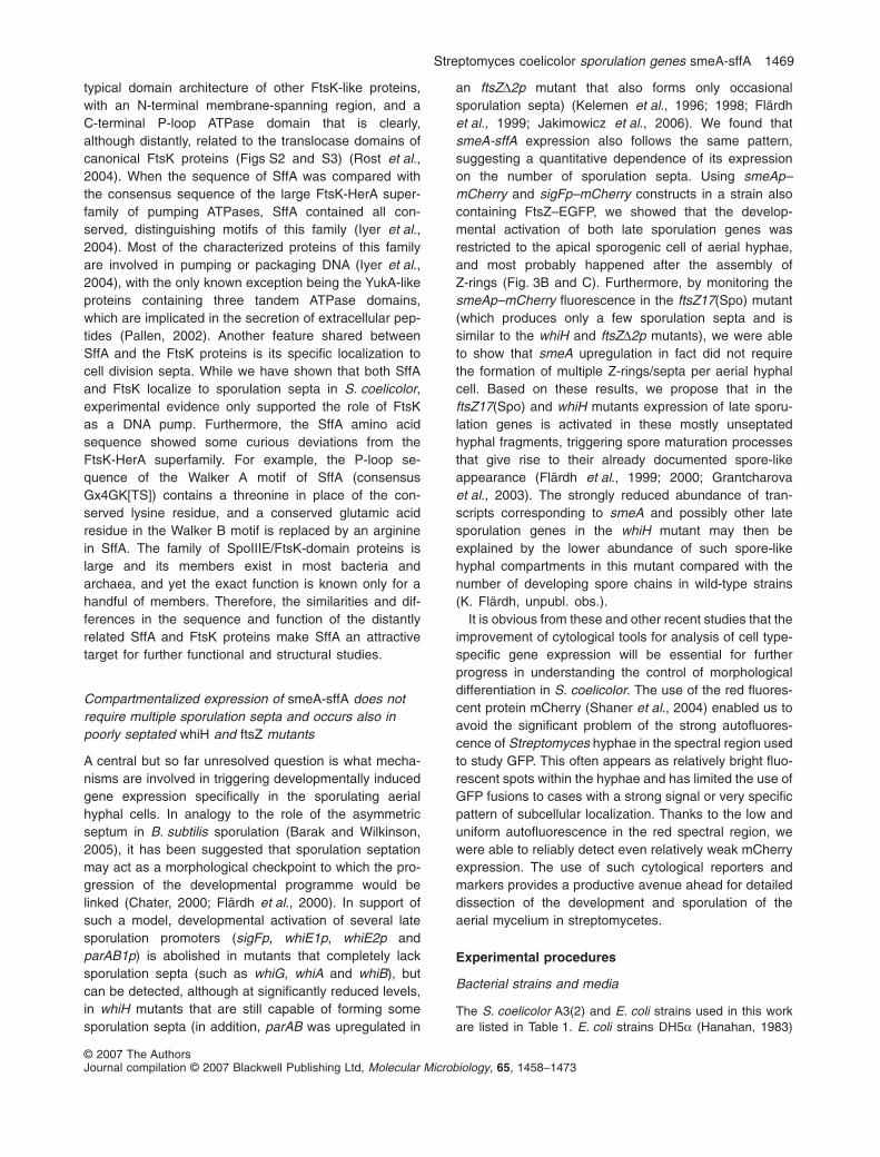
middle of cell division or sporulation septa and areinvolved in active translocation of DNA across the closingseptum as well as in resolution of chromosomes (Bathet al., 2000; Aussel et al., 2002). A recent report estab-lished that the SCO5750 protein has an FtsK-like functionin S. coelicolor, affecting segregation and/or stability ofchromosomes (Wang et al., 2007). Therefore, it wasimportant to establish whether SffA and FtsK have over-lapping or perhaps redundant functions. To this end, weconstructed deletion strains where ftsK was replaced byan apramycin resistance cassette both in the wild-type(M145) and in the DsmeA-sffA backgrounds, yieldingDftsK and DsmeA-sffA DftsK mutants (K161 and K169,Table 1). In contrast to Escherichia coli and Bacillus sub-tilis, in which FtsK and SpoIIIE have essential functionsfor viability and sporulation, respectively, the S. coelicolorDftsK strain grew as well as the wild-type parent and
sporulated abundantly, but showed an increased occur-rence of aberrant colonies upon restreaking, confirmingprevious observations (Wang et al., 2007). However, fluo-rescence microscopy of DAPI-stained spore chainsshowed a subtle but revealing difference between strainswith and without ftsK. In the wild-type strain, 88% (14 outof 16) of the occasional anucleate spore compartmentswere flanked by a spore compartment containing approxi-mately twice as much DNA as an average spore. Similarly,in the DsmeA-sffA strain, which has a higher occurrenceof anucleate spores, 87% of those (27 out of 31) wereflanked by a spore with two chromosomes (exampleshown in Fig. 5A). Only situations where an anucleatespore was flanked by another spore on both sides wereconsidered. In the DftsK and DsmeA-sffA DftsK strains,however, spores with severely reduced DNA content werereadily detected, but spores that were completely devoid
Fig. 4. mCherry–SmeA localizes to themembrane of sporulating hyphae andSffA–GFP localizes to sporulation septa in aSmeA-dependent manner.A. Phase-contrast (right) and fluorescence(left) images of spore chains of strain M145containing plasmid pNA547 expressing SmeAwith internally inserted mCherry. Peripherallylocalized fluorescence indicates a closeassociation of mCherry–SmeA with the cellmembrane.B–D. Different sporulating aerial hyphae ofthe sffA–gfp strain in developmentalsuccession from smooth to stronglyconstricted state showing accumulation andlocalization of SffA–GFP. Overlay offluorescence and phase-contrast images in(D) shows that the localization of SffA–GFPfoci coincides with cell division sites.E–G. Images of smooth and constricted aerialhyphae of the DsmeA sffA–gfp strain showingthat SffA–GFP is upregulated but delocalizedin the sporulating part. Arrows indicatevegetative-type septa to where SffA–GFP islocalized also in the absence of SmeA.H. A spore chain of the DsmeA sffA–gfp straincomplemented by smeA-sffA in trans, showingfull restoration of SffA–GFP localization toconstricting sporulation septa. GFPfluorescence (left) and correspondingphase-contrast images (right or middle) of livecells from MS agar cultures are shown in allpanels.Size bar corresponds to 5 mm.
of DNA were not observed, even though more than10 000 spores were examined for each strain. In clearcontrast to the situation in the ftsK + counterparts, in eitherthe DftsK strain or the DsmeA-sffA DftsK strain none of 43observed spores with strongly reduced DNA content wereflanked by spores with double DNA content (Fig. 5A). Ourinterpretation of these results is that the cross-septumDNA translocase activity of FtsK normally contributes tothe formation of unigenomic spores by removing chromo-somal DNA that becomes trapped under a closing septumto the proper adjacent spore compartment. However, onoccasions when septum placement or chromosome posi-tioning is impaired, the trapped chromosome may bepumped in the ‘wrong’ direction resulting in an emptyspore flanked by a spore with two chromosomes. Theseoccasions are rare in the wild type, but become morefrequent when chromosome positioning is disturbed. Insupport of this hypothesis, the DsmeA-sffA strain, whichshowed chromosome segregation and septum placementdefects, showed proportionally more occasions of zero/
double chromosome pairs. This resembles the function ofB. subtilis SpoIIIE in moving trapped chromosomes out ofclosing cell division septa (Sharpe and Errington, 1995).As most empty spores of the DsmeA-sffA strain were alsoflanked by spores with double chromosomes, it seemslikely that the clearing of DNA from closing septa is depen-dent on FtsK activity and not that of SffA.
We also replaced ftsK by ftsK–mCherry in the wild-typeand DsmeA-sffA background. Like SffA–EGFP, FtsK–mCherry localized to the sporulation septa in the wild-typebackground (Fig. 5B), confirming the FtsK localizationreported by Wang et al. (2007). The same localizationpattern was observed in the DsmeA-sffA background(Fig. 5B), indicating that, in contrast to SffA, the localiza-tion of FtsK was independent of SmeA. Thus, we haveshown that the SffA and FtsK putative translocase pro-teins both localize to sporulation septa, but their localiza-tion mechanisms and their cellular functions are different.FtsK appears to be involved in DNA translocationbetween spore compartments, but is not directly involvedin regulating sporulation. SffA, on the other hand, appearsto be functionally related to SmeA, which has a pleiotropicrole in spore maturation.
Discussion
In this article we have identified and characterized twodedicated sporulation genes whose expression is specifi-cally activated in sporogenic aerial hyphae. In contrastto several previously characterized sporulation-specificgenes (whi genes) which encode regulators of geneexpression, the gene products of smeA and sffA are notlikely to be directly involved in transcriptional control. Nev-ertheless, the small membrane protein SmeA was shownto influence several processes during spore development,including septum placement, DNA segregation and con-densation, spore wall thickening and spore separation.Although the deletion of sffA caused a subtle phenotype,its specific activation in sporulating aerial hyphae, as wellas its genetic and functional coupling to SmeA, suggesteda role for SffA in sporulation as well. Understanding thefunctions of SmeA and SffA in more detail will help to shednew light on the molecular mechanisms underlyingsporulation.
smeA-sffA organization is conserved
BLAST and PSI-BLAST searches revealed that the se-quenced Streptomyces species S. coelicolor, S. avermi-tilis and S. scabies contain four, seven and six smeA-likegenes, respectively, and several self-transmissible plas-mids from different Streptomyces species harbouredsingle copies of smeA homologues (Fig. 6). Furthermore,two sequenced Frankia strains also encoded one SmeA-
Fig. 5. FtsK is targeted to sporulation septa independently ofSmeA and its activity as a cross-septum translocase is notredundant with the function of SffA.A. Overlays of phase-contrast and DAPI fluorescence images(yellow) of spore chain segments of the DsmeA-sffA and the doublemutant DsmeA-sffA DftsK strains, containing anucleate spores.Numbers below the images indicate the mean intensity offluorescence signal (reflecting the DNA content) in a correspondingspore compartment. The anucleate spore compartment of theDsmeA-sffA strain, but not the partially DNA-free spore of theDsmeA-sffA DftsK strain, is flanked by a spore with twochromosomes.B. Overlays of phase-contrast and FtsK–mCherry fluorescence(red) images of live aerial hyphae of the ftsK–mCherry and theDsmeA-sffA ftsK–mCherry strains. In both strains FtsK–mCherry,which is replacing the wild-type FtsK, shows similar localization inthe middle of sporulation septa.Size bar corresponds to 5 mm.
like protein each, but we did not find homologoues in othercurrently available actinomycete genomes. In total, wehave identified 22 members of the Sme family, but wesuspect that more distantly related proteins may not havebeen detected using BLAST, due to the short length of thequery sequence (Fig. 6). All members of the SmeA familycontain a transmembrane domain predicted with highprobability by most algorithms. Furthermore, the SignalPalgorithm recognized a putative N-terminal secretionsignal in several, but not in all SmeA homologues (Nielsenet al., 1997). For example, the probabilities for the fourS. coelicolor SmeA proteins to possess an N-terminalsignal sequence vary between 0.684 and 0.995 accordingto the hidden Markov model prediction (Nielsen andKrogh, 1998). Each of the three sequenced Streptomycesspecies encodes one canonical SmeA-SffA pair in a con-served genetic environment. Strikingly, 12 of the 22members of the sme family are adjacent to genes encod-ing SpoIIIE/FtsK-family proteins, including the single smefamily genes of both Frankia species and all sme genesharboured on plasmids (indicated by asterisks in Fig. 6).Given that conservation of genetic linkage often indicatesa functional relationship, and as the plasmid-encodedSpoIIIE/FtsK-family proteins (called Tra proteins) functionas DNA translocases during conjugal plasmid transfer(Grohmann et al., 2003), the plasmid-encoded SmeA-likeproteins may have a function associated with DNAtransfer. Several of the chromosomally encoded Sme-Sff-like pairs appear to be part of integrated plasmids, basedon neighbouring genes encoding typical plasmid func-
tions; however, the chromosomal neighbourhood ofsmeA-sffA in S. coelicolor does not contain plasmid-related genes. Thus, an intriguing possibility is that SmeAand SffA function in some aspects of moving or organizingchromosomal DNA during sporulation.
What could be the cellular role of SmeA?
One consequence of SmeA deletion is the inability tocorrectly localize SffA to the sporulation septa. However,the complex phenotype of smeA deletion could not beexplained solely by mislocalization of SffA. PerhapsSmeA is needed for proper localization or membraneassembly of sporulation-specific proteins in addition toSffA. Such a function would be reminiscent of the roles ofseveral small membrane proteins in other bacteria. InE. coli, for example, CcmD (69 aa) has been proposed tofunction as an assembly factor for the formation of amembrane-bound multiprotein complex functioning in thehaem delivery process (Ahuja and Thony-Meyer, 2005),while in B. subtilis, the membrane-associated proteinSpoVM (29 aa) tethers the spore coat to the outer sporemembrane (Ramamurthi et al., 2006).
Are SmeA and SffA involved in DNA translocation?
In addition to the effects of DsmeA-sffA and DsffA muta-tions on the accuracy of chromosome segregation indeveloping spore chains, several observations support arole for SffA as a DNA translocase. SffA shares the
Fig. 6. The SmeA family of small Actinomycete membrane proteins. MULTALIN (Corpet, 1988) alignment of the SmeA-like proteins fromStreptomyces coelicolor (designated by their respective sequence tags SCO), S. avermitilis (sequence tags SAV), S. scabies (arbitrarily namedSscab1–6), two proteins with locus tags Francci3_0137 and Franean1DRAFT_3434 from Frankia sp. strains CcI3 and EAN1pec respectively.pFP1119c, pSV2p52c and SAP1p50 represent plasmid-borne SmeA homologues from Streptomyces sp. circular plasmid pFP11, Streptomycesviolaceoruber linear plasmid pSV2 and S. avermitilis linear plasmid SAP1 respectively. The predicted secretion signal sequence and theinternal transmembrane domain of SmeA are shown by grey bars above the alignment. A star designates close genetic linkage of therespective gene to a gene encoding a SpoIIIE/FtsK-domain protein. Multiple alignment was performed with default settings and the outputorder reflects the relatedness of the input sequences.
typical domain architecture of other FtsK-like proteins,with an N-terminal membrane-spanning region, and aC-terminal P-loop ATPase domain that is clearly,although distantly, related to the translocase domains ofcanonical FtsK proteins (Figs S2 and S3) (Rost et al.,2004). When the sequence of SffA was compared withthe consensus sequence of the large FtsK-HerA super-family of pumping ATPases, SffA contained all con-served, distinguishing motifs of this family (Iyer et al.,2004). Most of the characterized proteins of this familyare involved in pumping or packaging DNA (Iyer et al.,2004), with the only known exception being the YukA-likeproteins containing three tandem ATPase domains,which are implicated in the secretion of extracellular pep-tides (Pallen, 2002). Another feature shared betweenSffA and the FtsK proteins is its specific localization tocell division septa. While we have shown that both SffAand FtsK localize to sporulation septa in S. coelicolor,experimental evidence only supported the role of FtsKas a DNA pump. Furthermore, the SffA amino acidsequence showed some curious deviations from theFtsK-HerA superfamily. For example, the P-loop se-quence of the Walker A motif of SffA (consensusGx4GK[TS]) contains a threonine in place of the con-served lysine residue, and a conserved glutamic acidresidue in the Walker B motif is replaced by an argininein SffA. The family of SpoIIIE/FtsK-domain proteins islarge and its members exist in most bacteria andarchaea, and yet the exact function is known only for ahandful of members. Therefore, the similarities and dif-ferences in the sequence and function of the distantlyrelated SffA and FtsK proteins make SffA an attractivetarget for further functional and structural studies.
Compartmentalized expression of smeA-sffA does notrequire multiple sporulation septa and occurs also inpoorly septated whiH and ftsZ mutants
A central but so far unresolved question is what mecha-nisms are involved in triggering developmentally inducedgene expression specifically in the sporulating aerialhyphal cells. In analogy to the role of the asymmetricseptum in B. subtilis sporulation (Barak and Wilkinson,2005), it has been suggested that sporulation septationmay act as a morphological checkpoint to which the pro-gression of the developmental programme would belinked (Chater, 2000; Flärdh et al., 2000). In support ofsuch a model, developmental activation of several latesporulation promoters (sigFp, whiE1p, whiE2p andparAB1p) is abolished in mutants that completely lacksporulation septa (such as whiG, whiA and whiB), butcan be detected, although at significantly reduced levels,in whiH mutants that are still capable of forming somesporulation septa (in addition, parAB was upregulated in
an ftsZD2p mutant that also forms only occasionalsporulation septa) (Kelemen et al., 1996; 1998; Flärdhet al., 1999; Jakimowicz et al., 2006). We found thatsmeA-sffA expression also follows the same pattern,suggesting a quantitative dependence of its expressionon the number of sporulation septa. Using smeAp–mCherry and sigFp–mCherry constructs in a strain alsocontaining FtsZ–EGFP, we showed that the develop-mental activation of both late sporulation genes wasrestricted to the apical sporogenic cell of aerial hyphae,and most probably happened after the assembly ofZ-rings (Fig. 3B and C). Furthermore, by monitoring thesmeAp–mCherry fluorescence in the ftsZ17(Spo) mutant(which produces only a few sporulation septa and issimilar to the whiH and ftsZD2p mutants), we were ableto show that smeA upregulation in fact did not requirethe formation of multiple Z-rings/septa per aerial hyphalcell. Based on these results, we propose that in theftsZ17(Spo) and whiH mutants expression of late sporu-lation genes is activated in these mostly unseptatedhyphal fragments, triggering spore maturation processesthat give rise to their already documented spore-likeappearance (Flärdh et al., 1999; 2000; Grantcharovaet al., 2003). The strongly reduced abundance of tran-scripts corresponding to smeA and possibly other latesporulation genes in the whiH mutant may then beexplained by the lower abundance of such spore-likehyphal compartments in this mutant compared with thenumber of developing spore chains in wild-type strains(K. Flärdh, unpubl. obs.).
It is obvious from these and other recent studies that theimprovement of cytological tools for analysis of cell type-specific gene expression will be essential for furtherprogress in understanding the control of morphologicaldifferentiation in S. coelicolor. The use of the red fluores-cent protein mCherry (Shaner et al., 2004) enabled us toavoid the significant problem of the strong autofluores-cence of Streptomyces hyphae in the spectral region usedto study GFP. This often appears as relatively bright fluo-rescent spots within the hyphae and has limited the use ofGFP fusions to cases with a strong signal or very specificpattern of subcellular localization. Thanks to the low anduniform autofluorescence in the red spectral region, wewere able to reliably detect even relatively weak mCherryexpression. The use of such cytological reporters andmarkers provides a productive avenue ahead for detaileddissection of the development and sporulation of theaerial mycelium in streptomycetes.
Experimental procedures
Bacterial strains and media
The S. coelicolor A3(2) and E. coli strains used in this workare listed in Table 1. E. coli strains DH5a (Hanahan, 1983)
and TOP10 (Invitrogen) were used for cloning, and strainET12567/pUZ8002 was used to drive conjugative transferof non-methylated DNA from E. coli to S. coelicolor asdescribed previously (Kieser et al., 2000). Strain GM2929was used to prepare non-methylated plasmid DNA for directtransformation of S. coelicolor protoplasts. Strain EKF104was constructed by integrating the cre expression cassettefrom p705-Cre (Zhang et al., 1998) into E. coli strain LE392.Cultivation of E. coli strains was performed as described inSambrook et al. (1989). S. coelicolor strains were grown onmannitol soy flour agar plates (MS agar), in yeast extract-maltextract medium (YEME), in tryptone soy broth (TSB) or onR2YE agar (Kieser et al., 2000).
Construction of S. coelicolor mutants
The PCR-targeting procedure was used for generationof Streptomyces gene knockout mutants essentially asdescribed in Gust et al. (2003). The primers used are listed inTable S1. The apramycin resistance cassette was obtainedfrom plasmid pIJ773 (for replacement of sffA and ftsK) andthe chloramphenicol resistance cassette from plasmid pLox-Cat2 (for the in-frame deletions of smeA and smeA-sffA)(Palmeros et al., 2000). Target genes were first replaced byresistance cassettes on cosmids 6D7 (smeA, sffA) and 7C7(ftsK) (Redenbach et al., 1996) in the E. coli strain DY380,which contains an inducible l RED system (Yu et al., 2000).The LoxCat2 cassette that replaced smeA or smeA-sffA wassubsequently excised by inducing the Cre recombinase instrain EKF104, leaving only a short sequence consisting ofthe loxP sequence and flanking restriction sites. Mutatedcosmids were introduced into S. coelicolor wild-type ormutant strains by conjugation or protoplast transformationaccording to established protocols (Kieser et al., 2000) andscreened for clones where a double recombination event hadreplaced the target gene by a respective mutant allelepresent on the cosmid. Mutants were verified by diagnosticPCR.
Plasmid construction
The plasmids used are listed in Table 1. DNA manipulationand cloning were carried out according to standard proto-cols (Sambrook et al., 1989). All primers used for cloningare listed in Table S1. Plasmid constructs were verifiedby DNA sequencing. Inserts for construction of plasmidspNA276 (pSmeA-SffA), pNA485 (pSmeA) and pNA277(pSffA) were obtained by PCR using chromosomal DNA ofstrain M145, or mutated derivatives of cosmid 6D7 as tem-plates, as shown in Fig. 1. Four hundred and thirty-five basepires of smeA upstream sequence and 317 bp of sffA down-stream sequence were included in the clones. For EGFPand mCherry fusion constructs 996 bp of sffA and 1042 bpof ftsK encoding the C-terminal halves of the proteins werefused in frame to egfp or mCherry, respectively, on plasmidpEGFP-N2, creating plasmids pNA303 and pNA585, whichwere then used to transform S. coelicolor protoplasts.Transformants were selected where homologous recombi-nation via a single crossing-over event had created a full-length recombinant fusion allele in the wild-type locus under
control of the native promoter, as well as a second dis-rupted copy of gene. For insertion of mCherry (Shaneret al., 2004) internally into smeA, a KpnI site was introducedinto smeA between regions encoding the putative signalsequence and the putative transmembrane helix by PCR-based mutagenesis of plasmid pNA276 (pSmeA-SffA). Thissite was then used for in-frame insertion of PCR-amplifiedmCherry lacking its start and stop codons, resulting inplasmid pNA547. Red fluorescent promoter probe con-structs were constructed by cloning PCR-amplified promoterregions (660 bp for smeA and 494 bp for sigF) and includ-ing respective ribosome binding sites and start codons ofsmeA and sigF in frame with the mCherry ORF in plasmidpSET152, resulting plasmids pNA473 and pNA540.
Light microscopy
Samples for light and fluorescence microscopy wereobtained either by pressing a coverslip on developing colo-nies on MS agar or by growing the strains in the anglebetween an inserted coverslip and the MS agar surface(Kieser et al., 2000). GFP and mCherry fluorescence wasobserved directly after mounting these coverslips on a glassslide with 50% glycerol in phosphate-buffered saline. Forvisualization of nucleoids, samples were first methanol-fixedand stained with 4′,6-diamidino-2-phenylindole (DAPI) asdescribed previously (Flärdh et al., 1999). All fluorescenceand phase-contrast microscopy was performed using anAxioplan II imaging fluorescence microscope equippedwith appropriate filter sets, an Axiocam charge-coupleddevice camera and Axiovision software (Carl Zeiss LightMicroscopy). Digital images were processed using AdobePhotoshop CS version 8.0 software.
Electron microscopy
For SEM the samples were pre-fixed with 2.5% glutaralde-hyde, washed three times in phosphate-buffered saline andfixed in 1% osmium tetraoxide. After dehydration in ethanolthe samples were injected through nucleopore filters with0.2 mm pore size, critical point-dried, mounted on Cambridgealloy stubs, silver sputtered and examined in a Zeiss Supra35-VP field emission SEM equipped with a STEM detector,EDAX Genesis 4000 EDS.
For TEM the samples were pre-fixed with 2.5% glutaralde-hyde, washed in phosphate-buffered saline and fixed in 1%osmium tetraoxide. After dehydration in ethanol (20–100%)and infiltration in acetone-resin 1:1, the samples were infil-trated in pure resin and then embedded in silicon plates.Sections (60 nm thick) were made on a LKB-ultramicrotomewith a diamond knife from Dupont, stained with lead citrateand uranyl acetate and examined in a Zeiss Supra 35-VPfield emission SEM equipped with a STEM detector, EDAXGenesis 4000 EDS.
described previously (Flärdh et al., 2000). The probe wasprepared by PCR using primers KF129 (aggcggcgagcatgacgagg) and 5′ radiolabelled KF130 (cgagcaggaccacgcctgag).
5′RACE was carried out using the 5′RACE kit, version 2.0(Invitrogen), according to instructions from the manufac-turer. The template RNA for this analysis was prepared fromS. coelicolor strain M145 grown on a cellophane membraneon MS agar for 48 h, using RNeasy Protect Bacteria kit(Qiagen) according the protocol from The S. coelicolorMicroarray Resource, University of Surrey (http://www.surrey.ac.uk/SBMS/Fgenomics/Microarrays/html_code/ExperimentalDesign.html). The amplified 5′RACE productswere cloned using the TOPO TA cloning kit (Invitrogen) and10 randomly chosen plasmids were investigated byDNA sequencing to determine the 5′ ends of the smeAtranscript.
Acknowledgements
We are grateful to Francisco Bolivar, Helen Kieser, FrancisStewart and Roger Tsien for gifts of strains. Jakob Engman isacknowledged for assistance with the 5′RACE. This workwas supported by Carl Trygger Foundation, a Swedish-UKjoint research project from the Royal Swedish Academy ofScience and grants from the Swedish Research Council(Grant No. 621-2004-4023 to N.A. and Grant No. 621-2004-4454 to K.F.).
References
Ahuja, U., and Thony-Meyer, L. (2005) CcmD is involved incomplex formation between CcmC and the heme chaper-one CcmE during cytochrome c maturation. J Biol Chem280: 236–243.
Ainsa, J.A., Parry, H.D., and Chater, K.F. (1999) A responseregulator-like protein that functions at an intermediatestage of sporulation in Streptomyces coelicolor A3(2). MolMicrobiol 34: 607–619.
Ainsa, J.A., Ryding, N.J., Hartley, N., Findlay, K.C., Bruton,C.J., and Chater, K.F. (2000) WhiA, a protein of unknownfunction conserved among gram-positive bacteria, isessential for sporulation in Streptomyces coelicolor A3(2).J Bacteriol 182: 5470–5478.
Aussel, L., Barre, F.X., Aroyo, M., Stasiak, A., Stasiak, A.Z.,and Sherratt, D. (2002) FtsK is a DNA motor protein thatactivates chromosome dimer resolution by switching thecatalytic state of the XerC and XerD recombinases. Cell108: 195–205.
Barak, I., and Wilkinson, A.J. (2005) Where asymmetry ingene expression originates. Mol Microbiol 57: 611–620.
Bath, J., Wu, L.J., Errington, J., and Wang, J.C. (2000) Roleof Bacillus subtilis SpoIIIE in DNA transport across themother cell-prespore division septum. Science 290: 995–997.
Bierman, M., Logan, R., O’Brien, K., Seno, E.T., Rao, R.N.,and Schoner, B.E. (1992) Plasmid cloning vectors for theconjugal transfer of DNA from Escherichia coli to Strepto-myces spp. Gene 116: 43–49.
Chater, K.F. (1972) A morphological and genetic mapping
study of white colony mutants of Streptomyces coelicolor.J Gen Microbiol 72: 9–28.
Chater, K.F. (2000) Developmental decisions during sporu-lation in the aerial mycelium in Streptomyces. In Prokary-otic Development. Brun, Y.V., and Shimkets, L.J. (eds).Washington, DC: American Society for Microbiology Press,pp. 33–48.
Chater, K.F. (2001) Regulation of sporulation in Streptomy-ces coelicolor A3(2): a checkpoint multiplex? Curr OpinMicrobiol 4: 667–673.
Chater, K.F., and Losick, R. (1997) Multicellular lifestyle ofStreptomyces coelicolor A3(2) and its relatives. In Bacteriaas Multicellular Organisms. Shapiro, J.A., and Dworkin, M.(eds). New York, NY: Oxford University Press, pp. 149–182.
Chater, K.F., Bruton, C.J., Plaskitt, K.A., Buttner, M.J.,Mendez, C., and Helmann, J.D. (1989) The developmentalfate of S. coelicolor hyphae depends upon a gene producthomologous with the motility sigma factor of B. subtilis. Cell59: 133–143.
Corpet, F. (1988) Multiple sequence alignment with hierarchi-cal clustering. Nucleic Acids Res 16: 10881–10890.
Cserzo, M., Wallin, E., Simon, I., von Heijne, G., and Elofs-son, A. (1997) Prediction of transmembrane alpha-helicesin prokaryotic membrane proteins: the dense alignmentsurface method. Protein Eng 10: 673–676.
Elliot, M.A., Buttner, M.J., and Nodwell, J.R. (2007) Multicel-lular development in Streptomyces. In Multicellularityand Differentiation among the Myxobacteria and TheirNeighbors. Kaplan, H.B., and Whitworth, D.E. (eds). Wash-ington, DC: American Society for Microbiology Press.(in press).
Errington, J., Bath, J., and Wu, L.J. (2001) DNA transport inbacteria. Nat Rev Mol Cell Biol 2: 538–545.
Flärdh, K. (2003) Growth polarity and cell division inStreptomyces. Curr Opin Microbiol 6: 564–571.
Flärdh, K., and Van Wezel, G.P. (2003) Cell division duringgrowth and development of Streptomyces. Recent ResDevel Bacteriol 1: 71–90.
Flärdh, K., Findlay, K.C., and Chater, K.F. (1999) Associationof early sporulation genes with suggested developmentaldecision points in Streptomyces coelicolor A3(2). Microbi-ology 145: 2229–2243.
Flärdh, K., Leibovitz, E., Buttner, M.J., and Chater, K.F. (2000)Generation of a non-sporulating strain of Streptomycescoelicolor A3(2) by the manipulation of a developmentallycontrolled ftsZ promoter. Mol Microbiol 38: 737–749.
Gao, B., Paramanathan, R., and Gupta, R.S. (2006) Signa-ture proteins that are distinctive characteristics of Actino-bacteria and their subgroups. Antonie Van Leeuwenhoek90: 69–91.
Grantcharova, N., Ubhayasekera, W., Mowbray, S.L.,McCormick, J.R., and Flärdh, K. (2003) A missense muta-tion in ftsZ differentially affects vegetative and develop-mentally controlled cell division in Streptomyces coelicolorA3(2). Mol Microbiol 47: 645–656.
Grantcharova, N., Lustig, U., and Flärdh, K. (2005) Dynamicsof FtsZ assembly during sporulation in Streptomyces coeli-color A3(2). J Bacteriol 187: 3227–3237.
Grohmann, E., Muth, G., and Espinosa, M. (2003) Conjuga-tive plasmid transfer in gram-positive bacteria. MicrobiolMol Biol Rev 67: 277–301.
Gust, B., Challis, G.L., Fowler, K., Kieser, T., and Chater,K.F. (2003) PCR-targeted Streptomyces gene replacementidentifies a protein domain needed for biosynthesis of thesesquiterpene soil odor geosmin. Proc Natl Acad Sci USA100: 1541–1546.
Hanahan, D. (1983) Studies on transformation of Escherichiacoli with plasmids. J Mol Biol 166: 557–580.
Iyer, L.M., Makarova, K.S., Koonin, E.V., and Aravind, L.(2004) Comparative genomics of the FtsK-HerA superfam-ily of pumping ATPases: implications for the origins ofchromosome segregation, cell division and viral capsidpackaging. Nucleic Acids Res 32: 5260–5279.
Jakimowicz, D., Mouz, S., Zakrzewska-Czerwinska, J., andChater, K.F. (2006) Developmental control of a parAB pro-moter leads to formation of sporulation-associated ParBcomplexes in Streptomyces coelicolor. J Bacteriol 188:1710–1720.
Kelemen, G.H., Brown, G.L., Kormanec, J., Potuckova, L.,Chater, K.F., and Buttner, M.J. (1996) The positions of thesigma-factor genes, whiG and sigF, in the hierarchy con-trolling the development of spore chains in the aerialhyphae of Streptomyces coelicolor A3(2). Mol Microbiol 21:593–603.
Kelemen, G.H., Brian, P., Flärdh, K., Chamberlin, L., Chater,K.F., and Buttner, M.J. (1998) Developmental regulation oftranscription of whiE, a locus specifying the polyketidespore pigment in Streptomyces coelicolor A3(2). J Bacteriol180: 2515–2521.
Kieser, T., Bibb, M.J., Buttner, M.J., Chater, K.F., andHopwood, D. (2000) Practical Streptomyces Genetics.Norwich: The John Innes Foundation.
Kim, H.J., Calcutt, M.J., Schmidt, F.J., and Chater, K.F.(2000) Partitioning of the linear chromosome during sporu-lation of Streptomyces coelicolor A3(2) involves an oriC-linked parAB locus. J Bacteriol 182: 1313–1320.
Kormanec, J., and Sevcikova, B. (2002) The stress-responsesigma factor sigma(H) controls the expression of ssgB, ahomologue of the sporulation-specific cell division genessgA, in Streptomyces coelicolor A3(2). Mol Genet Genom-ics 267: 536–543.
Lee, E.C., Yu, D., Martinez de Velasco, J., Tessarollo, L.,Swing, D.A., Court, D.L., et al. (2001) A highly efficientEscherichia coli-based chromosome engineering systemadapted for recombinogenic targeting and subcloning ofBAC DNA. Genomics 73: 56–65.
Margolin, W. (2005) FtsZ and the division of prokaryotic cellsand organelles. Nat Rev Mol Cell Biol 6: 862–871.
Mazza, P., Noens, E.E., Schirner, K., Grantcharova, N.,Mommaas, A.M., Koerten, H.K., et al. (2006) MreB ofStreptomyces coelicolor is not essential for vegetativegrowth but is required for the integrity of aerial hyphae andspores. Mol Microbiol 60: 838–852.
McVittie, A. (1974) Ultrastructural studies on sporulation inwild-type and white colony mutants of Streptomycescoelicolor. J Gen Microbiol 81: 291–302.
Miguelez, E.M., Rueda, B., Hardisson, C., and Manzanal,M.B. (1998) Nucleoid partitioning and the later stagesof sporulation septum synthesis are closely associatedevents in the sporulating hyphae of Streptomycescarpinensis. FEMS Microbiol Lett 159: 59–62.
Molle, V., Palframan, W.J., Findlay, K.C., and Buttner, M.J.
(2000) WhiD and WhiB, homologous proteins required fordifferent stages of sporulation in Streptomyces coelicolorA3(2). J Bacteriol 182: 1286–1295.
Nielsen, H., and Krogh, A. (1998) Prediction of signalpeptides and signal anchors by a hidden Markov model.Proc Int Conf Intell Syst Mol Biol 6: 122–130.
Nielsen, H., Engelbrecht, J., Brunak, S., and von Heijne, G.(1997) Identification of prokaryotic and eukaryotic signalpeptides and prediction of their cleavage sites. Protein Eng10: 1–6.
Noens, E.E., Mersinias, V., Traag, B.A., Smith, C.P., Koerten,H.K., and van Wezel, G.P. (2005) SsgA-like proteins deter-mine the fate of peptidoglycan during sporulation of Strep-tomyces coelicolor. Mol Microbiol 58: 929–944.
Pallen, M.J. (2002) The ESAT-6/WXG100 superfamily – anda new Gram-positive secretion system? Trends Microbiol10: 209–212.
Palmeros, B., Wild, J., Szybalski, W., Le Borgne, S.,Hernandez-Chavez, G., Gosset, G., et al. (2000) A familyof removable cassettes designed to obtain antibiotic-resistance-free genomic modifications of Escherichia coliand other bacteria. Gene 247: 255–264.
Potuckova, L., Kelemen, G.H., Findlay, K.C., Lonetto, M.A.,Buttner, M.J., and Kormanec, J. (1995) A new RNA poly-merase sigma factor, sigma F, is required for the latestages of morphological differentiation in Streptomycesspp. Mol Microbiol 17: 37–48.
Ramamurthi, K.S., Clapham, K.R., and Losick, R. (2006)Peptide anchoring spore coat assembly to the outer fore-spore membrane in Bacillus subtilis. Mol Microbiol 62:1547–1557.
Redenbach, M., Kieser, H.M., Denapaite, D., Eichner, A.,Cullum, J., Kinashi, H., and Hopwood, D.A. (1996) A set ofordered cosmids and a detailed genetic and physical mapfor the 8 Mb Streptomyces coelicolor A3(2) chromosome.Mol Microbiol 21: 77–96.
Rost, B., Yachdav, G., and Liu, J. (2004) The PredictProteinserver. Nucleic Acids Res 32: W321–W326.
Ryding, N.J., Kelemen, G.H., Whatling, C.A., Flärdh, K.,Buttner, M.J., and Chater, K.F. (1998) A developmentallyregulated gene encoding a repressor-like protein is essen-tial for sporulation in Streptomyces coelicolor A3(2). MolMicrobiol 29: 343–357.
Sambrook, J., Fritsch, E.F., and Maniatis, T. (1989) Molecu-lar Cloning: A Laboratory Manual. Cold Spring Harbor, NY:Cold Spring Harbor Laboratory Press.
Schwedock, J., McCormick, J.R., Angert, E.R., Nodwell, J.R.,and Losick, R. (1997) Assembly of the cell division proteinFtsZ into ladder-like structures in the aerial hyphae ofStreptomyces coelicolor. Mol Microbiol 25: 847–858.
Sevcikova, B., and Kormanec, J. (2003) The ssgB gene,encoding a member of the regulon of stress-responsesigma factor sigmaH, is essential for aerial mycelium sep-tation in Streptomyces coelicolor A3(2). Arch Microbiol180: 380–384.
Shaner, N.C., Campbell, R.E., Steinbach, P.A., Giepmans,B.N., Palmer, A.E., and Tsien, R.Y. (2004) Improved mono-meric red, orange and yellow fluorescent proteins derivedfrom Discosoma sp. red fluorescent protein. Nat Biotechnol22: 1567–1572.
Sharpe, M.E., and Errington, J. (1995) Postseptational chro-
mosome partitioning in bacteria. Proc Natl Acad Sci USA92: 8630–8634.
Soliveri, J., Brown, K.L., Buttner, M.J., and Chater, K.F.(1992) Two promoters for the whiB sporulation gene ofStreptomyces coelicolor A3(2) and their activities in relationto development. J Bacteriol 174: 6215–6220.
Soliveri, J.A., Gomez, J., Bishai, W.R., and Chater, K.F.(2000) Multiple paralogous genes related to the Strepto-myces coelicolor developmental regulatory gene whiBare present in Streptomyces and other actinomycetes.Microbiology 146: 333–343.
Sun, J., Kelemen, G.H., Fernandez-Abalos, J.M., and Bibb,M.J. (1999) Green fluorescent protein as a reporter forspatial and temporal gene expression in Streptomycescoelicolor A3(2). Microbiology 145: 2221–2227.
Tian, Y., Fowler, K., Findlay, K., Tan, H., and Chater, K.F.(2007) An unusual response regulator influences sporula-tion at early and late stages in Streptomyces coelicolor.J Bacteriol 189: 2873–2885.
Wang, L., Yu, Y., He, X., Zhou, X., Deng, Z., Chater, K.F., andTao, M. (2007) Role of an FtsK-like protein in geneticstability in Streptomyces coelicolor A3(2). J Bacteriol 189:2310–2318.
Wildermuth, H., and Hopwood, D.A. (1970) Septation duringsporulation in Streptomyces coelicolor. J Gen Microbiol 60:51–59.
Yu, D., Ellis, H.M., Lee, E.C., Jenkins, N.A., Copeland, N.G.,and Court, D.L. (2000) An efficient recombination system
for chromosome engineering in Escherichia coli. Proc NatlAcad Sci USA 97: 5978–5983.
Zhang, Y., Buchholz, F., Muyrers, J.P., and Stewart, A.F.(1998) A new logic for DNA engineering using recombina-tion in Escherichia coli. Nat Genet 20: 123–128.
Supplementary material
The following supplementary material is available for thisarticle:Fig. S1. DNA microarray analysis of transcription of selectedgenes during development of the S. coelicolor strain M145and its non-sporulating mutants on MS agar.Fig. S2. Schematic representation of the domain architec-tures of the SffA and FtsK proteins.Fig. S3. MULTALIN alignment of the translocase domains ofSffA and of some selected FtsK/SpoIIIE-family proteins.Table S1. Primers used in this work.
This material is available as part of the online article from:http://www.blackwell-synergy.com/doi/abs/10.1111/j.1365-2958.2007.05877.x(This link will take you to the article abstract).
Please note: Blackwell Publishing is not responsible for thecontent or functionality of any supplementary materials sup-plied by the authors. Any queries (other than missing material)should be directed to the corresponding author for the article.