64


To Lass and Renass


List of Papers
I. Khoschnau S, Melhus H, Jacobson A, Rahme H, Bengtsson H, Ri-bom E, Grundberg E, Mallmin H, Michaëlsson K. Type I collagen alpha1 Sp1 polymorphism and the risk of cruciate ligament ruptures or shoulder dislocations. Am J Sports Med. 2008 Dec;36(12):2432-6.
II. Khoschnau S, Fahlgren A, Aspenberg P, Rahme H. Improved heal-ing of ligament to bone with point fixation in rabbits. Acta Orthop. 2006 Dec;77(6):967-72.
III. Khoschnau S, Milosavljevic J, Sahlstedt B, Rylance R, Rahme H. High prevalence of rotator cuff tears in a population who never sought for shoulder problems: a clinical, ultrasonographic and radio-graphic screening study. Submitted
IV. Khoschnau S, Larsson H, Elhami H, Rylance R, Rahme H. MRI without contrast is an unreliable method for detection of capsular re-action following shoulder surgery. Submitted


Contents
Introduction ................................................................................................... 11Collagen .................................................................................................... 13Tendons .................................................................................................... 14Ligaments ................................................................................................. 16Biomechanics ............................................................................................ 17Ligament and tendon healing process ...................................................... 18
Rotator cuff tear prevalence .......................................................................... 19Ultrasound examination of the rotator cuff .............................................. 19
Standardized technique for examination of the shoulder ..................... 20
Adhesive capsulitis (frozen shoulder) ........................................................... 22Pathogenesis ............................................................................................. 23Diagnosis .................................................................................................. 23MRI and capsulitis .................................................................................... 23Treatment .................................................................................................. 25
Aims of the studies ........................................................................................ 27Study I ....................................................................................................... 27Study II ..................................................................................................... 27Study III .................................................................................................... 27Study IV .................................................................................................... 27
Materials ........................................................................................................ 28Study I ....................................................................................................... 28Study II ..................................................................................................... 28Study III .................................................................................................... 29Study IV .................................................................................................... 29
Methods ......................................................................................................... 30Study I ....................................................................................................... 30
Genotyping for the collagen Ια1 Sp1 polymorphism .......................... 30Study II ..................................................................................................... 31
Types of fixations ................................................................................ 31Suture Anchors ..................................................................................... 31Suture tags ............................................................................................ 31Surgical procedure and treatment ........................................................ 32Evaluation ............................................................................................ 32

Study III .................................................................................................... 34Study IV .................................................................................................... 34
Surgical technique ................................................................................ 35Follow-up assessments ......................................................................... 35
Statistics ........................................................................................................ 36Study I ....................................................................................................... 36Study II ..................................................................................................... 36Study III .................................................................................................... 36Study IV .................................................................................................... 37
Results ........................................................................................................... 38Study I ....................................................................................................... 38Study II ..................................................................................................... 39Study III .................................................................................................... 40Study IV .................................................................................................... 41
General discussion ......................................................................................... 43
Conclusions ................................................................................................... 49Study I ....................................................................................................... 49Study II ..................................................................................................... 49Study III .................................................................................................... 49Study IV .................................................................................................... 49
Acknowledgements ....................................................................................... 50
References ..................................................................................................... 52

Abbreviations
ACL MCL COL1A1 COL1A2 MRI PCR OR CI SNP CS SSV
Anterior Cruciate Ligament Medial Collateral Ligament Collagen 1 Alpha 1 Collagen 1 Alpha 2 Magnetic Resonance Imaging Polymerase Chain Reactions Odds Ratio Confidence Interval Single-nucleotide Polymorphism Constant score Subjective Shoulder Value


11
Introduction
The glenohumeral joint is unique compared to other joints in the body in that it is highly dependent on the soft tissue envelope surrounding it. There are three groups of muscles, with their tendons, acting on the joint: the muscles going from the trunk to the scapulae, the axiohumeral group and the scapulohumeral group. The joint has a loose joint capsule including the glenohumeral ligaments and the labrum. Shoulder morbidity often depends on pathological changes in the joint capsule, labrum and tendons surround-ing the glenohumeral joint. Ligaments and tendons are soft connective tissues, which transmit forces from bone to bone, and muscle to bone, respectively. These tissues serve essential roles for biomechanical function of the musculoskeletal system by stabilizing and guiding the motion of joints. Ligaments and tendons consist of highly aligned collagen in the form of fibrils, fascicles, fibers, and the associated extracellular matrix that accompanies the collagen fibers. Liga-ments and tendons are one of the strongest tissues in the body. Nevertheless, these tissues are frequently injured due to repetition and overuse, eccentric activities, and quick, cutting motions that involve acceleration and decelera-tion. These injuries often upset the balance between mobility and stability of the joint, and the results are often pain and other morbidity, such as osteoar-thritis1-4. The numbers on incidence of ligament and tendon injuries are huge. For example, it is estimated that tendinopathy accounts for 30% to 50% of all injuries related to sports, plus over 48% in occupational disorders5-7. Similar-ly, the incidence of knee ligament ruptures, primarily involving the anterior cruciate ligament (ACL) and the medial collateral ligament (MCL), is esti-mated to be 2 per 1,000 people per year in the general population8,9. In the shoulder, injuries to the ligaments and capsule is approximately 8 per 100,000 person-years10, and the prevalence of rotator cuff tears is more than 30% in people over 60 years of age11,12. Both ligaments and tendons are collagenous bands of fibrils consisting of various collagen types, proteoglycans and glycoproteins13. Type I collagen is the major protein component of ligaments and constitutes 70–80% of its dry weight14. The type I collagen molecule is a heterotrimer consisting of two α1

12
and one α2 chains, which are encoded for by the COL1A1 and COL1A2 genes, respectively15. In addition to the reported association between the COL1A1 Sp1 binding site polymorphism and cruciate ligament ruptures16, mutations within the COL1A1 gene have been shown to cause monogenetic connective tissue disorders such as osteogenesis imperfecta and Ehlers–Danlos syndrome15. The functional Sp1 binding site polymorphism has also been shown to be associated with other multifactorial disorders such as osteoporotic frac-tures17, bone mineral density disorders17,18, osteoarthritis19, myocardial in-farction20, lumber disc disease21 and stress urinary incontinence22. It was proposed that the G→T substitution within the intronic Sp1 binding site increases the affinity for the transcription factor Sp1, resulting in increased COL1A1 gene expression18. The healing of ligament and tendon injuries is usually slow. In these injuries it can take up to 12 months for the pain to subside before one can return to physical and sports activity5. Although a ruptured MCL can generally heal spontaneously and sufficiently well such that nonsurgical management has become the treatment of choice23-27, its remodeling process takes years, and its mechanical properties remain inferior to those for the normal MCL23,28-32. It is also known that a midsubstance ACL tear has limited healing capabil-ity25,33,34 and reconstruction by replacement grafts has been regularly per-formed in order to regain knee function35-38. Little is known about the epidemiology of rotator cuff problems, since con-clusions are based on patients with shoulder problems. Ultrasound imaging of the rotator cuff has been used since the mid-1980s39-41. Mack et al.40 com-pared ultrasound evaluation with surgical and arthrographic findings; they found 95% accuracy when ultrasound was compared with surgical observa-tions of full thickness tears and a 91% correlation when it was compared to arthrography. In an ultrasound study of an asymptomatic population Milgrom et al. found a prevalence of 50% cuff injuries in patients in their seventh decade and 80% in their ninth decade. This study indicated that rota-tor cuff lesions might be regarded as a natural correlate of ageing, with linear increases after the fifth decade of life42. Adhesive capsulitis is a syndrome defined as painful restriction of shoulder movement43. Inflammation combined with a fibrotic reaction is a major pathologic change leading to thickening, contraction, and subsequent adhe-sion of the capsule, synovium and even the surrounding ligamentous struc-tures44-46. Magnetic resonance (MR) arthrography allows excellent visualiza-tion of capsulolabral and other intra-articular structures as well as the rotator cuff, which are often not visualized well in the absence of the significant

13
quantities of native joint fluid on conventional MR imaging47-52. The rotator cuff interval, known to be important in the motion of the glenohumeral joint, has been implicated in the pathogenesis of adhesive capsulitis in recent stud-ies53-55. Thickening and contraction of the rotator cuff interval act as a tight check-rein that prevents external rotation of the arm55-57. Emig et al. found a correlation between joint capsule and synovium thickness greater than 4mm and clinical diagnosis of adhesive capsulitis46. MRI of the shoulder is an effective and non-invasive means of diagnosing suspected cases and also provides information that may assist the physician in differentiating between the early and late stages. Capsule and synovial thickness, as measured in the axillary pouch, demonstrates the greatest correlation with clinical stage of adhesive capsulitis. Earlier, more hypervascular stages exhibit greater com-bined synovial and capsular thickening, while later, more fibrotic stages demonstrate only capsular thickening58.
Collagen
The collagen fibers The molecular and packing structures of collagen have eluded scientists for decades; the first evidence that it possesses a regular structure at the molecu-lar level was presented in the mid-1930s 59,60. Collagen is considered as one of the most interesting proteins found in the body, and it is the most abundant protein in mammals. Collagen is found in many places in the body, including tendons, ligaments, bone, connective tissue, skin, blood vessels and the lens and cornea of the eye.
In each case its presence increases the tensile strength and provides support to the tissue of which it is a part. Collagen occurs in many places throughout the body. So far, 29 types of collagen have been identified and described. Over 90% of the collagen in the body is of type I, II, III, or IV.
• Collagen I: skin, tendon, blood vessels, ligaments and bone. • Collagen II: cartilage. • Collagen III: reticulate (main component of reticular fibers), com-
monly found alongside type I in skin, tendons, vessels, ligaments and bone.
• Collagen IV: forms bases of cell basement membrane • Collagen V: Cells surfaces, hair and placenta.
The main subunit of collagen type 1 is called tropocollagen. It consists of three polypeptide chains wrapped around one another, forming a triple helix.

14
This unit is 300 nm long and 1.5 nm in diameter. Each polypeptide contains about 1000 amino acids, and the three interacting chains of the helix are stabilized by hydrogen bonds between them. The COL1A1 gene produces a component of type I collagen, called the pro-alpha1 chain. This chain combines with one other pro-alpha1 chain and also with a pro-alpha2 chain (produced by the COL1A2 gene) to make a mole-cule of type I procollagen. The amino group in the amino acid is important. One amino acid can react with the carboxyl group of another amino acid during a dehydration reaction, releasing a molecule of H2O. The resulting covalent bond is known as a peptide bond. Once 10 or more amino acids are linked by peptide bonds, the chain may be referred to as a polypeptide. This will contain a free amino group at one end (N-terminus) and a carboxyl group at the other end (C-terminus). The polypeptides are first synthesized by fibroblasts as even longer units called procollagen. These are secreted into extracellular spaces, where they are cleaved and shortened at both the N-terminus and C-terminus by specific enzymes called procollagen peptidases. Once tropocollagen has been formed these triple-helical chains associate with one another spontaneously to form dense collagen fibers. The association follows an orderly pattern, where rows of end-to-end molecules line up in a staggered fashion next to each another. In each row a gap of approximately 40 nm exists between each tro-pocollagen unit. This complex structural arrangement creates protein fibers that strengthen and support a variety of tissues. The COL1A1 gene is located on the long (q) arm of chromosome 17 be-tween positions 21.3 and 22.1, from base pair 45,616,455 to base pair 45,633,991. There are many disorders associated with defects in collagen synthesis, such as Ehlers-Danlos syndrome, osteogenesis imperfecta and Marfan syndrome. These disorders are inherited as autosomal dominant traits. It is estimated that one per 10000-20000 individuals worldwide suffer from osteogenesis imperfecta, while the classic type of Ehlers-Danlos syn-drome occurs in one in 20000-40000 people.
Tendons
Tendons connect muscle to bone and allow transmission of forces generated by muscle to bone, resulting in joint movement. Tendon injuries produce considerable morbidity1. Healthy tendons are white in color and have a fibroelastic texture. Tendons demonstrate marked variation in form; they can be rounded cords, straplike

15
bands, or flattened61. Within the extracellular matrix network, tenoblasts and tenocytes constitute about 90%to 95% of the cellular elements of tendons62. Tenoblasts are immature tendon cells. The remaining 5% to 10% of the cel-lular elements of tendons consists of chondrocytes at the bone attachment and insertion sites, synovial cells of the tendon sheath, and vascular cells, including capillary endothelial cells and smooth muscle cells of arterioles. Tenocytes are active in energy generation using the aerobic Krebs cycle, anaerobic glycolysis, and the pentose phosphate shunt, and they synthesize collagen and all components of the extracellular matrix network63-65. The oxygen consumption of tendons and ligaments is 7.5 times lower than that of skeletal muscles. The low metabolic rate and well-developed anaero-bic energy-generation capacity are essential to carry loads and maintain ten-sion for long periods, reducing the risk of ischemia and subsequent necrosis. However, a low metabolic rate results in slow healing after injury66.
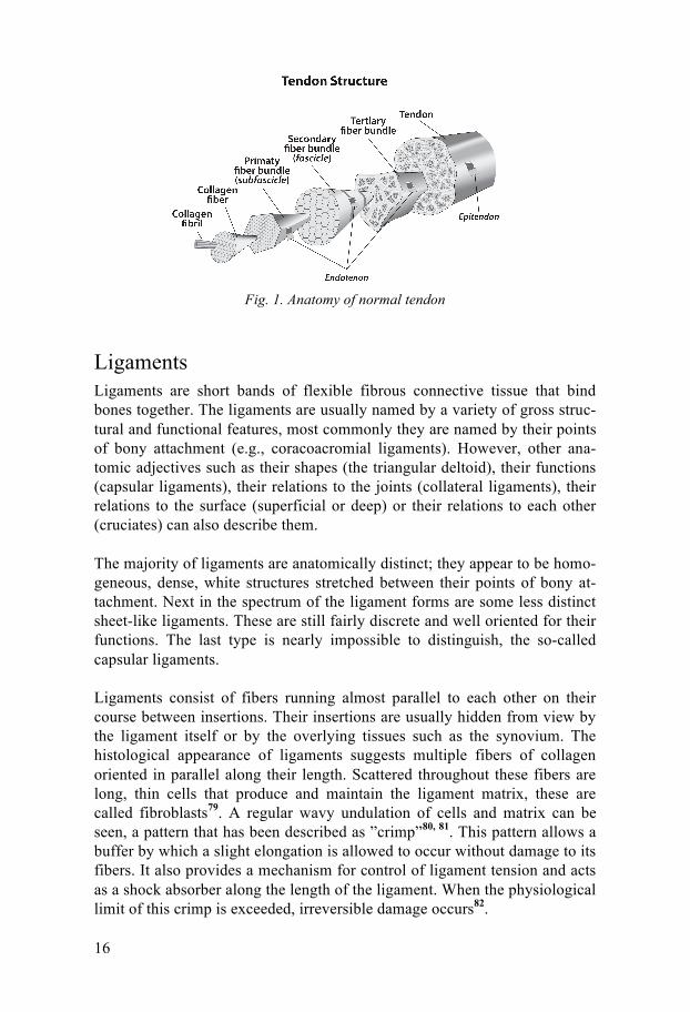
The dry mass of human tendons is approximately 30% of the total tendon mass, with water accounting for the remaining 70%. Collagen type I ac-counts for 65% to 80%, and elastin accounts for approximately 2% of the dry mass of tendons63,67-69. Collagen is arranged in hierarchical levels of increasing complexity, begin-ning with tropocollagen, a triple-helix polypeptide chain, which unites into fibrils, fibers (primary bundles), fascicles (secondary bundles), tertiary bun-dles and the tendon itself (Fig. 1)70-72. A collagen fiber is the smallest tendon unit that can be tested mechanically and is visible under light microscopy. The epitenon, a fine, loose connective-tissue sheath containing the vascular, lymphatic, and nerve supply to the tendon, covers the whole tendon and ex-tends deep within it between the tertiary bundles as the endotenon73,74. Su-perficially, the epitenon is surrounded by paratenon, a loose areolar connec-tive tissue consisting of type-I and type-III collagen fibrils, some elastic fi-brils, and an inner lining of synovial cells75. Synovial tendon sheaths are found in areas subjected to increased mechanical stress, such as tendons of the hands and feet, where efficient lubrication is required. The osteotendinous junction is composed of four zones: a dense tendon zone, fibrocartilage, mineralized fibrocartilage, and bone76. The specialized structure of the osteotendinous junction prevents collagen or fiber bending, fraying, shearing, and failure77,78.
16
Fig. 1. Anatomy of normal tendon
Ligaments
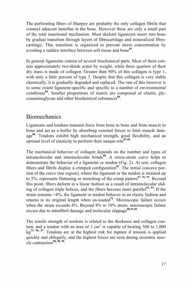
Ligaments are short bands of flexible fibrous connective tissue that bind bones together. The ligaments are usually named by a variety of gross struc-tural and functional features, most commonly they are named by their points of bony attachment (e.g., coracoacromial ligaments). However, other ana-tomic adjectives such as their shapes (the triangular deltoid), their functions (capsular ligaments), their relations to the joints (collateral ligaments), their relations to the surface (superficial or deep) or their relations to each other (cruciates) can also describe them. The majority of ligaments are anatomically distinct; they appear to be homo-geneous, dense, white structures stretched between their points of bony at-tachment. Next in the spectrum of the ligament forms are some less distinct sheet-like ligaments. These are still fairly discrete and well oriented for their functions. The last type is nearly impossible to distinguish, the so-called capsular ligaments. Ligaments consist of fibers running almost parallel to each other on their course between insertions. Their insertions are usually hidden from view by the ligament itself or by the overlying tissues such as the synovium. The histological appearance of ligaments suggests multiple fibers of collagen oriented in parallel along their length. Scattered throughout these fibers are long, thin cells that produce and maintain the ligament matrix, these are called fibroblasts79. A regular wavy undulation of cells and matrix can be seen, a pattern that has been described as ”crimp”80, 81. This pattern allows a buffer by which a slight elongation is allowed to occur without damage to its fibers. It also provides a mechanism for control of ligament tension and acts as a shock absorber along the length of the ligament. When the physiological limit of this crimp is exceeded, irreversible damage occurs82.
17
The perforating fibers of Sharpey are probably the only collagen fibrils that connect adjacent lamellae in the bone. However these are only a small part of the total insertional mechanism. Most skeletal ligaments insert into bone by gradual transition through layers of fibrocartilage and mineralized fibro-cartilage. This transition is organized to prevent stress concentration by avoiding a sudden interface between soft tissue and bone83. In general ligaments consist of several biochemical parts. Most of them con-tain approximately two-thirds water by weight, while three quarters of their dry mass is made of collagen. Greater than 90% of this collagen is type 1, with only a little percent of type 3. Despite that this collagen is very stable chemically, it is gradually degraded and replaced. The rate of this turnover is to some extent ligament-specific and specific to a number of environmental conditions84. Smaller proportions of matrix are composed of elastin, gly-cosaminoglycan and other biochemical substances85.
Biomechanics Ligaments and tendons transmit force from bone to bone and from muscle to bone and act as a buffer by absorbing external forces to limit muscle dam-age86. Tendons exhibit high mechanical strength, good flexibility, and an optimal level of elasticity to perform their unique role87-89. The mechanical behavior of collagen depends on the number and types of intramolecular and intermolecular bonds90. A stress-strain curve helps to demonstrate the behavior of a ligament or tendon (Fig. 2). At rest, collagen fibers and fibrils display a crimped configuration81. The initial concave por-tion of the curve (toe region), where the ligament or the tendon is strained up to 2%, represents flattening or stretching of the crimp pattern67, 91, 92. Beyond this point, fibers deform in a linear fashion as a result of intramolecular slid-ing of collagen triple helices, and the fibers become more parallel93, 94. If the strain remains <4%, the ligament or tendon behaves in an elastic fashion and returns to its original length when un-loaded71. Microscopic failure occurs when the strain exceeds 4%. Beyond 8% to 10% strain, macroscopic failure occurs due to intrafibril damage and molecular slippage88,91,95.
The tensile strength of tendons is related to the thickness and collagen con-tent, and a tendon with an area of 1 cm2 is capable of bearing 500 to 1,000 kg73, 96, 97. Tendons are at the highest risk for rupture if tension is applied quickly and obliquely, and the highest forces are seen during eccentric mus-cle contraction90, 98, 99.

18
Parameters obtained from the curve (Fig. 2) representing structural proper-ties of the ligament or the tendon includes stiffness, ultimate load, ultimate elongation and energy absorbed at failure.
Fig 2. Stress-strain curve demonstrating the basic physical properties of a tendon.
Ligament and tendon healing process Tendon healing can be largely divided into 3 overlapping phases: inflamma-tion, repair and remodelling phases. During the initial inflammatory phase, which lasts about 24 hours, erythrocytes, platelets and inflammatory cells (eg: neutrophils, monocytes and macrophages) migrate to the wound site and clean the site of necrotic materials by phagocytosis. In the meantime, these cells release vasoactive and chemotactic factors, which recruit fibroblasts to begin collagen synthesis and deposition. A few days after the injury, the repairing phase begins. In this phase, which lasts a few weeks, tendon fibroblasts synthesize abundant collagen and other extracellular matrix components such as proteoglycans. These are deposited at the wound site. After about 6 weeks, the remodelling phase starts. This phase is character-ized by decreased cellularity and decreased collagen and glycosaminoglycan synthesis. During this period, the repair tissue changes to fibrous tissue, and then this changes to scar-like ligament or tendon tissue after 10 weeks. Dur-ing the later remodelling phase covalent bonding between the collagen fibers increases, and this results in repaired tissue with higher stiffness and tense or strength. During this phase both the metabolism of the tenocytes and tendon vascularity decline23.

19
Rotator cuff tear prevalence
A rotator cuff tear is a disorder associated with pain and dysfunction in the shoulder. There are many reports regarding the prevalence of rotator cuff tears that were revealed in cadaver dissections. However, the frequency of tears varies from 5 to 39%100-105. This variation might be due to the differ-ences in the subject population. In addition, the background of the patients with rotator cuff tears, such as the symptoms involving their shoulder and a history of trauma, is unknown due to the limitations of cadaveric research. There are other reports revealing people with asymptomatic rotator cuff tears11,42, 106-109. Many conventional reports have focused on the patients with symptoms, which may be misleading with regard to the entire clinical pic-ture of a rotator cuff tear. Milgrom et al.42 reported an increased prevalence of partial- and full-thickness rotator cuff tears with increasing age in asymp-tomatic adults. There was a 50% rate of tears in subjects aged 70 to 79 years, and an 80% rate of tears in subjects > 80 years old. The relatively small number of subjects weakens the validity of this study. The study comprised 90 subjects between the ages of 30 and 89 years. Tempelhof et al.108 revealed an increased prevalence of rotator cuff tears with increased age without any correlation to sex, dominant arm or level of activity. Furthermore, the epi-demiology of rotator cuff tears has not been elucidated.
Ultrasound examination of the rotator cuff Rotator cuff disease is one of the most common reasons for using ultrasound, and many authors recommend it as a primary imaging technique for soft tissue injuries of the shoulder110. The main advantages are the ability to per-form dynamic examinations and conduct side-to-side comparisons. Many studies reveal excellent sensitivity and specificity in diagnosing rotator cuff tears111-116. The overall accuracy might reach up to 96%117,118. There are stud-ies showing a comparable accuracy between ultrasound and magnetic reso-nance imaging for diagnosis and measurement of rotator cuff tears119. An-other advantage of ultrasound examination is that the orthopedic surgeons can do it in the office119.

20
Standardized technique for examination of the shoulder The shoulder is a complicated joint to examine. Proper positioning of the patient is important for successful examination. The examination should be systematic with predetermined structures scanned step by step. Typically it begins with the long head of the biceps tendon, which is used as a reference landmark. It is examined both longitudinally and transversely with the pa-tient’s forearm resting in a supine position on the thigh. Medially to the biceps tendon is the subscapularis, which is best examined with the arm in external rotation. Infraspinatus and teres minor are examined by putting the patient’s arm across the chest with the hand on the opposite shoulder. Imaging the supraspinatus tendon is obstructed by the overlying acromion. The best way to expose it is to have the patient put his/her hand on the back pocket with the palm toward the gluteal muscle while keeping the elbow directed posteriorly. The tendon is examined in a perpendicular plane, keeping in mind that the axis of the tendon is approximately 45o be-tween the sagittal and coronal planes of the body (Fig. 3). Because the cuff tendons inserting onto the greater tuberosity are relatively indistinct from each other, it is difficult to distinguish them. One way to separate them is by sequential measurements. The supraspinatus is approxi-mately 1.5-2 cm from the edge of biceps tendon, and infraspinatus forms the next 1.5 cm posteriorly.

21
Fig.3. Sequence for rotator cuff ultrasound. 1) Ultrasound transducer placement for imaging the biceps tendon. 2) Ultrasound transducer placement for imaging the subscapularis. 3) Ultrasound transducer placement for imaging the supraspinatus. 4) Ultrasound transducer placement for imaging the infraspinatus and teres minor.
22
Adhesive capsulitis (frozen shoulder)
Duplay in 1872 first described adhesive capsulitis as periarthritis scapulo-humerale. In 1934 Codman120 used the term “frozen shoulder”. Neviaser121 in 1945 was the first to use the term adhesive capsulitis. Along the years several terms have been used: frozen shoulder, adhesive capsulitis, periar-thritis and pericapsulitis. Considering the pathological changes found in the joint capsule, adhesive capsulitis should preferably be used to describe this condition. This disease is characterized by a spontaneous onset of shoulder pain accompanied by progressive limitation of both active and passive glenohumeral joint movements. The natural history of idiopathic frozen shoulder syndrome is considered benign. Codman120 and Grey122 concluded that the course of frozen shoulder is benign and self-limiting with complete recovery from pain and return of range of motion within a maximum of 2 years from the onset of symptoms. Shaffer et al.123, on the other hand, reported persistence of symptoms and impaired range of movement in over 50% of their cases when followed up at 3 and 11 years. This long period of pain and disability has been the reason for many different types of intervention. Frozen shoulder may occur as an idiopathic process or as a result of an un-derlying disorder that leads to disuse. Rotator cuff tendinopathy, acute sub-acromial bursitis, fractures about the humeral head and neck, and paralytic stroke are relatively common predisposing factors for the development of frozen shoulder. Diabetes mellitus is also considered as a frequent cause of adhesive capsulitis124. The prevalence of adhesive capsulitis is 2–5% in a normal population125,126. It is most frequent in females and in patients over 40 years121. A genetic127 component is reported even if the direct mechanisms by which genes might influence soft tissue disorders are still unknown. Contra-lateral shoulder involvement occurs in up to 20–30% of the patients. Recurrence in the ipsi-lateral shoulder is rare126.

23
Pathogenesis The etiology of adhesive capsulitis is still unknown. Bulgen in 1976128 found HLA B27 more common in patients with adhesive capsulitis, but this has not been confirmed in subsequent studies129. Rodeo et al.130 in 1997 demonstrat-ed increased deposition of cytokines as transforming growth factor, platelet derived growth factor and tumor necrosis factor-alpha in the synovium and in the capsule of the adhesive capsulitis group compared to a control group. They postulated that cytokines might be involved in the fibrotic and inflam-matory process. Especially the matrix-bound transforming growth factor-beta may act as a persistent stimulus, resulting in a capsular fibrosis. Lundberg131 documented periarticular inflammatory changes and thickening of the joint capsule without intra-articular adhesions. Rizk et al.132 discov-ered thickening and constriction of the capsule. Ozaki133 found a contracted and hypertrophied coracohumeral ligament. Neviaser134 described the hy-pothesis that the underlying pathological changes are synovial inflammation with subsequent reactive capsular fibrosis.
Diagnosis The most used criteria are the following:
• Painful stiff shoulder for at least 4 weeks • Severe shoulder pain that interferes with activities of daily living • Night pain • Painful restriction of both active and passive range of motion • Normal plain x-ray • Arthrography was the investigation of choice for years. Joint volume
less than 10 ml and a marked loss of the normal axillary fold122
MRI and capsulitis The imaging findings of adhesive capsulitis have been previously limited to conventional arthrography. The arthrographic criteria of adhesive capsulitis include the following: limited injectable fluid capacity of the glenohumeral joint (7–10 ml), a small dependent axillary fold, and irregularity of the ante-rior capsular insertion at the anatomic neck of the humerus135,145. Recently, MRI features have been helpful in the diagnosis of adhesive cap-sulitis. The overall specificity has been relatively disappointing for the dis-ease46, 137-139. Most of the reports have included MRI images obtained with either indirect (intravenous) or direct (intraarticular) arthrography140-142. Em-ig et al.46 reported a thickening of the joint capsule and synovial membrane in the axillary recess in a T1 oblique coronal plane of adhesive capsulitis

24
shoulders without intravenous Gd-chelate injection. Tamai and Yamato139, using dynamic MRI, showed that the synovium in adhesive capsulitis dif-fered from that in normal shoulders. Thickening of the joint capsule and synovial membrane: – In the axillary recess: measured by the widest portion of the capsule and synovial membrane at its insertion at the humeral head perpendicular to the adjacent cortical bone, according to Emig46, on a coronal T1-weighted spin-echo and a coronal T1-weighted spin-echo Gd-chelate-enhanced sequence (Fig. 4a). Normal reference ranges, however, for asymptomatic joint capsule and synovial thickness, have been previously determined to be 2.9mm or less46. – In the rotator interval: measured by the widest portion of the capsule and synovium at the central part of the rotator interval perpendicular to the adja-cent humeral head cortex, according to Emig, Vahlensieck, Tetro and Meri-la46,143-145, on a sagittal T1-weighted spin-echo Gd-chelate-enhanced se-quence (Fig. 4b). The rotator interval is located in the concavity of the cora-coid process. Its contents include the coracohumeral and superior gleno-humeral ligaments, which are distinct structures surrounded by fatty tissue143.
Fig 4a Fig 4b Coronal oblique MR image taken poste-riorly shows thickened axillary pouch (arrows).
MR image shows enhancement of the rotator interval lesion (arrow) which sits above the subscapularis tendon

25
Treatment Several treatment modalities have been tried over the years. The use of corti-costeroids did not make any difference in long-term outcome as compared to physiotherapy even if they could provide some pain-relief146. Arslan et al.147 reported that local steroid injection therapy was as effective as physical ther-apy for the treatment of adhesive capsulitis. The improvement in range of motion at the end of the study was similar in both groups. Physiotherapy alone is an effective treatment, but it is also a complement to other therapies, especially to improve the range of motion in external rotation148-150. There are many controlled studies describing the effectiveness of physiotherapy in patients with adhesive capsulitis of the shoulder. However, methodological flaws, such as small number of subjects, high dropout rates and a short fol-low-up, limit the interpretation of the results of all of these studies148,151-153. Distension arthrography described by Andren154 in 1965, appears to be an-other good therapeutic intervention for achieving rapid symptomatic relief from adhesive capsulitis154,155. It consists of an injection of a saline solution causing the rupture or dilatation of the capsule by hydrostatic pressure. This might be combined with corticosteroid injections in some cases. Manipulation under anesthesia is normally used in patients resistant to phys-iotherapy, and it can reduce the period of pain and disability156,157. However, this might be associated with some risks such as dislocation, fracture, nerve palsy and rotator cuff tear. Reichmister and Friedman158 performed a retrospective study of 38 shoulder manipulations in 32 patients. These patients were followed for an average of 58 months. The patients were examined in a follow-up for combined shoul-der range of motion, external and internal rotation strength and status of the long head of the biceps. In this series, 97% of patients had relief of pain and recovery of near complete range of motion. Hill and Bogumill159 studied 17 patients who were followed up for a mean of 22 months, and they found that 70% had returned to work in less than 6 months and had improved motion and no complications. A recent study Farrell et al.156 showed that manipulation under anesthesia leads to sustained improvement in shoulder motion and function at a mean of 15 years after the procedure. Arthroscopic capsular release has become a reliable method for restoring range of motion in patients for which physical therapy and manipulation have failed160. Arthroscopy has been considered useful to confirm the diag-nosis, to exclude other significant pathologies, to classify the stage of the

26
condition, and to treat the stiff shoulder in combination with or without ma-nipulation161. It might also be recommended in diabetics or in patients with post-operative or post-fracture frozen shoulder162. Segmuller et al.163 treated 24 patients with 26 shoulders by arthroscopic cap-sular release. At 3.5-month follow-up 76% demonstrated return to normal or near normal function, 50% had residual loss of internal rotation and 87% achieved a good/excellent result using the Constant scoring system. Other treatments, such as the suprascapular nerve block, might also be useful in some patients in combination with steroids, especially when pain control is particularly difficult164,165. Dahan et al.166 stated that the use of bupivacaine suprascapular nerve blocks was effective in reducing the pain at 1-month follow-up.

27
Aims of the studies
Study I Since collagen type I is a major protein constituent of cruciate ligaments, joint capsules and tendons, we hypothesized that a polymorphism in COLIA1, might be associated with soft tissue injuries such as cruciate liga-ment ruptures and shoulder dislocations.
Study II To test the hypothesis that point fixation, as with suture anchors, might result in better healing and better biomechanical properties than when the soft tis-sue is compressed against the bone with a flat surface, e.g. with a screws with washers or tags.
Study III To assess the prevalence of rotator cuff tears in a population aged 50-75 years, who had not previously sought for complaints from their shoulders. To determine if there is any relationship between dysfunction measured by Constant score and the presence of cuff tears detected by ultrasound, and to identify any characteristic changes on plain radiology that might correlate to rotator cuff tears.
Study IV To study if MRI can demonstrate reactions of the shoulder joint capsule and the cause of persistent suffering of some patients following arthroscopic subacromial decompression with or without concomitant resection of the acromioclavicular joint. To correlate the MRI findings with the clinical as-sessments over time.

28
Materials
Study I All patients treated between 1999 and through 2003, at the Orthopedic De-partment of Uppsala University Hospital due to an established cruciate liga-ment rupture or confirmed shoulder dislocation, were invited to participate in our study. A total of 358 patients (age 15-60 years) accepted, whereas only three declined participation. Of the 358 cases, 233 suffered from damage of the cruciate ligament and 126 had had a shoulder dislocation, i.e., one had both diagnoses. As a control group for the present investigation we used a previously described cohort randomly selected from the population regis-ter167, 325 females, aged 19-39 years with whole blood samples.
Study II 10 skeletally mature New Zealand White rabbits (5 male) weighing 4.4 (4.0–4.8) kg were used. Special plates were designed and made by an instrument maker. The plates were made of stainless steel and shaped to conform to the proximal part of the rabbit tibia, where the medial collateral ligament inserts. The plates were identical in shape, except that half of the plates had 5 pegs of 1 mm height and diameter on the undersurface, resembling point fixation (Fig.5a). The other half had a smooth flat undersurface, resembling fixation with a compression device (Fig.5b). The plates were fixed to the tibia with two 1.5-mm AO cortical screws.
Fig. 5a. Plate with pegged undersurface Fig 5b. Plate with flat undersurface

29
Study III Between September 2007 and December 2009, subjects for this study were recruited through a questionnaire that was given to patients and the relatives who accompanied them when they sought our hospital outpatient clinics for specialities other than orthopedics. A written questionnaire was specifically directed to elicit any history of pain from their shoulders. All subjects aged 50-75 (median 66) years, were contacted and asked to participate in this research project. The participation was voluntary. Those who had previously received medical care or any kind of treatment for their shoulder pain were excluded. 106 (54 female and 52 male) subjects met the criteria to be en-rolled in the study.
Study IV Forty-eight consecutive patients aged 33-77 (median 56) years that had sub-acromial impingement with or without concomitant acromioclavicular joint arthritis, were included in this study. Patients with clinical signs of rotator cuff tear or adhesive capsulitis were excluded. Twenty-two patients had a painful acromioclavicular joint with positive compression sign. The domi-nant side was affected in 26 patients.
30
Methods
Study I Blood samples were collected from the participants to determine the geno-type at the collagen 1α1 Sp1 polymorphism. Joint laxity according to Carter-Wilkinson was determined168. Leisure physical activity (none, < 1 hour per week, 1-2 hours per week or >2 hours per week) in recent years and during the teenage was determined by a questionnaire. The gene for the col-lagen 1 type 1α is located on the autosomal chromosome 17, and not on the sex chromosomes. Therefore, the collagen gene is inherited in a similar way in both men and women. Therefore, we did not feel that it would pose a problem to compare the results of the (all female) control group with those from the (both male and female) participants. Weight and height were meas-ured for all participants.
Genotyping for the collagen Ια1 Sp1 polymorphism Genomic DNA from each individual was extracted from 3 ml of whole blood using a Wizard Genomic DNA purification kit (Promega Corporation, Madison, WI, USA). The genotype of each individual was determined using solid phase minisequencing 169-171. Polymerase chain reactions (PCR) were run on a Gene Amp PCR system-9700 robot using Ampli-Taq Gold® kits and standard reagents (Perkin Elmer Co, Norwalk CT, USA.). The pol-ymorphic nucleotide was detected in the captured DNA strand by single-base extension of the primer GTCCAGCCCTCATCCCGCCC with 3H-labeled nucleotides, the primer anneals immediately adjacent to the poly-morphic site. The genotype of the individual is defined by the ratio between incorporated 3H-labelled nucleotides.

31
Study II
Types of fixations A good healing of ligaments and tendons to bone is best achieved by close fixation to the bone. This might be done either by pressing the tissue against the bone or suturing the tissue to the bone.
Suture Anchors Suture anchors are very useful fixation devices for fixing tendons and liga-ments to bone. They are made up of:
1. The Anchor - which is inserted into the bone. This may be a screw mechanism or interference fit. They may be made of metal, plastic or biodegradable material (which dissolves in the body over time).
2. The Eyelet - is a hole or a loop in the anchor through which the su-ture passes. This links the anchor to the suture.
3. The Suture - is attached to the anchor by passing through the eyelet of the anchor. It also may be a non-absorbable material or absorbable material (Fig.6).
Suture tags Suture tags achieve fixation by pressing the soft tissue to the bone. They can be made of either nonabsorbable or biodegradable materials (Fig.7).
Fig 6. Suture anchors Fig 7. Suture tags

32
Surgical procedure and treatment The rabbits were sedated with fentanyl fluanisone (Hypnorm Janssen Phar-maceutica, Beerse, Belgium; 0.1–0.2 mg/kg body weight) subcutaneously. Under general anesthesia using isoflurane gas (Forene; Abbot Scandinavia, Solna, Sweden) and oxygen, the rabbits were operated on both the right and left knee. Surgery was performed under standard aseptic conditions. The skin was shaved, and prophylactic antibiotics (dicloxacillin) and analgesics in the form of 0.015 mg buprenorphine (Temgesic; Schering-Plough, Brus-sels, Belgium) were given preoperatively by subcutaneous injection. A 3-cm skin incision was made midway between the medial collateral ligament (MCL) and the patellar tendon insertion. The MCL was exposed, the synovi-al bursa under the ligament removed and the cortical bone roughened with a rasp proximal to the distal insertion of the ligament. A 3-0 nonabsorbable suture was passed under the ligament and pulled distally as far as possible to mark the insertion on the tibia without compromising it. The plate then fixed the ligament portion proximal to the suture and beneath the joint line. The plate was fixed to the bone with two 1.5-mm AO screws, one on each side of the ligament. The plate with pegs was used on the right side, and on the left side the plate with a flat undersurface was used. The skin was closed with a 4-0 etylon suture intracutanously. Minimal bleeding was observed during the operations. After the operation, the rabbits were housed 1 per cage (0.5 m2) and activity was allowed only in the cages. They received analgesia with Temgesic, 0.03 mg/kg twice daily for 3 days postoperatively. The condition of each rabbit was documented in an individual rabbit journal on a daily basis.
Evaluation The rabbits were sacrificed 28 days after the operation using an overdose of pentabarbitural injected intravenously. Each knee joint was harvested by transecting the femur just below the trochanteric region and the tibia above the ankle. The MCL attached to the femur and the tibia was isolated, while the remaining soft tissue was removed. Before removing the plate, a trans-verse cut was made through the MCL adjacent to the distal border of the plate, proximal to the original insertion of the MCL (marked with the non-absorbable suture) (Fig. 8a). This enabled us to test the mechanical proper-ties of the ligament fixation in the area under the plate. The knee was fixed to a material testing machine (100R, DDL Inc., Eden Praire, Mn, USA) us-ing 1.6 mm Kirschner wires inserted into drill-holes made in the femur and tibia (Fig. 8b). Testing was performed within a few hours after the rabbit had been killed. In that time interval it was kept cool and wet. A constant disten-sion in the direction of the MCL with a speed of 1 mm/sec was applied until failure. We recorded the peak force at failure, stiffness and energy uptake until the force dropped to 90% of maximum (Fig. 9).

33
Fig 8a. MCL after preparation, before Fig 8b. Mechanical testing mechanical testing
Fig 9. Load-displacement curve for a specimen with pegged plate

34
Study III Clinical examination was performed and consisted of measurement of Con-stant score on each shoulder. Bilateral ultrasound examination was per-formed according to a standard protocol using a Philips HDI 5000 (Bothell, WA, USA), with a 12 MHz linear-array transducer. All tendons were exam-ined, both in longitudinal and transverse plane. Then a conventional x-ray examination of each shoulder was done. It included a standard anterioposte-rior view with the glenoid in its absolute profile and the head of the humerus in neutral position. We listed different areas to be examined, including the presence of osteoarthritis of the glenohumeral and acromioclavicular joints, the acromion index (AI) according to Nyffler et al.172 (Fig. 10) and the sub-acromial index (SAI) (Fig. 11).
Fig. 10 Fig. 11 Measurement of acromion index Measurement of subacromial index AI=J/K SAI=L/E
Study IV An MRI of the affected shoulder was done preoperatively and at 3 months after surgery. Preoperatively, patients with cuff tears, osteoarthritis of the glenohumeral joint, labrum injuries due to instability, diabetes mellitus or rheumatoid arthritis were excluded from the study. A scoring system for the MRI was obtained, giving two points for edema of the axillary capsule, two points for thickening of the axillary capsule, two points for the pericapsular edema and one point for the rotator interval ede-ma. A total score of seven points indicated a maximum value for adhesive capsulitis.

35
Surgical technique The operative procedure was a modification of the technique described by Ellman173. All procedures were performed in the beach chair position, under general anaesthesia or long acting scalene block, with the arm in forward traction. The passive range of motion was first assessed without any attempt for manipulation. The arthroscope was introduced through the posterior por-tal and the subacromial space inspected. An anterior acromioplasty was per-formed with a motorized resector. The adequacy of the decompression was judged by introducing a straight blunt probe through the posterior portal. This determined whether the undersurface of the acromion was flat and the anterior hook of the acromion had been eliminated. For those patients who had symptomatic arthritic changes in the acromioclavicular joint, an arthro-scopic resection of the lateral end of the clavicle was done through an anteri-or portal.
Follow-up assessments The clinical assessments by measuring the Constant score and Subjective Shoulder Value were done before surgery, at 3 months and at 6 months after surgery. A follow-up interview of the Subjective Shoulder Value was done by an independent secretary two years after surgery. Two musculoskeletal radiologists independently evaluated the MR images. The images were evaluated in the same manor regardless if they were pre- or postoperative.

36
Statistics
Study I The injury risk, associated with the three genotypes of the collagen 1α1 gene, was analyzed with the SS genotype as the reference. For these associa-tions, we used age-adjusted unconditional logistic regression to estimate odds ratios (OR) and 95% confidence intervals (CIs). We also considered including body weight and height, or body mass index, in the age-adjusted model, but that gave only marginal influences on our estimates. Consequent-ly, we only present results from the age-adjusted model, with age in continu-ous form. Statistical analyses were carried out using SAS software (version 9.1, SAS institute Inc., Cary, NC, USA).
Study II We used Wilcoxon’s signed-rank sum test to compare the flat and pegged sides. Confidence intervals for the median difference between the sides are based on the observations with rank 2 and 9, which yields approximately 90% confidence174.
Study III Each subject contributed with both shoulders in the study. A mixed model approach was used to address the issue of the correlated shoulders and adjust for the variance of the bilateral observations. Mixed effects logistic regres-sion was implemented to model the relationship between the variables. Many of the analysis dealt with the Constant score as the dependent variable with one other factor as an independent variable. Unpaired t-test was used for the correlation between the presence of full-thickness cuff tear and osteoarthri-tis. The significant level was set to p<0.05.

37
Study IV The data were set up as longitudinal data with each visit corresponding to a visit on a timeline. A biased corrected weighted kappa score using boot-strapping was used to assess the correlation between the MRI scores of the two raters pre- and post surgery. Spearman’s test was used for the correlation between the MRI scores and Constant score at baseline and three months after surgery. Standard ANOVA was used to evaluate a possible difference in the mean values for the three Constant scores. The same analysis was done for the mean values for the Subjective Shoulder Values at each time point. For the relationship between the change in external rotation and the change in MRI score from the baseline to three months, simple linear regres-sion was used.

38
Results
Study I The control group consisted only of females whereas the case group is pre-sented according to sex. The genotype distribution among the controls was 71% SS, 25% heterozygotes Ss and only 4% ss. The polymorphism genotype in the study cohorts was in Hardy-Weinberg equilibrium. This equilibrium is a common test in genetic epidemiology. It measures the chance of a skewed genotype distribution. If the genotype distribution is not in Hardy-Weinberg equilibrium, there is a risk of selection bias in the study. Cases and controls had a similar age distribution. Female and male cases had a similar genotype distribution, and only one female and one male case had the ss genotype. There was an 85% reduced odds ratio (95% CI 34-97%) of an injury for those with the rare ss genotype as compared to those with the genotype SS (Table 1). We observed a similar reduction in risk for cruciate ligament rup-tures and shoulder dislocations. No significant difference in injury risk was observed among those with the Ss genotype as compared with the homozy-gotes with the SS genotype.
Table 1. Age-adjusted odds ratios with 95% confidence intervals (95% CI) of having a cruciate ligament injury or a shoulder dislocation according to procollagen Ia1 Sp1 genotype. Injury Genotype Cases Controls Odd ratio (95% CI) All SS 257 230 1.0 Ss 99 83 1.06 (0.76-1.49)
ss 2 12 0.15 (0.03-0.68) Cruciate ligament SS 162 230 1.0 rupture Ss 70 83 1.19 (0.82-1.75) ss 1 12 0.12 (0.02-0.92) Shoulder dislocation SS 95 230 1.0 Ss 30 83 0.88 (0.54-1.41) ss 1 12 0.20 (0.03-1.56)

39
Study II No animals were excluded. All the wounds healed uneventfully, and the rabbits loaded their hind limbs immediately postoperatively. Gross inspec-tion of the knee joints at the harvest showed that the plates were covered by fibrous material similar to tendon callus (Table 2). Rabbit no. 7 was an out-lier, probably due to malplacement of one screw through the ligament on the pegged plate side. In the other 9 animals, the force at failure, stiffness and energy uptake were always higher on the side with the pegged plate. Analyz-ing all 10 animals, the force was 133% higher on the pegged side (range –24 to 342; 90% CI for median 51–322%). Stiffness and energy were increased by 75% and 210%, respectively. (Stiffness: range –22% to 426%; 90% CI for median 8–260%. Energy uptake: range 79% to 659%; 90% CI for median 90–515%). Table 2. Mechanical results of flat or pegged plates in all rabbits
Animal Force at failure (N)
Energy uptake (Nmm)
Stiffness (N/mm)
Attachment plate
flat pegged diff flat pegged diff flat pegged diff
1 16 39 23 9 57 48 16 17 1 2 9 39 30 7 29 22 8 29 21 3 19 29 10 21 19 -2 15 26 11 4 7 24 17 12 21 9 3 18 15 5 18 38 20 16 28 12 16 28 12 6 19 37 18 21 29 8 21 29 8 7 69 51 -18 79 62 -17 35 27 -8 8 17 77 60 18 91 73 15 38 23 9 11 33 22 14 34 20 10 23 13 10 36 69 33 30 93 63 26 31 5 Min 7 24 -18 7 21 -17 3 17 -8 Med 18 39 21 16 32 29 15 28 16 Max 69 77 60 79 93 73 35 38 23

40
Study III Among 106 subjects, 64 (60%) had complaints from their shoulders. 19 had bilateral symptoms, 32 on the right and 13 on the left side. There were 25 men and 39 women. The prevalence of a full-thickness cuff tear was 30% (32/106) and a partial-thickness tear, 20% (22/106). All shoulders with full-thickness tears had tears on the supraspinatus tendon. When the prevalence was calculated on the bases of each shoulder, we found a prevalence of 21% (44/212) for full-thickness tears and 14% (29/212) for partial-thickness tears. A significant result was found between complaints and full-thickness tears. Those who had full-thickness tears had 3.7 times higher odds of having complaints than those without cuff tears (CI 1.4-10.0). No relationship was found between shoulder complaints and partial tears (Table 3). Constant score was higher in shoulders without complaints than in shoulders with complaints. Those who had complaints had a Constant score that was 7.3 (CI 4.0-10.6) points lower than those without complaints. The difference was significant between the full-thickness tear group and the other two groups (Table 4). The average Constant score for subjects with full-thickness cuff tears was 7.9 (CI 3.7-12.1) points lower. No correlation was found for the acromion index among the subjects with full-thickness supraspinatus tears, subjects with partial-thickness tears, and those without tears. However, the subacromial index was correlated with full thickness supraspinatus tears (Table 5). The ratio was 0.025 (CI 0.002-0.048) lower for full-thickness tears. The acromion index was lower for the group with primary osteoarthritis than the group with full-thickness tears (p<0.01), but the subacromial index did not differ significantly (p=0.6) (Ta-ble 5).
Table 3. Numbers of subjects with/without symptoms in relation to cuff tears and arthritis. Symptomatic (n.) Asymptomatic (n.) Total (n.) Normal cuff 47 92 139 Partial tears 9 20 29 Full-thickness tears 27 17 44 GH osteroarthritis 9 + 2 tear arthropathy 8 19 AC joint arthritis 28 40 68 GH = glenohumeral. n = number of patients.

41
Table 4. Constant score in relation to symptom complaints and rotator cuff tears. Number of shoulders (n.) Constanta score No complaints 129 82 (CI 80-84) Complaints 83 68 (CI 64-71) No cuff tears 139 78 (CI 76-81) Partial-thickness tears 29 80 (CI 80-84) Full-thickness tears 44 69 (CI 63-74) CI = Confidence Interval. Table 5. Acromion and subacromial indices in relation to rotator cuff tears and glenohumeral arthritis. Acromion Index (AI) Subacromial index (SAI)
No tears or partial tears
0.65 (CI 0.63-0.66) 0.28 (CI 0.27-0.29)
Full-thickness tears 0.66 (CI 0.64-0.68) 0.26 (CI 0.24-0.28)
Glenohumeral arthritis 0.60 (CI 0.56-0.64) 0.27 (CI 0.24-0.30)
CI = Confidence Interval.
Study IV Although there were significant differences between the MRI scores pre and post surgery for both raters, this was not clinically important since the Kappa scores for the two MRI raters were 0.498 (CI 0.305-0.690) before surgery and 0.295 (CI -0.006-0.490), which shows a poor reliability between the raters. The relationship for the Constant score and MRI scores after surgery was not significant. The differences between the average Constant scores preoperatively, at 3 months, and at 6 months after surgery were significant, 46 (CI 41-51), 70 (CI 66-74) and 77 (CI 73-81), respectively. In addition, the differences in average Subjective Shoulder Values were significant for all visits and even for the two-year follow-up, 50 (CI 45-55), 69 (CI 63-75), 76 (CI 70-82) and 97 (CI 96-99), respectively. No relationship was found be-tween the changes in external rotation and MRI scores over the time period studied (Fig. 12, 13).

42
Fig12. Correlation between the change in external rotation and MRI score pre- and at three months postoperatively for rater one
Figure 13. Correlation between the change in external rotation and MRI score pre- and at three months postoperatively for rater two.
-10
12
34
Cha
nge
in M
RI s
core
-40 -20 0 20 40Change in External rotation
Fitted values delta_MRIh
MRI rater 1
-20
24
6
Cha
nge
in M
RI s
core
-40 -20 0 20 40Change in External rotation
Fitted values delta_MRIl
MRI rater 2
43
General discussion
The shoulder is one of the largest and most complex joints in the body. It is a multi-axial ball-and-socket type of synovial joint with a large range of movement, and this is due to the limited interface between the head of hu-merus and the shallow glenoidal fossa. The joint is protected against dis-placement by numbers of tendons and ligaments, which surround it. The behavior of the soft tissues plays an important role in the overall function of the shoulder joint. The available information on the soft tissue structural and biomechanical properties is mainly based on animal models, which may be of limited value. However, with newly developed techniques it is possible to study the genetics of soft tissue injuries and the characteristics of the healing properties of both tendons and ligaments. This thesis presents new information regarding the structural and mechanical properties of tendinous injuries, based on understanding the genetic proper-ties of the collagen, which is the main protein constituent of the tendon and ligament structures. Besides this, this thesis improves understanding of the mechanical properties of different types of repair methods and the post-repair reactions of these structures. Understanding the biology of tendon-to-bone healing may offer novel thera-peutic options to improve the rate of long-term structural healing. Rotator cuff healing occurs by reactive scar formation rather than by regeneration of a histologically normal enthesis. The poor healing response is multifactorial but could be related to insufficient and disorganized expression of cytokines to direct formation of a complex healing structure. Other factors may include the presence of inflammatory cells at the tendon-bone interface that precipi-tates scar formation. The inflammatory response that occurs in adult healing leads to a gene expression program that results in scar-based healing rather than the formation of the native insertion site, unlike the complex signaling that occurs during embryologic development175. During the healing process, the activation of fibroblasts results in the expression of many cytokines, which enhances healing in different biological processes. These cytokines have the potential to improve tendon-to-bone healing through their role in cell proliferation, matrix synthesis and cell differentiation176.

44
Remarkable advances have been made in understanding the human gene contribution to health and disease. Large scale genetic studies have made it possible to measure associations between single-nucleotide polymorphisms and the occurrence of genetically complex diseases. Type 1 collagen is the most abundant protein in the body, and it is synthesized by fibroblasts, oste-oblasts and odontoblasts177. Although bones, ligaments and joint cartilage contain this common collagen type, various reports have described different locations for the regulation of the promoter region that control transcription of COLIA1177. One such location is the Sp1 transcription factor-binding site of the COLIA1 gene. One of the most widely studied polymorphisms in relation to bone quality is the Sp1 polymorphism in the COLIA1 gene178, which has been associated with low bone mass179-181, osteoporotic fracture risk in postmenopausal women182 and female children183 and osteoarthritis19. Functional analysis has shown that the osteoarthritis- and osteoporosis-related s allele of the Sp1 polymorphism is associated with increased DNA–protein binding, increased transcription from the s allele, and increased pro-duction of the collagen type I α1 mRNA and protein18. Even though the s allele of the Sp1 polymorphism is associated with an in-creased production of collagen type 1 α1 chains, it is still unclear how the ss genotype would reduce the odds of having a soft tissue injury. Nevertheless, the results from our study on Sp1 polymorphism in the COLIA1 gene are in accordance with studies done in South Africa on anterior cruciate ligament and achilles tendon ruptures16,183. This type of polymorphism is not only associated with musculo-skeletal diseases. Over one fourth of women with cervical insufficiency have a fami-ly history of cervical insufficiency, and the COLIA1 Sp1 binding site poly-morphism has been associated with the condition184. Furthermore, otosclero-sis, the single most common cause of hearing impairment in white adults, has also been associated with polymorphisms in the COLIA1 gene185. Soft tissue injuries might be polygenic with different types of polymor-phisms in nature. Nowadays, it is possible to perform whole-genome association studies and not just the candidate SNP approach used in our preliminary report. There-fore, attempts to identify several possible genes and genetic pathways by whole genome sequencing associated with these soft tissue injuries are fea-sible in order to define genetic mechanisms behind the predisposition to soft-tissue injuries. Secondarily, there might also be an interest to find out the genetic susceptibility of healing impairment of these injuries. These types of investigations will increase the understanding of possible intrinsic risk fac-tors for these injuries and may influence prevention, treatment and rehabili-tation programs for these injuries.

45
The rotator cuff is a composite of tendons around the shoulder joint. Rotator cuff insufficiency has been defined as a condition in which interference with the cuff’s function prevents it from fulfilling its physiological role. There are many reports regarding the prevalence of cuff tears that were revealed by cadaveric dissections, but most of these were deficient from data about the history and level of activity before death. Recently MRI or ultrasound imag-ing has enabled in-vivo assessment of the rotator cuff tear prevalence.
Tempelhof108 conducted an ultrasound prevalence study of asymptomatic shoulders, and he found prevalences from 23 to 51%, increasing over the age from 50-80 years of age. Milgorm42 demonstrated that the prevalence of partial- or full-thickness tears increases significantly after the age of 50 years. The prevalence in our study was based on a population who had not sought any medical advice for their shoulders. The prevalence was compara-ble with previous reports42,108. We believe that this prevalence would be even higher if we include a real true population regardless if they hade sought before or not for shoulder complaints. This means that the adage “grey hair equals cuff tear” could be true, and that the cuff tears are to be regarded as a normal part of the aging process. We also found that shoulders with full-thickness cuff tears had lower func-tional outcome measured by Constant score. This can be supported by the facts reported in the literature107,108. They noted that the Constant score was lower for patients who had tears in rotator cuff despite that they were asymp-tomatic in their shoulders. These findings may raise the question of whether the symptoms may develop over time in patients who previously did not have symptoms. Yamaguchi186 studied this fact and found that 51% of these patients developed symptoms and had a significant decrease in activity of daily living over a mean of 2.8 years. Regarding partial-thickness cuff tears, however, there were no differences in Constant score when compared with shoulders without any cuff tears. We often notice in our practice these types of tears on MRI, but these tears have no relevance for the shoulder dysfunction, and they do not need to be re-paired. Partial-thickness cuff tears could be compared to degenerative me-niscal tears in the knee, which are considered part of the degenerative osteo-arthritic changes associated with aging187. Plain x-ray is a simple investigation, and it is often the first modality em-ployed to investigate patients seeking for shoulder complaints. A lot of in-formation could be gained from this investigation such as changes associated with rotator cuff tears. Neer103 established a correlation between subacromial impingement and changes on x-ray. Morrison and Bigliani188 reported in-creased hooked acromions in cadavers with cuff tears. They stated that the

46
acromion types were innate anatomical variations and remained unchanged during life. These morphological classifications are often criticized as being subjective and showing poor interobsever reliability. Zuckerman189 reported only a fair level of agreement between three orthopaedic surgeons when they assessed the acromion types. Bright190 found only 18% agreement between six observers. Recently some authors have concentrated on both the morphology of the acromion in relation to the humeral head and the biomechanical effect, which is expected from this morphology. Nyffler et al172 described the ratio between lateral extension of acromion and humeral head width as a predictor of rotator cuff tears. They predicted that it is not the frictional effect that is considered to be the primary cause of the changes in the rotator cuff but ra-ther a biomechanical one. The lateral extension of the acromion influences the orientation of the deltoid muscle force vector. The larger the lateral ex-tension the higher the ascending force component of the force vector. Small lateral extension leads to larger compressive force, which could be a predis-posing factor for the development of glenohumeral osteoarthritis. The as-cending force can cause upward migration of the humeral head. Depending on this theory and the fact that one of the outcomes after rotator cuff tear is that the humeral head migrates upward, which leads to a narrowing of the subacromial space, we tried to find a predictive factor on x-ray depending on the measurement of the space between the under surface of the acromion and the top of humeral head. To avoid the bias, which can occur by different magnifications of x-rays, we calculated a ratio between the subacromial space and the length of the glenoid (subacromial index). We found a lower index for full-thickness cuff tears than for partial-thickness tears or shoulders without tears. We took into consideration that partial tears will not affect the wideness of the subacromial space. We also found a significant difference between shoulders with full-thickness tears and those, which had osteoar-thritic changes. More research is needed to evaluate these two indices, espe-cially to find a breakpoint along the range of indices that can predict the presence and absence of rotator cuff tears. The healing property of tendons and ligaments to bone is an important issue for the orthopaedic surgeons in order to obtain an optimal result. A contro-versy exists regarding the most favorable fixation technique. Regarding the rotator cuff, some studies have reported that suture anchors are weaker than interosseous tunnels191, while others have reported higher fixation strength with suture anchors192. Previously the repair of the rotator cuff was achieved by osteosutures. After the development of arthroscopic technique many kinds of fixation materials have been developed, such as suture anchors. The arthroscopic technique is more difficult than open repairs because all surgi-cal steps have to be performed through cannulae and holes through the mus-

47
cle layers. Therefore, the repaired tissue will usually be attached to the bone surface rather than to a trough in the bone. The healing might be expected to be more difficult to achieve, because the bone surface appears less vascular-ized, and, if the periosteum is scraped off, there are probably fewer progeni-tor cells. However, in a goat model, no difference was found in the healing process or in the mechanical properties between tendon healing to cortical and cancellous bone193. There are several key biomechanical factors that may influence the results of soft tissue repair. The repair should be strong enough to resist the early structural failure and high footprint contact pressure to assist biological heal-ing. This will be achieved by creating multiple contact points between the soft tissue and the bone surface. A literature review found significantly low-er re-tear rates for double-row repairs of the rotator cuff tears as compared with single-row for all tears greater than 1 cm194. As mentioned before, there seems to be a genetic predisposition for the sus-ceptibility of soft tissue injuries. We know that the mechanical properties after repair will remain inferior to the original, despite intensive remodelling. Complete regeneration of tendons and ligaments is never achieved. The character of the collagen fibrils will alter. There will be reduction in the pro-portion of type 1 collagen and increase in the amount of type 3 collagen. Type 3 collagen is responsible for the reduced tensile strength of the tissue due to reduced number of cross links compared with type 1 collagen195. Additional studies are necessary to confirm SNPs, to identify genetic path-ways that predispose to soft tissue injuries, and to identify genetic and life-style components that affect the healing process and the mechanical proper-ties of soft tissue repair. The goal is to gain a personalized medicine ap-proach to the healing of soft tissue injuries. The inflammatory processes that occur in and on the soft tissues after surgi-cal procedures cause excessive suffering for patients. In the shoulder joint this presents like a type of capsulitis, causing pain and stiffness. It is known that capsulitis has a genetic predisposition. Bulgen in 1976128 found HLA B27 more common in patients with adhesive capsulitis. There is also a rela-tion between capsulitis and both diabetes and Dupuytren disease. In paper IV we tried to find a diagnostic modality through MRI in order to visualize the changes that occur in the shoulder capsule after a common surgical proce-dure, subacromial decompression with or without concomitant acromiocla-vicular joint resection. We investigated different parts of the joint capsule with MRI, and we found significant differences in MRI scores before and after surgery. However, the reliability between results of the radiologists was poor. This indicates that it is difficult to achieve an exact measurement of the

48
edema and thickness of the joint capsule. There were relations between the MRI scores and the functional outcome of the patients measured by Constant score and Subjective Shoulder Value. Previous studies have discussed the role of MRI in the diagnosis of adhesive capsulits, but there is still discrep-ancy about the areas that should be included in the diagnosis of cap-sulitis46,53,142.

49
Conclusions
Study I In conclusion, we found that the COLIA1 Sp1 ss genotype was associated with a substantially reduced odds ratio of cruciate ligament ruptures and shoulder dislocations. Since our influential genotype is rare, the absolute risk of cruciate ligament injuries and shoulder dislocations seems to be only mar-ginally affected by COLIA1 Sp1 polymorphism. Accordingly, this particular SNP polymorphism has limited clinical relevance as a predictor for these injuries.
Study II Using the pegged plate roughly doubled the mechanical variables. One pos-sible explanation might be that the flat plate compromises the blood supply, leading to tissue necrosis and thereby impairing the healing process. There-fore our recommendation is to use suture anchors or devices with a pegged undersurface for fixation of soft tissue to a flat bone surface.
Study III The prevalence of rotator cuff tears was high in our study population who had never sought for their shoulder problems. The presence of a full-thickness cuff tear affects shoulder function. Both degenerative changes in the acromioclavicular joint and the existence of partial-thickness cuff tears are considered to be part of the natural aging process. The subacromial index can be used as a predictor for full-thickness cuff tears.
Study IV MRI had a low reliability for assessment of capsular reactions after surgery. Subacromial decompression with or without acromioclavicular joint resec-tion is a good surgical procedure with high patient satisfaction.

50
Acknowledgements
I wish to express my sincere gratitude to all who have taken part in the dif-ferent aspects of this work, and special thanks to: Hans Rahme, my very best friend and tutor, for introducing me to science, for professional guidance and encouragement throughout this work. Karl Michaelsson, my co-supervisor, for encouragement and for always providing me with well considered comments and suggestions. Stefan Nilsson, head of Elisabeth Hospital, for all support, for providing a good working condition during the years, and taking my scientific ambitions into consideration. My colleagues at hospital for their support and encouragement and for their help with collecting study subjects and for being tremendous co-workers. My co-authors, Hojat Elhami, Hans Larsson, Bo Sahlstedt, Rebecca Rylance. Jugoslav Milosvljevic for their invaluable contribution to this work. Håkan Melhus, for invaluable lessons in Genetics. Per Aspenberg and Anna Fahlgren, for their contribution and help with my second paper. Berith Svensson and Ann Cavallin, physiotherapists at Elisabeth Hospital for their help with scorings and the assessments related with study subjects. Eva Ericsson and Monika Jansson, secretaries at Elisabeth Hospital for all help with the documents and solving the entanglements. Shirin Manee and Lena Nordström, staff of radiology section at Elisabeth Hospital, for helping me with examination of the study subjects. Ann-Charlott Lundin and Ann-Sofie Silfverswärd for their help with col-lecting study subjects.

51
The staff of Elisabeth Hospital for being always co-operative and encourag-ing. My friends, for their encouragement and high evaluation of my work. My parents, Rasheed and Fatima, for their never ending support and en-couragement. My dear wife, Jian, for her enthusiasm and endless support and for her un-derstanding of the time I spend with my work. My wonderful sons, Lass and Renass, for their encouragement, their for-giveness and patience with my business with work, for giving me so much joy and happiness. You are the best.

52
References
1. Aglietti P, Buzzi R, Giron F, Simeone AJ, Zaccherotti G: Arthroscopic-assisted anterior cruciate ligament reconstruction with the central third patel-lar tendon. A 5–8-year follow-up. Knee Surg Sports Traumatol Arthrosc 1997;5:138-144.
2. Bach BR Jr, Tradonsky S, Bojchuk J, Levy ME, Bush-Joseph CA, Khan NH: Arthroscopically assisted anterior cruciate ligament reconstruction using pa-tellar tendon autograft. Five- to nine year follow-up evaluation. Am J Sports Med 1998;26:20-29.
3. Jomha NM, Borton DC, Clingeleffer AJ, Pinczewski LA: Long-term osteoar-thritic changes in anterior cruciate ligament reconstructed knees. Clin Orthop Relat Res 1999;358:188-193.
4. Jomha NM, Pinczewski LA, Clingeleffer A, Otto DD: Arthroscopic recon-struction of the anterior cruciate ligament with patellar- tendon autograft and interference screw fixation. The results at seven years. J Bone Joint Surg Br 1999;81:775-779.
5. Woo SL-Y, Renstrom P, Arnoczky SP, Eds: Tendinopathy in Atletes. Black-well Publishing; 2007.
6. Kannus P: Tendons–a source of major concern in competitive and recreation-al athletes. Scand J Med Sci Sports 1997:7:53-54.
7. Renstrom P: Sports traumatology today. A review of common current sports injury problems. Ann Chir Gynaecol 1991;80:81-93.
8. Miyasaka KC, Daniel DM, Stone ML, et al.: The incidence of knee ligament injuries in the general population. Am J Knee Surg 1991; 4:3-8.
9. Beaty J: Knee and leg: soft tissue trauma. In OKU Orthopaedic Knowledge Update 1st edition. Edited by: Arendt EA. Rosemont, IL: American Academy of Orthopaedic Surgeons 1999;xix, 442.
10. Simonet WT, Melton LJ 3rd, Cofield RH, Ilstrup DM: Incidence of anterior shoulder dislocation in Olmsted County, Minnesota. Clin Orthop Relat Res. 1984;186:186-191.
11. Sher JS, Uribe JW, Posada A, Murphy BJ, Zlatkin MB: Abnormal findings on magnetic resonance images of asymptomatic shoulders. J Bone Joint Surg Am 1995;77:10-15.
12. Lehman C, Cuomo F, Kummer FJ, Zuckerman JD: The incidence of full thickness rotator cuff tears in a large cadaveric population. Bull Hosp Jt Dis 1995;54:30-31.
13. Hoffmann A, Gross G: Tendon and ligament engineering in the adult organ-ism: mesenchymal stem cells and gene-therapeutic approaches. Int Orthop 2007;3:791-797.
14. Frank CB: Ligament structure, physiology and function. J Musculoskelet Neuronal Interact 2004;4:199-201.

53
15. Myllyharju J, Kivirikko KI: Collagens and collagen-related diseases. Ann Med 2001;33:7–21.
16. Posthumus M, September AV, Keegan M, O'Cuinneagain D, Van der Merwe W, Schwellnus MP, Collins M: Genetic risk factors for anterior cruciate liga-ment ruptures: COL1A1 gene variant.Br J Sports Med 2009 May;43:352-356.
17. Mann V, Ralston SH: Meta-analysis of COL1A1 Sp1 polymorphism in relation to bone mineral density and osteoporotic fracture. Bone 2003;32:711-717.
18. Mann V, Hobson EE, Li B, et al.: A COL1A1 Sp1 binding site polymor-phism predisposes to osteoporotic fracture by affecting bone density and quality. J Clin Invest 2001;107:899-907.
19. Lian K, Zmuda JM, Nevitt MC, et al.: Type I collagen alpha1 Sp1 transcrip-tion factor binding site polymorphism is associated with reduced risk of hip osteoarthritis defined by severe joint space narrowing in elderly women. Ar-thritis Rheum 2005;52:1431-1436.
20. Speer G, Szenthe P, Kosa JP, et al.: Myocardial infarction is associated with Spl binding site polymorphism of collagen type 1A1 gene. Acta Cardiol 2006;61:321-325.
21. Tilkeridis C, Bei T, Garantziotis S, et al.: Association of a COL1A1 poly-morphism with lumbar disc disease in young military recruits. J Med Genet 2005;42:e44.
22. Skorupski P, Krol J, Starega J, et al.: An alpha-1 chain of type I collagen Sp1-binding site polymorphism in women suffering from stress urinary in-continence. Am J Obstet Gynecol 2006;194:346-350.
23. Frank C, Woo SL-Y, Amiel D, Harwood F, Gomez M, Akeson W: Medial collateral ligament healing. A multidisciplinary assessment in rabbits. Am J Sports Med 1983;11:379-389.
24. Indelicato PA: Non-operative treatment of complete tears of the medial col-lateral ligament of the knee. J Bone Joint Surg Am 1983; 65:323-329.
25. Jokl P, Kaplan N, Stovell P, Keggi K: Non-operative treatment of severe in-juries to the medial and anterior cruciate ligaments of the knee. J Bone Joint Surg Am 1984;66:741-744.
26. Kannus P: Long-term results of conservatively treated medial collateral liga-ment injuries of the knee joint. Clin Orthop Relat Res 1988;226;103-112
27. Weiss JA, Woo SL-Y, Ohland KJ, Horibe S, Newton PO: Evaluation of a new injury model to study medial collateral ligament healing: primary repair versus nonoperative treatment. J Orthop Res 1991;9:516-528.
28. Woo SL-Y, Gomez MA, Inoue M, Akeson WH: New experimental proce-dures to evaluate the biomechanical properties of healing canine medial col-lateral ligaments. J Orthop Res 1987;5:425-432.
29. Hart RA, Woo SL-Y, Newton PO: Ultrastructural morphometry of anterior cruciate and medial collateral ligaments: an experimental study in rabbits. J Orthop Res 1992;10:96-103.
30. Niyibizi C, Kavalkovich K, Yamaji T, Woo SL-Y: Type V collagen is in-creased during rabbit medial collateral ligament healing. Knee Surg Sports Traumatol Arthrosc 2000;8:281-285.
31. Abramowitch SD, Papageorgiou CD, Debski RE, Clineff TD, Woo SLY: A biomechanical and histological evaluation of the structure and function of the healing medial collateral ligament in a goat model. Knee Surg Sports Trau-matol Arthrosc 2003;11:155-162.
54
32. Woo SL-Y, Niyibizi C, Matyas J, Kavalkovich K, Weaver-Green C, Fox RJ: Medial collateral knee ligament healing. Combined medial collateral and an-terior cruciate ligament injuries studied in rabbits. Acta Orthopaedica Scan-dinavica 1997;68:142-148.
33. Buss DD, Min R, Skyhar M, Galinat B, Warren RF, Wickiewicz TL: Nonop-erative treatment of acute anterior cruciate ligament injuries in a selected group of patients. Am J Sports Med 1995;23:160-165.
34. Ciccotti MG, Lombardo SJ, Nonweiler B, Pink M: Non-operative treatment of ruptures of the anterior cruciate ligament in middle-aged patients. Results after long-term follow-up. J Bone Joint Surg Am 1994;76:1315-1321.
35. Fetto JF, Marshall JL: The natural history and diagnosis of anterior cruciate ligament insufficiency. Clin Orthop Relat Res 1980;147;29-38.
36. Hirshman HP, Daniel DM, Miyasaka K: The fate of the unoperated knee lig-ament injuries. In Knee Ligaments: Structure, Function, Injury, and Repair Edited by: Daniel DM, Akeson WH, O'Connor JJ. New York: Raven Press 1990;481-503.
37. Kannus P, Jarvinen M: Conservatively treated tears of the anterior cruciate ligament. Long-term results. J Bone Joint Surg Am 1987;69:1007-1012.
38. Noyes FR, Mooar PA, Matthews DS, Butler DL: The symptomatic anterior cruciate-deficient knee. Part I: the long-term functional disability in athleti-cally active individuals. J Bone Joint Surg Am 1983;65:154-162.
39. Crass KR, Craig EV, Thompson RC, Feinberg SB: Ultrasonography of the rotator cuff: surgical correlation. JCU 1984;12:487-491.
40. Mack LA, Gannon MK, Kilcoyne RF, Matsen RA: Sonographic evaluation of the rotator cuff: accuracy in patients without prior surgery. Clin Orthop Relat Res,1988;234;21-27.
41. Middelton WD, Reinus WR, Totty WG, et al.: Ultrasonographic evalutaion of the rotator cuff and biceps tendon. J Bone Joint Surgery (Am) 1986;68:440-450.
42. Milgrom C, Schaffler M, Gilbert S, van Holsbeeck M: Rotator cuff changes in asymptomatic adults. The effect of age, hand domnance and gender. J Bone Joint Surg Br1995 Mar;77:296-298.
43. Siegel LB, Cohen NJ, Gall EP: Adhesive capsulitis: a sticky issue. Am Fam Physician. 1999;59:1843-1852.
44. Hulstyn MJ, Weiss A: Adhesive capsulitis of the shoulder. Orthop Rev 1993;22:425-433.
45. Neviaser RJ, Neviaser TJ: The frozen shoulder diagnosis and management. Clin Orthop 1987;223:59–64.
46. Emig EW, Schweitzer ME, Karasick D, et al.: Adhesive capsulitis of the shoulder: MR diagnosis. Am J Roentgenol. 1995;164:1457-1459.
47. Palmer WE, Brown JH, Rosenthal DI: Rotatar cuff: evaluation with fat sup-pressed MR arthrography. Radiology 1993;188:683-687.
48. Karzel RP, Snyder SJ: Magnetic resonance arthrography of the shoulder. Clin Sports Med 1993;12:123-136.
49. Tirman PFJ, Palmer WE, Feller JF: MR arthrography of the shoulder. Magn Reson Imaging Clin N Am. 1997;5:811–839.
50. Pfirrmann CWA, Zanetti M, Weishaupt D, et al.: Subscapularis tendon tears: detection and grading at MR arthrography. Radiology. 1999;213:709-714.

55
51. Zanetti M, Weishaupt D, Gerber C, et al.: Tendinopathy and rupture of the tendon of the long head of the biceps brachii muscle: evaluation with MR ar-thrography. Am J Roentgenol 1998;170:1557-1561.
52. Flannigan B, Kursunoglu-Brahme S, Synder S, et al.: MR arthrography of the shoulder: comparison with conventional MR imaging. Am J Roentgenol. 1990;155:829-832.
53. Connell D, Padmanabhan R, Buchbinder R: Adhesive capsulitis: role of MR imaging in differential diagnosis. Eur Radiol. 2002;12:2100-2106.
54. Uitvlugt G, Detrisac DA, Johnson LL, et al.: Arthroscopic observation before and after manipulation of frozen shoulder. Arthroscopy. 1993;9:181-185.
55. Buner TD, Anthony PP: The pathology of frozen shoulder. A Dupuytren like disease. J Bone Joint Surg Br 1995;77:677-683.
56. Ozaki J, Nakagawa Y, Sakurai G, et al.: Recalcitrant chronic adhesive cap-sulitis of the shoulder. J Bone Joint Surg Am. 1989;71:1511-1515.
57. Wiley AM: Arthroscopic appearance of frozen shoulder. Arthroscopy 1991;7:138-143.
58. Carolyn M. Sofka, Gina A. Ciavarra, Jo A. Hannafin, Frank A. Cordasco, Hollis G. Potter: Magnetic resonance imaging of adhesive capsulitis: Correla-tion with clinical staging. MDHSSJ2008;4:164-169
59. Wyckoff, R., R. Corey, and J. Biscoe: X-ray reflections of long spacing from tendon. Science 1935;82:175-176.
60. Clark, G., Parker, E., Schaad, J. and Warren, W.J: New measurements of previously unknown large interplanar spacings in natural materials. J Amer Chem Soc 1935;57:1509-1509.
61. Benjamin M, Ralphs J: Functional and developmental anatomy of tendons and ligaments. In: Gordon SL, Blair SJ, Fine LJ, editors. Repetitive Motion Disorders of the Upper Extremity. Rosemont, IL: American Academy of Or-thopaedic Surgeons 1995; p 185-203.
62. Kannus P, Jozsa L, Jarvinnen M: Basic science of tendons. In: Garrett WE Jr, Speer KP, Kirkendall DT, editors. Principles and Practice of Orthopaedic Sports Medicine. Philadelphia: Lippincott Williams and Wilkins 2000; p 21-37.
63. O’Brien M: Structure and metabolism of tendons. Scand J Med Sci Sports 1997;7:55-61.
64. Jozsa L, Balint JB, Reffy A, Demel Z: Histochemical and ultrastructural study of adult human tendon. Acta Histochem 1979;65:250-257.
65. Kvist M, Jozsa L, Jarvinen MJ, Kvist H: Chronic Achilles paratenonitis in athletes: a histological and histochemical study. Pathology 1987;19:1-11.
66. Williams JG: Achilles tendon lesions in sport. Sports Med 1986;3:114-135. 67. Hess GP, Cappiello WL, Poole RM, Hunter SC: Prevention and treatment of
overuse tendon injuries. Sports Med 1989;8:371-384. 68. Jozsa L, Lehto M, Kannus P, Kvist M, Reffy A, Vieno T, Jarvinen M, Demel
S, Elek E: Fibronectin and laminin in Achilles tendon. Acta Orthop Scand 1989; 60:469-471.
69. Tipton CM, Matthes RD, Maynard JA, Carey RA: The influence of physical activity on ligaments and tendons. Med Sci Sports 1975;7:165-175.
70. Astrom M: On the nature and etiology of chronic achilles tendinopathy [the-sis]. Lund, Sweden: University of Lund; 1997.
71. Jozsa LG, Kannus P: Human Tendons: Anatomy, Physiology, and Pathology. Champaign, IL: Human Kinetics 1997.

56
72. Movin T, Kristoffersen-Wiberg M, Shalabi A, Gad A, Aspelin P, Rolf C: In-tratendinous alterations as imaged by ultrasound and contrast medium-enhanced magnetic resonance in chronic achillodynia. Foot Ankle Int 1998;19:311-317.
73. Elliott DH: Structure and function of mammalian tendon. Biol Rev Camb Philos Soc 1965;40:392-421.
74. Kastelic J, Galeski A, Baer E: The multicomposite structure of tendon. Con-nect Tissue Res 1978;6:11-23.
75. Kvist M, Jozsa L, Jarvinen M, Kvist H: Fine structural alterations in chronic Achilles paratenonitis in athletes. Pathol Res Pract 1985;180:416-423.
76. Benjamin M, Ralphs JR: Fibrocartilage in tendons and ligaments—an adapta-tion to compressive load. J Anat 1998;193:481-494.
77. Benjamin M, Qin S, Ralphs JR: Fibrocartilage associated with human ten-dons and their pulleys. J Anat 1995;187:625-633.
78. Evans EJ, Benjamin M, Pemberton DJ: Fibrocartilage in the attachment zones of the quadriceps tendon and patellar ligament of man. J Anat 1990;171:155-162.
79. Akeson W, Woo SLY, Amiel D, and Frank CB; The biology of ligaments. In Hunter, L.Y., and Funk, F.H. (Eds.), Rehabilitation of Injured Knee. St. Lou-is, C.V, Mosby, 1984;pp. 93-148.
80. Dale W D, Baer E, Keller A, Kohn R R: On the ultrastructure of mammalian tendon. Experientia 1970;28:1293-1295.
81. Diamant J, Keller A, Baer E, Litt M, Arridge R: Collagen; Ultrastructure and its relation to mechanical properties as a function of aging process. R Soc Lond (Biol) 1972 Mar 14;180:293-315.
82. Viidik A: Simultaneous mechanical and light microscopic studies of collagen fibers. Z Anat Entwicklungsgesch 1972:136;204-212.
83. Cooper R, Misol S: Tendon and ligament insertion. A light and electron mi-croscopic study. J Bone Joint Surg 1970;52:1-20.
84. Amiel D, Akeson W, Harwood F, Frank C: Stress deprivation effect on met-abolic turnover of the medial collateral ligament collagen; A comparison be-tween nine and 12 week immobilization. Clin Orthop 1983;172:265-270.
85. Amiel D, Frank C, Harwood F, Fronek J, Akeson W: Tendons and ligaments – A morphological and biochemical comparison. J Orthop Res 1984;1:257-265.
86. Best TM, Garrett WE: Basic science of soft tissue: muscle and tendon. In: DeLee JC, Drez D Jr, editors. Orthopaedic Sports Medicine: Principles and Practice. Philadelphia: WB Saunders; 1994. p 1.
87. Kirkendall DT, Garrett WE: Function and biomechanics of tendons. Scand J Med Sci Sports 1997;7:62-66.
88. O’Brien M: Functional anatomy and physiology of tendons. Clin Sports Med 1992;11:505-520.
89. Oxlund H: Relationships between the biomechanical properties, composition and molecular structure of connective tissues. Connect Tissue Res 1986;15:65-72.
90. Carlstedt CA, Nordin M: Biomechanics of tendons and ligaments. In: Nordin M, Frankel VH, editors. Basic Biomechanics of the Musculoskeletal System. 2nd ed. Philadelphia: Lea and Febiger; 1989; p 59-74.
91. Butler DL, Grood ES, Noyes FR, Zernicke RF: Biomechanics of ligaments and tendons. Exerc Sport Sci Rev 1978;6:125-181.

57
92. Viidik A: Functional properties of collagenous tissues. Int Rev Connect Tis-sue Res 1973;6:127-215.
93. Zernicke RF, Loitz BJ: Exercise-related adaptations in connective tissue. In: Komi PV, editor. The Encyclopaedia of Sports Medicine. Strength and Power in Sport. Volume 3. Boston: Blackwell Scientific Publications 2002; p 93-113.
94. Mosler E, Folkhard W, Knorzer E, Nemetschek-Gansler H, Nemetschek T, Koch MH: Stress-induced molecular rearrangement in tendon collagen. J Mol Biol 1985;182:589-596.
95. Kastelic J, Baer E: Deformation in tendon collagen. Symp Soc Exp Biol 1980;34:397-435.
96. Oakes BW, Singleton C, Haut RC: Correlation of collagen fibril morphology and tensile modulus in the repairing and normal rabbit patella tendon. Trans Orthop Res Soc 1998;23:24.
97. Shadwick RE: Elastic energy storage in tendons: mechanical differences re-lated to function and age. J Appl Physiol 1990;68:1033-1040.
98. Barfred T: Experimental rupture of the Achilles tendon. Comparison of various types of experimental rupture in rats. Acta Orthop Scand 1971;42:528-543.
99. Stanish WD, Curwin S, Rubinovich M: Tendinitis: the analysis and treatment for running. Clin Sports Med 1985;4:593-609.
100. Codman EA, Akerson IB: The pathology associated with rupture of the su-praspinatus tendon. Ann Surg 1931;93:348-359.
101. Cotton RE, Rideout DF: Tears of the humeral rotator cuff. A radiological and pathological necropsy survey. J Bone Joint Surg Br 1964;46:314-328.
102. Keyes EL: Observations on rupture of the supraspinatus tendon: Based upon a study of seventy-three cadavers. Ann Surg 1933;97:849-856.
103. Neer CS II : Impingement lesions. Clin Orthop Relat Res 1983;173:70-77. 104. Ozaki J, Fujimoto S, Nakagawa Y, Masuhara K, Tamai S: Tears of the rota-
tor cuff of the shoulder associated with pathological changes in the acromion. A study in cadavera. J Bone Joint Surg Am 1988;70:1224-1230.
105. Petersson CJ, Gentz CF: Ruptures of the supraspinatus tendon. The signifi-cance of distally pointing acromioclavicular osteophytes. Clin Orthop Relat Res 1983;174:143-148.
106. Connor PM, Banks DM, Tyson AB, Coumas JS, D’Alessandro DF: Magnetic resonance imaging of the asymptomatic shoulder of overhead athletes: a 5-year follow-up study. Am J Sports Med 2003;31:724-727.
107. Schibany N, Zehetgruber H, Kainberger F, Wurnig C, Ba-Ssalamah A, Her-neth AM, et al.: Rotator cuff tears in asymptomatic individuals: a clinical and ultrasonographic screening study. Eur J Radiol 2004;51:263-268.
108. Tempelhof S, Rupp S, Seil R: Age-related prevalence of rotator cuff tears in asymptomatic shoulders. J Shoulder Elbow Surg 1999;8:296-299.
109. Yamaguchi K, Ditsios K, Middleton WD, Hildebolt CF, Galatz LM, Teefey SA: The demographic and morphological features of rotator cuff disease. A comparison of asymptomatic and symptomatic shoulders. J Bone Joint Surg Am 2006;88:1699-1704.
110. Matsen FA, Arntz CT, Lippitt SB: Rotator cuff: imaging techniques, In Rockwood CA, Matsen FA (eds): The Shoulder. Philadelphia, PA, W.B. Saunders Co., 2006, pp 789-793.

58
111. Hedtmann A, Fett H: Ultrasonography of the shoulder in subacromial syn-dromes with disorders and injuries of the rotator cuff [in German]. Orthopade 1995;24:498-508.
112. Middleton WD, Reinus WR, Totty WG, Melson CL, Murphy WA: Ultraso-nographic evaluation of the rotator cuff and biceps tendon. J Bone Joint Surg Am 1986;68:440-450.
113. Read JW, Perko M: Shoulder ultrasound: diagnostic accuracy for impinge-ment syndrome, rotator cuff tear, and biceps tendon pathology. J Shoulder Elbow Surg 1998;7:264-271.
114. Roberts CS, Walker JA, Seligson D: Diagnostic capabilities of shoulder ul-trasonography in the detection of complete and partial rotator cuff tears. Am J Orthop 2001;30:159-162.
115. Van Holsbeeck MT, Kolowich PA, Eyler WR, et al.: US depiction of partial-thickness tear of the rotator cuff. Radiology 1995;197:443-446.
116. Van Moppes FI, Veldkamp O, Roorda J: Role of shoulder ultrasonography in the evaluation of the painful shoulder. Eur J Radiol 1995;19:142-146.
117. Teefey SA, Hasan SA, Middleton WD, Patel M, Wright RW, Yamaguchi K: Ultrasonography of the rotator cuff. A comparison of ultrasonographic and arthroscopic findings in one hundred consecutive cases. J Bone Joint Surg Am 2000;82:498-504.
118. Teefey SA, Rubin DA, Middleton WD, Hildebolt CF, Leibold RA, Yamagu-chi K: Detection and quantification of rotator cuff tears. Comparison of ultra-sonographic, magnetic resonance imaging, and arthroscopic findings in sev-enty-one consecutive cases. J Bone Joint Surg Am 2004;86:708-716.
119. Iannotti JP, Ciccone J, Buss DD, et al.: Accuracy of office based ultrasonog-raphy of the shoulder for the diagnosis of rotator cuff tears. J Bone Joint Surg Am 2005;87:1305-1311.
120. Codman EA: The Shoulder. Rupture of the Supraspinatus Tendon and Other Lesions in or about the Subacromial Bursa. Boston, privately printed, 1934; pp 216–224.
121. Neviaser JS: Adhesive capsulitis of the shoulder. Study of pathological find-ings in periarthritis of the shoulder. J Bone Joint Surg 1945;27A:211-222.
122. Grey RG: The natural history of “idiopathic” frozen shoulder. J Bone Joint Surg Am 1978;60:564-564.
123. Shaffer B, Tibone JE, Kerlane RK: Frozen shoulder. A long term follow up. J Bone Joint Surg Am 1992;74:738-746.
124. Lequesne, M, Dang, N, Bensasson, M, Mery, C.: Increased association of di-abetes mellitus with capsulitis of the shoulder and shoulder-hand syndrome. Scand J Rheumatol 1977;6:53-56.
125. Hannafin JA, Chiaia TA: Adhesive capsulitis: a treatment approach. Clin Or-thop 2000;372:95-109.
126. Ricci M, Castellarin G, Vecchini E, Sembenini P, Vangelista A: Adhesive capsulitis of the shoulder: arthroscopic and rehabilitative treatment. GIOT 2004;30:60-64.
127. Hakim AJ, Cherkas LF, Spector TD, MacGregor AJ: Genetic associations between frozen shoulder and tennis elbow: a female twin study. Rheumatolo-gy 2003;42:739-742.
128. Bulgen DY, Hazelman BL, Voak D: HLA B27 and frozen shoulder. Lancet 1976;1(7968):1042-1044.

59
129. Miller MD, Wirth MA, Rockwood CA Jr: Thawing the frozen shoulder: the “patient” patient. Orthopoedics 1996: 19:849-853.
130. Rodeo SA, Hannafin JA, Tom J, et al.: Immunolocalization of cytokines and their receptors in adhesive capsulitis of the shoulder. J Orthop Res 1997;15:427-436.
131. Lundberg J: The frozen shoulder. Clinical and radiographical observations. The effect of manipulation under general anesthesia. Structure and glycosa-minoglycan content of the joint capsule. Local bone metabolism. Acta Orthop Scand 1969:119:1-59.
132. Rizk TE, Pinals RS, Talavier AS: Corticosteroid injections in adhesive cap-sulitis: investigation of their value and site. Arch Phys Med Rehabil 1991:72:20-22.
133. Ozaki J, Yoshiyuki N, Sakurai G, et al.: Recalcitrant chronic adhesive cap-sulitis of the shoulder. JBJS 1989;71-A:1511-1515.
134. Neviaser RJ, Neviaser TJ: The frozen shoulder. Diagnosis and management. Clin Orthop 1987;223:59-64.
135. Loyd JA, Loyd HM: Adhesive capsulitis of the shoulder: arthrographic diag-nosis and treatment. South Med J 1983;76:879-883.
136. Neviaser TJ: Arthrography of the shoulder. Orthop Clin North Am 1980;11:205-217.
137. Connell D, Padmanabhan R, Buchbinder R: Adhesive capsulitis: role of MRI imaging differential diagnosis. Eur Radiol 2002;12:2100-2106.
138. Manton GL, Schweitzer ME, Weishpaut D, Karasick D: Utility of MRI ar-thrography in the diagnosis of adhesive capsulitis. Skeletal Radiol 2001;30:326-330.
139. Tamai K, Yamato M: Abnormal synovium in frozen shoulder: a preliminary report with dynamic magnetic resonance imaging. J Shoulder Elbow Surg 1997;6:534-543.
140. Jung JY, Jee WH, Chun HJ, Kim YS, Chung YG, Kim JM: Adhesive cap-sulitis of the shoulder: evaluation with MR arthrography. Eur Radiol 2006;16:791-796.
141. Lefevre-Colau MM, Drape JL, Fayad F, et al.: Magnetic resonance imaging of shoulders with idiopathic adhesive capsulitis: reliability of measures. Eur Radiol 2005;15:2415-2422.
142. Mengiardi B, Pfirrmann CW, Gerber C, Hodler J, Zanetti M: Frozen shoul-der: MR arthrographic findings. Radiology 2004;233:486-492.
143. Vahlensieck M: MRI of the shoulder. Eur Radiol 2000;10:242-249. 144. Tetro AM, Bauer G, Hollstien SB, Yamaguchi K: Arthroscopic release of the
rotator interval and coracohumeral ligament: an anatomic study in cadavers. Arthroscopy 2002;18:145-150.
145. Merila M, Leibecke T, Gehl HB, Busch LC, Russlies M, Eller A, Haviko T, Kolts I: The anterior glenohumeral joint capsule: macroscopic and MRI anat-omy of the fasciculus obliquusor so-called ligamentum glenohumeralespirale. Eur Radiol 2004;14:1421-1426.
146. Dierks RL, Stevens M: Gentle thawing of frozen shoulder: a prospective study of supervised neglect versus intensive physical therapy in seventy-seven patients with frozen shoulder syndrome followed up for two years. J Shoulder Elbow Surg 2004;13:499-502.

60
147. Arslan S, Celiker R: Comparison of the efficacy of local corticosteroid injec-tion and physical therapy for the treatment of adhesive capsulitis. Rheumatol Int 2001;21:20-23.
148. Ginn KA, Cohen ML: Exercise therapy for shoulder pain aimed at restoring neuromuscular control: a randomized comparative clinical trial. J Rehabil Med 2005;37:115-122.
149. Griggs SM, Ahn A, Green A: Idiopathic adhesive capsulitis. A prospective functional outcome study of non-operative treatment. J Bone Joint Surg Am 2000;82:1398-1407
150. Bulgen DY, Binder AI, Hazleman BL, et al.: Frozen shoulder: prospective clinical study with an evaluation of three treatment regimens. Ann Rheum Dis 1984;43:353-360.
151. Dacre JE, Beeney N, Scott DL: Injections and physiotherapy for the painful stiff shoulder. Ann Rheum Dis 1989;48:322-325.
152. Maricar NN, Chok B: A comparison of the effect of manual therapy with ex-ercise therapy and exercise therapy alone for stiff shoulders. Physiother Sin-gap 1999;2:99-104.
153. Nicholson GG: The effect of passive joint mobilization on pain and hypomo-bility associated with adhesive capsulitis of the shoulder. J Orthop Sports Phys Ther 1985;6:238-246.
154. Andren L, Lundberg BJ: Treatment of rigid shoulder by joint distension dur-ing arthrography. Acta Orthop Scand 1965;36:45-53.
155. Gavant ML, Rizk TE, Gold RE, Flick PA: Distention arthrography in the treatment of adhesive capsulitis of the shoulder. J Vasc Interv Radiol 1994;5:305-308.
156. Farrell CM, Sperling JW, Cofield RH: Manipulation for frozen shoulder: long-term results. Mayo Clinic, Rochester, MN 55905, USA. J Shoulder El-bow Surg 2005;14:480-484.
157. Olgivie-Harris DJ, Biggs DJ, Fitsialos DP, et al.: The resistant frozen shoulder. Manipulation versus arthroscopic release. Clin Orthop 1995:319:238-248.
158. Reichmister JP, Friedman SL: Long-term functional results after manipula-tion of the frozen shoulder. Md Med J 1999;48:7-11.
159. Hill JJ Jr, Bogumill H: Manipulation in the treatment of frozen shoulder. Or-thopedics 1988;11:1255-1260.
160. Warner JJ, Allen A, Marks PH, et al.: Arthroscopic release for chronic, re-fractory adhesive capsulitis of the shoulder. J Bone Joint Surg Am 1996;78:1808-1816.
161. Waldburger M, Meier JL, Gobelet C: The frozen shoulder: diagnosis and treatment. Prospective study of 50 cases of adhesive capsulitis. Clin Rheu-matol 1992;11:20-22.
162. Holloway BG, Schenk T, Williams GR, et al.: Arthroscopic capsular release for the treatment of refractory postoperative or post-fracture shoulder stiff-ness. J Bone Joint Surg Am 2001;83:1682-1687.
163. Segmuller HE, Taylor DE, Hogan CS, Saies AD, Hayes MG: Arthroscopic treatment of adhesive capsulitis. J Shoulder Elbow Surg 1995;4:403-408,
164. Jones DS, Chattopadhyay C: Suprascapular nerve block for the treatment of frozen shoulder in primary care: a randomized trial. Br J Gen Pract 1999;49:39-41.

61
165. Karatas GK, Meray J: Suprascapular nerve block for pain relief in adhesive capsulitis: comparison of 2 different techniques. Arch Phys Med Rehabil 2002;83:593-597.
166. Dahan TH, Fortin L, Pelletier M, Petit M, Vadeboncoeur R, Suissa S: Double blind randomized clinical trial examining the efficacy of bupivacaine su-prascapular nerve blocks in frozen shoulder. J Rheumatol 2000;27:1464-1469.
167. Grundberg E, Brändström H, Ribom EL, Ljunggren O, Kindmark A, Mallmin H: A poly adenosine repeat in the human vitamin D receptor gene is associated with bone mineral density in young Swedish women. Calcif Tis-sue Int 2003;73:455-462.
168. Carter C, Wilkinson J: Persistent joint laxity and congenital dislocation of the hip. J Bone Joint Surg Br1964;46:40-45.
169. Liljedahl U, Karlsson J, Melhus H, Kurland L, Lindersson M, Kahan T, Ny-ström F, Lind L, Syvänen AC: A microarray minisequencing system for pharmacogenetic profiling of antihypertensive drug response. Pharmaco-genetics. 2003 Jan;13:7-17
170. Syvänen AC: From gels to chips: "minisequencing" primer extension for analysis of point mutations and single nucleotide polymorphisms. Hum Mu-tat. 1999;13:1-10.
171. Wadelius M, Sörlin K, Wallerman O, et al.: Warfarin sensitivity related to CYP2C9, CYP3A5, ABCB1 (MDR1) and other factors. Pharmacogenomics J 2004;4:40-48.
172. Nyffeler RW, Werner CM, Sukthankar A, Schmid MR, Gerber C: Associa-tion of a large lateral extension of the acromion with rotator cuff tears. J Bone Joint Surg Am. 2006;88:800-5.
173. Ellman H: Arthroscopic subacromial decompression: analysis of one to three year results. Arthroscopy 1987;3:173-181.
174. Altman D G: Practical statistics for medical research. Table B11: Ranks for obtaining a confidence interval for the median. Textbook, Chapman & Hall/CRC, London1999; p 535
175. AsheeshBedi, Travis Maak, Christopher Walsh, Scott A. Rodeo, Dan Grande, David M. Dines, Joshua S. Dines: Cytokines in rotator cuff degeneration and repair. J Shoulder Elbow Surg. 2012 ;21(2):218-27
176. Manning CN, Kim HM, Sakiyama-Elbert S, Galatz LM, Havlioglu N, Thomo-poulos S: Sustained delivery of transforming growth factor beta three enhances tendon-to-bone healing in a rat model. J Orthop Res 2011;29:1099-105
177. Rossert J, Terraz C, Dupont S: Regulation of type I collagen genes expres-sion. Nephrol Dial Transplant. 2000;15 Suppl 6:66-68.
178. Thijssen JH: Gene polymorphisms involved in the regulation of bone quality. Gynecol Endocrinol 2006;22:131-139.
179. Grant SF, Reid DM, Blake G, Herd R, Fogelman I, Ralston SH: Reduced bone density and osteoporosis associated with a polymorphic Sp1 binding site in the collagen type Iα1 gene. Nat Genet. 1996;14:203-205.
180. Keen RW, Woodford-Richens KL, Grant SF, Ralston SH, Lanchbury JS, Spector TD: Association of polymorphism at the type I collagen (COL1A1) locus with reduced bone mineral density, increased fracture risk, and in-creased collagen turnover. Arthritis Rheum. 1999;42:285-290.
62
181. Weichetová M, Stepán JJ, Michalská D, Haas T, Pols HA, Uitterlinden AG: COLIA1 polymorphism contributes to bone mineral density to assess preva-lent wrist fractures. Bone 2000;26:287-290.
182. Garnero P, Borel O, Grant SF, Ralston SH, Delmas PD: Collagen Iα1 Sp1 polymorphism, bone mass, and bone turnover in healthy French premenopau-sal women: the OFELY study. J Bone Miner Res 1998;13:813-817.
183. Posthumus M, September AV, Schwellnus MP, et al.: Investigation of the Sp1-binding site polymorphism within the COL1A1 gene in participants with Achilles tendon injuries and controls. J Sci Med Sport 2008;12:184 189.
184. Warren JE, Silver RM, Dalton J, Nelson LT, Branch DW, Porter TF: Colla-gen 1Alpha1 and transforming growth factor-beta polymorphisms in women with cervical insufficiency. Obstet Gynecol 2007;110:619-624.
185. Chen W, Meyer NC, McKenna MJ, et al.: Single-nucleotide polymorphisms in the COL1A1 regulatory regions are associated with otosclerosis. Clin Ge-net 2007;71:406-414.
186. Yamaguchi K, Tetro AM, Blam O, Evanoff BA, Teefey SA, Middleton WD: Natural history of asymptomatic rotator cuff tears: a longitudinal analysis of asymptomatic tears detected sonographically. J Shoulder Elbow Surg. 2001;10:199-203.
187. Englund M, Guermazi A, Gale D, Hunter DJ, Aliabadi P, Clancy M, Felson DT: Incidental meniscal findings on knee MRI in middle-aged and elderly persons. N Engl J Med. 2008 ;359:1108-15.
188. Bigliani L, Morrison D, April E: The morphology of the acromion and its re-lationship to rotator cuff tears. Orthop Trans 1986;10:228.
189. Zuckerman, J.D., et al.: Interobserver reliability of acromial morphology classification: an anatomic study. J Shoulder Elbow Surg, 1997;6:286-7.
190. Bright, A.S., et al.: Reliability of radiographic evaluation for acromial mor-phology. Skeletal Radiol, 1997;26:718-21.
191. Rossouw D J, McElory B J, Amis A A, Emery R J: A biomechanical evalua-tion of suture anchors in repair of the rotator cuff. J Bone Joint Surg (Br) 1997;79:458-61.
192. Reed S C, Glossop N, Ogilvie-Harris D J: Full-thickness rotator cuff tears. A biomechanical comparison of suture versus bone anchor techniques. Am J Sports Med 1996;24:46-8.
193. St Pierre P, Olson E J: Tendon-healing to cortical bone compared with heal-ing to a cancellous trough. A biomechanical and histological evaluation in goats. J Bone Joint Surg (Am) 1995;77: 1858-1866.
194. Duquin TR, Buyea C, Biasson LJ: Which method of rotator cuff repair leads to the highest rate of structural healing? a systemic review. Am J Sports Med. 2010;38:835-841
195. Maffulli N, Ewen SW, Waterston SW, et al.: Tenocytes from ruptured and tendinopathic Achilles tendons produce greater quantities of type III collagen than tenocytes from normal achilles tendons. An in vitro model of human tendon healing. Am J Sports Med2000;28:499–505.


