Page 1
1
SOGP Recommendation for the Diagnosis and Management of Iron
Deficiency Anaemia in Pregnancy and Postpartum
Authors Contribution
Prof. Muhammad Al Fareed Zafar, Prof. Arif Tajammul, Prof. Sonia Naqvi, Prof. Huma
Quddusi, Prof. Sadaqat Jabeen, Prof. Yousaf Latif Khan
Scope and Purpose
These guidelines were formulated under the chair of Prof. Muhammad Al Fareed Zafar with a
team of experts from across Pakistan. Prof. Sonia Naqvi oversaw and coordinated for the
preparation of this document. The technical contributions came from the team members
comprising: Prof. Muhammad Al Fareed Zafar, Prof. Sonia Naqvi, Prof. Arif Tajammul, Prof.
Huma Quddusi, Prof. Sadaqat Jabeen, and Prof. Yousaf Latif Khan. We would like to express
our gratitude to the members of this committee for their technical support throughout the
process.
We also gratefully acknowledge the technical inputs of Professor Olus Api, MD, Yeditepe
University Hospital, Clinic of Gynecology and Obstetrics, İstanbul, Turkey.
Page 2
2
Introduction
Global Burden of Disease
The prevalence data for anaemia is an important indicator in public health as it is related to
morbidity and mortality in the most vulnerable population groups including pregnant women and
children under five.1 Three groups show the highest prevalence for anaemia: pregnant women
(42%), women of reproductive age (30%) and children under 5 years of age (47%) with iron
deficiency accounting for almost 50% of cases as the most common cause.2 Anaemia during
pregnancy is a public health problem especially in developing nations and is associated with
adverse outcomes in pregnancy.3 The World Health Organization (WHO) has defined the cut off
value for anaemia in pregnancy as the haemoglobin (Hb) concentration of less than 11 g/dl
during the first and third trimester of gestation is lower than 11g/dl whereas in the second
trimester of pregnancy, the haemoglobin concentration further decreases by approximately
0.5g/dL.4,5
The global statistics for mean haemoglobin improved between 1995 and 2011 in non-pregnant
women from 12.5 g/dL to 12.6 g/dL, in pregnant women from 11.2 g/dL to 11.4 g/dL, and in
children from 10.9 g/d to 11.1 g/dL and concurrently the prevalence of anaemia fell from 33% to
29% in non-pregnant women, from 43% to 38% in pregnant women, and from 47% to 43% in
children.6 The WHO country estimates for Pakistan (2011) report noted anaemia as a severe
public health problem: the mean blood Hb levels for pregnant women aged 15–49 years were
10.9 g/dL, percentage of pregnant women with blood Hb level (<11.0 g/dL) were 50%, and
percentage of pregnant women with blood Hb levels (<7.0 g/dL) were 2.1%.7
Globally, iron deficiency anaemia (IDA) in women of reproductive age, affects 17% of women
including 15% (248 million) of non-pregnant and 19% (16.2 million) of pregnant women.8,9
It
affects nearly 700 to 800 million people worldwide. In the developing countries the rate of
anaemia in pregnant women stands at 56% and almost 65% of pregnant women in South Asia
suffer from IDA. Notably 88% women in the Indian subcontinent area develop IDA during
pregnancy. 10,11
Page 3
3
The reported prevalence of IDA in Pakistani women is between 30-60%.12
Prevalence of
anaemia among women of reproductive age (% of women ages 15-49) in Pakistan was 52% as of
2016. Its highest value over the past 26 years was 53.60% in 1990, while its lowest value was
48.80% in 2001.13
The National nutrition survey 2011 of Pakistan reported that 51% of pregnant
mothers suffered from IDA.14
Numerous small studies showed a great variation in the extent of
prevalence of IDA in Pakistan e.g. 48.2% of the pregnant women were shown to be anemic
while 90.5% of the total tested pregnant women suffered from IDA.15
Etiology
Anaemia among pregnant women in developing counties presents a multifactorial etiology.16
Increased iron needs may be due to a variety of reasons including: the increasing needs of the
body as part of development, blood loss, worm infestation, pregnancy, infections, poor
absorption of iron due to diets high in phytates, inflammatory bowel disease or blood donation.
Furthermore, there is an additional higher risk of developing anaemia in women who have
gynecological diseases or have heavy blood loss during menstruation. Genetic disorders like
thalassemia and sickle cell disease are also associated with and aggravated by the presence of
anaemia.17
Bone marrow diseases that cause suppression of red cells synthesis, chronic renal
failure, rheumatoid arthritis and tuberculosis are some additional causes that lead to anaemia.18
The causes of IDA in Pakistan are numerous. A few amongst the many include: early marriage
and multiple childbirths, malnutrition and poor diet, lack of awareness, illiteracy, improper
supplementation of iron in pregnancy (late registration and poor follow-up), untreated systemic
disease, menstrual disorders, poor socio-economic conditions, high consumption of cereals,
legumes, and plant-based diets. However, data are scant to precisely gauge the level of IDA
prevalence relevant to these determinants in Pakistan.10
Numerous studies from Pakistan report
that amongst the many risk factors associated with IDA the main ones are; household food
insecurity, nutritional deprivation, low meat intake, absence of eggs in diet or consumption less
than twice a week during pregnancy, drinking more than three cups of tea per day before
pregnancy, consumption of clay or dirt during pregnancy, pica, worm infestation, pan &
manpuri, acute blood loss during pregnancy because of antepartum haemorrhage or hemorrhoids,
education, absence of iron folic acid supplementation during the last pregnancy, multiparity of
four or more pregnancies, birth spacing of less than 24 months, low monthly income and
presence of clinical anaemia.12,15,19,20
Page 4
4
Sign and Symptoms
As mild anaemia may be asymptomatic it may get diagnosed during screening on routine
prenatal checkup for haemoglobin levels. As anaemia advances to moderate and severe literature
review shows that it can present with various common and typical signs and symptoms amongst
many such as: pale looking patient with dizziness, fatigue, lethargy and generalized weakness,
irritability, cold intolerance, poor concentration, shortness of breath, frequent minor infections,
sore throats, headache (frontal), pica (unusual craving for non-food items such as ice and dirt),
decreased appetite and dysphagia (owing to postcricoid oesophageal web), GI discomfort and
weight loss, ringing in the ears. Other clinical signs of anaemia include pallor, blue sclera, pale
conjunctiva, skin and nail changes (brittle nails, koilonychia), leg oedema, gum and tongue
changes (smooth tongue/ glossitis and stomatitis), tachycardia and functional heart
murmur.21,22,23,24,25
Consequences
In Asia, anaemia (irrespective of the severity) is the second leading cause of maternal death
accounting for 12.8% independent of deaths due to postpartum haemorrhage.26
Similarly, other
studies quote that about 20% of maternal deaths are caused by anaemia and hence anaemia is an
additional risk factor in contributing 50% to all maternal deaths. Anaemic pregnant women are
prone to severe morbidity and mortality as well as poor fetal outcomes in developing countries.
As stated by the State of the World Population 2017 maternal mortality ratio of Pakistan was 178
deaths per 100,000 live births.11,27
Various surveys have shown different rates but they are all
high and are a need for concern. According to Pakistan demographic and health survey 2012-
2013, in rural areas, 26 percent of women made at least 4 antenatal care (ANC) visits compared
to 62 percent in urban areas. Coverage of the skilled attendant at birth is 44 percent in rural areas,
compared to 71 percent in urban areas.28
Iron deficiency contributes to maternal morbidity through effects on immune function with
increased susceptibility and severity of infections, poor work capacity, low performance and
postpartum cognition and emotions. Maternal iron depletion also leads to significant neonatal
morbidity in the form of increased risk of iron deficiency in the first 3 months of life, impaired
psychomotor and/or mental development and can also negatively contribute to the infants social
Page 5
5
emotional behaviour. There is also evidence of the association between maternal iron deficiency
and preterm delivery, low birth weight, possibly placental abruption and increased peripartum
blood loss.21
Several large epidemiological studies have demonstrated that maternal anaemia is
associated with premature delivery, low birth weight, and increased perinatal infant mortality.29
Guideline Development Methodology
A group of experts developed the present evidence based recommendations. The steps in this
process included: identification of priority questions and outcomes, retrieval and assessment of
the evidence, formulation of the recommendations and also considered is its dissemination and
implementation and later on impact evaluation and updating of the guideline as deemed
necessary and appropriate.30
This consensus group consisted of content experts, methodologists, and representatives of
potential stakeholders and beneficiaries. This expert group participated in a technical
consultation, concerning these consensus recommendations, held on 05–07 November 2018 in
Istanbul, Turkey with inputs from international experts. Additional, support was also taken from
WHO materials as referenced at the end of this document.
Diagnosis
First Line Investigations:
1. Haemoglobin
Haemoglobin is the first screening test. The cut off limit has been identified by WHO and RCOG
as 11g/dL.31, 32
But CDC (Centre for Disease Control) has established the value trimester wise as
well as in the postnatal period i.e., 11g/dL in first and third trimester, 10.5 g/dL in second
trimester and 10 g/dL in postnatal period.33
Severity of anaemia have also been described by
WHO as mild 10-10.9 g/dL, moderate 7-9.9 g/dL and severe < 7g/dL.31
Haematocrit or packed
cell volume have no advantage over haemoglobin measurement.31,34
2. Full blood count, red cell indices and RBC’s morphology
Full blood count, red cell indices and RBC’s morphology should be the 1st line of investigation.
35
These should be advised at booking and at 28 weeks.34,36
Red Cell Indices can provide sensitive
Page 6
6
indication of iron deficiency anaemia in the absence of chronic disease or haemoglobinopathy.37
Decreased RBC’s count, WBC’s count and platelet count will direct to the diagnosis of aplastic
anaemia and confirmation of diagnosis will be done by bone marrow study.
RBC’s morphology i.e. MCV, MCH, MCHC helps in the diagnosis of different types of anaemia
and will help in the selection of next line of investigations. Microcytic, hypochromic i.e.
decreased MCV, MCH and MCHC (MCV < 76fl, MCH < 27 pg) shows iron deficiency anaemia,
while decreased MCH, MCV and normal/decreased MCHC will identify haemoglobinopathies,
whereas normochromic normocytic (Normal MCV, MCH and MCHC) will point towards acute
blood loss or chronic illness like acute chronic inflammation, malignancy, liver disease and renal
disease. Other tests either assess iron stores or the adequacy of iron supply to the tissue.21
3. Serum Ferritin
Serum Ferritin is the most specific biochemical test for total body iron stores in the absence of
inflammatory changes.36-38
It is not effected by recent iron ingestion.32
Ferritin levels below
30µg/l should prompt treatment and levels below 15µg/l are diagnostic of established iron
deficiency.39
Commonly reported threshold of 15 µg/l is specific but can be expected to miss as
many as half the cases of iron deficiency anaemia.40
A serum ferritin concentration < 30 μg/l
together with a Hb concentration < 11 g/dL during the 1st trimester, < 10.5 g/dL during the 2
nd
trimester, and < 11 g/dL during the 3rd
trimester are diagnostic for iron deficiency anaemia
during pregnancy.41
Serum Ferritin may give falsely high values, when there is coexisting
infection or inflammation and being an acute phase reactant it may be normal or even elevated in
inflammatory conditions despite the presence of anaemia.21,37
Therefore in such conditions serum
Ferritin < 100 µg/l would be suggestive of iron deficiency.42
Second Line Investigations:
1. Other indicators of iron deficiency anaemia
Other indicators include low transferrin saturation, low serum iron level, raised total iron binding
capacity, raised red cells zinc protoporphyrin and increased serum transferrin receptors (sTfR).37
Percentage saturation of transferrin iron and free erythrocytic protoporphyrin values do not
become abnormal until tissue stores are depleted of iron.42
Transferrin saturation (TSAT) can be
considered as an alternate or complementary to serum Ferritin.38
These tests can be used to
Page 7
7
determine iron status but also present challenges in interpretation. 42
Serum transferrin receptor is
not widely used in clinical practice in many countries.34
2. Functional iron deficiency anaemia
Functional iron deficiency anaemia occurs when there is inadequate iron supply to bone marrow
in the presence of storage iron in the cells37
such as in chronic diseases; CRP (C Reactive
Protein) helps in differentiating between iron deficiency anaemia and other conditions such as
inflammatory bowel disease, kidney disease, aplastic anaemia, autoimmune disease, hepatitis,
HIV, radiation and chemotherapy. Percentage of hypochromic red cells (% HRC) is the best
established variable and reticulocyte count is the next most established option.43
Serum Ferritin >
100 µg/l, CRP > 30, Iron < 7µmol/L, iron saturation < 20, TIBC < 45 µmol/L, are suggestive of
chronic diseases.44
Page 8
8
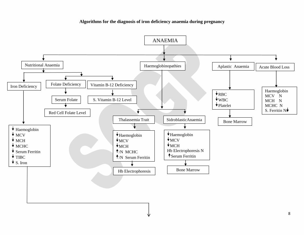
Algorithms for the diagnosis of iron deficiency anaemia during pregnancy
Acute Blood Loss
RBC
WBC
Platelet
Red Cell Folate Level
Thalassemia Trait
Vitamin B-12 Deficiency
S. Vitamin B-12 Level
Bone Marrow
Serum Folate
Folate Deficiency Iron Deficiency
Aplastic Anaemia Haemoglobinopathies Nutritional Anaemia
Haemoglobin
MCV N
MCH N
MCHC N
S. Ferritin N/
Haemoglobin
MCV
MCH
Hb Electrophoresis N
Serum Ferritin
Haemoglobin
MCV
MCH
/N MCHC
/N Serum Ferritin
Haemoglobin
MCV
MCH
MCHC
Serum Ferritin
TIBC
S. Iron
SidroblasticAnaemia
ANAEMIA
Bone Marrow Hb Electrophoresis
Page 9
9
Haemoglobin
< 11 g/dL 1st and 3
rd Trimester
< 10.5 g/dL 2nd
Trimester
< 10 g/dL Postnatal
Complete Blood Count
RBC Morphology
Red Cell Indices
RBC
WBC
Platelet
RBC’s
MCV
MCH
/N MCHC
MCV N
MCH N
MCHC N
MCV (> 96fl)
S. Folate Level S. Vitamin B-12 Level
Iron Deficiency
Thalassaemia Trait
Chronic Disease
Sidroblastic Anaemia
S. Ferritin Level
< 30 µg/L
Iron Deficiency Anaemia 30–100 µg/L
Iron Studies
> 100 µg/L
S. Ferritin Level
Haemoglobinopathies
Thalassaemia Trait
Sidroblastic Anaemia
Non Haematological Causes
Acute / chronic illness
Inflammation
Malignancies
Liver disease
Renal disease Hb Electrophoresis
Abnormal Thalassaemia Trait Normal Sidroblastic
Anaemia
CPR >30
Iron < 7 umol/L
Iron Saturation < 20%
TIBC < 45 umol/L
Functional Iron Deficiency
CRP Normal/
Iron > 7 umol/L
Iron Saturation > 20%
TIBC > 45 umol/L
Pancytopenia
Bone Marrow Study
Confirm by Bone Marrow
Iron replete
Page 10
10
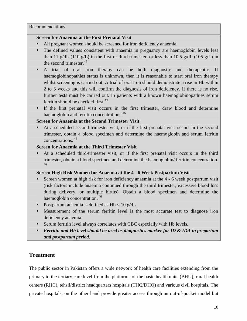
Recommendations
Screen for Anaemia at the First Prenatal Visit
All pregnant women should be screened for iron deficiency anaemia.
The defined values consistent with anaemia in pregnancy are haemoglobin levels less
than 11 g/dL (110 g/L) in the first or third trimester, or less than 10.5 g/dL (105 g/L) in
the second trimester.45
A trial of oral iron therapy can be both diagnostic and therapeutic. If
haemoglobinopathies status is unknown, then it is reasonable to start oral iron therapy
whilst screening is carried out. A trial of oral iron should demonstrate a rise in Hb within
2 to 3 weeks and this will confirm the diagnosis of iron deficiency. If there is no rise,
further tests must be carried out. In patients with a known haemoglobinopathies serum
ferritin should be checked first.39
If the first prenatal visit occurs in the first trimester, draw blood and determine
haemoglobin and ferritin concentrations.46
Screen for Anaemia at the Second Trimester Visit
At a scheduled second-trimester visit, or if the first prenatal visit occurs in the second
trimester, obtain a blood specimen and determine the haemoglobin and serum ferritin
concentrations. 46
Screen for Anaemia at the Third Trimester Visit
At a scheduled third-trimester visit, or if the first prenatal visit occurs in the third
trimester, obtain a blood specimen and determine the haemoglobin/ ferritin concentration.
46
Screen High Risk Women for Anaemia at the 4 - 6 Week Postpartum Visit
Screen women at high risk for iron deficiency anaemia at the 4 - 6 week postpartum visit
(risk factors include anaemia continued through the third trimester, excessive blood loss
during delivery, or multiple births). Obtain a blood specimen and determine the
haemoglobin concentration. 46
Postpartum anaemia is defined as Hb < 10 g/dL
Measurement of the serum ferritin level is the most accurate test to diagnose iron
deficiency anaemia
Serum ferritin level always correlates with CBC especially with Hb levels.
Ferritin and Hb level should be used as diagnostics marker for ID & IDA in prepartum
and postpartum period.
Treatment
The public sector in Pakistan offers a wide network of health care facilities extending from the
primary to the tertiary care level from the platforms of the basic health units (BHU), rural health
centers (RHC), tehsil/district headquarters hospitals (THQ/DHQ) and various civil hospitals. The
private hospitals, on the other hand provide greater access through an out-of-pocket model but
Page 11
11
offer mostly tertiary care. The diagnostic services vary from facility to facility in the public
sector both in terms of standards and variety of tests. The private sector, however, provides a
greater network of collection services and standardized labs for improved wider spectrum of
diagnostics. Therefore, as diagnostics and consequent treatment protocols may not be
harmonious across the country, patients do not have access to standard antenatal, intrapartum and
postpartum care resulting in high maternal mortality. This number varies according to the
particular survey and is stated as 260 / 100,000 live births by the recent Pakistan Demographic
and Health Survey (PDHS 2017/18). 47
IDA is the most prevalent nutritional deficiency in Pakistan. Amongst the many factors, food
insecurity, poor nutrition, repeated pregnancies and unhealthy dietary habits have been
associated with its onset amongst the vulnerable populations as stated earlier. Pregnant women
are at special risk of iron deficiency anaemia due to increased requirements which are difficult to
cover with our current diet alone. Most women in our country embark on pregnancy with
depleted iron stores.
A haemoglobin level of less than 11 g/dL in the antenatal period should be suspected as iron
deficiency anaemia (IDA) unless proven otherwise (such as Thalassaemia). The most sensitive
and specific test to diagnose iron deficiency is serum ferritin level with a cut off value of less
than 30 µg/L, indicating poor iron stores. Daily iron and folic acid supplementation is
recommended as part of the antenatal care to reduce the risk of maternal anaemia and iron
deficiency leading to increased maternal, fetal and new born complications.
Strategies to combat IDA include counselling services, food fortification, dietary modification,
supplementation, and deworming. It is imperative to provide dietary advice to pregnant women
at the time of booking, especially those belonging to the poor socio-economic strata. 10
Oral iron supplementation is usually the first choice for the treatment of iron deficiency anaemia
(IDA) because of its effectiveness and low cost. But unfortunately in many iron deficient
conditions, oral iron is a less than the ideal treatment mainly because of adverse events related to
the gastrointestinal tract as well as the long course required to treat anaemia and replenish body
iron stores.
Because of the high rate of gastrointestinal AE’s (35% to 59%) with ferrous sulfate, new
compounds containing either the ferric or ferrous salt forms have been developed as different
preparations (amino acid chelates, carbonyl iron, iron III polymaltose complex [IPC], extended
Page 12
12
release products) and are approved for clinical use. Among these, IPC is the most studied and
used for IDA patients based on its better tolerability leading to higher compliance rates and
improved effectiveness.48
Treatment with IV iron is clearly superior to oral iron and presents several advantages such as
quicker and higher increase in Hb levels and replenishment of body iron stores. The ferric
hydroxide preparation was the first iron compound for parenteral use but had severe toxic
reactions. This was followed by the high molecular weight iron dextran (HMW-ID) which was
associated with an elevated risk of anaphylactic reactions and low molecular weight iron dextran
(LMW-ID) requiring a test dose and had black box warnings. Ferric gluconate (FG) and iron
sucrose (IS) offered safer alternatives to HMW/LMW-ID. By far the greatest experience in
published literature is with IS. The more recent products ferric carboxymaltose (FCM), iron
isomaltoside, and ferumoxytol offer better safety profiles than the more traditional IV
preparations, particularly because these products may be given more rapidly and in larger doses
than their predecessors with the possibility of complete replacement of iron in 15-60 minutes. A
warning has been issued for ferumoxytol about potentially life threatening events. FCM has an
efficiency ratio over 10 times better than IS and does not have the requirement of a test dose.48
1. Prenatal treatment:
1st Trimester
Most women in Pakistan are not able to see the health care provider in the 1st trimester of their
pregnancy, and this problem varies with the level of education and urban–rural setting.
Furthermore the symptoms of mild anaemia can be vague but as anemia progresses more
symptoms set in. Signs and symptoms include fatigue, low physical and mental capacity,
headache, vertigo, leg cramps, pagophagia, cold intolerance, koilonychia, mucosal patches and
angular stomatitis.
Classical laboratory investigations include haemoglobin level, serum iron concentration, serum
transferrin and serum ferritin. However, due to limited resources we are not able to do all the
above for our patients. Hence, it is recommended that we do at least serum ferritin levels at the
1st antenatal visit along with a complete blood count. Management will depend upon whether we
are using iron for treatment or prophylactic purposes. Prophylactic therapy is given to all
pregnant patients in our setting.
Page 13
13
Women should be counselled on how to take oral iron supplementation correctly. It should be on
an empty stomach, 1 hour before meals, with a source of vitamin C to maximize absorption.
Other medications or antacids, tea or coffee should not be taken at the same time.
2nd
and 3rd
Trimester
CBC should be repeated and parenteral iron should be given if haemoglobin is below 10.5 g/dL
during 2nd
trimester and 11 g/dL in 3rd
trimester. There is no need to repeat the serum ferritin
level if they were normal in 1st trimester. Severe anaemia (< 7 g/dL) in unbooked patients
presenting at or near term should be managed by giving a blood transfusion (This refers to those
patients who seek help of their consulting physician for the first time and did not have any earlier
antenatal care).
Page 14
14
Recommendations for Iron deficiency anaemia
Cut off value for Hb < 11 g/dl in 1st and 3
rd trimester, 10.5g/dl in 2
nd trimester
A prophylactic daily dose of 60 mg of elemental iron is recommended for non-anaemic
pregnant mothers. 53
High dose IV iron such as Iron sucrose or Ferric Carboxymaltose (FCM) should be given
to pregnant mothers with Hb < 10.5 g/dL in the 2nd
trimester and < 11 g/dl in 3rd
trimester
Where I/V iron is not available anaemic pregnant woman should be treated with daily iron
(120 mg of elemental iron) and folic acid (400 μg or 0.4 mg) supplementation until her
haemoglobin concentration rises to normal. She can then switch to the standard antenatal
dose to prevent recurrence of anaemia. 53
If Hb level is < 7g/dL in 2nd
and 3rd
trimester of pregnancy blood transfusion should be
recommended after the evaluation of risks & benefits of blood transfusion.
Folic acid should be commenced as early as possible (ideally before conception) to
prevent neural tube defects
* In settings where anaemia in pregnant women is a severe public health problem (40% or higher),
a daily dose of 60 mg of elemental iron is preferred over a lower dose. Higher doses are
recommended for Pakistan [51]. 53
2. Post Natal Treatment
Postpartum iron deficiency anaemia is a major public health problem and is an important clinical
entity as demonstrated by several studies that it is linked to postpartum anaemia, postpartum
depression and fatigue, as well as other cognitive sequelae. It is associated with adverse future
pregnancy outcomes for subsequent short inter pregnancies.81
All patients should have an active
management of the third stage of labour to the baby’s minimize the blood loss at the time of
delivery. If the baby’s APGAR score is good delayed cord clamping and waiting till the
pulsations have stopped further helps the placental blood to reach the baby so that anaemia may
be prevented.
Page 15
15
Recommendations
Immediate postpartum care (within 24 hours): If Hb level is < 10 g/dL patient should not
be discharged from a hospital such patients should be given a single shot of high dose IV
iron such as FCM/Iron Sucrose OR if patient’s Hb level is > 10 g/dL after delivery, normal
ferritin levels and haemodynamically stable, she should be discharged with advice of oral
iron therapy at the discretion of health professional.
Late postpartum care (after 24 hours): Patients who report back after delivery with Hb <
10 g/dL should also be given IV iron such as FCM and/or Iron Sucrose.
□ Oral iron supplementation, either alone or in combination with folic acid (120 mg of
elemental iron plus 400 µg folic acid), may be provided to postpartum women
(haemodynamically stable) for 6–12 weeks following delivery for reducing the risk of
anaemia in settings where gestational anaemia is of public health concern.
□ All women with Hb < 7 g/dL should be considered for transfusion to achieve Hb > 7
g/dl.
□ Contraceptive advice is mandatory to all patients.
Page 16
16
Algorithm for the treatment of iron deficiency anaemia during 1st, 2
nd, and 3
rd trimester of
pregnancy
1st Trimester 2
nd & 3
rd Trimester
Haemoglobin
Haemoglobin
< 11 g/dL 1st and 3
rd Trimester
< 10.5 g/dL 2nd
Trimester
Postpartum < 10 g/dL
Haemoglobin
< 7 g/dl
Iron Deficiency
Anaemia
Severe Anaemia
Blood Transfusion
Oral Iron Therapy Various ferrous and ferric options are
available. IPC offers a safe option.
- Compliance Issues
- Inadequate Response
- Intolerance
Intravenous Iron Therapy
Few I/V options available. FCM or
Iron sucrose is safe and effective.
option
Haemoglobin
< 10.5 g/dL – 2nd
trimester
< 11 g/dL – 3rd
trimester
Page 17
17
Algorithm for the treatment of iron deficiency anaemia during postpartum period
Haemoglobin
Haemoglobin <10g/dL
Haemoglobin
< 7 g/dl
Haemoglobin >10g/dL Severe Anaemia
Blood Transfusion Immediate postpartum
care (within 24 hours)
- Give single shot of high
dose IV iron such as FCM
/Iron Sucrose
Late postpartum care
(after 24 hours)
- Patients who reports
back after delivery with
Hb < 10 g/dL should also
be given IV iron such as
FCM or Iron Sucrose.
Oral iron supplementation, either alone or in
combination with folic acid (120 mg of
elemental iron plus 400 µg folic acid), should
give to postpartum women (hemodynamically
stable) for 6–12 weeks following delivery.
Immediate postpartum
care (within 24 hours)
-Discharge patient if
ferritin levels normal and
hemodynamically stable,
with advice of oral iron
therapy
Intravenous IV Therapy
No
t
To
lera
ted
Page 18
18
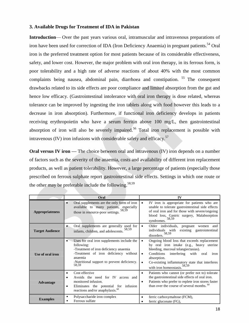
3. Available Drugs for Treatment of IDA in Pakistan
Introduction— Over the past years various oral, intramuscular and intravenous preparations of
iron have been used for correction of IDA (Iron Deficiency Anaemia) in pregnant patients.54
Oral
iron is the preferred treatment option for most patients because of its considerable effectiveness,
safety, and lower cost. However, the major problem with oral iron therapy, in its ferrous form, is
poor tolerability and a high rate of adverse reactions of about 40% with the most common
complaints being nausea, abdominal pain, diarrhoea and constipation. 55
The consequent
drawbacks related to its side effects are poor compliance and limited absorption from the gut and
hence low efficacy. (Gastrointestinal intolerance with oral iron therapy is dose related, whereas
tolerance can be improved by ingesting the iron tablets along with food however this leads to a
decrease in iron absorption). Furthermore, if functional iron deficiency develops in patients
receiving erythropoietin who have a serum ferritin above 100 mg/L, then gastrointestinal
absorption of iron will also be severely impaired.56
Total iron replacement is possible with
intravenous (IV) iron infusions with considerable safety and efficacy.57
Oral versus IV iron — The choice between oral and intravenous (IV) iron depends on a number
of factors such as the severity of the anaemia, costs and availability of different iron replacement
products, as well as patient tolerability. However, a large percentage of patients (especially those
prescribed on ferrous sulphate report gastrointestinal side effects. Settings in which one route or
the other may be preferable include the following.58,59
Oral IV
Appropriateness
Oral supplements are the only form of iron
available to many patients, especially
those in resource-poor settings. 58,59
IV iron is appropriate for patients who are
unable to tolerate gastrointestinal side effects
of oral iron and for those with severe/ongoing
blood loss, Gastric surgery, Malabsorption
syndromes. 58,59
Target Audience
Oral supplements are generally used for
infants, children, and adolescents. 58,59
Older individuals, pregnant women and
individuals with existing gastrointestinal
disorders. 58,59
Use of oral iron
Uses for oral iron supplements include the
following:
-Treatment of iron deficiency anaemia
-Treatment of iron deficiency without
anaemia
-Nutritional support to prevent deficiency.
58,59
Ongoing blood loss that exceeds replacement
by oral iron intake (e.g., heavy uterine
bleeding, mucosal telangiectasias).
Conditions interfering with oral iron
absorption.
Co-existing inflammatory state that interferes
with iron homeostasis.58,59
Advantage
Cost effective
Avoids the need for IV access and
monitored infusion.
Eliminates the potential for infusion
reactions and/or anaphylaxis.60
Patients who cannot (or prefer not to) tolerate
the gastrointestinal side effects of oral iron.
Patients who prefer to replete iron stores faster
than over the course of several months. 60
Examples Polysaccharide iron complex
Ferrous sulfate ferric carboxymaltose (FCM),
ferric gluconate (FG),
Page 19
19
Ferrous fumarate – 324 or 325 mg tablet
(contains 106 mg elemental iron per tablet)
Ferrous gluconate
Relatively inexpensive Over-The-Counter
(OTC) preparations includes: heme iron
polypeptide, carbonyl iron, ferric citrate,
ferrous ascorbate, and ferrous succinate.61
ferumoxytol,
iron sucrose (IS),
iron isomaltoside (not available in the United
States),
Low molecular weight iron dextran (LMW
ID).
Side Effects
Gastrointestinal side effects are extremely
common with oral iron administration.
These include metallic taste, nausea,
flatulence, constipation, diarrhea,
epigastric distress, and/or vomiting.62
Infusion reactions and potential anaphylaxis, shock,
and death, although this risk is exceedingly low.63
I. ORAL IRON SALTS such as ferrous fumarate, ferrous gluconate, and ferrous sulfate have
been the mainstay of oral iron supplementation because they are inexpensive and effective at
restoring iron balance with a favorable overall safety and tolerability profile. However, as
discussed earlier, in some patients, absorption of oral iron salts is inadequate and poor tolerance
results in reduced adherence to therapy. The newer polysaccharide iron complex and heme iron
polypeptide products now offer alternative therapies with improved absorption and tolerability
profile compared to the traditional iron salts. 64
A. Iron (III) hydroxide polymaltose complex (IPC) is an iron preparation with non-ionic iron
and polymaltose in a stable complex. Numerous clinical trials in men, women, children and
infants have shown that IPC is effective in treating iron deficiency anaemia (IDA). IPC is best
given with meals, and probably in a slightly higher iron dose than that of the classical salts. Its
acceptance and compliance in patients shows an advantage over ferrous salts as reflected in
several studies demonstrating a lower rate of treatment interruption related to a lower incidence
of upper GIT adverse events. 49
Meta-analysis of studies conducted in adult patients with iron deficiency anaemia, comparing
IPC with ferrous sulfate in equivalent doses, suggested similar efficacy however, the tolerance of
IPC in adults and its profile for adverse events was clearly better than that of ferrous sulfate 50
B. Ferrous Gluconate is used to prevent or treat low iron blood levels in iron deficiency
anaemia. Studies have shown that Oral ferrous gluconate treatment for severe iron deficiency
anaemia is well tolerated and highly effective in premenopausal women. The treated women did
not exhibit any major side effect, only one serious side effect was reported (erythema nodosum).
Mild self-limiting side effects including black discolouration of stools in 54% cases, nausea in
6%, constipation in 2% and diarrhoea in 2% were observed.65
Page 20
20
C. Ferrous sulfate is used to prevent or treat low iron blood levels in iron deficiency anaemia. It
is the cheapest and most commonly prescribed oral iron supplement, showing a rapid rise in both
serum iron concentration and Non-transferrin bound serum iron (NTBI) but the greatest
frequency of adverse events at the same time.49
This may lead to poor compliance and hence
poor results.
D. Ferrous Fumarate is used to prevent or treat low iron blood levels in iron deficiency
anaemia. Ferrous fumarate, the least toxic iron (II) compound, causes fewer adverse events
because of its low solubility and slow dissolution rate after oral administration. 49
A 2013
systematic review included over 10,000 patients receiving different oral iron formulations.
Gastrointestinal adverse effects were seen with all oral formulations (ferrous fumarate, 43
percent; ferrous gluconate, 31 percent; ferrous sulfate, 30 percent). Other supplements such as
iron protein succinylate or ferrous glycine sulfate, which have enteric coatings (that reduces
absorption) had lower frequencies of adverse effects.66
II. INTRAMUSCULAR (IM) IRON THERAPY
Some preparations such as iron sorbitex, high molecular weight iron dextran, and low molecular
weight iron dextran can be given intramuscularly (IM). However, as IM injections have some
significant drawbacks they are generally not recommended. IM iron is painful and produces
permanent skin staining. It is not safer than IV infusion. Its use is therefore discouraged.41
III. SPECIFIC IV IRON PREPARATIONS
A. Low Molecular Weight iron dextran (LMW-ID) is the least expensive of the IV iron
formulations that can be administered in a large dose (total dose infusion) and is seen as safe and
effective in the settings of heavy uterine bleeding, pregnancy, postpartum, inflammatory bowel
disease, gastric bypass, hereditary hemorrhagic telangiectasia, chronic kidney disease, and
restless leg syndrome. A test dose is required prior to the first dose of LMW-ID.67, 68
There are a
limited number of studies available on the use of LMW-ID during pregnancy and the postpartum
period. It is contraindicated during the 1st trimester. No serious adverse events have been
associated with its use, but mild adverse events have been observed in 5% of cases.41
B. Iron sucrose (IS) also called iron saccharate, is given over multiple infusions, with a
maximum individual dose of 10 to 15 ml (equivalent to 200 to 300 mg elemental iron, based on a
concentration of 20 mg elemental iron per ml). A test dose is recommended if the patient has a
Page 21
21
history of drug allergies; otherwise, a test dose is not required. For patients with cancer receiving
ESAs, 10 ml may be infused over 60 minutes every two to three weeks. Larger doses (i.e., doses
above 300 mg) are not recommended. This product cannot be given intramuscularly. Numerous
studies on its efficacy and safety, as compared with iron dextran and ferrous gluconate, reported
that it was well tolerated; No hypersensitivity reactions or fatal events were observed except
urticaria.69
Iron Sucrose are safe for use in: settings of dialysis, non-dialysis chronic kidney
disease, inflammatory bowel disease, chemotherapy-induced anaemia, the peripartum period,
gastric bypass, heavy uterine bleeding, and a host of other conditions associated with iron
deficiency.70
C. Ferric Carboxymaltose (FCM) is a colloidal iron hydroxide complex with tighter binding of
elemental iron to the carbohydrate polymer than some other IV iron preparations and it can be
given in large doses equivalent to 1000 mg of elemental iron. It may be given in short durations
such as a 15-minute infusion.71
Its use has been approved in pregnant women from the 2nd
trimester. Studies comparing IV versus oral iron therapies show that ferric carboxymaltose was
associated with a higher rate of: patient tolerability, patient compliance, and target value
achievement at lower doses with safety equal to but more effective than iron sucrose.69
Several
trials establish the efficacy and safety of FCM in iron-deficient patients in many settings such as:
heavy uterine bleeding, postpartum women, non-dialysis dependent chronic renal failure,
inflammatory bowel disease, heart failure, chemotherapy associated anaemia without
concomitant use of an erythropoietin stimulating agent (ESA), and patients non-responsive to
oral iron. 71
Numerous studies have also reported the safety profile use of FCM during pregnancy
without serious adverse events.72
Initial experiences with FCM raised concerns about
hypophosphatemia following administration but subsequently, only rare reports of clinical
sequelae related to hypophosphatemia have been reported.73,74
Serum phosphate levels may need
to be monitored in selected populations such as those with borderline phosphate levels at the
baseline. 75
FCM offered an efficacious and time-efficient correction of IDA in women during the late stages
of pregnancy along with improvements in QoL and significant decrease in gastrointestinal side
effects. In patients who are intolerant and need a quick correction of iron deficit prior to delivery
(and identification or non-correction of IDA), FCM is an option for anaemia correction with a
safety profile similar to oral iron.51
Efficacy and Safety of ferric carboxymaltose and other
formulations in iron deficient patients have been widely studied in randomized controlled trials,
Page 22
22
showing significant improvements in serum ferritin (µg/L) with ferric carboxymaltose compared
to oral iron (delta 172.8; 95 % CI 66.7–234.4) and in haemoglobin (g/dL) levels with respect to
ferric gluconate (delta 0.6; 95 % CI 0.2–0.9), oral iron (delta 0.8; 95 % CI 0.6–0.9) and placebo
(delta 2.1; 95 % CI 1.2–3.0). The conclusion was that all currently available intravenous iron
preparations appear to be safe and effective, but ferric carboxymaltose seems to provide a better
and quicker correction of haemoglobin and serum ferritin levels in iron deficient patients.52
D. Iron isomaltoside has a matrix structure that results in tight iron binding and slow release of
labile free iron, allowing administration in a single infusion, at a dose of 20 mg/kg, over 15
minutes without a test dose. A 2017 trial comparing a single infusion of iron isomaltoside (500
or 1000 mg, depending on body weight and haemoglobin level) or multiple infusions of iron
sucrose (given as 200 mg infusions, for a total dose based on body weight and haemoglobin),
showed that a greater number of patients in the iron isomaltoside group had an increase in
haemoglobin of ≥ 2 g/dL (69 versus 52 percent; p <0.0001). Rates of serious adverse events did
not differ (0.6 percent in both groups).76
E. Iron Sorbitol is a complex of ferric, sorbitol and citric acid, stabilized with dextrin and
sorbitol. Its use is only in the treatment of proven iron deficiency anaemia, where oral therapy is
ineffective or inappropriate through parenteral route. Severe anaphylactic reactions may occur
after parenteral administration. As evident from the study, the practice of prescribing iron
sorbitol in a dose of 10 ampoules (75 mg/ampoule) with continuing oral iron therapy throughout
pregnancy does not fulfill the goal of iron therapy. It is impractical to check the compliance in
these patients, and the therapy is cumbersome and prolonged.77
IV. OTHER IV IRON PREPARATIONS NOT AVAILABLE IN PAKISTAN
A. Ferric Gluconate also called ferric gluconate complex, can be given over multiple infusions.
A test dose is recommended if the patient has a history of drug allergies. 69
There few studies on
its use during pregnancy and there are data concerning its use during the neonatal period. It is
contraindicated during the 1st trimester. No serious adverse events have been linked with its use.
41,69
B. Ferumoxytol is composed of super paramagnetic iron oxide nanoparticles coated with a
LMW semi synthetic carbohydrate.60,78
In a 2018 trial of ferumoxytol (two infusions of 510 mg
Page 23
23
for a total of 1020 mg) versus ferric carboxymaltose (two infusions of 750 mg for a total of 1500
mg), showed both groups to have comparable increases in haemoglobin (approximate increase,
1.5 g/dL) with no clinical sequelae related to hypophosphatemia in either group.79
Ferumoxytol can cause a brighter signal on magnetic resonance imaging (MRI) scans, therefore
if an MRI is planned within three months of administration, the radiologist should be notified
accordingly. There are no published data on the safety and efficacy of ferumoxytol in pregnancy.
C. Iron Polymaltose there is a limited number of studies on the use of iron polymaltose during
pregnancy and the neonatal period, but no serious adverse events have been reported. Its
administration is limited to the IM route.49
Prevention of Anaemia in Pregnancy
1. Health and nutrition education of women
Women should be educated to improve over all dietary intake and consumption of iron and
folate rich food stuffs. In addition, knowledge and practice enhancement through counselling
and behaviour change communication are vital aspects of improving diet.
Women should be encouraged to take diet which includes meat, poultry, fish, beans, nuts,
seeds, leafy greens, eggs, fruits like banana and melons. They should be educated about
supplement high in a vitamin C like tomato juice or oranges; this will help absorption of iron.
2. Optimal birth spacing
Women should be counselled to have birth spacing to prevent maternal anaemia and low
birth weight babies.
3. Fortification of food
It can be a cost effective way to improve iron status and is available for wheat, maize, flour
and rice.
4. Haemoglobin estimation
Haemoglobin estimation in adolescent school girls and women intending to become pregnant
for detection and treatment of anaemia prior to pregnancy
5. Iron Folic acid (IFA)supplementation
Women in the postpartum period should have iron and folic acid supplementation so that she
can replenish iron stores to prevent anaemia.
o WHO recommendations - 2016
Page 24
24
o Daily oral iron and folic acid supplementation with 30 mg to 60 mg of elemental
iron* and 400 µg (0.4 mg) folic acid** is recommended for pregnant women to
prevent maternal anaemia, puerperal sepsis, low birth weight, and preterm birth.***
*The equivalent of 60 mg of elemental iron is 300 mg ferrous sulfate
heptahydrate, 180 mg ferrous fumarate or 500 mg of ferrous gluconate.
** Folic acid should be commenced as early as possible (ideally before
conception) to prevent neural tube defects.
*** This recommendation supersedes the previous recommendation found
within the WHO guideline ‘Daily iron and folic acid supplementation in
pregnant women’ (2012).
6. Prevention of other nutritional deficiencies
Other nutritional supplements should be given such as vitamin B12 and vitamin A.
7. Promoting safe water, sanitation and hygiene (WASH)
WASH has a great impact on the general nutrition status and would also indirectly affect the
anaemia status of the women.
8. Control of helminthic infestation
Periodic treatment (deworming medication such as albendazole and mebendazole as a routine
part where hookworm prevalence is >20%) with improvement of water and sanitation and
health education can reduce the transmission of soil-transmitted helminths and
schistosomiasis infections.
9. Prevention and treatment of infections such as malaria and tuberculosis are vital for the
prevention of anaemia.
10. Treatment of conditions causing blood loss
This includes conditions of blood loss such as haemorrhoids, menorrhagia due to fibroid or
dysfunctional uterine bleeding and requires prompt medically or surgically.
11. Community-based distribution (home visits) of iron and other micronutrient supplements
to the most vulnerable groups, particularly where health services are not well utilized, and other
high-risk group.80
12. Promote use of ITB (bed nets).80
13. Reproductive health
Promote the care of women (raising awareness about danger signs during pregnancy and
postpartum periods and what to do about them; the need for antenatal and postpartum care and
adequate diet for women).80
Page 25
25
14. Private sector (food and pharmaceutical manufacturers, marketers, distributors)
Promote production and sale of iron and micronutrient supplements, fortified foods, ITB
(Bed nets) and family planning by private vendors, marketers, distributors.80
Conclusion
The role of I/V iron as a safe and effective option for quick correction of iron deficiency and iron
deficiency anaemia along with its protective role in the postpartum period needs to be
underscored as part of Pakistan guidelines for management of iron deficiency and iron deficiency
anaemia. This can also serve for guidance of various societies as well as physicians in improving
health outcomes in pregnant women.
REFERENCES
1. Anlaakuu P, Anto F. Anaemia in pregnancy and associated factors: a cross sectional study of
antenatal attendants at the Sunyani Municipal Hospital, Ghana. BMC Res Notes 2017;10(1):402-
9.
2. WHO/UNICEF/UNU. Iron deficiency anaemia assessment, prevention, and control: a guide
for programme managers. Geneva, World Health Organization, 2001. [online] Available at:
http://www.who.int/nutrition/publications/micronutrients/anaemia_iron_deficiency/WHO_NHD
_01.3/en/ [Accessed 26 Sep. 2018].
3. Stephen G, Mgongo M, Hussein Hashim T, Katanga J, Stray-Pedersen B, Msuya SE. Anaemia
in pregnancy: prevalence, risk factors, and adverse perinatal outcomes in Northern Tanzania.
Anaemia. [Internet]. 2018 May 02 [cited 2018 Sep 4]:Doc No 1846280 [about 9 screens].
Available from: https://www.hindawi.com/journals/anaemia/2018/1846280/cta/
4. Gupta A, Gadipudi A. Iron Deficiency Anaemia in Pregnancy: Developed Versus Developing
Countries. EMJ Hematol. 2018;6[1]:101-109.
5. World Health Organization. Guideline: daily iron and folic acid supplementation in pregnant
women;2012. [online] Available at: http://www.who.int/iris/handle/10665/77770 [Accessed 06
Sep. 2018].
6. Stevens GA, Finucane MM, De-Regil LM, Paciorek CJ, Flaxman SR, Branca F, etal. Global,
regional, and national trends in haemoglobin concentration and prevalence of total and severe
anaemia in children and pregnant and non-pregnant women for 1995-2011: a systematic analysis
of population-representative data. Lancet Glob Health. 2013 Jul;1(1):e16-25. doi:
10.1016/S2214-109X(13)70001-9.
Page 26
26
7. World Health Organization. The global prevalence of anaemia in 2011. 2015. [online]
Available at:
http://www.who.int/nutrition/publications/micronutrients/global_prevalence_anaemia_2011/en/
[Accessed 01 Sep. 2018].
8. World Health Organization. Anaemia policy brief. 2014. Available from:
http://www.who.int/nutrition/topics/globaltargets_anaemia_policybrief.pdf. [Accessed 01 Sep.
2018].
9. World Health Organization. Global nutrition targets 2025: policy brief series. 2014. Available
from: http://www.who.int/nutrition/publications/globaltargets2025_policybrief_overview/en/.
[Accessed 11 Oct. 2018].
10. Akhtar S, Ahmed A, Ahmad A, Ali Z, Riaz M, Ismail T. Iron status of the Pakistani
population-current issues and strategies. Asia Pac J Clin Nutr. 2013;22(3):340-7.
11. Sanghvi TG, Harvey PW,Wainwright E. Maternal iron–folic acid supplementation programs:
evidence of impact and implementation. Food Nutr Bull. 2010;31(2 Suppl):100-7.
12. Habib MA, Raynes-Greenow C, Soofi SB, Ali N, Nausheen S, Ahmed I, et al. Prevalence
and determinants of iron deficiency anaemia among non-pregnant women of reproductive age in
Pakistan. Asia Pac J Clin Nutr. 2018;27(1):195-203.
13. World Health Organization, Global Health Observatory Data Repository/World Health
Statistics.2015. Available from: http://apps.who.int/gho/data/node.main.1?lang=en. [Accessed
21 Aug. 2018].
14. Bhutta Z, Soofi S, Zaidi S, Habib A, Hussain M. Pakistan National Nutrition Survey, 2011.
Available from:
https://ecommons.aku.edu/cgi/viewcontent.cgi?article=1262&context=pakistan_fhs_mc_women
_childhealth_paediatr. [Accessed 05 Sep. 2018].
15. Baig-Ansari N, Badruddin SH, Karmaliani R, Harris H, Jehan I, Pasha O et al. Prevalence
and risk factors in pregnant women in an urban area of Pakistan. Food Nutr Bull. 2008;29:132-9.
16. Van den Broek N. Anaemia in pregnancy in developing countries. Br J Obstet Gynaecol
1998;105:385–90.
17. Maryam M, Zahid M, Khurshid M, Ahmad MQ, Yasmin K, SarfrazK, et al. Factors
Associated with Iron Deficiency Anaemia among Pregnant Women Visiting Outpatient
Department of Jinnah Hospital Lahore, Pakistan. J Hematol Transfus 2018;6(1): 1080-85.
18. Mawani M, Ali SA, Bano G, Ali SA. Iron Deficiency Anaemia among Women of
Reproductive Age, an Important Public Health Problem: Situation Analysis. Reprod Syst Sex
Disord. [Internet]. 2016 Aug 09 [cited 2018 Sep 23]:Doc No 100187 [about 6 screens]. Available
from: https://www.hindawi.com/journals/anaemia/2018/1846280/cta/
Page 27
27
19. Huma Naz, Bushra Begum. Prevalence and associated risk factors of anaemia in pregnant
women in a teaching hospital, Korangi Industrial Area. Pak J Surg 2013; 29(2):131-33
20. Khalid R, Irshad J, Saleem A, Ashraf S. Risk factors for Anaemia in Pregnant Women in a
rural area of Bahawalnagar Pakistan- a Descriptive Cross Sectional Study. P J M H S
2017;11(4): 1238-42
21. Pavord S, Myers B, Robinson S, Allard S, Strong J, Oppenheimer C. UK guidelines on the
management of iron deficiency in pregnancy. Brit J Haematology 2012;156:588–600.
22. Talaulikar VS. Anaemia in pregnancy. [Internet]. [cited 2018 Aug 13]. Available from:
https://www.glowm.com/Critical_current_issue/page/25
23. Jamali NH, Mahesar H, Bhutto MA. Prevalence of iron deficiency anaemia in school and
college going students of district Shaheed Benazirabad Sindh province, Pakistan. Open Journal
of Blood Diseases 2016;(6):67-78.
24. Shah SZA, Baloch GH, Yousfani ZA, Abbas SA, Baloch ZAQ, Sumera Bukhari S, et al.
Clinical profile of patients with iron deficiency anaemia at tertiary care teaching hospital. Indo
Am. J. P. Sci 2017; 4(02):373-77
25. Khan HMS, Sohail M , Ali A, Akhtar N, Khan H, Fatima Rasool F. Symptoms-Based
Evaluation of Iron Deficiency Anaemia in Students of Bahawalpur Correlated with their Eating
Habits. Trop J Pharm Res 2014; 13 (5): 769-72
26. Khaskheli MN, Baloch S, Sheeba A, Baloch S, Khaskheli FK. Iron deficiency anaemia is still
a major killer of pregnant women. Pak J Med Sci. 2016;32(3):630-34.
27. Galloway R, Dusch E, Elderet L. Women’s perceptions of iron deficiency and anaemia
prevention and control in eight developing countries. Sci Dir Soc Sci Med. 2002;55(4):529–544
28. National Institute of Population Studies (NIPS) [Pakistan] and ICF International. 2013.
Pakistan Demographic and Health Survey 2012-13. Islamabad, Pakistan, and Calverton,
Maryland, USA: NIPS and ICF International. [online] Available at:
https://www.nips.org.pk/abstract_files/PDHS%20Final%20Report%20as%20of%20Jan%2022-
2014.pdf. [Accessed 19 Oct. 2018].
29. Panda BK, Taralekar VS, Mishra A, Srivastava P. Anaemia in pregnancy: improving
adherence with interventions. Biopharm Journal 2015;1(1):33-40
30. World Health Organization. Guideline: Iron supplementation in postpartum women. 2016.
Available from: http://apps.who.int/iris/handle/10665/249242 [Accessed 11 Nov. 2018].
31. World Health Organization. Assessing the iron status of population: report of a joint world
health organization / centre for disease control and prevention technical consultation on the
assessment of iron status at the population level. 2007. Available from:
Page 28
28
https://www.who.int/nutrition/publications/micronutrients/anaemia_iron_deficiency/9789241596
107/en/ [Accessed 29 Aug. 2018].
32. World health organization. Haemoglobin concentrations for the diagnosis of anaemia and
assessment of severity vitamin and mineral nutrition information system. 2011. Available from:
https://www.who.int/vmnis/indicators/haemoglobin/en/ [Accessed 17 Aug. 2018].
33. Centers for disease control and prevention (CDC). CDC criteria for anaemia in children and
childbearing aged women MMWR. Morbidity and mortality weekly report.989;38(22):400-404.
34. National Collaborating Centre for Women's and Children's Health (UK). Antenatal Care:
Routine Care for the Healthy Pregnant Woman. London: RCOG Press; 2008 Mar. (NICE
Clinical Guidelines, No. 62.) Available from: https://www.ncbi.nlm.nih.gov/books/NBK51886/
35. Iron deficiency anaemia guidelines. BMJ best practice. Available from:
https://bestpractice.bmj.com/topics/en-us/94 [Accessed 9 Sep. 2018].
36. Anaemia in pregnancy. South Australian Perinatal Practice Guidelines. 2016. Available
from:
https://www.sahealth.sa.gov.au/wps/wcm/connect/public+content/sa+health+internet/resources/p
olicies/anaemia+in+pregnancy+-+sa+perinatal+practice+guidelines [Accessed 03 Aug. 2018].
37. Goddard A, James M, Mesintyre A, Scott B. On behalf of the British society of
gastroenterology. Guidelines for the management of iron deficiency anaemia. Gut 2011; 60:
1309-16.
38. Peyrin-Biroulet L, Williet N, Cacoub P. Guidelines on the diagnosis and treatment of iron
deficiency across indications: a systematic review. Am J Clin Nutr. 2015;102(6):1585-94.
39. South West Regional Transfusion Committee. Regional template / guideline for the
management of anaemia in pregnancy and postnatally.2014. Available from:
https://www.transfusionguidelines.org/document-library/documents/rtc-sw_2014 [Accessed 12
Aug. 2018].
40. Daru J, Colman K, Stanworth SJ, De La Salle B, Wood EM, Pasricha SR. Serum ferritin as
an indicator of iron status: what do we need to know? Am J Clin Nutr. 2017;106(Suppl 6):1634-
39. S
41. Api O, Breyman C, Çetiner M, Demir C, Ecder T. Diagnosis and treatment of iron deficiency
anaemia during pregnancy and the postpartum period: Iron deficiency anaemia working group
consensus report. Turk J Obstet Gynecol. 2015;12(3):173-81.
42. Toward Optimized Practice (TOP) iron deficiency anaemia. Clinical practice guideline.
March 2018. Available from: http://www.topalbertadoctors.org/cpgs/ .[Accessed 25 Sep. 2018].
43. Sharran Grey, Helen Wright, Muhammad Athar. Management of anaemia in primary care
pathway NHS foundation trust. Bolton NHS version1 ate Nov; 2015.Aaavilable from:
Page 29
29
http://hospital.blood.co.uk/media/28019/bolton-anaemia-management-in-primary-care-pathway-
_final-december-2015.pdf. [Accessed 06 Oct. 2018].
44. Investigation and Management of the Adult Patient with Anaemia. Guidelines and audit
implementation network. 2015. Available from:
https://www.rqia.org.uk/getattachment/1e2a9adc-7517-4a47-858a-5192b0746456/Investigation-
Management-of-Adult-Anaemia-patients-August-2015.pdf.aspx. [Accessed 23 Oct. 2018].
45. Baker RD, Greer FR. Diagnosis and prevention of iron deficiency and iron-deficiency
anaemia in infants and young children (0-3 years of age). Pediatrics. 2010;126(5):1040-50
46. Institute of Medicine (US) Committee on the Prevention, Detection, and Management of Iron
Deficiency Anaemia Among U.S. Children and Women of Childbearing Age; Earl R, Woteki
CE, editors. Iron Deficiency Anaemia: Recommended Guidelines for the Prevention, Detection,
and Management Among U.S. Children and Women of Childbearing Age. Washington (DC):
National Academies Press (US); 1993. Recommended Guidelines For Preventing And Treating
Iron Deficiency Anaemia In Pregnant Women. Available from:
https://www.ncbi.nlm.nih.gov/books/NBK236485. [Accessed 14 Aug. 2018].
47. National Institute of Population Studies (NIPS) [Pakistan] and ICF. 2018. Pakistan
Demographic and Health Survey 2017-18. Islamabad, Pakistan, and Rockville, Maryland, USA:
NIPS and ICF.
48. Cançado RD, Muñoz M. Intravenous iron therapy: how far have we come?. Rev Bras
Hematol Hemoter. 2011;33(6):461-9.
49. Geisser P. Safety and efficacy of iron(III)-hydroxide polymaltose complex / a review of over
25 years experience.Arzneimittelforschung. 2007;57(6A):439-52.
50. Toblli JE, Brignoli R. Iron(III)-hydroxide polymaltose complex in iron deficiency anaemia /
review and meta-analysis. Arzneimittelforschung. 2007;57(6A):431-8.
51. Shim JY, Kim MY, Kim YJ, Young Lee Y, Lee JL, Jun JK, et al. Efficacy and safety of
ferric carboxymaltose versus ferrous sulfate for iron deficiency anaemia during pregnancy:
subgroup analysis of Korean women. BMC Pregnancy Childbirth. 2018;18(1):349-56.
52. Rognoni C, Venturini S, Meregaglia M, Marmifero M, Tarricone R. Efficacy and Safety of
Ferric Carboxymaltose and Other Formulations in Iron-Deficient Patients: A Systematic Review
and Network Meta-analysis of Randomised Controlled Trials. Clin Drug Investig. 2016
Mar;36(3):177-94
53. WHO. Guideline: Daily iron and folic acid supplementation in pregnant women. Geneva,
World Health Organization, 2012. Available from:
http://apps.who.int/iris/bitstream/10665/77770/1/9789241501996_eng.pdf. [Accessed 21 Oct.
2018].
Page 30
30
54. Bayoumeu F. Subiran-Buisset c, Baka NE, Legagneur H, Monnier-Barbarino P, Laxenaire
MC. Iron therapy in iron deficiency anaemia in pregnancy: intravenous route versus oral route.
Am J Obstet Gynecol. 2002;186:518–22.
55. Sharma JB, Soni D. Oxidative stress in pregnancy - Role of iron therapy: oral iron therapy
and risk of free radicals. Obs and Gynac. 2001;VI(12):705–6.
56. Macdougall. Strategies for iron supplementation: Oral versus intravenous. Kidney Int Suppl.
1999;69:61-6.
57. Lee T, Clavel T, Smirnov K, Schmidt A, Lagkouvardos I, Walker A, et al. Oral versus
intravenous iron replacement therapy distinctly alters the gut microbiota and metabolome in
patients with IBD. Gut 2017;66(5):863-71.
58. Bregman DB, Morris D, Koch TA, He A, Goodnough LT. Hepcidin levels predict
nonresponsiveness to oral iron therapy in patients with iron deficiency anaemia. Am J Hematol
2013; 88(2):97-101
59. Werner E, Kaltwasser JP, Ihm P. Oral iron treatment: intestinal absorption and the influence
of a meal (author's transl)]. Dtsch Med Wochenschr 1977; 102(29):1061-4.
60. Auerbach M, Adamson JW. How we diagnose and treat iron deficiency anaemia. Am J
Hematol 2016; 91(10):31-8
61. Powers JM, Buchanan GR, Adix L, Zhang S, Gao A, McCavit TL. Effect of Low-Dose
Ferrous Sulfate vs Iron Polysaccharide Complex on Haemoglobin Concentration in Young
Children with Nutritional Iron-Deficiency Anaemia: A Randomized Clinical Trial. JAMA. 2017
Jun 13;317(22):2297-2304
62. Tolkien Z, Stecher L, Mander AP, et al. Ferrous sulfate supplementation causes significant
gastrointestinal side-effects in adults: a systematic review and meta-analysis. PLoS One 2015;
10:e0117383.
63. Litton E, Xiao J, Ho KM. Safety and efficacy of intravenous iron therapy in reducing
requirement for allogeneic blood transfusion: systematic review and meta-analysis of randomised
clinical trials. BMJ 2013; 347:f4822.
64. Oral Iron for Anaemia: A Review of the Clinical Effectiveness, Cost-effectiveness and
Guidelines [Internet]. Ottawa (ON): Canadian Agency for Drugs and Technologies in Health;
2016 Jan 6. Available from: https://www.ncbi.nlm.nih.gov/books/NBK343969/. [Accessed 11
Oct. 2018].
65. Tazeen Fatima Munim TF, Shaista Rashid S. The Efficacy and Safety of Oral Ferrous
Gluconate in Premenopausal Women with Severe Iron Deficiency Anaemia. Ann Abbasi
Shaheed Hosp Karachi Med Dent Coll 2017;22(1):5-11
Page 31
31
66. Cancelo-Hidalgo MJ, Castelo-Branco C, Palacios S, Haya-Palazuelos J, Ciria-Recasens M,
Manasanch J, et al. Tolerability of different oral iron supplements: a systematic review. Curr
Med Res Opin. 2013;29(4):291-303.
67. Auerbach M, Pappadakis JA, Bahrain H, Auerbach SA, Ballard H, Dahl NV. Safety and
efficacy of rapidly administered (one hour) one gram of low molecular weight iron dextran
(INFeD) for the treatment of iron deficient anaemia. Am J Hematol 2011; 86(10):860-2.
68. Auerbach M, Winchester J, Wahab A, Richards K, McGinley M, Hall F, et al. A randomized
trial of three iron dextran infusion methods for anaemia in EPO-treated dialysis patients. Am J
Kidney Dis 1998; 31(1):81-6.
69. Miller HJ, Hu J, Valentine JK, Gable PS. Efficacy and tolerability of intravenous ferric
gluconate in the treatment of iron deficiency anaemia in patients without kidney disease. Arch
Intern Med 2007; 167(12):1327-8.
70. Macdougall IC, Roche A. Administration of intravenous iron sucrose as a 2-minute push to
CKD patients: a prospective evaluation of 2,297 injections. Am J Kidney Dis 2005; 46(2):283-9.
71. Steinmetz T, Tschechne B, Harlin O, Klement B, Franzem M, J. Wamhoff J,et al. Clinical
experience with ferric carboxymaltose in the treatment of cancer- and chemotherapy-associated
anaemia. Ann Oncol 2013; 24(2):475-82.
72. Froessler B, Collingwood J, Hodyl NA, Dekker G. Intravenous ferric carboxymaltose for
anaemia in pregnancy. BMC Pregnancy Childbirth 2014. 25;14:115. doi: 10.1186/1471-2393-14-
115.
73. Smyth B, Ong S. Severe hypocalcaemia and hypophosphataemia following intravenous iron
and denosumab: a novel drug interaction. Intern Med J. 2016 Mar;46(3):360-3.
74. Blazevic A, Hunze J, Boots JM. Severe hypophosphataemia after intravenous iron
administration. Neth J Med. 2014 Jan;72(1):49-53.
75. Huang LL, Lee D, Troster SM, Kent AB, Roberts MA, Macdougall IC, et al. A controlled
study of the effects of ferric carboxymaltose on bone and haematinic biomarkers in chronic
kidney disease and pregnancy. Nephrol Dial Transplant. 2017 Nov 17. doi: 10.1093/ndt/gfx310.
76. Derman R, Roman E, Modiano MR, et al. A randomized trial of iron isomaltoside versus iron
sucrose in patients with iron deficiency anaemia. Am J Hematol 2017; 92(3):286-91.
77. Wali A, Mushtaq A, Nilofar. Comparative study-efficacy, safety andcompliance of
intravenous iron sucrose and intramuscular iron sorbitolin iron deficiency anaemia of pregnancy.
J Pak Med Assoc 2002; 52(5):392-5.
78. Macdougall IC, Strauss WE, McLaughlin J, Li Z, Dellanna F, Hertel J. A randomized
comparison of ferumoxytol and iron sucrose for treating iron deficiency anaemia in patients with
CKD. Clin J Am Soc Nephrol. 2014;9(4):705-12.
Page 32
32
79. Adkinson NF, Strauss WE, Macdougall IC, Bernard KE, Auerbach M, Kaper RF, et al.
Comparative safety of intravenous ferumoxytol versus ferric carboxymaltose in iron deficiency
anaemia: A randomized trial. Am J Hematol. 2018 May;93(5):683-690.
80. At a glance series – Anaemia. World Bank Health-Nutrition-Population. Available from:
www.worldbank.org/hnp. [Accessed 5 Oct,2018].
81. Prabhu M, Bateman BT. Postpartum anaemia: missed opportunities for prevention and
recognition. Transfusion 2017; 57(1): 3-5.