Journal of Sciences, Islamic Republic of Iran 26(3): 281 - 285 (2015) http://jsciences.ut.ac.ir University of Tehran, ISSN 1016-1104 281 Sol-Gel Synthesis of Zinc Oxide (ZnO) Nanoparticles: Study of Structural and Optical Properties S. Jurablu, M. Farahmandjou * , and T. P. Firoozabadi Department of Physics, Faculty of Science, Islamis Azad University, Varamin, Islamic Republic of Iran Received: 18 February 2015 / Revised: 31 May 2015 / Accepted: 17 August 2015 Abstract Zinc oxide (ZnO) nanopowders were synthesized by the sol–gel method from an ethanol solution of zinc sulfate heptahydrate in the presence of diethylene glycol surfactant. Detailed structural and microstructural investigations were carried out using x-ray diffraction (XRD), high-resolution transmission electron microscopy (HRTEM), field emission scanning electron microscopy (FE-SEM), Fourier transform infrared spectroscopy (FTIR) and UV-Vis spectrophotometer. XRD pattern showed that the zinc oxide nanoparticles exhibited hexagonal wurtzite structure. The average particle size of ZnO was achieved around 28 nm as estimated by XRD technique and direct HRTEM observation. The surface morphological studies from SEM and TEM depicted spherical particles with formation of clusters. The sharp peaks in FTIR spectrum determined the purity of ZnO nanoparticles and absorbance peak of UV-Vis spectrum showed the wide bandgap energy of 3.49 ev. Keywords: ZnO nanoparticles; Sol-gel method; Crystal structure; Optical properties. * Corresponding author: Tel: +982122340298; Fax: +982122482091; Email: [email protected]Introduction Nanocrystalline materials have attracted a wide attention due to their unique properties and immense potential application in nano device fabrication. Zinc oxides of particle size in nanometer range have been paid more attention for their unique properties. They are widely used for solar energy conversion, non-linear optics, catalysis, varistors, pigments, gas sensors, cosmetics etc.[1-9]. Zinc oxide (ZnO) is an unexpensive, n-type semiconductor with a wide band gap having optical transparency in the visible range. It crystallizes in a hexagonal wurtzite structure (zincite) with lattice parameters as c = 5.205 Å, a = 3.249 Å. The n-type semiconductor behavior is due to the ionization of excess zinc atoms in interstitial positions and the oxygen vacancies [10]. Surface defects play an important role in the photocatalytic activities of metal oxides as they increase the number of the active sites [11,12]. The structure of ZnO can be described as a number of alternating planes composed of tetrahedrally coordinated O 2− and Zn 2+ stacked alternately along the c-axis, as shown in Figure 1. Various techniques for the preparation of ZnO nanopowders have been applied: sol–gel method [13-16], co-precipitation method [15,17] etc. Among the different methods, the sol-gel approach appears to be one of the most promising methods to prepare ZnO nanoparticles. Some of the most important advantages of the sol-gel method are: easiness of the synthesis, low temperature of decomposition and control on the chemical composition. These advantages make the sol-gel technique a very attractive preparation
Transcript
Journal of Sciences, Islamic Republic of Iran 26(3): 281 - 285 (2015) http://jsciences.ut.ac.irUniversity of Tehran, ISSN 1016-1104
281
Sol-Gel Synthesis of Zinc Oxide (ZnO) Nanoparticles:Study of Structural and Optical Properties
S. Jurablu, M. Farahmandjou*, and T. P. Firoozabadi
Department of Physics, Faculty of Science, Islamis Azad University, Varamin, Islamic Republic of Iran
Received: 18 February 2015 / Revised: 31 May 2015 / Accepted: 17 August 2015
AbstractZinc oxide (ZnO) nanopowders were synthesized by the sol–gel method from an
ethanol solution of zinc sulfate heptahydrate in the presence of diethylene glycolsurfactant. Detailed structural and microstructural investigations were carried out usingx-ray diffraction (XRD), high-resolution transmission electron microscopy (HRTEM),field emission scanning electron microscopy (FE-SEM), Fourier transform infraredspectroscopy (FTIR) and UV-Vis spectrophotometer. XRD pattern showed that the zincoxide nanoparticles exhibited hexagonal wurtzite structure. The average particle size ofZnO was achieved around 28 nm as estimated by XRD technique and direct HRTEMobservation. The surface morphological studies from SEM and TEM depicted sphericalparticles with formation of clusters. The sharp peaks in FTIR spectrum determined thepurity of ZnO nanoparticles and absorbance peak of UV-Vis spectrum showed the widebandgap energy of 3.49 ev.
IntroductionNanocrystalline materials have attracted a wide
attention due to their unique properties and immensepotential application in nano device fabrication. Zincoxides of particle size in nanometer range have beenpaid more attention for their unique properties. Theyare widely used for solar energy conversion, non-linearoptics, catalysis, varistors, pigments, gas sensors,cosmetics etc.[1-9]. Zinc oxide (ZnO) is anunexpensive, n-type semiconductor with a wide bandgap having optical transparency in the visible range. Itcrystallizes in a hexagonal wurtzite structure (zincite)with lattice parameters as c = 5.205 Å, a = 3.249 Å. Then-type semiconductor behavior is due to the ionizationof excess zinc atoms in interstitial positions and the
oxygen vacancies [10]. Surface defects play animportant role in the photocatalytic activities of metaloxides as they increase the number of the active sites[11,12]. The structure of ZnO can be described as anumber of alternating planes composed of tetrahedrallycoordinated O2− and Zn2+ stacked alternately along thec-axis, as shown in Figure 1. Various techniques for thepreparation of ZnO nanopowders have been applied:sol–gel method [13-16], co-precipitation method [15,17]etc. Among the different methods, the sol-gel approachappears to be one of the most promising methods toprepare ZnO nanoparticles. Some of the most importantadvantages of the sol-gel method are: easiness of thesynthesis, low temperature of decomposition andcontrol on the chemical composition. These advantagesmake the sol-gel technique a very attractive preparation
Vol. 26 No. 3 Summer 2015 S. Jurablu, et al. J. Sci. I. R. Iran
282
method, especially in the case of photocatalytically ac-tive ZnO powders [15]. In this work, a simple sol gelmethod was used to prepare ZnO nanoparticles. The aimof this study was to synthesize zinc oxide of lowdimension and investigation of morphologicalproperties and surfactant effect on the particle size. Thismethod has novel features which are of considerableinterest due to its low cost, easy preparation andindustrial viability. Synthesis of ZnO nanoparticles bysol gel technique is reported by ZnSO4.7H2O precursorand calcined at 500oC. The structural and opticalproperties of ZnO have been studied by XRD, HRTEM,SEM, FTIR and UV-visible analyses.
Materials and MethodsZnO nanoparticles were synthesized by a new sol gel
approach according to the following manner. The 0.015mol ZnSO4.7H2O (Merck) and 1.2 g diethylene glycol
(Merck) were added to mixed solution of 10mL ethanol(99.7% Merck) and 300 mL distilled water. The mixedsolution stirred with a magnetic stirrer at 850C for 2hours to obtain the gel. The obtained gel was dried at2200C for 1 hour then ground into a fine particle. Thetemperature of the dried precursor powder wasincreased at the rate of 1oC/min to attain the requiredtemperature and then allowed the sample to stay at5000C for 3 hours to obtain the final product (i.e., ZnOnanoparticles).The specification of the size, structureand optical properties of the as-synthesis and annealedZnO nanoparticles were carried out. X-raydiffractometer (XRD) was used to identify thecrystalline phase and to estimate the crystalline size.The XRD pattern were recorded with 2θ in the range of4-85o with type X-Pert Pro MPD, Cu-Kα: λ = 1.54 Å.The morphology was characterized by field emissionscanning electron microscopy (SEM) with type KYKY-EM3200, 25 kV and transmission electron microscopy(TEM) with type Zeiss EM-900, 80 kV. The opticalproperties of absorption were measured by ultraviolet–visible spectrophotometer (UV–Vis) with optima SP-300 plus, and Fourier transform infrared spectroscopy(FTIR) with WQF 510. All the measurements werecarried out at room temperature.
ResultsX-ray diffraction (XRD) at 40Kv was used to
identify crystalline phases and to estimate the crystallinesizes. Figure 2(a) shows the XRD morphology of as-prepared ZnO nanoparticles and Figure 2(b) showsannealed sample at 500 oC for 3 hours. In our case all
Figure 1. The wurtzite structure model of ZnO.
Figure 2. XRD pattern of ZnO nanoparticles: (a) as-synthesized and (b) annealed samples at 500 oC for 3 hours
Sol-Gel Synthesis of Zinc Oxide (ZnO) Nanoparticles …
283
the diffraction peaks at angles (2θ) of 31.36o, 34.03o,35.8 o, 47.16o, 56.26o, 62.54o, 67.64o, 68.79o, 69.45o,72.82o and 77.33o correspond to the reflection from(100), (002), (101), (102), (110), (103), (200), (112),(201), (004) and (202) crystal planes of the hexagonalwurtzite zinc oxide structure. The mean size of theordered ZnO nanoparticles has been estimated from fullwidth at half maximum (FWHM) and Debye-Sherrerformula according to equation the following:
cos89.0
BD (1)
where, 0.89 is the shape factor, λ is the x-raywavelength, B is the line broadening at half themaximum intensity (FWHM) in radians, and θ is theBragg angle. The mean size of as-prepared ZnOnanoparticles was around 25 nm from this Debye-Sherrer equation.
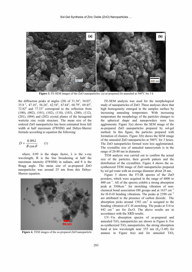
FE-SEM analysis was used for the morphologicalstudy of nanoparticles of ZnO. These analyses show thathigh homogeneity emerged in the samples surface byincreasing annealing temperature. With increasingtemperature the morphology of the particles changes tothe spherical shape and nanopowders were lessagglomerate. Figure 3(a) shows the SEM image of theas-prepared ZnO nanoparticles prepared by sol-gelmethod. In this figure, the particles prepared withformation of clusters. Figure 3(b) shows the SEM imageof the annealed ZnO nanoparticles at 500oC for 3 hours.The ZnO nanoparticles formed were less agglomerated.The crystallite size of annealed nanocrystals is in therange of 20-80 nm in diameter.
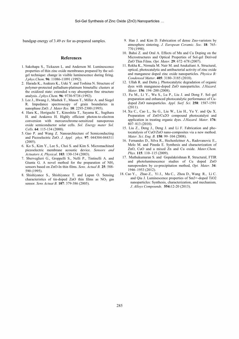
TEM analysis was carried out to confirm the actualsize of the particles, their growth pattern and thedistribution of the crystallites. Figure 4 shows the as-synthesized TEM image of ZnO nanoparticles preparedby sol gel route with an average diameter about 28 nm.
Figure 5 shows the FT-IR spectra of the ZnOpowders, which were acquired in the range of 4000 to400 cm−1. All of the spectra exhibit a strong absorptionpeak at 3508cm−1 for stretching vibration of non-chemical bond association OH groups and at 1637 cm−1
for H-O-H bending vibrations. The peaks at 2390 cm−1
are attributed to the presence of carbon dioxide. Theabsorption picks around 1392 cm-1 is assigned to thebending vibration of C-H stretching. The peaks at 514 to442 cm−1 are for Zn-O. The above results are inaccordance with the XRD results.
UV–Vis absorption spectra of as-prepared andannealed TiO2 nanoparticles are shown in Figure 6. Foras-synthesized TiO2 nanoparticles, the strong absorptionband at low wavelength near 355 nm (Eg=3.49) foranatase in Figure 6(a) and for annealed TiO2
Figure 3. FE-SEM images of the ZnO nanoparticles: (a) as-prepared (b) annealed at 5000C for 3 h
Figure 4. TEM images of the as-prepared ZnO nanoparticle
Vol. 26 No. 3 Summer 2015 S. Jurablu, et al. J. Sci. I. R. Iran
284
nanoparticles the strong absorption band at lowwavelength near 410 nm (Eg=3.02) for rutile phase inFigure 6(b) indicate the presence of phase transition ofanatase to rutile for TiO2 nanoparticles under heattreatment at 500oC [18]. The absorption edge extends tolonger wavelengths for TiO2 nanoparticles, andabsorption tail in the visible-light region over 350–500nm and strong absorption band in the UV-light region isclearly observed. It indicates the absorption positionsdepend on the morphologies and sizes of ZnO. The UVabsorption ability of ZnO is related with band gapenergy. The UV-absorption edge provides a reliableestimate of the band gap of any system.
DiscussionIn summary, we have analyzed the composition of
ZnO particles synthesized by a new sol gel methodusing ZnSO4 ·7H2O and diethylene glycol as surfactant.The XRD and TEM results show that thesenanoparticles are hexagonal wurtzite in phase ZnO witha mean grain size of about 28 nm. From SEM images, itis clear that with increasing temperature the morphologyof the particles changes to the spherical shape andnanopowders were less agglomerate. TEM imageexhibits that the as-synthesized ZnO nanoparticlesprepared by sol gel route with an average diameterabout 28 nm. From the FTIR data, it is shown thepresence of Zn-O stretching mode of ZnO. Theabsorbance peak of UV-Vis spectrum showed the wide
Figure 5. The FTIR pattern of the zinc oxide nanoparticles
Figure 6. UV–Vis absorption spectra of TiO2 nanoparticles: (a) as-prepared and (b) annealed samples
Sol-Gel Synthesis of Zinc Oxide (ZnO) Nanoparticles …
285
bandgap energy of 3.49 ev for as-prepared samples.
References
1. Sakohapa S., Tickazen L. and Anderson M. Luminescenceproperties of thin zinc oxide membranes prepared by the sol-gel technique: change in visible luminescence during firing.J.phys.Chem. 96: 11086-11091 (1992).
2. Harada K., Asakura K., Ueki Y. and Toshina N. Structure ofpolymer-protected palladium-platinum bimetallic clusters atthe oxidized state: extended x-ray absorption fine structureanalysis. J.phys.Chem. 96: 9730-9738 (1992).
3. Lee J., Hwang J., Mashek T., Mason T., Miller A. and SiegelR. Impedance spectroscopy of grain boundaries innanophase ZnO. J. Mater Res. 10: 2295-2300 (1995).
4. Hara K., Horiguchi T., Kinoshita T., Sayama K., SugiharaH. and Arakawa H. Highly efficient photon-to-electronconversion with mercurochrome-sensitized nanoporousoxide semiconductor solar cells. Sol. Energy mater Sol.Cells. 64: 115-134 (2000).
5. Gao P. and Wang Z. Nanoarchtecture of Semiconductingand Piezoelectric ZnO. J. Appl. phys. 97: 044304-044311(2005).
6. Ko S., Kim Y., Lee S., Choi S. and Kim S. Micromachinedpiezoelectric membrane acoustic device. Sensors andActuators A, Physical. 103: 130-134 (2003).
7. Sberveglieri G., Groppelli S., Nelli P., Tintinelli A. andGiunta G. A novel method for the preparation of NH3sensors based on ZnO-In thin films. Sens. Actual B. 25: 588-590 (1995).
8. Shishiyance S., Shishiyance T. and Lupan O. Sensingcharacteristics of tin-doped ZnO thin films as NO2 gassensor. Sens Actuat B. 107: 379-386 (2005).
9. Han J. and Kim D. Fabrication of dense Zno-varistors byatmosphere sintering. J. European Ceramic. Soc. 18: 765-770 (1998).
10. Bahsi Z. and Oral A. Effects of Mn and Cu Doping on theMicrostructures and Optical Properties of Sol-gel DerivedZnO Thin Films. Opt. Mater. 29: 672–678 (2007).
11. Rekha K., Nirmala M. Nair M. and Anukaliani A. Structural,optical, photocatalytic and antibacterial activity of zinc oxideand manganese doped zinc oxide nanoparticles. Physica B:Condensed Matter. 405: 3180–3185 (2010).
12. Ullah R. and Dutta j. Photocatalytic degradation of organicdyes with manganese-doped ZnO nanoparticles. J.Hazard.Mater. 156: 194–200 (2008).
13. Fu M., Li Y., Wu S., Lu P., Liu J. and Dong F. Sol–gelpreparation and enhanced photocatalytic performance of Cu-doped ZnO nanoparticles. Appl. Surf. Sci. 258: 1587-1591(2011).
14. Xu C., Cao L., Su G., Liu W., Liu H., Yu Y. and Qu X.Preparation of ZnO/Cu2O compound photocatalyst andapplication in treating organic dyes. J.Hazard. Mater. 176:807–813 (2010).
15. Liu Z., Deng J., Deng J. and Li F. Fabrication and pho-tocatalysis of CuO/ZnO nano-composites via a new method.Mater. Sci. Eng. B. 150: 99–104 (2008).
16. Fernandes D., Silva R., Hechenleitner A., Radovanovic E.,Melo M. and Pineda E. Synthesis and characterization ofZnO, CuO and a mixed Zn and Cu oxide. Mater.Chem.Phys. 115: 110–115 (2009).
17. Muthukumaran S. and Gopalakrishnan R. Structural, FTIRand photoluminescence studies of Cu doped ZnOnanopowders by co-precipitation method, Opt. Mater. 34:1946–1953 (2012).
18. Cao Y., Zhao Z., Yi J., Ma C., Zhou D., Wang R., Li C.and Qiu J. Luminescence properties of Sm3+-doped TiO2nanoparticles: Synthesis, characterization, and mechanism,J. Alloys Compounds. 554:12-20 (2013).