2028 ’ ACCOUNTS OF CHEMICAL RESEARCH ’ 2028–2036 ’ 2013 ’ Vol. 46, No. 9 Published on the Web 07/23/2013 www.pubs.acs.org/accounts 10.1021/ar300292p & 2013 American Chemical Society Solid-State NMR Approaches to Internal Dynamics of Proteins: From Picoseconds to Microseconds and Seconds ALEXEY KRUSHELNITSKY, DETLEF REICHERT, AND KAY SAALW € ACHTER* Institut f€ ur Physik NMR, Martin-Luther-Universit € at Halle-Wittenberg, Betty-Heimann-Str. 7, D-06120 Halle (Saale), Germany RECEIVED ON OCTOBER 23, 2012 CONSPECTUS S olid-state nuclear magnetic resonance (NMR) spectroscopy has matured to the point that it is possible to determine the structure of proteins in immobilized states, such as within microcrystals or embedded in membranes. Currently, researchers continue to develop and apply NMR techniques that can deliver site-resolved dynamic information toward the goal of understanding protein function at the atomic scale. As a widely-used, natural approach, researchers have mostly measured longitudinal (T 1 ) relaxation times, which, like in solution-state NMR, are sensitive to picosecond and nanosecond motions, and motionally averaged dipolar couplings, which provide an integral amplitude of all motions with a correlation time of up to a few microseconds. While overall Brownian tumbling in solution mostly precludes access to slower internal dynamics, dedicated solid- state NMR approaches are now emerging as powerful new options. In this Account, we give an overview of the classes of solid-state NMR experiments that have expanded the accessible range correlation times from microseconds to many milliseconds. The measurement of relaxation times in the rotating frame, T 1F , now allows researchers to access the microsecond range. Using our recent theoretical work, researchers can now quantitatively analyze this data to distinguish relaxation due to chemical-shift anisotropy (CSA) from that due to dipoledipole couplings. Off-resonance irradiation allows researchers to extend the frequency range of such experiments. We have built multidimensional analogues of T 2 -type or line shape experiments using variants of the dipolar-chemical shift correlation (DIPSHIFT) experiment that are particularly suited to extract intermediate time scale motions in the millisecond range. In addition, we have continuously improved variants of exchange experiments, mostly relying on the recoupling of anisotropic interactions to address ultraslow motions in the ms to s ranges. The NH dipolar coupling offers a useful probe of local dynamics, especially with proton-depleted samples that suppress the adverse effect of strong proton dipolar couplings. We demonstrate how these techniques have provided a concise picture of the internal dynamics in a popular model system, the SH3 domain of r-spectrin. T 1 -based methods have shown that large-amplitude bond orientation fluctuations in the picosecond range and slower 10 ns low-amplitude motions coexist in these structures. When we include T 1F data, we observe that many residues undergo low amplitude motions slower than 100 ns. On the millisecond to second scale, mostly localized but potentially cooperative motions occur. Comparing different exchange experiments, we found that terminal NH 2 groups in side chains can even undergo a combination of ultraslow large-angle two-site jumps accompanied by small-angle fluctuations that occur 10 times more quickly.
Transcript
2028 ’ ACCOUNTS OF CHEMICAL RESEARCH ’ 2028–2036 ’ 2013 ’ Vol. 46, No. 9 Published on the Web 07/23/2013 www.pubs.acs.org/accounts10.1021/ar300292p & 2013 American Chemical Society
Solid-State NMR Approaches to InternalDynamics of Proteins: From Picoseconds
to Microseconds and SecondsALEXEY KRUSHELNITSKY, DETLEF REICHERT,
AND KAY SAALW€ACHTER*Institut f€ur Physik � NMR, Martin-Luther-Universit€at Halle-Wittenberg,
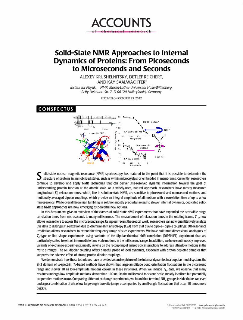
S olid-state nuclear magnetic resonance (NMR) spectroscopy has matured to the point that it is possible to determine thestructure of proteins in immobilized states, such as within microcrystals or embedded in membranes. Currently, researchers
continue to develop and apply NMR techniques that can deliver site-resolved dynamic information toward the goal ofunderstanding protein function at the atomic scale. As a widely-used, natural approach, researchers have mostly measuredlongitudinal (T1) relaxation times, which, like in solution-state NMR, are sensitive to picosecond and nanosecond motions, andmotionally averaged dipolar couplings, which provide an integral amplitude of all motions with a correlation time of up to a fewmicroseconds. While overall Brownian tumbling in solution mostly precludes access to slower internal dynamics, dedicated solid-state NMR approaches are now emerging as powerful new options.
In this Account, we give an overview of the classes of solid-state NMR experiments that have expanded the accessible rangecorrelation times from microseconds to many milliseconds. The measurement of relaxation times in the rotating frame, T1F, nowallows researchers to access the microsecond range. Using our recent theoretical work, researchers can now quantitatively analyzethis data to distinguish relaxation due to chemical-shift anisotropy (CSA) from that due to dipole�dipole couplings. Off-resonanceirradiation allows researchers to extend the frequency range of such experiments. We have built multidimensional analogues ofT2-type or line shape experiments using variants of the dipolar-chemical shift correlation (DIPSHIFT) experiment that areparticularly suited to extract intermediate time scale motions in the millisecond range. In addition, we have continuously improvedvariants of exchange experiments, mostly relying on the recoupling of anisotropic interactions to address ultraslow motions in thems to s ranges. The NH dipolar coupling offers a useful probe of local dynamics, especially with proton-depleted samples thatsuppress the adverse effect of strong proton dipolar couplings.
We demonstrate how these techniques have provided a concise picture of the internal dynamics in a popular model system, theSH3 domain of r-spectrin. T1-based methods have shown that large-amplitude bond orientation fluctuations in the picosecondrange and slower 10 ns low-amplitude motions coexist in these structures. When we include T1F data, we observe that manyresidues undergo low amplitude motions slower than 100 ns. On the millisecond to second scale, mostly localized but potentiallycooperative motions occur. Comparing different exchange experiments, we found that terminal NH2 groups in side chains can evenundergo a combination of ultraslow large-angle two-site jumps accompanied by small-angle fluctuations that occur 10 times morequickly.
Vol. 46, No. 9 ’ 2013 ’ 2028–2036 ’ ACCOUNTS OF CHEMICAL RESEARCH ’ 2029
NMR Approaches to Internal Dynamics of Proteins Krushelnitsky et al.
1. IntroductionConformational transitions within a protein structure repre-
sent the basic processes mediating its function. Therefore,
detailed knowledge of the molecular-level dynamics of
proteins is required for an in-depth understanding of their
biological activity. Proteins are not at all static structures:
driven by fast thermal motion of their structural elements
and within the hydration shell,1 the cooperativity arising
from their polymeric nature leads to dynamics covering a
vast spectral range. Internal motions on the microsecond to
second time scale are of particular interest, as this is the time
scale of many biologically relevant events, such as catalysis,
recognition, folding, and so forth. It is well-known that, in
solution, the direct NMR assessment of such slowmotions is
challenged by the overall Brownian tumbling that averages
anisotropic interactions, and enables observation of mo-
tions on the time scale slower than the Brownian tumbling
only by means of isotropic chemical shift exchange-based
methods. This restriction is lifted in the solid state, and
consequently, developing and applying corresponding
NMR approaches using the named interactions to address
thewhole frequency range of internal protein dynamics is at
the focus of current intense activities.
To date, most studies of site-resolved internal dynamics
in solid proteins have been limited to the picosecond to
microsecond range,2�5 relying mostly on 13C or 15N T1relaxation times. For the lower end of this window and its
extension to still slower dynamics, there is still a consider-
able lack of experimental tools that can provide reliable
quantitative information. In this Account, we review a num-
ber of methodological approaches and applications thereof,
focusing on molecular motions on the microsecond to
second time scales. We place a specific focus on the use of
heteronuclear 13C�1H and in particular 15N�1H dipole�dipole couplings to detect the changes in the respective bond
orientation. Their use is on the one hand convenient, be-
cause these interactions are readily measurable in samples
with the common isotope labeling schemes. The respective
tensor magnitude and orientation is very well-known, re-
quiring no additional knowledge as is for instance the case
for the chemical-shift anisotropy. By way of their magnitude
in the 10�20 kHz range, the effects of XH dipole�dipole
interactions on transverse and rotating-frame relaxation
phenomena are on the other hand particularly sensitive to
slow (more specifically: “intermediate”) motions in the reci-
procal time range, that is, about 1�100 μs. The qualitative
presence of any appreciably faster motions is further identi-
fied by fast-limit motional averaging and thus reduced
couplings, most often quantified by an order parameter
S < 1. Slower motions are the domain of specific exchange
experiments, which can of course also be conducted on the
basis of isotropic chemical-shift changes, which is the other
most useful NMR interaction. 2H NMR based on the quad-
rupolar interaction allows for very detailed insights into
localized dynamics,6 but requires specific isotope labeling
and is thus less attractive as a potential routine tool.
2. New and Not-so-New Solid-State NMRTools to Study Slow Protein Dynamics
2.1. The Fast End of Slow: Spin�Lattice Relaxation in
the Rotating Frame. The measurement of the relaxation
time T1F (or its inverse, the corresponding rate R1F) is a well-
known routine tool for studying molecular motions. The
frequency window of this method is determined by the
nutation frequency of the rf spin-lock field which usually
has a value of several tens of kilohertz. Since the amplitude
of the spin-lock field can be varied within a relatively wide
range, one can measure T1F dispersions, which significantly
enhances the capability of this technique. Here, we shall
discuss a few important challenges and limitations.
In practice, a quantitative interpretation of T1F is often
challenged by the so-called spin�spin (or coherent) contri-
bution to the relaxation rate arising from the additional
relaxation pathway from the rotating frame Zeeman reser-
voir to the lattice through the proton dipolar reservoir.7 It is
further enhanced by (easily avoided) rotational resonance
conditions. The proton-related coherent contribution strongly
depends on the ratio between spin-lock and local dipolar
field: the higher this ratio, the smaller this contribution. For
rigid molecules, this contribution can be quite appreciable
even for relatively strong spin-lock fields, and thus, one
should always be aware of this effect and try to set the spin-
lock field as high as possible. This leads to the self-evident
limitsof theapplicationof strongand long rf pulses: theycanbe
dangerousboth for thehardwareand for thesample.However,
using the resonance offset, the effective amplitude of the spin-
lock pulse can be increased without increasing the rf power.
The alternative approach to suppress the undesired con-
tribution is proton decoupling during the spin-lock pulse
applied on X nuclei.7 This enables decreasing the amplitude
of the spin-lock pulse and thus studying slowermotions. This
type of experiment is a heteronuclear equivalent of the
homonuclear relaxation in the doubly rotating frame.8
However, a theoretical background of this experiment has
not been worked out yet. The H�X dipolar relaxation
mechanism vanishes to first order in this case, but still it is
2030 ’ ACCOUNTS OF CHEMICAL RESEARCH ’ 2028–2036 ’ 2013 ’ Vol. 46, No. 9
NMR Approaches to Internal Dynamics of Proteins Krushelnitsky et al.
effective in higher orders, and the quantitative description of
this is still missing. Another option to suppress the spin�spin
contribution to the relaxation rate is to perform the T1Fexperiments under very fast MAS. At spinning rates of
50�60 kHz and more, the interproton dipolar interaction
is scaled down to a significant degree, and thus the couplings
within the proton dipolar reservoir effectively diminish. The
applicability of this approachhasbeen recently demonstrated9
and reviewed in this issue.10
The spin�spin contribution can also be suppressed by the
replacement of protons by deuterons.11 This alsomakes the
proton dipolar reservoir and the dipole�dipole couplings
within it negligible and enables analyses of T1F relaxation
times of X nuclei at low spin-lock fields.12 This claimmay be
challenged by the observed residual MAS rate dependence
of T1F in such as samples,9,10 yet is on the other hand
convincingly validated by the fact that the proton line width
is only 20�30 Hz at 10�20 kHz spinning in partially deu-
terated proteins,11 while it is still around 150�200 Hz at
60 kHzMAS in a fully protonated protein.13 Since proton line
width is a direct measure of the 1H�1H coupling and hence
the efficiency of the spin�spin contribution, its almost
1 order of magnitude lower value in deuterated proteins
even at moderate MAS renders the contribution safely
negligible. Any residual R1F dependence on the MAS rate
(ωR = 2πνR) can straightforwardly be explained by its addi-
tivity to the effective spin lock fieldωe in the spectral density
functions, J(ωe ( ωR) and J(ωe ( 2ωR), which further depend
on both amplitude and time scale ofmolecularmotion. Also,
rotary resonance effects may play a role.10 Recently, the R1Fequations for the CSA and heteronuclear dipolar relaxation
mechanisms were rigorously derived,14 taking into account
(i) the resonance offset of the spin-lock pulse and (ii) spin-
lock and MAS frequencies of the same order of magnitude.
The latter is especially important for the experiments at high
MAS rates. It is noted that coherent effects at the (relatively
narrow) rotary resonance conditions9 are not included in the
treatment and should thus be avoided.9,14
2.2. Intermediate Dynamics: Motions on the Interaction
Time Scale. Besides T1F experiments, the toolbox of solid-
state NMR contains line shape- and transverse-evolution-
based experiments, and, as highlighted here, separated-local-
field (SLF) methods, in order to address intermediate motions.
Lineshape experiments are among the earliest dynamic solid-
state experiments performed in biological solids.15 They rely
on the partial averaging of the quadrupolar interactions or the
CSA. The sensitive dynamic range is about 2 orders of magni-
tude in correlation time, centered around the inverse of the
interaction constant. For a specifically labeled sample, even a
single static NMR spectrummay already reflect the occurrence
of a dynamic process and its characteristic time. However, the
inherently low signal-to-noise ratio of such experiments, in
combination with the requirement for site-selective isotope
labels, renders line-shape experiments hardly suitable to ob-
tain a comprehensive picture of the dynamics of a protein.
A more economic alternative is to apply separated-local
field (SLF) experiments which provide information on the
(R2) dispersion technique, providing information on isotropic
chemical-shift exchange and being frequently applied in
solution-state NMR studies of conformational dynamics,20
can also be applied to solid proteins.21 In the same refer-
ence, Schanda and co-workers have further demonstrated
that the difference in R2 of XH zero- and double-quantum
coherences is in principle sensitive to intermediate-time
scale changes of not only the isotropic chemical shift, but
also of the CSA tensors of the two involved nuclei. Its
application to a conformational exchange process in micro-
crystalline ubiquitin suggests that in this case, isotropic shift
variations of the involved 15N and 1H nuclei dominate over
reorientations of either of the two CSA tensors.21
2.3. UltraslowMotions: ExchangeNMR. In the exchange
experiment, the precession frequencies of a nucleus are
compared before and after the mixing time tm; see Figure 1.
If a magnetic interaction determining the precession fre-
quency ismodulated bymolecularmotion during themixing
time, then the precession phases are not equal anymore
(φ1 6¼ φ2), and the signal decreases with increasing tm,
permitting the determination of the exchange rate.
Apart from the J-coupling, all other relevant NMR inter-
actions, such as dipolar couplings, the (isotropic and
anisotropic) chemical shift can be modulated by molecular
reorientations and thus can be used in this experiment.
Different modifications of the exchange experiment select
different useful interactions and suppress the remaining
ones. Again, heteronucleus-based detection under high-
resolution MAS conditions is imperative. As 2D exchange
spectroscopy based upon, for example, the observation of
exchange signals among the CSA sideband manifold is not
economic, many different 1D versions became available,
which commonly work with fixed evolution times for the
phases φ1 and φ2, and create a stimulated-echo type of signal
function. One specific variant, the so-called time-reversed
ODESSA experiment, was the first to be applied to solid
proteins.22
Soon after, the development of the now most popular
centerband-only detection of exchange (CODEX) experi-
ment by Schmidt-Rohr and colleagues23 permitted the ac-
curate determination of both exchange rate and amplitude
of motion. This approach was quickly applied to biopoly-
meric systems.24,25 In the original CODEX, the CSA interac-
tion is reintroduced during the encoding periods by a train of
180� pulses applied every half of the MAS rotor period in
analogy to the well-known REDOR pulse sequence; see
Figure 1. Putting one of the 180� pulses to another rf
channel, the CSA-based CODEX can be turned into its dipolar
variant,26,27 enabling a simpler interpretation of the results,
which are now based upon the reorientation of a XH or XY
dipolar tensor. In this case, the cosine and sine contributions
to the stimulated echo (see caption of Figure 1) are asso-
ciated with in-phase and antiphase magnetization terms,
respectively. A last important variant of a MAS exchange
experiment is of course based on isotropic shifts, detecting
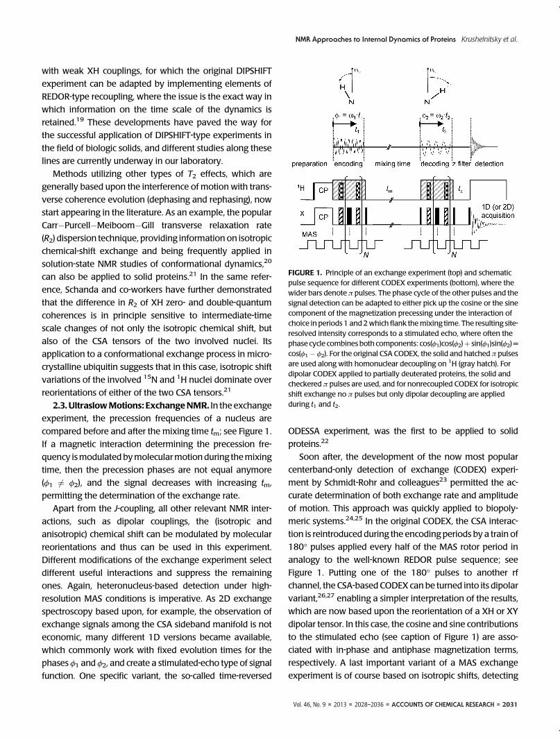
FIGURE 1. Principle of an exchange experiment (top) and schematicpulse sequence for different CODEX experiments (bottom), where thewider bars denote π pulses. The phase cycle of the other pulses and thesignal detection can be adapted to either pick up the cosine or the sinecomponent of the magnetization precessing under the interaction ofchoice in periods 1 and 2which flank themixing time. The resulting site-resolved intensity corresponds to a stimulated echo, where often thephase cycle combines both components: cos(φ1)cos(φ2)þ sin(φ1)sin(φ2) =cos(φ1� φ2). For the original CSA CODEX, the solid and hatched π pulsesare used along with homonuclear decoupling on 1H (gray hatch). Fordipolar CODEX applied to partially deuterated proteins, the solid andcheckered π pulses are used, and for nonrecoupled CODEX for isotropicshift exchange no π pulses but only dipolar decoupling are appliedduring t1 and t2.
2032 ’ ACCOUNTS OF CHEMICAL RESEARCH ’ 2028–2036 ’ 2013 ’ Vol. 46, No. 9
NMR Approaches to Internal Dynamics of Proteins Krushelnitsky et al.
simple chemical exchange, that is, the modulation of the
isotropic chemical shifts by conformational transitions. In
this case, simply no recoupling pulses are applied, but either
one 180� pulse on the proton channel in the middle of the
recoupling periods or conventional dipolar decoupling in
order to removeundesirable J-coupling. The dipolar andCSA
interactions are then averaged out by MAS. This version of
the exchange experimentmay be referred as “nonrecoupled
CODEX”, yet it is fully equivalent to the many well-known
variants of stimulated echo experiments.
There are some important issues for the CODEX variants
and related exchange experiments that deserve someatten-
tion. First, one must distinguish between the exchange
process (i.e., molecular motion) and longitudinal relaxation
that both simultaneously occur during the mixing time. A
correction of the data for T1 relaxation can be done either by
swapping the z storage/filter periods in a pulse sequence of
the type shown in Figure 1,23,27 or via a separate measure-
ment.26 Second, the observed exchange process can be
caused not only by molecular motion but also by spin
diffusion. The latter can be reduced by suitable isotopic
dilution, and/or can be identified and corrected for via its
usually absent temperature dependence. Third, the ex-
change process can also be caused by the so-called RIDER
(relaxation-induced dipolar exchange with recoupling)
effect,28 which is based on the loss of heteronuclear spin
states (antiphasemagnetization) during themixing time due
to the longitudinal relaxation of adjacent, not irradiated but
dipolar-coupled spins (often quadrupolar nuclei such as 14N).
The recipe to suppress this important effect is either dipolar
decoupling from these additional nuclei, or accumulation of
only the cosine (in-phase) components of themagnetization.26
2.4. Tackling Side Effects Related to 1H�1H Dipole�Dipole Couplings. One of the substantial difficulties in the
NMR-characterization of the protein dynamics is proton
driven spin diffusion (PDSD) between 13C/15N nuclei. While
the effect is helpful for structural studies, it is a severe
obstacle in dynamic NMR since (i) PDSD averages the relaxa-
tion rates of various nuclei in a protein if the PDSD rate is
faster or comparable to the relaxation rates, thus deteriorat-
ing the selectivity of the dynamic information; and (ii) in the
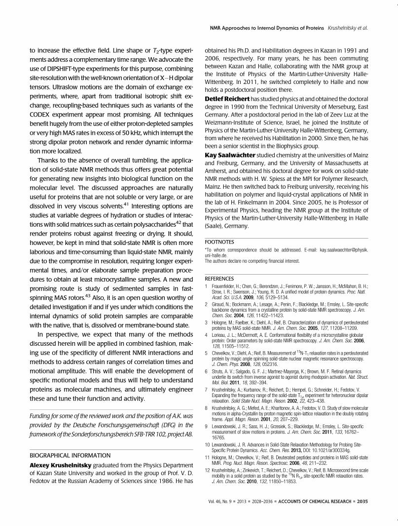
were mainly observed for the terminal NH2 groups of two
Gln residues. The comparison of the dipolar CODEX “ampli-
tudes” anddipolar order parameter data for the sameprotein
presented in Figure 3 shows that the mobilities on the sub-
microsecond (fast-limit averaging) and millisecond to second
time scales are essentially different phenomena: there is no
correlationbetween theamplitudeof such fastmotions and the
presenceof slowermotions. At the same time, CODEX, fast-limit
dipolar order parameters and also R1F data complement each
other, providing a clearer picture of the protein dynamics.37
A comparative analysis of different experiments con-
ductedon the same sample in general canbeavery effective
tool for revealing details of protein dynamics. Herewe stress
the use of the simultaneous analysis of different variants of
the CODEX experiment on the example of the side chain
NH2 group of Gln 50 in SH3. Figure 4 presents the mixing
time dependencies of intensity of the peak in 2D 15N�1H
correlation spectrum assigned to this group for the dipolar
and the nonrecoupled CODEX experiments, the latter being
a simple stimulated echo based upon isotropic shift exchange.
It is seen that both experiments reveal motions of this group in
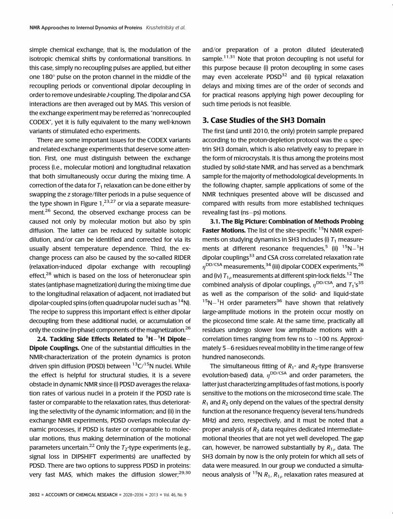
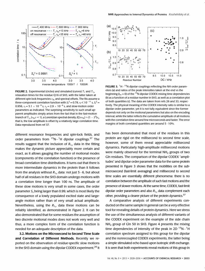
FIGURE 2. Experimental (circles) and simulated (curves) T1 and T1Frelaxation times for the residue Q16 of SH3, with the latter taken atdifferent spin lock frequencies νSL and spectral offsets. The fits assume athree-component correlation function with Sf
2 = 0.78, τf < 10�11 s, Si2 =
0.956, τi = 5.1 � 10�9 s, τs = 2.9 � 10�6 s, and slow-motion orderparameters as indicated. The surprising sensitivity to such small ap-parent amplitudes simply arises from the fact that in the fast-motionbranch of T1F (νSLτ, 1), a Lorentzian spectral density J(2πνSL)≈ (1� S2)τs,that is, the low amplitude is offset by a relatively large correlation time.Data reproduced from ref 37.
FIGURE 3. 1H�15N dipolar couplings reflecting the NH order param-eters (a) and ratios of the peak intensities taken at the end vs thebeginning (tm = 0) of the 15N dipolar CODEXmixing time dependencies(b) as a function of a residue number in SH3, as well as a correlation plotof both quantities (c). The data are taken from refs 26 and 33, respec-tively. The physical meaning of the CODEX intensity ratio is similar to adipolar order parameter, yet it is not fully equivalent since the formerdepends not only on themotional parameters but also on the encodinginterval, while the latter reflects the cumulative amplitude of allmotionswith the correlation time around fewmicroseconds and faster. The errormargins of both correlated quantities are around 5�10%.
2034 ’ ACCOUNTS OF CHEMICAL RESEARCH ’ 2028–2036 ’ 2013 ’ Vol. 46, No. 9
NMR Approaches to Internal Dynamics of Proteins Krushelnitsky et al.
themillisecond time scale, but the correlation times determined
from the exchange decays differ by 1 order of magnitude.
This surprising result can be reasonably explained by
considering that the two experiments are sensitive to differ-
ent types of reorientations and that the NH2 group simply
takes part in two independentmotions: first, a jumplike 180�turn around the C�N bond, and second a faster small-
amplitude reorientation of this bond. The dipolar CODEX
detects the 15N�1H bond reorientation, and thus, the faster
small-amplitude motion cannot reliably be detected before
the background of the 180� jumps because of an insufficient
S/N ratio. On the other hand, isotropic shift exchange is not
sensitive to the 180� jumps since it does not change the
molecular conformation and the isotropic chemical shift of15N remains constant. We stress that without the compara-
tive analysis of two types of exchange experiments the
conclusion on two independent millisecond time scale mo-
tions would be impossible. Note that H/D exchangewith the
D2O reservoir cannot explain the observation, because it
would render the corresponding signal undetectable (the
proton has to be in place before and after tm). Thus, potential
H/D exchange would only decrease the apparent T1 relaxa-
tion time, which is accounted for in the analyses.
Finally, we address the (potential) differences of internal
protein dynamics in the solid and solution states. While the
solution statemight provide the most “realistic” surrounding
of a given functional protein, we stress that not only dedi-
cated solid-state NMR data but also B-factors from X-ray
crystallography have frequently been interpreted as carrying
meaningful dynamic information. Thus, we comment on a
recent critical assessment38 which revealed that there is
essentially no general correlation between B-factors and true
dynamic information fromsolid-stateNMR. The reason is that
B-factors report on variations of atomic coordinates from one
unit cell to the next without time scale information: static
disorder and very fast conformational fluctuations contribute
equally to large B-factors. Another point is that NMR is mainly
sensitive to rotations, which may only involve significant
displacements of weakly scattering hydrogen atoms. B-factors
are thus to be interpreted with great care.
3.3. Solid- versus Solution-State Dynamics. Turning to
more meaningful comparisons of dynamic information from
solution- and solid-state NMR, relaxation experiments have
demonstrated that the parameters of the fast (ps�ns time scale)
internal dynamics of SH3 in the solid and liquid states are
essentially the same.39,40 As for the longer time scale, such a
comparison is impossible because of the isotropic Brownian
tumbling: the motions slower than the tumbling are not ob-
servable using the experiments employing anisotropic interac-
tions. The only option for a direct comparison between the two
is using isotropic chemical shift as a probe for conformational
dynamics, as it is accessible in both the solid and liquid states.
Using the mentioned nonrecoupled CODEX version, we have
conducted such a study for SH3 in both states (to be published
elsewhere), indicating that the millisecond motions are not
identical in the two states. In solution, we observe no millise-
cond motions; however, they can be easily identified for some
residues in the solid state.
These findings are in an agreementwith the recent results
of Schanda and co-workers.21 Using the techniquementioned
in section 2.2, they studied conformational exchange in ubi-
quitin in themicrocrystalline stateand insolution, and revealed
that slow internalmotions aremuch slower in the former. This
means that intermolecular contacts inmicrocrystallineproteins
enhance energy barriers between conformational substates,
which affect the rates of slow internal mobility. The experi-
mental evidence of this phenomenon is still sparse and its
detailed description is not yet available. Still, it is clear that one
should be cautious in generalizing solid-state NMR results for
an interpretation of protein behavior in solution.
4. ConclusionsWe have reviewed a number of established as well as novel
solid-state NMR techniques suited to study internal protein
dynamicson themicrosecond to second range.Rotating-frame
T1F experiments cover the range below microsecond to milli-
seconds, and benefit from off-resonance spin lock irradiation
FIGURE4. Dipolar and nonrecoupled CODEX (see Figure 1)mixing timedependencies of the side-chain 15N of Gln 50 in SH3, conducted at 24 �Cwith encoding periods of 0.8 and8ms, respectively. The exponential fits(lines) and a schematic representation of the two types of motion of theactual NHD group are also shown.
Vol. 46, No. 9 ’ 2013 ’ 2028–2036 ’ ACCOUNTS OF CHEMICAL RESEARCH ’ 2035
NMR Approaches to Internal Dynamics of Proteins Krushelnitsky et al.
to increase the effective field. Line shape or T2-type experi-
ments address a complementary time range.Weadvocate the
use of DIPSHIFT-type experiments for this purpose, combining
dures to obtain at least microcrystalline samples. A new and
promising route is study of sedimented samples in fast-
spinning MAS rotors.43 Also, it is an open question worthy of
detailed investigation if and if yes under which conditions the
internal dynamics of solid protein samples are comparable
with the native, that is, dissolved or membrane-bound state.
In perspective, we expect that many of the methods
discussed herein will be applied in combined fashion, mak-
ing use of the specificity of different NMR interactions and
methods to address certain ranges of correlation times and
motional amplitude. This will enable the development of
specific motional models and thus will help to understand
proteins as molecular machines, and ultimately engineer
these and tune their function and activity.
Funding for some of the reviewed work and the position of A.K. wasprovided by the Deutsche Forschungsgemeinschaft (DFG) in theframeworkof theSonderforschungsbereichSFB-TRR102, project A8.
BIOGRAPHICAL INFORMATION
Alexey Krushelnitsky graduated from the Physics Departmentof Kazan State University and worked in the group of Prof. V. D.Fedotov at the Russian Academy of Sciences since 1986. He has
obtained his Ph.D. and Habilitation degrees in Kazan in 1991 and2006, respectively. For many years, he has been commutingbetween Kazan and Halle, collaborating with the NMR group atthe Institute of Physics of the Martin-Luther-University Halle-Wittenberg. In 2011, he switched completely to Halle and nowholds a postdoctoral position there.
Detlef Reichert has studied physics at and obtained the doctoraldegree in 1990 from the Technical University of Merseburg, EastGermany. After a postdoctoral period in the lab of Zeev Luz at theWeizmann-Institute of Science, Israel, he joined the Institute ofPhysics of the Martin-Luther-University Halle-Wittenberg, Germany,from where he received his Habilitation in 2000. Since then, he hasbeen a senior scientist in the Biophysics group.
Kay Saalw€achter studied chemistry at the universities of Mainzand Freiburg, Germany, and the University of Massachusetts atAmherst, and obtained his doctoral degree for work on solid-stateNMR methods with H. W. Spiess at the MPI for Polymer Research,Mainz. He then switched back to Freiburg university, receiving hishabilitation on polymer and liquid-crystal applications of NMR inthe lab of H. Finkelmann in 2004. Since 2005, he is Professor ofExperimental Physics, heading the NMR group at the Institute ofPhysics of the Martin-Luther-University Halle-Wittenberg in Halle(Saale), Germany.
FOOTNOTES
*To whom correspondence should be addressed. E-mail: [email protected] authors declare no competing financial interest.
REFERENCES1 Frauenfelder, H.; Chen, G.; Berendzen, J.; Fenimore, P. W.; Jansson, H.; McMahon, B. H.;
Stroe, I. R.; Swenson, J.; Young, R. D. A unified model of protein dynamics. Proc. Natl.Acad. Sci. U.S.A. 2009, 106, 5129–5134.
2 Giraud, N.; Bockmann, A.; Lesage, A.; Penin, F.; Blackledge, M.; Emsley, L. Site-specificbackbone dynamics from a crystalline protein by solid-state NMR spectroscopy. J. Am.Chem. Soc. 2004, 126, 11422–11423.
3 Hologne, M.; Faelber, K.; Diehl, A.; Reif, B. Characterization of dynamics of perdeuteratedproteins by MAS solid-state NMR. J. Am. Chem. Soc. 2005, 127, 11208–11209.
4 Lorieau, J. L.; McDermott, A. E. Conformational flexibility of a microcrystalline globularprotein: Order parameters by solid-state NMR spectroscopy. J. Am. Chem. Soc. 2006,128, 11505–11512.
5 Chevelkov, V.; Diehl, A.; Reif, B. Measurement of 15N-T1 relaxation rates in a perdeuteratedprotein by magic angle spinning solid-state nuclear magnetic resonance spectroscopy.J. Chem. Phys. 2008, 128, 052316.
6 Struts, A. V.; Salgado, G. F. J.; Martnez-Mayorga, K.; Brown, M. F. Retinal dynamicsunderlie its switch from inverse agonist to agonist during rhodopsin activation. Nat. Struct.Mol. Biol. 2011, 18, 392–394.
7 Krushelnitsky, A.; Kurbanov, R.; Reichert, D.; Hempel, G.; Schneider, H.; Fedotov, V.Expanding the frequency range of the solid-state T1F experiment for heteronuclear dipolarrelaxation. Solid State Nucl. Magn. Reson. 2002, 22, 423–438.
8 Krushelnitsky, A. G.; Mefed, A. E.; Kharitonov, A. A.; Fedotov, V. D. Study of slow molecularmotions in alpha-Crystallin by proton magnetic spin-lattice relaxation in the doubly rotatingframe. Appl. Magn. Reson. 2001, 20, 207–229.
9 Lewandowski, J. R.; Sass, H. J.; Grzesiek, S.; Blackledge, M.; Emsley, L. Site-specificmeasurement of slow motions in proteins. J. Am. Chem. Soc. 2011, 133, 16762–16765.
10 Lewandowski, J. R. Advances in Solid-State Relaxation Methodology for Probing Site-Specific Protein Dynamics. Acc. Chem. Res. 2013, DOI: 10.1021/ar300334g.
11 Hologne, M.; Chevelkov, V.; Reif, B. Deuterated peptides and proteins in MAS solid-stateNMR. Prog. Nucl. Magn. Reson. Spectrosc. 2006, 48, 211–232.
12 Krushelnitsky, A.; Zinkevich, T.; Reichert, D.; Chevelkov, V.; Reif, B. Microsecond time scalemobility in a solid protein as studied by the 15N R1F site-specific NMR relaxation rates.J. Am. Chem. Soc. 2010, 132, 11850–11853.
2036 ’ ACCOUNTS OF CHEMICAL RESEARCH ’ 2028–2036 ’ 2013 ’ Vol. 46, No. 9
NMR Approaches to Internal Dynamics of Proteins Krushelnitsky et al.
13 Marchetti, A.; Jehle, S.; Felletti, M.; Knight, M. J.; Wang, Y.; Xu, Z.-Q.; Park, A. Y.; Otting,G.; Lesage, A.; Emsley, L.; Dixon, N. E.; Pintacuda, G. Backbone Assignment of FullyProtonated Solid Proteins by 1H Detection and Ultrafast Magic-Angle-Spinning NMRSpectroscopy. Angew. Chem., Int. Ed. 2012, 51, 10756–10759.
14 Kurbanov, R.; Zinkevich, T.; Krushelnitsky, A. The nuclear magnetic resonance relaxationdata analysis in solids: General R1/R1F equations and the model-free approach. J. Chem.Phys. 2011, 135, 184104.
15 Diverdi, J. A.; Opella, S. J. Dynamics of B-DNA in the solid-state. J. Mol. Biol. 1981, 149,307–311.
16 Huster, D.; Xiao, L.; Hong, M. Solid-state NMR investigation of the dynamics of the solubleand membrane-bound colicin Ia channel-forming domain. Biochemistry 2001, 40, 7662–7674.
17 deAzevedo, E. R.; Saalw€achter, K.; Pascui, O.; de Souza, A. A.; Bonagamba, T. J.; Reichert,D. Intermediate motions as studied by solid-state separated local field NMR experiments.J. Chem. Phys. 2008, 128, 104505.
18 Cobo, M. F.; Malinakova, K.; Reichert, D.; Saalw€achter, K.; deAzevedo, E. R. Intermediatemotions and dipolar couplings as studied by Lee-Goldburg cross-polarization NMR:Hartmann-Hahn matching profiles. Phys. Chem. Chem. Phys. 2009, 11, 7036–7047.
19 Cobo, M. F.; Achilles, A.; Reichert, D.; deAzevedo, E. R.; Saalw€achter, K. Recoupledseparated-local-field experiments and applications to study intermediate-regime molecularmotions. J. Magn. Reson. 2012, 221, 85–96.
20 Zeeb, M.; Balbach, J. NMR spectroscopic characterization of millisecond protein folding bytransverse relaxation dispersion measurements. J. Am. Chem. Soc. 2005, 127, 13207–13212.
21 Tollinger, M.; Sivertsen, A. C.; Meier, B. H.; Ernst, M.; Schanda, P. Site-resolvedmeasurement of microsecond-to-millisecond conformational-exchange processes inproteins by solid-state NMR spectroscopy. J. Am. Chem. Soc. 2012, 134, 14800–14807.
22 Krushelnitsky, A. G.; Reichert, D.; Hempel, G.; Fedotov, V. D.; Schneider, H.; Yagodina,L. O.; Schulga, A. A. Superslow backbone protein dynamics as studied by 1D solid-stateMAS exchange NMR spectroscopy. J. Magn. Reson. 1999, 138, 244–255.
23 deAzevedo, E. R.; Hu, W. G.; Bonagamba, T. J.; Schmidt-Rohr, K. Centerband-onlydetection of exchange: Efficient analysis of dynamics in solids by NMR. J. Am. Chem. Soc.1999, 121, 8411–8412.
24 Krushelnitsky, A. G.; Hempel, G.; Reichert, D. Simultaneous processing of solid-state NMRrelaxation and 1D-MAS exchange data: the backbone dynamics of free vs. binase-boundbarstar. Biochim. Biophys. Acta 2003, 1650, 117–127.
25 Reichert, D.; Pascui, O.; deAzededo, E. R.; Bonagamba, T. J.; Arnold, K.; Huster, D. A solid-state NMR study of the fast and slow dynamics of collagen fibrils at varying hydration levels.Magn. Reson. Chem. 2004, 42, 276–284.
26 Krushelnitsky, A.; deAzevedo, E. R.; Linser, R.; Reif, B.; Saalw€achter, K.; Reichert, D. Directobservation of millisecond to second motions in proteins by dipolar CODEX NMRspectroscopy. J. Am. Chem. Soc. 2009, 131, 12097–12099.
27 McDermott, A. E.; Li, W. Characterization of slow conformational dynamics in solids: dipolarCODEX. J. Biomol. NMR 2009, 45, 227–232.
28 Saalw€achter, K.; Schmidt-Rohr, K. Relaxation-induced dipolar exchange with recoupling -an MAS NMR method for determining heteronuclear distances without irradiating thesecond spin. J. Magn. Reson. 2000, 145, 161–172.
29 Krushelnitsky, A.; Br€auniger, T.; Reichert, D. 15N spin diffusion rate in solid-state NMR oftotally enriched proteins: the magic angle spinning frequency effect. J. Magn. Reson. 2006,182, 339–342.
30 Lewandowski, J. R.; Sein, J.; Sass, H. J.; Grzesiek, S.; Blackledge, M.; Emsley, L.Measurement of site-specific 13C spin-lattice relaxation in a crystalline protein. J. Am.Chem. Soc. 2010, 132, 8252–8254.
31 Schanda, P.; Meier, B. H.; Ernst, M. Quantitative analysis of protein backbone dynamics inmicrocrystalline ubiquitin by solid-state NMR spectroscopy. J. Am. Chem. Soc. 2010, 132,15957–15967.
32 Reichert, D.; Hempel, G.; Poupko, R.; Luz, Z.; Olejniczak, Z.; Tekely, P. MAS NMR studies ofcarbon-13 spin exchange in durene. Solid State Nucl. Magn. Reson. 1998, 13, 137–148.
33 Chevelkov, V.; Fink, U.; Reif, B. Accurate determination of order parameters from 1H,15Ndipolar couplings in MAS solid-state NMR experiments. J. Am. Chem. Soc. 2009, 131,14018–14022.
34 Chevelkov, V.; Reif, B. TROSY effects in MAS solid-state NMR. Concepts Magn. Reson.2008, 32A, 143–156.
35 Chevelkov, V.; Fink, U.; Reif, B. Quantitative analysis of backbone motion in proteins usingMAS solid-state NMR spectroscopy. J. Biomol. NMR 2009, 45, 197–206.
36 Chevelkov, V.; Xue, Y.; Linser, R.; Skrynnikov, N. R.; Reif, B. Comparison of solid-statedipolar couplings and solution relaxation data provides insight into protein backbonedynamics. J. Am. Chem. Soc. 2010, 132, 5015–5017.
37 Zinkevich, D.; Chevelkov, V.; Reif, B.; Saalw€achter, K.; Krushelnitsky, A. Internal proteindynamics on ps to μs timescales as studied by multi-frequency 15N solid-state NMRrelaxation. J. Biomol. NMR 2013, submitted.
38 Reichert, D.; Zinkevich, T.; Saalw€achter, K.; Krushelnitsky, A. The relation of the X- rayB-factor to protein dynamics: insights from recent dynamic solid-state NMR data. J. Biomol.Struct. Dyn. 2012, 30, 617.
39 Chevelkov, V.; Zhuravleva, A. V.; Xue, Y.; Reif, B.; Skrynnikov, N. R. Combined analysis of15N relaxation data from solid- and solution-state NMR Spectroscopy. J. Am. Chem. Soc.2007, 129, 12594–12595.
40 Agarwal, V.; Xue, Y.; Reif, B.; Skrynnikov, N. R. Protein side-chain dynamics as observed bysolution- and solid-state NMR spectroscopy: A similarity revealed. J. Am. Chem. Soc. 2008,130, 16611–16621.
41 Mainz, A.; Jehle, S.; van Rossum, B. J.; Oschkinat, H.; Reif, B. Large protein complexes withextreme rotational correlation times investigated in solution by magic-angle-spinning NMRspectroscopy. J. Am. Chem. Soc. 2009, 131, 15968–15969.
42 Hackel, C.; Zinkevich, T.; Belton, P.; Achilles, A.; Reichert, D.; Krushelnitsky, A. Thetrehalose coating effect on the internal protein dynamics. Phys. Chem. Chem. Phys. 2012,14, 2727–2734.
43 Bertini, I.; Luchinat, C.; Parigi, G.; Ravera, E.; Reif, B.; Turano, P. Solid-state NMR of proteinssedimented by ultracentrifugation. Proc. Natl. Acad. Sci. U.S.A. 2011, 108, 10396–10399.