Printed in Great BritainPublished by Bioscientifica Ltd.
Genetic basis of primary aldosteronism
f l fernandes-rosa, s boulkroun and othersReview
R47–R631
59
1
Jou
rnal
of
Mo
lecu
lar
End
ocr
ino
log
y59:
Somatic and inherited mutations in primary aldosteronism
Fabio Luiz Fernandes-Rosa1,2,3,*, Sheerazed Boulkroun1,2,* and Maria-Christina Zennaro1,2,3
1INSERM, UMRS_970, Paris Cardiovascular Research Center, Paris, France2University Paris Descartes, Sorbonne Paris Cité, Paris, France3Assistance Publique-Hôpitaux de Paris, Hôpital Européen Georges Pompidou, Service de Génétique, Paris, France*(F L Fernandes-Rosa and S Boulkroun contributed equally to this work)
Abstract
Primary aldosteronism (PA), the most common form of secondary hypertension, is caused
in the majority of cases by unilateral aldosterone-producing adenoma (APA) or bilateral
adrenal hyperplasia. Over the past few years, somatic mutations in KCNJ5, CACNA1D,
ATP1A1 and ATP2B3 have been proven to be associated with APA development,
representing more than 50% of sporadic APA. The identification of these mutations has
allowed the development of a model for APA involving modification on the intracellular
ionic equilibrium and regulation of cell membrane potential, leading to autonomous
aldosterone overproduction. Furthermore, somatic CTNNB1 mutations have also been
identified in APA, but the link between these mutations and APA development remains
unknown. The sequence of events responsible for APA formation is not completely
understood, in particular, whether a single hit or a double hit is responsible for both
aldosterone overproduction and cell proliferation. Germline mutations identified in
patients with early-onset PA have expanded the classification of familial forms (FH) of
PA. The description of germline KCNJ5 and CACNA1H mutations has identified FH-III and
FH-IV based on genetic findings; germline CACNA1D mutations have been identified
in patients with very early-onset PA and severe neurological abnormalities. This review
summarizes current knowledge on the genetic basis of PA, the association of driver gene
mutations and clinical findings and in the contribution to patient care, plus the current
understanding on the mechanisms of APA development.
Background
Arterial hypertension is a major cardiovascular risk factor with an estimated global age-standardized prevalence of 24.1% in adult males and 20.1% in adult females (NCD-RisC 2017). Detection of secondary hypertension is crucial for targeted management of the underlying disease and prevention of cardiovascular complications. Primary aldosteronism (PA) is the most common form of secondary hypertension with
an estimated prevalence of 5–10% in hypertensive patients (Mulatero et al. 2004, Plouin et al. 2004, Rossi et al. 2006, Douma et al. 2008, Hannemann & Wallaschofski 2012, Hannemann et al. 2012). PA is due to autonomous aldosterone production from the adrenal gland, leading to hypertension with an increased aldosterone-to-renin ratio and on occasion hypokalemia (Funder et al. 2016).
Journal of Molecular Endocrinology (2017) 59, R47–R63
Printed in Great BritainPublished by Bioscientifica Ltd.
R48Review f l fernandes-rosa, s boulkroun and others
Genetic basis of primary aldosteronism
59 1: R48Review
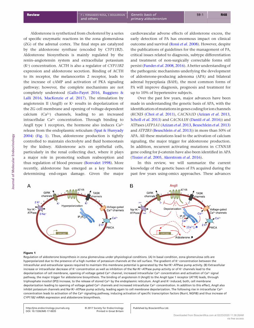
Aldosterone is synthetized from cholesterol by a series of specific enzymatic reactions in the zona glomerulosa (ZG) of the adrenal cortex. The final steps are catalyzed by the aldosterone synthase (encoded by CYP11B2). Aldosterone biosynthesis is mainly regulated by the renin–angiotensin system and extracellular potassium (K+) concentration. ACTH is also a regulator of CYP11B2 expression and aldosterone secretion. Binding of ACTH to its receptor, the melanocortin 2 receptor, leads to the increase of cAMP and activation of PKA signaling pathway; however, the complete mechanisms are not completely understood (Gallo-Payet 2016, Ruggiero & Lalli 2016, MacKenzie et al. 2017). The stimulation by angiotensin II (AngII) or K+ results in depolarization of the ZG cell membrane and opening of voltage-dependent calcium (Ca2+) channels, leading to an increased intracellular Ca2+ concentration. Through binding to AngII type 1 receptors, the hormone also induces Ca2+ release from the endoplasmic reticulum (Spat & Hunyady 2004) (Fig. 1). Thus, aldosterone production is tightly controlled to maintain electrolyte and fluid homeostasis by the kidney. Aldosterone acts on epithelial cells, particularly in the renal collecting duct, where it plays a major role in promoting sodium reabsorption and thus regulation of blood pressure (Bonvalet 1998). More recently, aldosterone has emerged as a key hormone determining end-organ damage. Given the major
cardiovascular adverse effects of aldosterone excess, the early detection of PA has enormous impact on clinical outcome and survival (Rossi et al. 2008). However, despite the publications of guidelines for the management of PA, critical issues related to diagnosis, subtype differentiation and treatment of non-surgically correctable forms still persist (Funder et al. 2008, 2016). A better understanding of the pathogenic mechanisms underlying the development of aldosterone-producing adenoma (APA) and bilateral adrenal hyperplasia (BAH), the most common forms of PA will improve diagnosis, prognosis and treatment for up to 10% of hypertensive subjects.
Over the past few years, major advances have been made in understanding the genetic basis of APA, with the identification of mutations in genes coding for ion channels (KCNJ5 (Choi et al. 2011), CACNA1D (Azizan et al. 2013, Scholl et al. 2013) and CACNA1H (Daniil et al. 2016)) and ATPases (ATP1A1 (Azizan et al. 2013, Beuschlein et al. 2013) and ATP2B3 (Beuschlein et al. 2013)) in more than 50% of APA. All these mutations lead to the activation of calcium signaling, the major trigger for aldosterone production. In addition, recurrent activating mutations in CTNN1B gene coding for β-catenin have also been identified in APA (Tissier et al. 2005, Akerstrom et al. 2016).
In this review, we will summarize the current knowledge of the genetic bases of PA acquired during the past few years using-omics approaches. These advances
Figure 1Regulation of aldosterone biosynthesis in zona glomerulosa under physiological conditions. (A) In basal condition, zona glomerulosa cells are hyperpolarized due to the presence of a high number of potassium channels at the cell surface. The gradient of K+ concentration between the intracellular and extracellular spaces required to maintain this membrane potential is generated by the Na+/K+-ATPase pump activity. (B) Extracellular increase or intracellular decrease of K+ concentration as well as inhibition of the Na+/K+-ATPase pump activity or of K+ channels lead to the depolarization of cell membrane, opening of voltage gated Ca2+ channel, increased intracellular Ca2+ concentration and activation of Ca2+ signal pathway, the major trigger for aldosterone biosynthesis. The binding of angiotensin II (AngII) to the AngII type 1 receptor (AT1R) leads, through triphosphate inositol (IP3) increase, to the release of stored Ca2+ by the endoplasmic reticulum. AngII and K+ induced, both, cell membrane depolarization leading to opening of voltage gated Ca2+ channels and increased intracellular Ca2+ concentration. In addition to this effect, AngII also inhibit potassium channels and Na+/K+-ATPase pump activity, leading again to cell membrane depolarization. The following rise in intracellular Ca2+ concentration leads to activation of the Ca2+ signaling pathway, inducing activation of specific transcription factors (NurrI, NGFIB) and thus increase of CYP11B2 mRNA expression and aldosterone biosynthesis.
Downloaded from Bioscientifica.com at 02/23/2020 11:38:26AMvia free access
Printed in Great BritainPublished by Bioscientifica Ltd.
R49Review f l fernandes-rosa, s boulkroun and others
Genetic basis of primary aldosteronism
PROOF ONLY
have highlighted the major role of the ionic equilibrium and regulation of cell membrane potential in autonomous aldosterone overproduction.
Somatic mutations in APA
Zona glomerulosa cells from the adrenal cortex are maintained in a hyperpolarized state, mainly due to the high expression of potassium channels. The study of mouse models in which the expression of specific potassium channels was invalidated has highlighted their crucial role in adrenal (Davies et al. 2008, Heitzmann et al. 2008, Guagliardo et al. 2012, Penton et al. 2012). Hence, the deletion of TWIK-related acid-sensitive K+ channel (Task)-1 (Task 1) and -3 (Task 3) leads to alteration in the membrane properties of ZG cells, inducing autonomous aldosterone production with clinical features typical of glucocorticoid-remediable aldosteronism (Heitzmann et al. 2008) or BAH (Davies et al. 2008). Task1−/− mice display severe hyperaldosteronism, hypokalemia and arterial low renin hypertension. Interestingly, this hyperaldosteronism was remediable by glucocorticoids due to ectopic expression of AS in ZF before puberty in both male and female and then only in adult female (Heitzmann et al. 2008). In contrast, the phenotype of Task3−/− mice recapitulates low renin and salt-sensitive hypertension (Guagliardo et al. 2012, Penton et al. 2012). Surprisingly, invalidation of both Task 1 and Task 3 in mice has no impact on cellular organization and zonation of the adrenal cortex, although aldosterone production by ZG cells is inappropriate for salt intake, recapitulating features of idiopathic primary aldosteronism (Davies et al. 2008). Invalidation of these different potassium channels leads to hyperaldosteronism due to abnormal depolarization of ZG cell membrane resulting in increased intracellular Ca2+ concentration and stimulation of aldosterone biosynthesis; however, formation of adrenal tumors has never been observed in these models, indicating that other mechanisms are required to promote increased cell proliferation in APA.
Mutations affecting cell membrane potential
The emergence of high throughput sequencing techniques has allowed major advances in the understanding of the genetic basis of many diseases. Thus in 2011, Choi and coworkers identified two different somatic mutations in the KCNJ5 gene in 8 of 22 APA, as well as an inherited mutation in the same gene in a Mendelian form of severe aldosteronism and massive bilateral adrenal hyper-plasia (see below in the Familial forms of PA section)
(Choi et al. 2011). KCNJ5 codes for the G protein-activated inward rectifier potassium channel 4 (GIRK4). The GIRK4 channel belongs to a large family of inwardly rectifying K+ channels. ‘Inward rectifiers’ are a class of K+ channels that conduct much larger inward currents at membrane voltages negative to the K+ equilibrium potential. The GIRK channel family comprises four members (GIRK1 to GIRK 4) that form heterotetramers or homotetramers. They are composed of two membrane-spanning domains (M1 and M2), one pore-forming region (H5) and cytoplasmic N- and C-termini that participate in the pore structure. The first two mutations reported by Choi and coworkers, p.Gly151Arg and p.Leu168Arg, are located near or within the selectivity filter of the channel (Choi et al. 2011). Subsequently, many other studies have reported additional somatic or germline mutations in or surrounding the selectivity filter (from Thr149 to Gly153). All these mutations leads to a loss of K+ selectivity and an increase of sodium (Na+) influx into the cells, producing a chronic depolarization of cell membrane, opening of voltage-gated Ca2+ channels, activation of calcium signaling, increase of aldosterone synthase expression and autonomous aldosterone production by the cells (Fig. 2). Overexpression of the GIRK4 p.Thr158Ala mutant in HAC cells resulted in increased aldosterone, cortisol and 18-oxocortisol levels, as observed in patients harboring this mutation, associated with increased expression of CYP11B2 and CYP11B1 and decreased expression of CYP17, but a decrease of cell proliferation (Oki et al. 2012). Scholl and coworkers also reported a decrease of cell viability in 293T cells overexpressing p.Gly151Glu or p.Gly151Arg (Scholl et al. 2012), due to increased Na+ influx into the cells via the mutated channels.
Figure 2Aldosterone biosynthesis in APA in presence of KCNJ5 mutations. Mutations affecting the GIRK4 potassium channel, encoded by KCNJ5, lead to increase of intracellular Na+ concentration, thus resulting in cell membrane depolarization and opening of voltage gated Ca2+ channel.
Downloaded from Bioscientifica.com at 02/23/2020 11:38:26AMvia free access
Printed in Great BritainPublished by Bioscientifica Ltd.
R50Review f l fernandes-rosa, s boulkroun and others
Genetic basis of primary aldosteronism
59 1: R50Review
This additional effect of KCNJ5 mutations may possibly be a mechanism limiting adrenocortical cell mass growth, but it also strongly suggests that other mechanisms are involved in cell proliferation.
In addition to affecting K+ cell permeability, two KCNJ5 mutations (p.Arg115Trp and p.Glu246Gly) decreased GIRK4 wt currents when coexpressed in HEK cells and altered the membrane abundance of the channels, suggesting that reduced GIRK4 in cell surface may lead to inappropriate hypersecretion of aldosterone and formation of adrenal adenoma (Cheng et al. 2015), although this is not the case for other mutations (Murthy et al. 2012). Although KCNJ5 mRNA expression is not affected by mutational status, at the protein level, decreased GIRK4 expression in APA compared with peritumoral ZG was observed in APA with KCNJ5 mutations compared to non-mutated APA or those with other mutations (Boulkroun et al. 2013, Cheng et al. 2015, Fernandes-Rosa et al. 2015a). KCNJ5 mutations were associated neither with specific adrenal cortex remodeling nor specific transcriptional profiles (Boulkroun et al. 2012, 2013). Slight modifications of gene expression patterns have been reported, such as increased expression of CYP11B2, Visinin-like 1 (VSNL1) or acyl-coA synthetase short-chain family member 3 (ACSS3) in APA harboring KCNJ5 expression (Monticone et al. 2012, Williams et al. 2012b, Zhou et al. 2016). VSNL-1 is a calcium-sensor protein that may protect cells from apoptosis induced by the overexpression of KCNJ5 mutants, counterbalancing to some extent the pro-apoptotic effect of overexpression of mutated GIRK4 channels in APA (Williams et al. 2012a).
More recently, exome sequencing approaches allowed the identification of mutations in ATP1A1, encoding the α1 subunit of the Na+/K+-ATPase in APA (Azizan et al. 2013, Beuschlein et al. 2013). The Na+/K+-ATPase is a member of the P-type ATPase family and is composed of two subunits, α and β. Using the energy derived from ATP hydrolysis, it generates electrochemical gradients for Na+ and K+ across the plasma membrane; hydrolysis of one ATP allows the exit of three Na+ and the entry of two K+, generating an electrogenic gradient. The α-subunit is formed of ten transmembrane domains (M1–M10) with intracellular N- and C-tails. Different somatic variants (p.Gly99Arg (Williams et al. 2014), p.Leu104Arg (Beuschlein et al. 2013), p.Val332Gly (Beuschlein et al. 2013)) or deletions (p.Phe100_Leu104del (Azizan et al. 2013, Beuschlein et al. 2013), p. Met102_Ile106del (Akerstrom et al. 2015), p.Leu103_Leu104del (Akerstrom et al. 2015), p.Phe956_Glu961 del (Akerstrom et al. 2015), p.Phe959_Glu961del (Akerstrom et al. 2015), p.Glu960_Leu964del
(Akerstrom et al. 2015), p.Glu960_Ala963del (Azizan et al. 2013)) were identified in M1, M4 and M9 domains. Residues mutated in M1 and M4 domains are essential for K+ binding and gating of the binding pocket, their mutations leading to a major decrease in Na+ and K+ binding affinity and thus a loss of pump function (Fig. 3) (Beuschlein et al. 2013, Williams et al. 2014). Remarkably, in H295R_S2 cells, although the expression of p.Leu104Arg or the p.Val332Gly mutants leads to cell membrane depolarization and impaired K+ sensitivity, there is no increase in resting Ca2+ activity but intracellular acidification of the cells due to H+ leak. Although this acidification may contribute to the autonomous aldosterone production, specific mechanism remains to be established (Stindl et al. 2015). Where residues are mutated in M9, these amino acids may be of importance for the Na+-specific site III, the Na+-binding site involving specific residues located in M5 (Tyr778), M6 (Gly813 and Thr814) and M9 (Glu 960 and Glu961) (Li et al. 2005, Azizan et al. 2013). Until now, germline mutations in ATP1A1 have never been reported, suggesting that loss of function of this protein would not be compatible with life.
The major trigger of aldosterone biosynthesis in response to increased plasma K+ and angiotensin II is an increase of calcium concentration in ZG cells (see above). Although mutations in KCNJ5 and ATP1A1 affect cell membrane potential, leading to increased intracellular Ca2+ concentration in case of KCNJ5 mutations or disturbance of the pH, in the case of ATP1A1 mutations, mutations
Figure 3Aldosterone biosynthesis in APA in presence of ATP1A1 mutations. Mutations affecting the Na+/K+-ATPase pump, encoded by ATP1A1, lead to increase of intracellular Na+ and H+ concentration, thus resulting in cell membrane depolarization and decrease of intracellular pH.
Downloaded from Bioscientifica.com at 02/23/2020 11:38:26AMvia free access
Printed in Great BritainPublished by Bioscientifica Ltd.
R51Review f l fernandes-rosa, s boulkroun and others
Genetic basis of primary aldosteronism
PROOF ONLY
identified in ATP2B3, CACNA1D and CACNA1H (see below, in the Familial forms of PA and germline mutations section) are directly involved in the control of intracellular calcium equilibrium (Fig. 4).
ATP2B3 codes for the plasma membrane calcium ATPase isoform 3 (PMCA3), a P-type ATPase family, like the Na+/K+-ATPase. PMCA3 is essential to extrude Ca2+ from the cytoplasm and thereby has an essential role in regulating intracellular calcium homeostasis (Di Leva et al. 2008). ATP2B3 is located on chromosome X, and germline mutations have been shown to be responsible for the development of X-linked congenital cerebellar ataxia (Zanni et al. 2012). Similar to the α subunit of the Na+/K+-ATPase, PMCA3 is composed of 10 transmembrane domains, a large cytoplasmic loop and cytoplasmic N- and C-tails. All the mutations identified in ATP2B3 are located in the M4 domain, deletions of at least two amino acids in a specific region between amino acids 425 and 433 (Beuschlein et al. 2013, Dutta et al. 2014, Fernandes-Rosa et al. 2014, Akerstrom et al. 2015, Scholl et al. 2015a, Zheng et al. 2015), a region crucial for the binding of Ca2+ ions but also close to the catalytic core of the pump (Di Leva et al. 2008). Electrophysiological studies have shown that the ATP2B3 p.Leu425_Val426del mutation leads to increased intracellular Ca2+ concentration in transfected human embryonic kidney (HEK-293) cells due to an increased Ca2+ influx and reduced capacity to export Ca2+, suggesting a loss of the physiological pump function (Tauber et al. 2016). In addition to the alteration of Ca2+ equilibrium, ATP2B3 mutations, like KCNJ5 but to a less extent, lead to Na+ and possibly Ca2+ influx into the cells, contributing to increased intracellular Ca2+ concentration also through depolarization of the cell membrane and opening of voltage-gated Ca2+ channel. These alterations
are associated to increased aldosterone production and CYP11B2 expression in adrenocortical cells (Tauber et al. 2016).
CACNA1D encodes the L-type Ca2+ channel α-subunit Cav1.3 (Azizan et al. 2013, Scholl et al. 2013). The channel contains four repeat domains (I–IV), each composed of six transmembrane segments (S1–S6), plus a membrane-associated loop between S5 and S6 (Catterall 2010). The S4 segment is involved in voltage sensing for activation, whereas S5, S6 and the loop between these two segments form the pore lining of the channel (Dutta et al. 2016). Cav1.3 is one of the most abundant calcium channels in normal human adrenal and in APA, predominantly expressed in ZG (Scholl et al. 2013, Daniil et al. 2016). Although Cav3.2, encoded by CACNA1H, has been shown necessary for membrane potential oscillations in ZG cells, the role of Cav1.3 in adrenal is less clear (Hu et al. 2012). In 2013, exome sequencing approaches identified somatic and germline mutations (see below) in CACNA1D (Azizan et al. 2013, Scholl et al. 2013). Nine different mutations were identified, all except two located specifically in S5, S6 or in the intracellular loop between the S5 and S6 segments. Electrophysiological studies showed different changes in channel properties depending on the mutations although all led to increased Ca2+ influx and activation of calcium signaling stimulating aldosterone production (Azizan et al. 2013, Scholl et al. 2013). By sequencing the entire gene, we identified nine additional mutations located in segments S4, S5 and S6, cytoplasmic segments or repeated region III (Fernandes-Rosa et al. 2014). Although the prevalence of the different mutations was not the same, the identification of all these new mutations suggests the necessity of sequencing the entire CACNA1D gene when searching for causative mutations for APA (Fernandes-Rosa et al. 2014).
Mutations affecting β-catenin activation
Activation of Wnt/β-catenin pathway is one of the major features in APA, being found in at least two-thirds of APA (Boulkroun et al. 2011, Berthon et al. 2012, Berthon & Stratakis 2014). The Wnt/β-catenin pathway plays an important role in embryonic development, stem cell maintenance and differentiation in many tissues, especially normal adrenal development and tumorigenesis (El Wakil & Lalli 2011). In the absence of ligand, β-catenin is a part of the axin complex consisting of adenomatous polyposis coli (APC), axin, glycogen synthase kinase-3β and casein kinase-1β. The phosphorylation of β-catenin in this complex results in its ubiquitination and degradation
Figure 4Aldosterone biosynthesis in APA in presence of mutations affecting intracellular calcium balance. Mutations in ATP2B3 coding for PMCA3, CACNA1D, encoding the α subunit of the L-type Ca2+ channel Cav1.3, and CACNA1H, encoding the α subunit of the T-type Ca2+ channel Cav3.2 affect directly intracellular Ca2+ concentrations.
Downloaded from Bioscientifica.com at 02/23/2020 11:38:26AMvia free access
Printed in Great BritainPublished by Bioscientifica Ltd.
R52Review f l fernandes-rosa, s boulkroun and others
Genetic basis of primary aldosteronism
59 1: R52Review
in the proteasome, preventing it from translocating to the nucleus and activating specific Wnt target genes. Wnt/β-catenin activation occurs via binding of ligand Wnt to its specific receptor frizzled resulting in the inhibition of its phosphorylation and its subsequent dissociation from the axin complex, accumulation in the cytoplasm and translocation into the nucleus, where it serves as a transcriptional coactivator of transcription factors of the T-cell factor (TCF)/lymphocyte enhancer factor (LEF) family.
Different mouse models have highlighted the role of this pathway in adrenal development and in aldosterone production. Wnt4−/− mice display abnormal differentiation of the definitive zone of the adrenal cortex and decrease of the number of zona glomerulosa cells leading to a decrease of aldosterone production (Heikkila et al. 2002, Jeays-Ward et al. 2003). The constitutive activation of β-catenin in the adrenal cortex results in adrenocortical zonation defects by ectopic activation of ZG differentiation and inhibition of orthotopic ZF differentiation. With time, these mice develop hyperaldosteronism, similar to that observed in mice expressing a defective APC allele (Bhandaru et al. 2009, Berthon et al. 2010).
Until recently, although the activation of Wnt/β-catenin was common in APA, only rare mutations in CTNN1B gene, coding for β-catenin have been described (Tissier et al. 2005, Tadjine et al. 2008). Different recent studies have reported known mutations in CTNNB1 gene in specific residues involved in the stability of the protein (p.Ser45Phe, p.Ser45Pro and p.Thr41Ala) (Akerstrom et al. 2016, Wu et al. 2017), leading to constitutive activation of the Wnt/β-catenin pathway. A novel frame-shift mutation has also been reported (A39Efs*3) located close to previously described mutation (Scholl et al. 2015a), resulting in an early termination of the protein.
Teo and coworkers reported the case of three women with PA due to an APA with somatic CTNN1B mutations (p.Ser33Cys, pSer45Phe and p.Gly34Arg), two diagnosed during pregnancy and the third after menopause (Teo et al. 2015). Their APA expressed high levels of luteinizing hormone–chorionic gonadotropin receptor (LHCGR) and gonadotropin-releasing hormone receptor (GNRHR), suggesting that pregnancy may reveal underlying PA (Teo et al. 2015). Although these data provide a substantial addition to our understanding of the mechanisms linking CTNNB1 mutation and APA development, the pathophysiology of APA harboring CTNNB1 mutations seems more complex. Previous studies have reported somatic CTNNB1 mutations in APA from male patients or non-pregnant females (Scholl et al. 2015a,
Akerstrom et al. 2016, Wu et al. 2017). Moreover, although GNRHR and LHCGR overexpression is found in more than 40% of APA (Nakamura et al. 2014, Nicolini et al. 2014), CTNNB1 mutations are identified only in 5% (Akerstrom et al. 2016), with immunohistochemical evidence for β-catenin activation observed in about two-thirds of APA (Berthon et al. 2014). Further studies are necessary to establish the complex mechanisms affected by CTNNB1 mutations in APA.
Prevalence and clinical correlates of somatic mutations in APA
Since the identification of KCNJ5 mutations in 2011, many studies have reported the prevalence and the clinical correlates of somatic mutations in APA (Akerstrom et al. 2012, Boulkroun et al. 2012, Azizan et al. 2013, Fernandes-Rosa et al. 2014, Kitamoto et al. 2014, Tauber et al. 2014). A first multicenter European study performed on 380 APA showed a prevalence of 34% of somatic KCNJ5 mutations (Boulkroun et al. 2012). More recently, a second European study performed on 474 APA identified somatic mutations in 54.3% of tumors: KCNJ5 mutations were found in 38% of APA, CACNA1D in 9.3%, ATP1A1 in 5.3% and ATP2B3 in 1.7% (Fernandes-Rosa et al. 2014); a similar prevalence was reported in the American population (Scholl et al. 2015a). The prevalence of somatic CTNNB1 mutations in APA ranged from 2.1% to 5.1 % (Scholl et al. 2015a, Akerstrom et al. 2016, Wu et al. 2017). Differences were observed in the distribution of mutations across the different centers participating in the multicenter European study: the prevalence of CACNA1D mutations was highest in the Paris cohort, whereas ATP2B3 mutation rate was highest in the Munich cohort (Fernandes-Rosa et al. 2014). In Asian populations, KCNJ5 mutations appear to have a higher prevalence, ranging from 59% to 76% (Taguchi et al. 2012, Wang et al. 2015, Wu et al. 2015, Zheng et al. 2015, Hong et al. 2016). A meta-analysis of somatic KCNJ5 mutations, comprising 1636 patients with APA, reported that the overall prevalence of somatic KCNJ5 mutations was 43%, ranging from 63% in Asia to 35% in Europe + the United States + Australia (Lenzini et al. 2015). This contrast in the prevalence could be explained by ethnic background, environmental differences or different selection criteria.
Various studies have tried to establish genotype–phenotype correlations or to find biomarkers to determine the mutational status of APA before surgery. Genotype–phenotype analysis performed in 199 patients from a cohort from Paris have shown that patients with
Downloaded from Bioscientifica.com at 02/23/2020 11:38:26AMvia free access
Printed in Great BritainPublished by Bioscientifica Ltd.
R53Review f l fernandes-rosa, s boulkroun and others
Genetic basis of primary aldosteronism
PROOF ONLY
KCNJ5 mutations are more frequently female, diagnosed younger and with higher minimal plasma potassium concentrations, and the presence of CACNA1D mutations associated with smaller adenomas (Fernandes-Rosa et al. 2014). No association was found between the mutation status and preoperative plasma aldosterone, renin levels, aldosterone-to-renin ratio or number of medications taken before surgery. Replication analyses, performed on APA from two other European cohorts, confirmed the association of KCNJ5 mutations with female sex and younger age of diagnosis (Fernandes-Rosa et al. 2014). The association of KCNJ5 mutations with female sex and younger age was also observed in a meta-analysis study comprising 1636 APA patients from 13 studies (Lenzini et al. 2015), as were higher plasma levels of aldosterone and larger tumors (Lenzini et al. 2015). Cardiovascular complications of aldosterone excess in APA in relation to the mutational status were evaluated in two studies (Kitamoto et al. 2014, Rossi et al. 2014). Left ventricular mass index (LVMI) was found to be higher in patients carrying KCNJ5 mutations (Rossi et al. 2014), and in both studies, LVMI was independently associated with KCNJ5 mutations (Kitamoto et al. 2014, Rossi et al. 2014). Finally, steroid profiles performed on adrenal and peripheral venous plasma samples of 79 APA patients by liquid chromatography–tandem mass spectrometry (LC–MS/MS) established that specific somatic mutations in APAs can be defined by distinct steroid profiles in adrenal vein plasma. This approach may translate to identifying specific steroid signatures in peripheral venous plasma, and be potentially useful in determining the underlying APA genotype (Williams et al. 2016).
An unanswered question concerns the influence of somatic mutations on the management of PA. Higher lateralization indices on adrenal venous sampling (AVS) were found in patients carrying somatic KCNJ5 mutations (Seccia et al. 2012). More recently, Osswald and coworkers observed no difference in steroid gradients during AVS as a function of the mutation status (Osswald et al. 2013). Another difficulty in establishing genotype–phenotype correlations or surrogate biomarkers of the mutational status is APA heterogeneity. Different somatic mutations have been identified in distinct nodules within the same adrenal, including mutations in different genes (Dekkers et al. 2014, Fernandes-Rosa et al. 2015b). Moreover, APA mutations are reported to occur only in aldosterone synthase (AS)-positive regions with two different mutations identified in distinct AS-positive regions within the same nodule (Nanba et al. 2016a),
pointing to the complexity of genetic determinants of APA.
Familial forms of PA and germline mutations
Familial forms are rare causes of PA that account for 1–5% of cases. Different forms displaying Mendelian inheritance have been described: familial hyperaldosteronism type I (FH-I) or glucocorticoid-remediable aldosteronism (GRA), familial hyperaldosteronism type II (FH-II), familial hyperaldosteronism type III (FH-III) with KCNJ5 mutations and familial hyperaldosteronism type IV (FH-IV) due to CACNA1H mutations. Germline KCNJ5, CACNA1D and CACNA1H mutations are also associated with sporadic PA and/or early-onset hypertension due to PA (Scholl et al. 2013, Murthy et al. 2014, Daniil et al. 2016). Furthermore, germline ARMC5 variants have been identified in African American patients with PA (Zilbermint et al. 2015).
Familial hyperaldosteronism type I
FH-I is characterized by clinical and biochemical findings of PA relieved by dexamethasone. The clinical characteristics comprise early and severe hypertension, biochemical abnormalities of PA, increased production of 18-hydroxycortisol and 18-oxocortisol, bilateral adrenal hyperplasia or adrenal nodules (Sutherland et al. 1966, New 1980). FH-I has an autosomal dominant mode of inheritance, accounting for 0.5–1.0% of PA in adult patients (Jackson et al. 2002, Pizzolo et al. 2005, Mulatero et al. 2011a) and for 3% in hypertensive children (Aglony et al. 2011). The genetic etiology is a crossover of the homologous genes coding for AS (CYP11B2) and 11β-hydroxylase (CYP11B1), creating a chimeric gene with the CYP11B1 promoter and CYP11B2 coding sequences juxtaposed. This crossing over leads to an ectopic expression of AS throughout the ZF, with aldosterone being produced not only in isolated cells from ZG (Lifton et al. 1992, Stowasser & Gordon 2000). The description of different crossover patterns of the hybrid gene suggests that mutations arise independently (Dluhy & Lifton 1995). The genetic diagnosis is usually made by Southern blot or long PCR, both techniques with high sensitivity and specificity (Funder et al. 2016).
The Endocrine Society Clinical Practice Guideline suggests that in patients with an onset of confirmed PA earlier than 20 years of age and in those who have a family history of PA or stroke at a young age (<40 years), genetic testing for FH-I should be performed
Downloaded from Bioscientifica.com at 02/23/2020 11:38:26AMvia free access
Printed in Great BritainPublished by Bioscientifica Ltd.
R54Review f l fernandes-rosa, s boulkroun and others
Genetic basis of primary aldosteronism
59 1: R54Review
(Funder et al. 2016). FH-I patients should be treated with glucocorticoid hormones, which suppress ACTH secretion leading to the reduction of aldosterone levels. Low-dose glucocorticoids are sufficient, as only partial suppression of adrenocorticotropin is required to correct hypertension over several years (Stowasser et al. 2000).
Familial hyperaldosteronism type II
FH-II is a non-glucocorticoid-remediable form of familial hyperaldosteronism first described by Gordon and coworkers in 1991 (Gordon et al. 1991), with transmission consistent with an autosomal dominant mode of inheritance (Stowasser & Gordon 2000, 2001), and his prevalence ranging from 1.2% to 6% in adult patients with PA (Stowasser & Gordon 2001, Medeau et al. 2005, Mulatero et al. 2011b, Pallauf et al. 2012). FH-II patients display a familial history of PA and different subtypes may be present within the same family (Stowasser & Gordon 2001, Stowasser 2009, Mulatero et al. 2011b).
The genetic bases of FH-II are not completely understood. No mutations in candidate genes including CYP11B2, AGTR1 and the p53 tumor-suppressor gene have been identified in FH-II patients (Ballantine et al. 1996a,b, Davies et al. 1997, Stowasser & Gordon 2001). On the other hand, some studies have established evidence for an association of a genetic locus on the chromosomal region 7p22 with FH-II in some affected families (Lafferty et al. 2000, So et al. 2005, Sukor et al. 2008). Candidate genes located in this region were sequenced in FH-II patients, including retinoblastoma-associated Kruppel-associated box gene (RBaK), postmeiotic segregation increased 2 (PMS2), guanine nucleotide-binding protein α-12 (GNA12), replication protein A3 (RPA3), zinc finger protein 12 (ZNF12), glucocorticoid-induced transcript 1 (GLCCI1), fascin 1 (FSCN1) and the cAMP-dependent protein kinase type I β-regulatory subunit (PRKAR1B), but no mutations were identified (Medeau et al. 2005, So et al. 2006, Jeske et al. 2008).
FH-II is clinically and biochemically indistinguishable from sporadic forms of PA. With the discovery in the last years of the genetic basis of FH-III and FH-IV (see below), the diagnosis of FH-II is made on the basis of two or more affected family members, without KCNJ5 and CACNA1H mutations. Advances in genetic techniques, with whole-exome and whole-genome sequencing studies, should allow the discovery of genes associated with familial PA and thus change the current definition of FH-II.
Familial hyperaldosteronism type III and germline KCNJ5 mutations
FH-III is a non-glucocorticoid-remediable form of FH first described in a father and two daughters with early-onset severe arterial hypertension and hypokalemia associated with marked hyperaldosteronism and very high levels of the hybrid steroids 18-oxocortisol and 18-hydroxycortisol (Geller et al. 2008). The patients showed massive BAH with bilateral adrenalectomy required to control blood pressure (Geller et al. 2008). The genetic background of FH-III is a mutation in the KCNJ5 gene (p.Thr158Ala) (Choi et al. 2011). This mutation is located near the region responsible for GIRK4 K+ selectivity (GYG motif), leading to a loss in K+ selectivity and increased influx of Na+ into the cytoplasm, resulting in the depolarization of the plasma membrane and opening of voltage-gated Ca2+ channels. The calcium signaling pathway is activated by increased intracellular Ca2+, resulting in an increase of steroidogenic enzyme expression and aldosterone production (Oki et al. 2012).
Different studies have identified germline KCNJ5 mutations in FH-III families with a phenotypic variability, ranging from spironolactone-responsive hyperaldosteronism to massive adrenal hyperplasia with drug-resistant hypertension (Choi et al. 2011, Charmandari et al. 2012, Mulatero et al. 2012, Mussa et al. 2012, Scholl et al. 2012, Monticone et al. 2013, 2015, Adachi et al. 2014, Tong et al. 2016). This variability was attributed to the type of KCNJ5 mutation, with a severe phenotype associated with GIRK4 p.Gly151Arg, p.Thr158Ala, p.Ile157Ser and p.Tyr152Cys mutations, and a milder phenotype associated with KCNJ5 p.Gly151Glu mutations. In vitro studies showed that the milder phenotype in patients carrying the KCNJ5 p.Gly151Glu mutation may be explained by rapid cell lethality due to much higher sodium conductance of the channel preventing adrenal hyperplasia (Scholl et al. 2012). A recent study, however, described a patient with sporadic spironolactone-responsive PA with a germline KCNJ5 mutation previously associated with a severe phenotype (p.Gly151Arg) (Adachi et al. 2014). In the same way, patients carrying the same germline KCNJ5 mutation may display differences in adrenal morphology, even within the same family (Adachi et al. 2014). These differences suggest that the clinical severity in FH-III cannot be defined only on the basis of the type of KCNJ5 mutation.
The complexity of the FH-III phenotype associated with KCNJ5 mutations has been highlighted by two recent
Downloaded from Bioscientifica.com at 02/23/2020 11:38:26AMvia free access
Printed in Great BritainPublished by Bioscientifica Ltd.
R55Review f l fernandes-rosa, s boulkroun and others
Genetic basis of primary aldosteronism
PROOF ONLY
studies. Combined aldosterone and cortisol oversecretion associated with massive bilateral adrenal hyperplasia was described in a patient carrying a germline KCNJ5 mutation (p.Glu145Gln) (Tong et al. 2016). In primary cell culture of the resected hyperplastic adrenal, calcium channel blockers inhibited the secretion of both aldosterone and cortisol and the mRNA expression of steroidogenic enzymes. The authors suggest that the hypersecretion of cortisol might be ascribed to overly large size of the hyperplastic adrenal (with low CYP11B1 expression) and, like aldosterone secretion, may be mediated by voltage-gated Ca2+ channels (Tong et al. 2016). A second study reported that adrenals from patients carrying a germline KCNJ5 mutation (p. p.Thr158Ala) were markedly enlarged with loss of zonation and cells expressing aldosterone synthase and/or 11β-hydroxylase, with and without 17α-hydroxylase, and some expressing all three enzymes (Gomez-Sanchez et al. 2017). GIRK4 was abundantly expressed in almost all cells, suggesting that the mutation not only altered the potassium filter selectivity of the channel, but also a signal that designated specificity for CYP11B2-expressing cells or the presence of a second mutation that abolishes selectivity of steroidogenesis on the basis of adrenal zonation (Gomez-Sanchez et al. 2017).
Germline KCNJ5 mutations have also been suggested to play a role in the common sporadic form of PA, although the functional consequences of these germline mutations are not completely established. Sequencing germline DNA from 251 subjects with sporadic PA, Murthy and coworkers identified three KCNJ5 missense mutations (p.Arg52His, p.Glu246Lys and p.Gly247Arg), located outside the selectivity filter of GIRK4, and a rare SNP (rs7102584, p.Glu282Gln) in 5% of patients (Murthy et al. 2014). The mutations p.Arg52His and p.Glu246Lys altered channel properties and the cells expressing GIRK4 channels carrying the SNP rs7102584 showed reduced viability, whereas channels carrying the p.Gly247Arg mutation were functionally indistinguishable from the wild-type GIRK4 (Murthy et al. 2014).
The role of common KCNJ5 SNPs was also assessed in cohorts of Asian patients with PA and essential hypertension (Li et al. 2013, Wang et al. 2017). Comparing 235 subjects with sporadic PA to 913 subjects with essential hypertension, Li and coworkers showed that in Chinese population, the minor allele of the KCNJ5 SNP rs2604204, located in the KCNJ5 3′-UTR, was more frequent in males with PA (43.4%) than that in males with essential hypertension (33.3%, P = 0.001) (Li et al. 2013), suggesting that KCNJ5 may be involved in the
pathogenesis of sporadic PA in Chinese patients. The same SNP was studied in 229 Chinese patients with newly diagnosed essential hypertension (Wang et al. 2017). KCNJ5 rs2604204 was related to increased plasma aldosterone, plasma renin, AngI and AngII levels in patients with essential hypertension (Wang et al. 2017). The mechanism underlying the association between a KCNJ5 3′-UTR polymorphism and activation of the renin–angiotensin–aldosterone system remains unknown.
Germline CACNA1D mutations in sporadic PA
De novo germline CACNA1D mutations have been described in two children with PASNA (primary aldosteronism, seizures and neurological abnormalities), a syndrome of PA and neuromuscular abnormalities (Scholl et al. 2013). The first subject, without family history of early-onset hypertension or seizures, showed hypertension with biventricular hypertrophy as a newborn, with the diagnostic of aldosteronism (high plasma aldosterone, suppressed renin and hypokalemia) at age 1 month. Neurological abnormalities (cerebral palsy, cortical blindness and complex neuromuscular abnormalities) were observed, with seizures starting at age 7 months. Treatment with a calcium channel blocker normalized blood pressure, with cure of hypertension and PA on follow-up (Korah & Scholl 2015). A germline mutation was identified in exon 8B of CACNA1D (p.Gly403Asp), in a residue also mutated in APA (p.Gly403Arg) (Azizan et al. 2013, Scholl et al. 2013). The second patient, also without family history of early-onset hypertension or seizures, was diagnosed at birth with cerebral palsy and complex seizures, arterial hypertension at the age of 5 years and hypokalemia at the age of 8 years due to hyperaldosteronism. No adrenal abnormality was observed on CT scan. The patient carried a germline CACNA1D mutation (p.Ile770Met) also found in somatic DNA from APA patients.
Familial hyperaldosteronism type IV (FH-IV) and germline CACNA1H mutations
To identify new genetic abnormalities associated to the development of early-onset hypertension due to PA, Scholl and coworkers performed whole-exome sequencing analysis of 40 unrelated patients. A recurrent germline mutation in CACNA1H (encoding the pore-forming α1 subunit of the T-type voltage-dependent calcium channel Cav3.2) (p.Met1549Val) was identified in 5 children with PA before age 10 years (Scholl et al. 2015b). Familial analysis
Downloaded from Bioscientifica.com at 02/23/2020 11:38:26AMvia free access
Printed in Great BritainPublished by Bioscientifica Ltd.
R56Review f l fernandes-rosa, s boulkroun and others
Genetic basis of primary aldosteronism
59 1: R56Review
showed autosomal dominant transmission of the mutation. Differences in clinical presentation (incomplete penetrance) were observed within family members, attributed to age-dependent variability or to genetic or environmental modifiers. Despite the previously described association between epilepsy and autism with CACNA1H mutations (Chen et al. 2003, Splawski et al. 2006, Heron et al. 2007), and the presence of severe neurological abnormalities observed in patients carrying CACNA1D mutations (Scholl et al. 2013), no neurological abnormalities or seizures were described in the patients carrying the CACNA1H p.Met1549Val mutation. However, developmental delay or attention deficit were reported in two mutation carriers (Scholl et al. 2015b). Functional studies demonstrated increased aldosterone production and increased CYP11B2 mRNA expression in cells transfected with Cav3.2 1549Val compared with cells transfected with WT Cav 3.2. In addition, the T-type calcium channel blocker Mibefradil reduced the increased aldosterone production and CYP11B2 expression caused by transfection of WT Cav3.2 and Cav3.2 1549Val in untreated cells, suggesting that T-type calcium channel inhibitors may be an useful treatment option for patients with CACNA1H gain-of-function mutations (Reimer et al. 2016).
Using a similar approach, we identified four germline CACNA1H mutations in PA patients with different phenotypic presentations. A de novo germline CACNA1H mutation (p.Met1549Ile) was identified in a patient with early-onset hypertension and hyperaldosteronism, associated with multiplex development disorders (Daniil et al. 2016). This mutation modifies the same amino acid as the CACNA1H p.Met1549Val variant reported in FH-IV (Scholl et al. 2015b). The germline CACNA1H variation p.Ser196Leu was identified in a patient with family history of HT and PA, diagnosed with HT and PA at 36 (HT) and 51 years of age (PA), and in her sister (HT and PA diagnosed at 35 years of age). A third CACNA1H variant (p.Pro2083Leu) was identified in a PA subject with familial history of PA and his brother. This variant is located in the C-terminal cytoplasmic domain of Cav3.2. Finally, in one patient with APA, a germline CACNA1H missense variant (p.Val1951Glu) was found, located in the C-terminal cytoplasmic domain of Cav3.2, in a region possibly implicated in fast channel activation. Electrophysiological analysis of these four mutant channels demonstrated that the Ca2+ current properties were affected, suggesting a gain-of-function phenotype. Additionally, results from functional studies in adrenocortical H295R-S2 cells
directly linked electrophysiological abnormalities to increased aldosterone production and/or expression of genes coding for steroidogenic enzymes (Daniil et al. 2016). Identification of new CACNA1H mutations associated with early-onset PA and multiplex developmental disorder or with familial forms diagnosed as FH-II or APA, broadens the phenotypic spectrum of PA associated with CACNA1H mutations.
ARMC5 variants
Germline and somatic mutations in the armadillo repeat containing 5 (ARMC5) gene causes hypercortisolism in primary bilateral macronodular adrenal hyperplasia (PBMAH) (Assie et al. 2013). ARMC5 belongs to the large family of armadillo (ARM)-repeat-containing proteins, of which the best-known member is β-catenin and is likely to be a tumor-suppressor gene (Berthon & Stratakis 2014). ARMC5 inactivation appears to affect steroidogenesis. Upon sequencing the ARMC5 gene in germline DNA from 56 PA patients, 12 different ARMC5 variants were identified in 20 unrelated and two related individuals (39%) (Zilbermint et al. 2015); in silico analysis revealed that six of these variants were predicted to be damaging. Interestingly, all carriers of these variants were African American. Tumor DNA sequencing confirmed the presence of the germline mutations but failed to identify additional somatic mutations in ARMC5. Furthermore, the silencing of ARMC5 in H295R cells decreased CYP11B2 expression (Zilbermint et al. 2015). The authors suggested that the increased adrenocortical mass leads to higher aldosterone secretion, despite a reduction of aldosterone-secretory capacity of individual zona glomerulosa cells, as seen with hypercortisolism in patients with PBMAH (Zilbermint et al. 2015). Recently, Mulatero and coworkers reported 18 ARMC5 variants, including 5 rare (allele frequence >1%) and 2 new variants (Mulatero et al. 2016). In silico analysis of these variants, however, did not detect sequence variations that were predicted to alter protein function (Mulatero et al. 2016). The link between ARMC5 mutations and PA is still to be determined.
Molecular and histopathological heterogeneity of APA
As APA may be composed of cells exhibiting characteristics of different zones of the adrenal cortex (Neville & O’Hare 1985, Azizan et al. 2012), the association between APA cellular composition and molecular markers of adrenal
Downloaded from Bioscientifica.com at 02/23/2020 11:38:26AMvia free access
Printed in Great BritainPublished by Bioscientifica Ltd.
R57Review f l fernandes-rosa, s boulkroun and others
Genetic basis of primary aldosteronism
PROOF ONLY
cortex with the mutation status was proposed, although conflictual results were observed among different studies. Some have described that APA harboring KCNJ5 mutations are mainly composed by cells resembling cortisol-secreting cells of the adrenal zona fasciculate (ZF-like) and APA harboring CACNA1D and ATP1A1 mutations are mainly composed by cells resembling the aldosterone-secreting cells of the adrenal zona glomerulosa (ZG-like) (Azizan et al. 2012, 2013). Accordingly to these results, CYP17A1 expression was found to be higher in APA harboring KCNJ5 mutations, suggesting that these adenomas arise from the ZF (Azizan et al. 2012, 2013). However, these results were not replicated in a large cohort of APA with known somatic mutational status. Analyzing histological characteristics of 78 APA, we found that 72% of APA were composed of a majority of ZF-like cells and no difference neither in the cellular composition of APA nor in CYP11B2 expression according to the mutation status was observed (Fernandes-Rosa et al. 2014). Furthermore, we have shown that APA, independently of the cellular composition phenotype, were composed of cells expressing markers of ZG (CYP11B2, Dab2 and CD56), and that both APA and adjacent ZG exhibit histological characteristics of stem/progenitor cells, which may underlie excessive proliferation and APA formation (Boulkroun et al. 2011). We have also shown that Dab2 is coexpressed with CYP11B2 in APA, suggesting that APA is formed by ZG cells acquiring morphological characteristics of ZF cells due to the excess of steroid production (Boulkroun et al. 2010).
Although the direct link between recurrent somatic mutations and aldosterone production is well established, the effects of these mutations on nodulation and cell proliferation remain to be clarified. KCNJ5 germline mutations originally described in FH-III were associated with massive bilateral hyperplasia, suggesting that the KCNJ5 mutation was responsible for increased cell proliferation and autonomous aldosterone production (Choi et al. 2011). On the other hand, patients with PASNA and germline CACNA1D mutations did not exhibit enlarged adrenals or adrenal nodules (Scholl et al. 2013), as also observed for some patients with germline KCNJ5 mutations (Mulatero et al. 2012, Adachi et al. 2014). Furthermore, in vitro studies demonstrated that cells overexpressing a mutant GIRK4 exhibited lower proliferation, or even apoptosis, due to increased intracellular Na+ concentration (Oki et al. 2012, Scholl et al. 2012). Although adrenal nodules can be observed in normal adrenals associated with older age and severity
of hypertension (Wolkersdorfer & Bornstein 1998), we found an increase in nodulation of the cortex adjacent to APA compared with control adrenals (Boulkroun et al. 2010). This raises the possibility that remodeling of the adrenal cortex precedes the development of an APA, and thus, that somatic mutations could be a secondary event in the APA development.
Different studies have provided strong arguments in favor of the hypothesis of independent events leading to nodulation and aldosterone overproduction. Recently, we and others have reported different somatic mutations in different CYP11B2-positive nodules within the same multinodular adrenal (Dekkers et al. 2014, Fernandes-Rosa et al. 2015b). Furthermore, a number of APA exhibited heterogeneity of expression of CYP11B2; among these APAs, recurrent somatic mutations were identified in 3 nodules of the same adrenal, localized only in positive CYP11B2 regions. Interestingly, in one APA, two different mutations were identified in two distinct CYP11B2-positive regions within the same APA (Nanba et al. 2016a). Moreover, the co-existence of two different adrenocortical adenomas within the same adrenal causing the concurrent diagnosis of PA and Cushing syndrome was recently reported (Nanba et al. 2016b). Taken together, all these results suggest that somatic mutations responsible for aldosterone biosynthesis could be a secondary event in APA development. In agreement with this possibility, we have described the concurrence of different germline and somatic mutations leading to APA development (Vouillarmet et al. 2016). A young patient with severe PA aged 26 years, bilateral macronodular adrenal hyperplasia seen on CT, and lateralized aldosterone secretion, showed clinical improvement of PA after right adrenalectomy. The resected adrenal contained three nodules, one expressing aldosterone synthase, with Weiss index of 3, and a somatic KNCJ5 mutation. A FDG-PET performed after surgery revealed abnormal rectal activity despite the absence of clinical symptoms, and gastrointestinal exploration revealed familial adenomatous polyposis. We identified the presence of a germline heterozygous APC gene mutation associated with nodular biallelic APC inactivation due to loss of heterozygosity (Vouillarmet et al. 2016). This case supports a two-hit model for APA formation whereby the first hit (APC germline mutation) drives the nodule development, whereas the second hit (KCNJ5 somatic mutation) specifies the pattern of hormonal secretion.
On the other hand, some authors suggest that APA could be derived from aldosterone-producing cell clusters (APCCs) (Nishimoto et al. 2010). APCCs are composed
Downloaded from Bioscientifica.com at 02/23/2020 11:38:26AMvia free access
Printed in Great BritainPublished by Bioscientifica Ltd.
R58Review f l fernandes-rosa, s boulkroun and others
Genetic basis of primary aldosteronism
59 1: R58Review
of morphological ZG cells in contact with the capsule and inner columnar ZF-like cells forming cords along sinusoids, staining positive for both CYP11B2 and CYP11B1 and found in both normal and PA adrenal tissue (Nishimoto et al. 2010, Boulkroun et al. 2011). The sequencing of DNA from 23 APCCs from normal adrenals showed known aldosterone driver mutations in CACNA1D, ATP1A1 and ATP2B3 in 35% of the APCCs; however, no KCNJ5 mutations, the most frequent alteration observed in APA, were found (Nishimoto et al. 2015). The authors suggest that as APCCs share the same ATP1A1, ATP2B3 and CACNA1D mutations as APA, they may represent a precursor population of cells that lead to APA. The lack of KCNJ5 mutations in APCC may reflect rapid progress of KCNJ5-mutated APCCs to APA, so that KCNJ5 mutations may only be rarely detected in APCCs (Nishimoto et al. 2015).
Conclusion
In the past five years, since the original article from Choi and coworkers (Choi et al. 2011), large advances have been done in the field of genetic causes of PA. Somatic mutations in five genes (KCNJ5, CACNA1D, ATP1A1, ATP2B3 and CTNNB1) are proven to be associated with APA development, explaining more than 50% of sporadic cases. Germline mutations identified in KCNJ5 and CACNA1H have expanded the classification of familial forms of PA, now based on genetic findings. Clinical studies to establish genotype–phenotype correlations and biomarkers associated with recurrent somatic mutations are yet largely unsuccessful; in vitro studies have found pharmacological characteristics of mutant channels, which may provide potential treatment options.
In future studies, larger numbers of samples should progressively explain the genetic basis of the remaining 50% of sporadic APA, as well as the familial forms of PA without genetic defects identified to date. The development of biomarkers or techniques allowing the identification of somatic mutations before surgery may open new options in PA diagnosis and treatment. Whether somatic mutations in APA are responsible for both cellular proliferation and aldosterone production or whether they are independent events leading to aldosterone production occurring in a previously altered adrenal cortex is yet to be verified. Finally, the existence of possible genetic determinants of BAH, as well as the presence of common mechanisms involved in APA and BAH development, is still unknown and constitutes a new field for PA research in future studies.
Declaration of interestThe authors declare that there is no conflict of interest that could be perceived as prejudicing the impartiality of this review.
FundingThis work was funded through institutional support from INSERM and by the Agence Nationale pour la Recherche (ANR Blanc 2011, No.: 11-BSV1 005 03, ANR-13-ISV1-0006-01), the Fondation pour la Recherche Médicale (DEQ20140329556), the Programme Hospitalier de Recherche Clinique (PHRC grant AOM 06179), and by grants from INSERM and Ministère Délégué à la Recherche et des Nouvelles Technologies. The laboratory of Dr Maria-Christina Zennaro is also partner of the H2020 project ENSAT-HT grant n° 633983.
References
Adachi M, Muroya K, Asakura Y, Sugiyama K, Homma K & Hasegawa T 2014 Discordant genotype-phenotype correlation in familial hyperaldosteronism type III with KCNJ5 gene mutation: a patient report and review of the literature. Hormone Research in Paediatrics 82 138–142. (doi:10.1159/000358197)
Aglony M, Martinez-Aguayo A, Carvajal CA, Campino C, Garcia H, Bancalari R, Bolte L, Avalos C, Loureiro C, Trejo P, et al. 2011 Frequency of familial hyperaldosteronism type 1 in a hypertensive pediatric population: clinical and biochemical presentation. Hypertension 57 1117–1121. (doi:10.1161/HYPERTENSIONAHA.110.168740)
Akerstrom T, Crona J, Delgado Verdugo A, Starker LF, Cupisti K, Willenberg HS, Knoefel WT, Saeger W, Feller A, Ip J, et al. 2012 Comprehensive re-sequencing of adrenal aldosterone producing lesions reveal three somatic mutations near the KCNJ5 potassium channel selectivity filter. PLoS ONE 7 e41926. (doi:10.1371/journal.pone.0041926)
Akerstrom T, Willenberg HS, Cupisti K, Ip J, Backman S, Moser A, Maharjan R, Robinson B, Iwen KA, Dralle H, et al. 2015 Novel somatic mutations and distinct molecular signature in aldosterone-producing adenomas. Endocrine-Related Cancer 22 735–744. (doi:10.1530/ERC-15-0321)
Akerstrom T, Maharjan R, Sven Willenberg H, Cupisti K, Ip J, Moser A, Stalberg P, Robinson B, Alexander Iwen K, Dralle H, et al. 2016 Activating mutations in CTNNB1 in aldosterone producing adenomas. Scientific Reports 6 19546. (doi:10.1038/srep19546)
Assie G, Libe R, Espiard S, Rizk-Rabin M, Guimier A, Luscap W, Barreau O, Lefevre L, Sibony M, Guignat L, et al. 2013 ARMC5 mutations in macronodular adrenal hyperplasia with Cushing’s syndrome. New England Journal of Medicine 369 2105–2114. (doi:10.1056/NEJMoa1304603)
Azizan EA, Lam BY, Newhouse SJ, Zhou J, Kuc RE, Clarke J, Happerfield L, Marker A, Hoffman GJ & Brown MJ 2012 Microarray, qPCR, and KCNJ5 sequencing of aldosterone-producing adenomas reveal differences in genotype and phenotype between zona glomerulosa- and zona fasciculata-like tumors. Journal of Clinical Endocrinology and Metabolism 97 E819–E829. (doi:10.1210/jc.2011-2965)
Azizan EA, Poulsen H, Tuluc P, Zhou J, Clausen MV, Lieb A, Maniero C, Garg S, Bochukova EG, Zhao W, et al. 2013 Somatic mutations in ATP1A1 and CACNA1D underlie a common subtype of adrenal hypertension. Nature Genetics 45 1055–1060. (doi:10.1038/ng.2716)
Ballantine DM, Klemm SA, Tunny TJ, Stowasser M & Gordon RD 1996a PCR-SSCP analysis of the p53 gene in tumours of the adrenal gland. Clinical and Experimental Pharmacology and Physiology 23 582–583. (doi:10.1111/j.1440-1681.1996.tb02786.x)
Ballantine DM, Klemm SA, Tunny TJ, Stowasser M & Gordon RD 1996b PCR-SSCP analysis of the promoter region of the renin gene in
Downloaded from Bioscientifica.com at 02/23/2020 11:38:26AMvia free access
Printed in Great BritainPublished by Bioscientifica Ltd.
R59Review f l fernandes-rosa, s boulkroun and others
Genetic basis of primary aldosteronism
PROOF ONLY
patients with aldosterone-producing adenomas. Clinical and Experimental Pharmacology and Physiology 23 584–586. (doi:10.1111/j.1440-1681.1996.tb02787.x)
Berthon A & Stratakis CA 2014 From beta-catenin to ARM-repeat proteins in adrenocortical disorders. Hormone and Metabolic Research 46 889–896. (doi:10.1055/s-0034-1389993)
Berthon A, Sahut-Barnola I, Lambert-Langlais S, de Joussineau C, Damon-Soubeyrand C, Louiset E, Taketo MM, Tissier F, Bertherat J, Lefrancois-Martinez AM, et al. 2010 Constitutive beta-catenin activation induces adrenal hyperplasia and promotes adrenal cancer development. Human Molecular Genetics 19 1561–1576. (doi:10.1093/hmg/ddq029)
Berthon A, Martinez A, Bertherat J & Val P 2012 Wnt/beta-catenin signalling in adrenal physiology and tumour development. Molecular and Cellular Endocrinology 351 87–95. (doi:10.1016/j.mce.2011.09.009)
Berthon A, Drelon C, Ragazzon B, Boulkroun S, Tissier F, Amar L, Samson-Couterie B, Zennaro MC, Plouin PF, Skah S, et al. 2014 WNT/beta-catenin signalling is activated in aldosterone-producing adenomas and controls aldosterone production. Human Molecular Genetics 23 889–905. (doi:10.1093/hmg/ddt484)
Beuschlein F, Boulkroun S, Osswald A, Wieland T, Nielsen HN, Lichtenauer UD, Penton D, Schack VR, Amar L, Fischer E, et al. 2013 Somatic mutations in ATP1A1 and ATP2B3 lead to aldosterone-producing adenomas and secondary hypertension. Nature Genetics 45 440–444, 444e1–444e2. (doi:10.1038/ng.2550)
Bhandaru M, Kempe DS, Rotte A, Rexhepaj R, Kuhl D & Lang F 2009 Hyperaldosteronism, hypervolemia, and increased blood pressure in mice expressing defective APC. American Journal of Physiology: Regulatory, Integrative and Comparative Physiology 297 R571–R575. (doi:10.1152/ajpcell.00087.2009)
Bonvalet JP 1998 Regulation of sodium transport by steroid hormones. Kidney International Supplements 65 S49–S56. (doi:10.1210/jc.82.2.611)
Boulkroun S, Samson-Couterie B, Dzib JF, Lefebvre H, Louiset E, Amar L, Plouin PF, Lalli E, Jeunemaitre X, Benecke A, et al. 2010 Adrenal cortex remodeling and functional zona glomerulosa hyperplasia in primary aldosteronism. Hypertension 56 885–892. (doi:10.1161/HYPERTENSIONAHA.110.158543)
Boulkroun S, Samson-Couterie B, Golib-Dzib JF, Amar L, Plouin PF, Sibony M, Lefebvre H, Louiset E, Jeunemaitre X, Meatchi T, et al. 2011 Aldosterone-producing adenoma formation in the adrenal cortex involves expression of stem/progenitor cell markers. Endocrinology 152 4753–4763. (doi:10.1210/en.2011-1205)
Boulkroun S, Beuschlein F, Rossi GP, Golib-Dzib JF, Fischer E, Amar L, Mulatero P, Samson-Couterie B, Hahner S, Quinkler M, et al. 2012 Prevalence, clinical, and molecular correlates of KCNJ5 mutations in primary aldosteronism. Hypertension 59 592–598. (doi:10.1161/HYPERTENSIONAHA.111.186478)
Boulkroun S, Golib Dzib JF, Samson-Couterie B, Rosa FL, Rickard AJ, Meatchi T, Amar L, Benecke A & Zennaro MC 2013 KCNJ5 mutations in aldosterone producing adenoma and relationship with adrenal cortex remodeling. Molecular and Cellular Endocrinology 371 221–227. (doi:10.1016/j.mce.2013.01.018)
Catterall WA 2010 Signaling complexes of voltage-gated sodium and calcium channels. Neuroscience Letters 486 107–116. (doi:10.1016/j.neulet.2010.08.085)
Charmandari E, Sertedaki A, Kino T, Merakou C, Hoffman DA, Hatch MM, Hurt DE, Lin L, Xekouki P, Stratakis CA, et al. 2012 A novel point mutation in the KCNJ5 gene causing primary hyperaldosteronism and early-onset autosomal dominant hypertension. Journal of Clinical Endocrinology and Metabolism 97 E1532–E1539. (doi:10.1210/jc.2012-1334)
Chen Y, Lu J, Pan H, Zhang Y, Wu H, Xu K, Liu X, Jiang Y, Bao X, Yao Z, et al. 2003 Association between genetic variation of CACNA1H
and childhood absence epilepsy. Annals of Neurology 54 239–243. (doi:10.1002/ana.10607)
Cheng CJ, Sung CC, Wu ST, Lin YC, Sytwu HK, Huang CL & Lin SH 2015 Novel KCNJ5 mutations in sporadic aldosterone-producing adenoma reduce Kir3.4 membrane abundance. Journal of Clinical Endocrinology and Metabolism 100 E155–E163. (doi:10.1210/jc.2014-3009)
Choi M, Scholl UI, Yue P, Bjorklund P, Zhao B, Nelson-Williams C, Ji W, Cho Y, Patel A, Men CJ, et al. 2011 K+ channel mutations in adrenal aldosterone-producing adenomas and hereditary hypertension. Science 331 768–772. (doi:10.1126/science.1198785)
Daniil G, Fernandes-Rosa FL, Chemin J, Blesneac I, Beltrand J, Polak M, Jeunemaitre X, Boulkroun S, Amar L, Strom TM, et al. 2016 CACNA1H mutations are associated with different forms of primary aldosteronism. EBioMedicine 13 225–236. (doi:10.1016/j.ebiom.2016.10.002)
Davies E, Bonnardeaux A, Plouin PF, Corvol P & Clauser E 1997 Somatic mutations of the angiotensin II (AT1) receptor gene are not present in aldosterone-producing adenoma. Journal of Clinical Endocrinology and Metabolism 82 611–615. (doi:10.1210/jcem.82.2.3764)
Davies LA, Hu C, Guagliardo NA, Sen N, Chen X, Talley EM, Carey RM, Bayliss DA & Barrett PQ 2008 TASK channel deletion in mice causes primary hyperaldosteronism. PNAS 105 2203–2208. (doi:10.1073/pnas.0712000105)
Dekkers T, ter Meer M Lenders JW, Hermus AR, Schultze Kool L, Langenhuijsen JF, Nishimoto K, Ogishima T, K Mukai, Azizan EA, et al. 2014 Adrenal nodularity and somatic mutations in primary aldosteronism: one node is the culprit? Journal of Clinical Endocrinology and Metabolism 99 E1341–E1351. (doi:10.1210/jc.2013-4255)
Di Leva F, Domi T, Fedrizzi L, Lim D & Carafoli E 2008 The plasma membrane Ca2+ ATPase of animal cells: structure, function and regulation. Archives of Biochemistry and Biophysics 476 65–74. (doi:10.1016/j.abb.2008.02.026)
Dluhy RG & Lifton RP 1995 Glucocorticoid-remediable aldosteronism (GRA): diagnosis, variability of phenotype and regulation of potassium homeostasis. Steroids 60 48–51. (doi:10.1016/0039-128X(94)00010-A)
Douma S, Petidis K, Doumas M, Papaefthimiou P, Triantafyllou A, Kartali N, Papadopoulos N, Vogiatzis K & Zamboulis C 2008 Prevalence of primary hyperaldosteronism in resistant hypertension: a retrospective observational study. Lancet 371 1921–1926. (doi:10.1016/S0140-6736(08)60834-X)
Dutta RK, Welander J, Brauckhoff M, Walz M, Alesina P, Arnesen T, Soderkvist P & Gimm O 2014 Complementary somatic mutations of KCNJ5, ATP1A1, and ATP2B3 in sporadic aldosterone producing adrenal adenomas. Endocrine-Related Cancer 21 L1–L4. (doi:10.1530/ERC-13-0466)
Dutta RK, Soderkvist P & Gimm O 2016 Genetics of primary hyperaldosteronism. Endocrine-Related Cancer 23 R437–R454. (doi:10.1530/ERC-16-0055)
El Wakil A & Lalli E 2011 The Wnt/beta-catenin pathway in adrenocortical development and cancer. Molecular and Cellular Endocrinology 332 32–37. (doi:10.1016/j.mce.2010.11.014)
Fernandes-Rosa FL, Williams TA, Riester A, Steichen O, Beuschlein F, Boulkroun S, Strom TM, Monticone S, Amar L, Meatchi T, et al. 2014 Genetic spectrum and clinical correlates of somatic mutations in aldosterone-producing adenoma. Hypertension 64 354–361. (doi:10.1161/HYPERTENSIONAHA.114.03419)
Fernandes-Rosa FL, Amar L, Tissier F, Bertherat J, Meatchi T, Zennaro MC & Boulkroun S 2015a Functional histopathological markers of aldosterone producing adenoma and somatic KCNJ5 mutations. Molecular and Cellular Endocrinology 408 220–226. (doi:10.1016/j.mce.2015.01.020)
Fernandes-Rosa FL, Giscos-Douriez I, Amar L, Gomez-Sanchez CE, Meatchi T, Boulkroun S & Zennaro MC 2015b Different somatic mutations in multinodular adrenals with aldosterone-producing
Downloaded from Bioscientifica.com at 02/23/2020 11:38:26AMvia free access
Funder JW, Carey RM, Fardella C, Gomez-Sanchez CE, Mantero F, Stowasser M, Young WF Jr & Montori VM 2008 Case detection, diagnosis, and treatment of patients with primary aldosteronism: an endocrine society clinical practice guideline. Journal of Clinical Endocrinology and Metabolism 93 3266–3281. (doi:10.1210/jc.2008-0104)
Funder JW, Carey RM, Mantero F, Murad MH, Reincke M, Shibata H, Stowasser M & Young WF Jr 2016 The management of primary aldosteronism: case detection, diagnosis, and treatment: an Endocrine Society Clinical Practice Guideline. Journal of Clinical Endocrinology and Metabolism 101 1889–1916. (doi:10.1210/jc.2015-4061)
Gallo-Payet N 2016 60 YEARS OF POMC: Adrenal and extra-adrenal functions of ACTH. Journal of Molecular Endocrinology 56 T135–T156. (doi:10.1530/JME-15-0257)
Geller DS, Zhang J, Wisgerhof MV, Shackleton C, Kashgarian M & Lifton RP 2008 A novel form of human mendelian hypertension featuring nonglucocorticoid-remediable aldosteronism. Journal of Clinical Endocrinology and Metabolism 93 3117–3123. (doi:10.1210/jc.2008-0594)
Gomez-Sanchez CE, Qi X, Gomez-Sanchez EP, Sasano H, Bohlen MO & Wisgerhof M 2017 Disordered zonal and cellular CYP11B2 enzyme expression in familial hyperaldosteronism type 3. Molecular and Cellular Endocrinology 439 74–80. (doi:10.1016/j.mce.2016.10.025)
Gordon RD, Stowasser M, Tunny TJ, Klemm SA, Finn WL & Krek AL 1991 Clinical and pathological diversity of primary aldosteronism, including a new familial variety. Clinical and Experimental Pharmacology and Physiology 18 283–286. (doi:10.1111/j.1440-1681.1991.tb01446.x)
Guagliardo NA, Yao J, Hu C, Schertz EM, Tyson DA, Carey RM, Bayliss DA & Barrett PQ 2012 TASK-3 channel deletion in mice recapitulates low-renin essential hypertension. Hypertension 59 999–1005. (doi:10.1161/HYPERTENSIONAHA.111.189662)
Hannemann A & Wallaschofski H 2012 Prevalence of primary aldosteronism in patient’s cohorts and in population-based studies – a review of the current literature. Hormone and Metabolic Research 44 157–162. (doi:10.1055/s-0031-1295438)
Hannemann A, Bidlingmaier M, Friedrich N, Manolopoulou J, Spyroglou A, Volzke H, Beuschlein F, Seissler J, Rettig R, Felix SB, et al. 2012 Screening for primary aldosteronism in hypertensive subjects: results from two German epidemiological studies. European Journal of Endocrinology 167 7–15. (doi:10.1530/EJE-11-1013)
Heikkila M, Peltoketo H, Leppaluoto J, Ilves M, Vuolteenaho O & Vainio S 2002 Wnt-4 deficiency alters mouse adrenal cortex function, reducing aldosterone production. Endocrinology 143 4358–4365. (doi:10.1210/en.2002-220275)
Heitzmann D, Derand R, Jungbauer S, Bandulik S, Sterner C, Schweda F, El Wakil A, Lalli E, Guy N, Mengual R, et al. 2008 Invalidation of TASK1 potassium channels disrupts adrenal gland zonation and mineralocorticoid homeostasis. EMBO Journal 27 179–187. (doi:10.1038/sj.emboj.7601934)
Heron SE, Khosravani H, Varela D, Bladen C, Williams TC, Newman MR, Scheffer IE, Berkovic SF, Mulley JC & Zamponi GW 2007 Extended spectrum of idiopathic generalized epilepsies associated with CACNA1H functional variants. Annals of Neurology 62 560–568. (doi:10.1002/ana.21169)
Hong AR, Kim JH, Song YS, Lee KE, Seo SH, Seong MW, Shin CS, Kim SW & Kim SY 2016 Genetics of aldosterone-producing adenoma in Korean patients. PLoS ONE 11 e0147590. (doi:10.1371/journal.pone.0147590)
Hu C, Rusin CG, Tan Z, Guagliardo NA & Barrett PQ 2012 Zona glomerulosa cells of the mouse adrenal cortex are intrinsic electrical oscillators. Journal of Clinical Investigation 122 2046–2053. (doi:10.1172/JCI61996)
Jackson RV, Lafferty A, Torpy DJ & Stratakis C 2002 New genetic insights in familial hyperaldosteronism. Annals of the New York Academy of Sciences 970 77–88. (doi:10.1111/j.1749-6632.2002.tb04414.x)
Jeays-Ward K, Hoyle C, Brennan J, Dandonneau M, Alldus G, Capel B & Swain A 2003 Endothelial and steroidogenic cell migration are regulated by WNT4 in the developing mammalian gonad. Development 130 3663–3670. (doi:10.1242/dev.00591)
Jeske YW, So A, Kelemen L, Sukor N, Willys C, Bulmer B, Gordon RD, Duffy D & Stowasser M 2008 Examination of chromosome 7p22 candidate genes RBaK, PMS2 and GNA12 in familial hyperaldosteronism type II. Clinical and Experimental Pharmacology and Physiology 35 380–385. (doi:10.1111/j.1440-1681.2008.04882.x)
Kitamoto T, Suematsu S, Matsuzawa Y, Saito J, Omura M & Nishikawa T 2014 Comparison of cardiovascular complications in patients with and without KCNJ5 gene mutations harboring aldosterone-producing adenomas. Journal of Atherosclerosis and Thrombosis 22 191–200. (doi:10.5551/jat.24455)
Korah HE & Scholl UI 2015 An update on familial hyperaldosteronism. Hormone and Metabolic Research 47 941–946. (doi:10.1055/s-0035-1564166)
Lafferty AR, Torpy DJ, Stowasser M, Taymans SE, Lin JP, Huggard P, Gordon RD & Stratakis CA 2000 A novel genetic locus for low renin hypertension: familial hyperaldosteronism type II maps to chromosome 7 (7p22). Journal of Medical Genetics 37 831–835. (doi:10.1136/jmg.37.11.831)
Lenzini L, Rossitto G, Maiolino G, Letizia C, Funder JW & Rossi GP 2015 A meta-analysis of somatic KCNJ5 K(+) channel mutations in 1636 patients with an aldosterone-producing adenoma. Journal of Clinical Endocrinology and Metabolism 100 E1089–E1095. (doi:10.1210/jc.2015-2149)
Li C, Capendeguy O, Geering K & Horisberger JD 2005 A third Na+-binding site in the sodium pump. PNAS 102 12706–12711. (doi:10.1073/pnas.0505980102)
Li NF, Li HJ, Zhang DL, Zhang JH, Yao XG, Wang HM, Abulikemu S, Zhou KM & Zhang XY 2013 Genetic variations in the KCNJ5 gene in primary aldosteronism patients from Xinjiang, China. PLoS ONE 8 e54051. (doi:10.1371/journal.pone.0054051)
Lifton RP, Dluhy RG, Powers M, Rich GM, Cook S, Ulick S & Lalouel JM 1992 A chimaeric 11beta-hydroxylase aldosterone synthase gene causes glucocorticoid-remediable aldosteronism and human hypertension. Nature 355 262–265. (doi:10.1038/355262a0)
MacKenzie SM, Freel EM, Connell JM, Fraser R & Davies E 2017 ACTH and polymorphisms at steroidogenic loci as determinants of aldosterone secretion and blood pressure. International Journal of Molecular Science [in press]. (doi:10.3390/ijms18030579)
Medeau V, Assie G, Zennaro MC, Clauser E, Plouin PF & Jeunemaitre X 2005 Familial aspect of primary hyperaldosteronism: analysis of families compatible with primary hyperaldosteronism type 2. Annales d’Endocrinologie 66 240–246. (doi:10.1016/S0003-4266(05)81756-1)
Monticone S, Hattangady NG, Nishimoto K, Mantero F, Rubin B, Cicala MV, Pezzani R, Auchus RJ, Ghayee HK, Shibata H, et al. 2012 Effect of KCNJ5 mutations on gene expression in aldosterone-producing adenomas and adrenocortical cells. Journal of Clinical Endocrinology and Metabolism 97 E1567–E1572. (doi:10.1210/jc.2011-3132)
Monticone S, Hattangady NG, Penton D, Isales CM, Edwards MA, Williams TA, Sterner C, Warth R, Mulatero P & Rainey WE 2013 A novel Y152C KCNJ5 mutation responsible for familial hyperaldosteronism type III. Journal of Clinical Endocrinology and Metabolism 98 E1861–E1865. (doi:10.1210/jc.2013-2428)
Monticone S, Bandulik S, Stindl J, Zilbermint M, Dedov I, Mulatero P, Allgaeuer M, Lee CC, Stratakis CA, Williams TA, et al. 2015 A case of severe hyperaldosteronism caused by a de novo mutation affecting a critical salt bridge Kir3.4 residue. Journal of Clinical Endocrinology and Metabolism 100 E114–E118. (doi:10.1210/jc.2014-3636)
Downloaded from Bioscientifica.com at 02/23/2020 11:38:26AMvia free access
Printed in Great BritainPublished by Bioscientifica Ltd.
R61Review f l fernandes-rosa, s boulkroun and others
Genetic basis of primary aldosteronism
PROOF ONLY
Mulatero P, Stowasser M, Loh KC, Fardella CE, Gordon RD, Mosso L, Gomez-Sanchez CE, Veglio F & Young WF Jr 2004 Increased diagnosis of primary aldosteronism, including surgically correctable forms, in centers from five continents. Journal of Clinical Endocrinology and Metabolism 89 1045–1050. (doi:10.1210/jc.2003-031337)
Mulatero P, Tizzani D, Viola A, Bertello C, Monticone S, Mengozzi G, Schiavone D, Williams TA, Einaudi S, La Grotta A, et al. 2011a Prevalence and characteristics of familial hyperaldosteronism: the PATOGEN study (Primary Aldosteronism in TOrino-GENetic forms). Hypertension 58 797–803. (doi:10.1161/HYPERTENSIONAHA. 111.175083)
Mulatero P, Williams TA, Monticone S & Veglio F 2011b Is familial hyperaldosteronism underdiagnosed in hypertensive children? Hypertension 57 1053–1055. (doi:10.1161/HYPERTENSIONAHA. 111.172916)
Mulatero P, Tauber P, Zennaro MC, Monticone S, Lang K, Beuschlein F, Fischer E, Tizzani D, Pallauf A, Viola A, et al. 2012 KCNJ5 mutations in european families with nonglucocorticoid remediable familial hyperaldosteronism. Hypertension 59 235–240. (doi:10.1161/HYPERTENSIONAHA.111.183996)
Mulatero P, Schiavi F, Williams TA, Monticone S, Barbon G, Opocher G & Fallo F 2016 ARMC5 mutation analysis in patients with primary aldosteronism and bilateral adrenal lesions. Journal of Human Hypertension 30 374–378. (doi:10.1038/jhh.2015.98)
Murthy M, Azizan EA, Brown MJ & O’Shaughnessy KM 2012 Characterization of a novel somatic KCNJ5 mutation delI157 in an aldosterone-producing adenoma. Journal of Hypertension 30 1827–1833. (doi:10.1097/HJH.0b013e328356139f)
Murthy M, Xu S, Massimo G, Wolley M, Gordon RD, Stowasser M & O’Shaughnessy KM 2014 Role for germline mutations and a rare coding single nucleotide polymorphism within the KCNJ5 potassium channel in a large cohort of sporadic cases of primary aldosteronism. Hypertension 63 783–789. (doi:10.1161/HYPERTENSIONAHA.113.02234)
Mussa A, Camilla R, Monticone S, Porta F, Tessaris D, Verna F, Mulatero P & Einaudi S 2012 Polyuric-polydipsic syndrome in a pediatric case of non-glucocorticoid remediable familial hyperaldosteronism. Endocrine Journal 59 497–502. (doi:10.1507/endocrj.EJ11-0406)
Nakamura Y, Hattangady NG, Ye P, Satoh F, Morimoto R, Ito-Saito T, Sugawara A, Ohba K, Takahashi K, Rainey WE, et al. 2014 Aberrant gonadotropin-releasing hormone receptor (GnRHR) expression and its regulation of CYP11B2 expression and aldosterone production in adrenal aldosterone-producing adenoma (APA). Molecular and Cellular Endocrinology 384 102–108. (doi:10.1016/j.mce.2014.01.016)
Nanba K, Chen AX, Omata K, Vinco M, Giordano TJ, Else T, Hammer GD, Tomlins SA & Rainey WE 2016a Molecular heterogeneity in aldosterone-producing adenomas. Journal of Clinical Endocrinology and Metabolism 101 999–1007. (doi:10.1210/jc.2015-3239)
Nanba K, Omata K, Tomlins SA, Giordano TJ, Hammer GD, Rainey WE & Else T 2016b Double adrenocortical adenomas harboring independent KCNJ5 and PRKACA somatic mutations. European Journal of Endocrinology 175 K1–K6. (doi:10.1530/EJE-16-0262)
NCD Risk Factor Collaboration (NCD-RisC) 2017 Worldwide trends in blood pressure from 1975 to 2015: a pooled analysis of 1479 population-based measurement studies with 19·1 million participants. Lancet 389 37–55. (doi:10.1016/S0140-6736(16)31919-5)
Neville AM & O’Hare MJ 1985 Histopathology of the human adrenal cortex. Clinics in Endocrinology and Metabolism 14 791–820. (doi:10.1016/S0300-595X(85)80078-5)
New M 1980 Hypertension of childhood with suppressed renin. Endocrine Reviews 1 421–430. (doi:10.1210/edrv-1-4-421)
Nicolini G, Balzan S, Morelli L, Iacconi P, Sabatino L, Ripoli A & Fommei E 2014 LH, progesterone, and TSH can stimulate aldosterone in vitro: a study on normal adrenal cortex and
aldosterone producing adenoma. Hormone and Metabolic Research 46 318–321. (doi:10.1055/s-0033-1358733)
Nishimoto K, Nakagawa K, Li D, Kosaka T, Oya M, Mikami S, Shibata H, Itoh H, Mitani F, Yamazaki T, et al. 2010 Adrenocortical zonation in humans under normal and pathological conditions. Journal of Clinical Endocrinology and Metabolism 95 2296–2305. (doi:10.1210/jc.2009-2010)
Nishimoto K, Tomlins SA, Kuick R, Cani AK, Giordano TJ, Hovelson DH, Liu CJ, Sanjanwala AR, Edwards MA, Gomez-Sanchez CE, et al. 2015 Aldosterone-stimulating somatic gene mutations are common in normal adrenal glands. PNAS 112 E4591–E4599. (doi:10.1073/pnas.1505529112)
Oki K, Plonczynski MW, Luis Lam M, Gomez-Sanchez EP & Gomez-Sanchez CE 2012 Potassium channel mutant KCNJ5 T158A expression in HAC-15 cells increases aldosterone synthesis. Endocrinology 153 1774–1782. (doi:10.1210/en.2011-1733)
Osswald A, Fischer E, Degenhart C, Quinkler M, Bidlingmaier M, Pallauf A, Lang K, Mussack T, Hallfeldt K, Beuschlein F, et al. 2013 Lack of influence of somatic mutations on steroid gradients during adrenal vein sampling in aldosterone-producing adenoma patients. European Journal of Endocrinology 169 657–663. (doi:10.1530/EJE-13-0551)
Pallauf A, Schirpenbach C, Zwermann O, Fischer E, Morak M, Holinski-Feder E, Hofbauer L, Beuschlein F & Reincke M 2012 The prevalence of familial hyperaldosteronism in apparently sporadic primary aldosteronism in Germany: a single center experience. Hormone and Metabolic Research 44 215–220. (doi:10.1055/s-0031-1299730)
Penton D, Bandulik S, Schweda F, Haubs S, Tauber P, Reichold M, Cong LD, El Wakil A, Budde T, Lesage F, et al. 2012 Task3 potassium channel gene invalidation causes low renin and salt-sensitive arterial hypertension. Endocrinology 153 4740–4748. (doi:10.1210/en.2012-1527)
Pizzolo F, Trabetti E, Guarini P, Mulatero P, Ciacciarelli A, Blengio GS, Corrocher R & Olivieri O 2005 Glucocorticoid remediable aldosteronism (GRA) screening in hypertensive patients from a primary care setting. Journal of Human Hypertension 19 325–327. (doi:10.1038/sj.jhh.1001821)
Plouin PF, Amar L & Chatellier G 2004 Trends in the prevalence of primary aldosteronism, aldosterone-producing adenomas, and surgically correctable aldosterone-dependent hypertension. Nephrology Dialysis Transplantation 19 774–777. (doi:10.1093/ndt/gfh112)
Reimer EN, Walenda G, Seidel E & Scholl UI 2016 CACNA1H (M1549V) mutant calcium channel causes autonomous aldosterone production in HAC15 cells and is inhibited by mibefradil. Endocrinology 157 3016–3022. (doi:10.1210/en.2016-1170)
Rossi GP, Bernini G, Caliumi C, Desideri G, Fabris B, Ferri C, Ganzaroli C, Giacchetti G, Letizia C, Maccario M, et al. 2006 A prospective study of the prevalence of primary aldosteronism in 1125 hypertensive patients. Journal of the American College of Cardiology 48 2293–2300. (doi:10.1016/j.jacc.2006.07.059)
Rossi GP, Bolognesi M, Rizzoni D, Seccia TM, Piva A, Porteri E, Tiberio GA, Giulini SM, Agabiti-Rosei E & Pessina AC 2008 Vascular remodeling and duration of hypertension predict outcome of adrenalectomy in primary aldosteronism patients. Hypertension 51 1366–1371. (doi:10.1161/HYPERTENSIONAHA.108.111369)
Rossi GP, Cesari M, Letizia C, Seccia TM, Cicala MV, Zinnamosca L, Kuppusamy M, Mareso S, Sciomer S, Iacobone M, et al. 2014 KCNJ5 gene somatic mutations affect cardiac remodelling but do not preclude cure of high blood pressure and regression of left ventricular hypertrophy in primary aldosteronism. Journal of Hypertension 32 1514–1521; discussion 1522. (doi:10.1097/HJH.0000000000000186)
Ruggiero C & Lalli E 2016 Impact of ACTH signaling on transcriptional regulation of steroidogenic genes. Frontiers in Endocrinology 7 24. (doi:10.3389/fendo.2016.00024)
Downloaded from Bioscientifica.com at 02/23/2020 11:38:26AMvia free access
Printed in Great BritainPublished by Bioscientifica Ltd.
R62Review f l fernandes-rosa, s boulkroun and others
Genetic basis of primary aldosteronism
59 1: R62Review
Scholl UI, Nelson-Williams C, Yue P, Grekin R, Wyatt RJ, Dillon MJ, Couch R, Hammer LK, Harley FL, Farhi A, et al. 2012 Hypertension with or without adrenal hyperplasia due to different inherited mutations in the potassium channel KCNJ5. PNAS 109 2533–2538. (doi:10.1073/pnas.1121407109)
Scholl UI, Goh G, Stolting G, de Oliveira RC, Choi M, Overton JD, Fonseca AL, Korah R, Starker LF, Kunstman JW, et al. 2013 Somatic and germline CACNA1D calcium channel mutations in aldosterone-producing adenomas and primary aldosteronism. Nature Genetics 45 1050–1054. (doi:10.1038/ng.2695)
Scholl UI, Healy JM, Thiel A, Fonseca AL, Brown TC, Kunstman JW, Horne MJ, Dietrich D, Riemer J, Kucukkoylu S, et al. 2015a Novel somatic mutations in primary hyperaldosteronism are related to the clinical, radiological and pathological phenotype. Clinical Endocrinology 83 779–789. (doi:10.1111/cen.12873)
Scholl UI, Stolting G, Nelson-Williams C, Vichot AA, Choi M, Loring E, Prasad ML, Goh G, Carling T, Juhlin CC, et al. 2015b Recurrent gain of function mutation in calcium channel CACNA1H causes early-onset hypertension with primary aldosteronism. eLife 4 e06315. (doi:10.7554/eLife.06315)
Seccia TM, Mantero F, Letizia C, Kuppusamy M, Caroccia B, Barisa M, Cicala MV, Miotto D & Rossi GP 2012 Somatic mutations in the KCNJ5 gene raise the lateralization index: implications for the diagnosis of primary aldosteronism by adrenal vein sampling. Journal of Clinical Endocrinology and Metabolism 97 E2307–E2313. (doi:10.1210/jc.2012-2342)
So A, Duffy DL, Gordon RD, Jeske YW, Lin-Su K, New MI & Stowasser M 2005 Familial hyperaldosteronism type II is linked to the chromosome 7p22 region but also shows predicted heterogeneity. Journal of Hypertension 23 1477–1484. (doi:10.1097/01.hjh.0000174299.66369.26)
So A, Jeske YW, Gordon RD, Duffy D, Kelemen L & Stowasser M 2006 No evidence for coding region mutations in the retinoblastoma-associated Kruppel-associated box protein gene (RBaK) causing familial hyperaldosteronism type II. Clinical Endocrinology 65 829–831. (doi:10.1111/j.1365-2265.2006.02674.x)
Spat A & Hunyady L 2004 Control of aldosterone secretion: a model for convergence in cellular signaling pathways. Physiological Reviews 84 489–539. (doi:10.1152/physrev.00030.2003)
Splawski I, Yoo DS, Stotz SC, Cherry A, Clapham DE & Keating MT 2006 CACNA1H mutations in autism spectrum disorders. Journal of Biological Chemistry 281 22085–22091. (doi:10.1074/jbc.M603316200)
Stindl J, Tauber P, Sterner C, Tegtmeier I, Warth R & Bandulik S 2015 Pathogenesis of adrenal aldosterone-producing adenomas carrying mutations of the Na(+)/K(+)-ATPase. Endocrinology 156 4582–4591. (doi:10.1210/en.2015-1466)
Stowasser M 2009 Update in primary aldosteronism. Journal of Clinical Endocrinology and Metabolism 94 3623–3630. (doi:10.1210/jc.2009-1399)
Stowasser M & Gordon RD 2000 Primary aldosteronism: learning from the study of familial varieties. Journal of Hypertension 18 1165–1176. (doi:10.1097/00004872-200018090-00002)
Stowasser M & Gordon RD 2001 Familial hyperaldosteronism. Journal of Steroid Biochemistry and Molecular Biology 78 215–229. (doi:10.1016/S0960-0760(01)00097-8)
Stowasser M, Bachmann AW, Huggard PR, Rossetti TR & Gordon RD 2000 Treatment of familial hyperaldosteronism type I: only partial suppression of adrenocorticotropin required to correct hypertension. Journal of Clinical Endocrinology and Metabolism 85 3313–3318. (doi:10.1210/jcem.85.9.6834)
Sukor N, Mulatero P, Gordon RD, So A, Duffy D, Bertello C, Kelemen L, Jeske Y, Veglio F & Stowasser M 2008 Further evidence for linkage of familial hyperaldosteronism type II at chromosome 7p22 in Italian as well as Australian and South American families. Journal of Hypertension 26 1577–1582. (doi:10.1097/hjh.0b013e3283028352)
Sutherland DJ, Ruse JL & Laidlaw JC 1966 Hypertension, increased aldosterone secretion and low plasma renin activity relieved by dexamethasone. Canadian Medical Association Journal 95 1109–1119.
Tadjine M, Lampron A, Ouadi L & Bourdeau I 2008 Frequent mutations of beta-catenin gene in sporadic secreting adrenocortical adenomas. Clinical Endocrinology 68 264–270. (doi:10.1111/j.1365-2265.2007. 03033.x)
Taguchi R, Yamada M, Nakajima Y, Satoh T, Hashimoto K, Shibusawa N, Ozawa A, Okada S, Rokutanda N, Takata D, et al. 2012 Expression and mutations of KCNJ5 mRNA in Japanese patients with aldosterone-producing adenomas. Journal of Clinical Endocrinology and Metabolism 97 1311–1319. (doi:10.1210/jc.2011-2885)
Tauber P, Penton D, Stindl J, Humberg E, Tegtmeier I, Sterner C, Beuschlein F, Reincke M, Barhanin J, Bandulik S, et al. 2014 Pharmacology and pathophysiology of mutated KCNJ5 found in adrenal aldosterone-producing adenomas. Endocrinology 155 1353–1362. (doi:10.1210/en.2013-1944)
Tauber P, Aichinger B, Christ C, Stindl J, Rhayem Y, Beuschlein F, Warth R & Bandulik S 2016 Cellular pathophysiology of an adrenal adenoma-associated mutant of the plasma membrane Ca(2+)-ATPase ATP2B3. Endocrinology 157 2489–2499. (doi:10.1210/en.2015-2029)
Teo AE, Garg S, Shaikh LH, Zhou J, Karet Frankl FE, Gurnell M, Happerfield L, Marker A, Bienz M, Azizan EA, et al. 2015 Pregnancy, primary aldosteronism, and adrenal CTNNB1 mutations. New England Journal of Medicine 373 1429–1436. (doi:10.1056/NEJMoa1504869)
Tissier F, Cavard C, Groussin L, Perlemoine K, Fumey G, Hagnere AM, Rene-Corail F, Jullian E, Gicquel C, Bertagna X, et al. 2005 Mutations of beta-catenin in adrenocortical tumors: activation of the Wnt signaling pathway is a frequent event in both benign and malignant adrenocortical tumors. Cancer Research 65 7622–7627. (doi:10.1158/0008-5472.CAN-05-0593)
Tong A, Liu G, Wang F, Jiang J, Yan Z, Zhang D, Zhang Y & Cai J 2016 A novel phenotype of familial hyperaldosteronism type III: concurrence of aldosteronism and Cushing’s syndrome. Journal of Clinical Endocrinology and Metabolism 101 4290–4297. (doi:10.1210/jc.2016-1504)
Vouillarmet J, Fernandes-Rosa F, Graeppi-Dulac J, Lantelme P, Decaussin-Petrucci M, Thivolet C, Peix JL, Boulkroun S, Clauser E & Zennaro MC 2016 Aldosterone-producing adenoma with a somatic KCNJ5 mutation revealing APC-dependent familial adenomatous polyposis. Journal of Clinical Endocrinology and Metabolism 101 3874–3878. (doi:10.1210/jc.2016-1874)
Wang B, Li X, Zhang X, Ma X, Chen L, Zhang Y, Lyu X, Tang Y, Huang Q, Gao Y, et al. 2015 Prevalence and characterization of somatic mutations in Chinese aldosterone-producing adenoma patients. Medicine 94 e708. (doi:10.1097/MD.0000000000000708)
Wang H, Weng C & Chen H 2017 Positive association between KCNJ5 rs2604204 (A/C) polymorphism and plasma aldosterone levels, but also plasma renin and angiotensin I and II levels, in newly diagnosed hypertensive Chinese: a case-control study. Journal of Human Hypertension [in press]. (doi:10.1038/jhh.2016.97)
Williams TA, Monticone S, Crudo V, Warth R, Veglio F & Mulatero P 2012a Visinin-like 1 is upregulated in aldosterone-producing adenomas with KCNJ5 mutations and protects from calcium-induced apoptosis. Hypertension 59 833–839. (doi:10.1161/HYPERTENSIONAHA.111.188532)
Williams TA, Monticone S, Urbanet R, Bertello C, Giraudo G, Vettor R, Fallo F, Veglio F & Mulatero P 2012b Genes implicated in insulin resistance are down-regulated in primary aldosteronism patients. Molecular and Cellular Endocrinology 355 162–168. (doi:10.1016/j.mce.2012.02.007)
Williams TA, Monticone S, Schack VR, Stindl J, Burrello J, Buffolo F, Annaratone L, Castellano I, Beuschlein F, Reincke M, et al. 2014 Somatic ATP1A1, ATP2B3, and KCNJ5 mutations in aldosterone-
Downloaded from Bioscientifica.com at 02/23/2020 11:38:26AMvia free access
Williams TA, Peitzsch M, Dietz AS, Dekkers T, Bidlingmaier M, Riester A, Treitl M, Rhayem Y, Beuschlein F, Lenders JW, et al. 2016 Genotype-specific steroid profiles associated with aldosterone-producing adenomas. Hypertension 67 139–145. (doi:10.1161/HYPERTENSIONAHA.115.06186)
Wolkersdorfer GW & Bornstein SR 1998 Tissue remodelling in the adrenal gland. Biochemical Pharmacology 56 163–171. (doi:10.1016/S0006-2952(98)00067-7)
Wu VC, Huang KH, Peng KY, Tsai YC, Wu CH, Wang SM, Yang SY, Lin LY, Chang CC, Lin YH, et al. 2015 Prevalence and clinical correlates of somatic mutation in aldosterone producing adenoma-Taiwanese population. Scientific Reports 5 11396. (doi:10.1038/srep11396)
Wu VC, Wang SM, Chueh SJ, Yang SY, Huang KH, Lin YH, Wang JJ, Connolly R, Hu YH, Gomez-Sanchez CE, et al. 2017 The prevalence of CTNNB1 mutations in primary aldosteronism and consequences for clinical outcomes. Scientific Reports 7 39121. (doi:10.1038/srep39121)
Zanni G, Cali T, Kalscheuer VM, Ottolini D, Barresi S, Lebrun N, Montecchi-Palazzi L, Hu H, Chelly J, Bertini E, et al. 2012 Mutation of plasma membrane Ca2+ ATPase isoform 3 in a family with X-linked congenital cerebellar ataxia impairs Ca2+ homeostasis. PNAS 109 14514–14519. (doi:10.1073/pnas.1207488109)
Zheng FF, Zhu LM, Nie AF, Li XY, Lin JR, Zhang K, Chen J, Zhou WL, Shen ZJ, Zhu YC, et al. 2015 Clinical characteristics of somatic mutations in Chinese patients with aldosterone-producing adenoma. Hypertension 65 622–628. (doi:10.1161/HYPERTENSIONAHA. 114.03346)
Zhou J, Lam B, Neogi SG, Yeo GS, Azizan EA & Brown MJ 2016 Transcriptome pathway analysis of pathological and physiological aldosterone-producing human tissues. Hypertension 68 1424–1431. (doi:10.1161/HYPERTENSIONAHA.116.08033)
Zilbermint M, Xekouki P, Faucz FR, Berthon A, Gkourogianni A, Helene Schernthaner-Reiter M, Batsis M, Sinaii N, Quezado MM, Merino M, et al. 2015 Primary aldosteronism and ARMC5 variants. Journal of Clinical Endocrinology and Metabolism 100 E900–E909. (doi:10.1210/jc.2014-4167)
Received in final form 5 April 2017Accepted 11 April 2017Accepted Preprint published online 11 April 2017
Downloaded from Bioscientifica.com at 02/23/2020 11:38:26AMvia free access