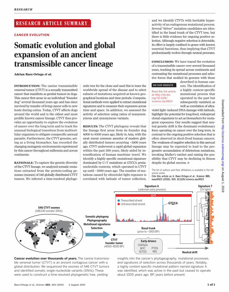
RESEARCH ARTICLE SUMMARY ◥ CANCER EVOLUTION Somatic evolution and global expansion of an ancient transmissible cancer lineage Adrian Baez-Ortega et al. INTRODUCTION: The canine transmissible venereal tumor (CTVT) is a sexually transmitted cancer that manifests as genital tumors in dogs. This cancer first arose in an individual “founder dog” several thousand years ago and has since survived by transfer of living cancer cells to new hosts during coitus. Today, CTVT affects dogs around the world and is the oldest and most prolific known cancer lineage. CTVT thus pro- vides an opportunity to explore the evolution of cancer over the long term and to track the unusual biological transition from multicel- lular organism to obligate conspecific asexual parasite. Furthermore, the CTVT genome, act- ing as a living biomarker, has recorded the changing mutagenic environments experienced by this cancer throughout millennia and across continents. RATIONALE: To capture the genetic diversity of the CTVT lineage, we analyzed somatic muta- tions extracted from the protein-coding ge- nomes (exomes) of 546 globally distributed CTVT tumors. We inferred a time-resolved phyloge- netic tree for the clone and used this to trace the worldwide spread of the disease and to select subsets of mutations acquired at known geo- graphical locations and time periods. Computa- tional methods were applied to extract mutational signatures and to measure their exposures across time and space. In addition, we assessed the activity of selection using ratios of nonsynon- ymous and synonymous variants. RESULTS: The CTVT phylogeny reveals that the lineage first arose from its founder dog 4000 to 8500 years ago, likely in Asia, with the most recent common ancestor of modern glob- ally distributed tumors occurring ~1900 years ago. CTVT underwent a rapid global expansion within the past 500 years, likely aided by in- tensification of human maritime travel. We identify a highly specific mutational signature dominated by C>T mutations at GTCCA penta- nucleotide contexts, which operated in CTVT up until ~1000 years ago. The number of mu- tations caused by ultraviolet light exposure is correlated with latitude of tumor collection, and we identify CTVTs with heritable hyper- activity of an endogenous mutational process. Several “driver” mutation candidates are iden- tified in the basal trunk of the CTVT tree, but there is little evidence for ongoing positive se- lection. Although negative selection is detectable, its effect is largely confined to genes with known essential functions, thus implying that CTVT predominantly evolves through neutral processes. CONCLUSION: We have traced the evolution of a transmissible cancer over several thousand years, tracking its spread across continents and contrasting the mutational processes and selec- tive forces that molded its genome with those described in human can- cers. The identification of a highly context-specific mutational process that operated in the past but subsequently vanished, as well as correlation of ultra- violet light–induced DNA damage with latitude, highlight the potential for long-lived, widespread clonal organisms to act as biomarkers for muta- genic exposures. Our results suggest that neu- tral genetic drift is the dominant evolutionary force operating on cancer over the long term, in contrast to the ongoing positive selection that is often observed in short-lived human cancers. The weakness of negative selection in this asexual lineage may be expected to lead to the pro- gressive accumulation of deleterious mutations, invoking Muller’s ratchet and raising the pos- sibility that CTVT may be declining in fitness despite its global success. ▪ RESEARCH Baez-Ortega et al., Science 365, 464 (2019) 2 August 2019 1 of 1 Transcribed strand Untranscribed strand Early drivers PTEN RB1 CDKN2A MYC SETD2 546 CTVT exomes (148,030 somatic SNVs) Somatic phylogeny Phylogeography Mutational signatures Selection CTVT founder tumor (4000–8500 BP) Neutral drift Basal trunk (prior to ~1900 BP) Signature A (unknown early process) Signature 7 (UV light) GTCCA C>T Cancer evolution over thousands of years. The canine transmissi- ble venereal tumor (CTVT) is an ancient contagious cancer with a global distribution. We sequenced the exomes of 546 CTVT tumors and identified somatic single-nucleotide variants (SNVs). These were used to construct a time-resolved phylogenetic tree, yielding insights into the cancer’ s phylogeography, mutational processes, and signatures of selection across thousands of years. Notably, a highly context-specific mutational pattern named signature A was identified, which was active in the past but ceased to operate about 1000 years ago. BP, years before present. ON OUR WEBSITE ◥ Read the full article at http://dx.doi. org/10.1126/ science.aau9923 .................................................. The list of authors and their affiliations is available in the full article online. Cite this article as A. Baez-Ortega et al., Science 365, eaau9923 (2019). DOI: 10.1126/science.aau9923 on August 1, 2019 http://science.sciencemag.org/ Downloaded from
Transcript
RESEARCH ARTICLE SUMMARY◥
CANCER EVOLUTION
Somatic evolution and globalexpansion of an ancienttransmissible cancer lineageAdrian Baez-Ortega et al.
INTRODUCTION: The canine transmissiblevenereal tumor (CTVT) is a sexually transmittedcancer thatmanifests as genital tumors in dogs.This cancer first arose in an individual “founderdog” several thousand years ago and has sincesurvived by transfer of living cancer cells to newhosts during coitus. Today, CTVT affects dogsaround the world and is the oldest and mostprolific known cancer lineage. CTVT thus pro-vides an opportunity to explore the evolutionof cancer over the long term and to track theunusual biological transition frommulticel-lular organism to obligate conspecific asexualparasite. Furthermore, the CTVT genome, act-ing as a living biomarker, has recorded thechangingmutagenic environments experiencedby this cancer throughoutmillennia and acrosscontinents.
RATIONALE: To capture the genetic diversityof the CTVT lineage, we analyzed somaticmuta-tions extracted from the protein-coding ge-nomes (exomes) of 546 globally distributedCTVTtumors. We inferred a time-resolved phyloge-
netic tree for the clone and used this to trace theworldwide spread of the disease and to selectsubsets of mutations acquired at known geo-graphical locations and time periods. Computa-tionalmethodswereapplied to extractmutationalsignatures and tomeasure their exposures acrosstime and space. In addition, we assessed theactivity of selection using ratios of nonsynon-ymous and synonymous variants.
RESULTS: The CTVT phylogeny reveals thatthe lineage first arose from its founder dog4000 to 8500 years ago, likely in Asia, with themost recent common ancestor of modern glob-ally distributed tumors occurring ~1900 yearsago. CTVT underwent a rapid global expansionwithin the past 500 years, likely aided by in-tensification of human maritime travel. Weidentify a highly specific mutational signaturedominated by C>T mutations at GTCCA penta-nucleotide contexts, which operated in CTVTup until ~1000 years ago. The number of mu-tations caused by ultraviolet light exposure iscorrelated with latitude of tumor collection,
and we identify CTVTs with heritable hyper-activity of an endogenousmutational process.Several “driver”mutation candidates are iden-tified in the basal trunk of the CTVT tree, butthere is little evidence for ongoing positive se-lection. Althoughnegative selection is detectable,its effect is largely confined to genes with knownessential functions, thus implying that CTVTpredominantly evolves through neutral processes.
CONCLUSION: We have traced the evolutionof a transmissible cancer over several thousandyears, tracking its spread across continents andcontrasting themutational processes and selec-tive forces that molded its genome with those
described in human can-cers. The identification ofa highly context-specificmutational process thatoperated in the past butsubsequently vanished, aswell as correlation of ultra-
violet light–inducedDNAdamagewith latitude,highlight the potential for long-lived, widespreadclonal organisms to act as biomarkers formuta-genic exposures. Our results suggest that neu-tral genetic drift is the dominant evolutionaryforce operating on cancer over the long term, incontrast to the ongoing positive selection that isoften observed in short-lived human cancers.Theweakness of negative selection in this asexuallineage may be expected to lead to the pro-gressive accumulation of deleterious mutations,invoking Muller’s ratchet and raising the pos-sibility that CTVT may be declining in fitnessdespite its global success.▪
RESEARCH
Baez-Ortega et al., Science 365, 464 (2019) 2 August 2019 1 of 1
Transcribed strandUntranscribed strand
Early drivers
PTENRB1
CDKN2AMYC
SETD2
546 CTVT exomes(148,030 somatic SNVs)
Somatic phylogenyPhylogeography
Mutational signaturesSelection
CTVTfounder tumor
(4000–8500 BP)
Neutral drift
Basal trunk(prior to ~1900 BP)
Signature A (unknown early process)
Signature 7 (UV light)
GTCCA
C>T
Cancer evolution over thousands of years. The canine transmissi-ble venereal tumor (CTVT) is an ancient contagious cancer with aglobal distribution. We sequenced the exomes of 546 CTVT tumorsand identified somatic single-nucleotide variants (SNVs). Thesewere used to construct a time-resolved phylogenetic tree, yielding
insights into the cancer’s phylogeography, mutational processes,and signatures of selection across thousands of years. Notably,a highly context-specific mutational pattern named signature Awas identified, which was active in the past but ceased to operateabout 1000 years ago. BP, years before present.
ON OUR WEBSITE◥
Read the full articleat http://dx.doi.org/10.1126/science.aau9923..................................................
The list of authors and their affiliations is available in the fullarticle online.Cite this article as A. Baez-Ortega et al., Science 365,eaau9923 (2019). DOI: 10.1126/science.aau9923
Somatic evolution and globalexpansion of an ancienttransmissible cancer lineageAdrian Baez-Ortega1, Kevin Gori1*, Andrea Strakova1*, Janice L. Allen2,Karen M. Allum3, Leontine Bansse-Issa4, Thinlay N. Bhutia5, Jocelyn L. Bisson1,6,Cristóbal Briceño7, Artemio Castillo Domracheva8, Anne M. Corrigan9, Hugh R. Cran10,Jane T. Crawford11, Eric Davis12, Karina F. de Castro13, Andrigo B. de Nardi14,Anna P. de Vos15, Laura Delgadillo Keenan16, Edward M. Donelan2,Adela R. Espinoza Huerta17, Ibikunle A. Faramade18, Mohammed Fazil19,Eleni Fotopoulou20, Skye N. Fruean21, Fanny Gallardo-Arrieta22, Olga Glebova23,Pagona G. Gouletsou24, Rodrigo F. Häfelin Manrique25, Joaquim J. G. P. Henriques26,Rodrigo S. Horta27, Natalia Ignatenko28, Yaghouba Kane29, Cathy King3,Debbie Koenig3, Ada Krupa30, Steven J. Kruzeniski17, Young-Mi Kwon1,Marta Lanza-Perea9, Mihran Lazyan31, Adriana M. Lopez Quintana32,Thibault Losfelt33, Gabriele Marino34, Simón Martínez Castañeda35,Mayra F. Martínez-López36,37, Michael Meyer38, Edward J. Migneco39,Berna Nakanwagi40, Karter B. Neal41, Winifred Neunzig3, Máire Ní Leathlobhair1,Sally J. Nixon42, Antonio Ortega-Pacheco43, Francisco Pedraza-Ordoñez44,Maria C. Peleteiro45, Katherine Polak46, Ruth J. Pye47, John F. Reece48,Jose Rojas Gutierrez49, Haleema Sadia50, Sheila K. Schmeling51, Olga Shamanova52,Alan G. Sherlock47, Maximilian Stammnitz1, Audrey E. Steenland-Smit4,Alla Svitich53, Lester J. Tapia Martínez17, Ismail Thoya Ngoka54, Cristian G. Torres55,Elizabeth M. Tudor56, Mirjam G. van der Wel57, Bogdan A. Viţălaru58,Sevil A. Vural59, Oliver Walkinton47, Jinhong Wang1, Alvaro S. Wehrle-Martinez60,Sophie A. E. Widdowson61, Michael R. Stratton62, Ludmil B. Alexandrov63,Iñigo Martincorena62, Elizabeth P. Murchison1†
The canine transmissible venereal tumor (CTVT) is a cancer lineage that aroseseveral millennia ago and survives by “metastasizing” between hosts through celltransfer. The somatic mutations in this cancer record its phylogeography andevolutionary history. We constructed a time-resolved phylogeny from 546 CTVT exomesand describe the lineage’s worldwide expansion. Examining variation in mutationalexposure, we identify a highly context-specific mutational process that operated earlyin the cancer’s evolution but subsequently vanished, correlate ultraviolet-lightmutagenesis with tumor latitude, and describe tumors with heritable hyperactivity of anendogenous mutational process. CTVT displays little evidence of ongoing positiveselection, and negative selection is detectable only in essential genes.We illustrate howlong-lived clonal organisms capture changing mutagenic environments, and reveal thatneutral genetic drift is the dominant feature of long-term cancer evolution.
Transmissible cancers are malignant somaticcell clones that spread between individualsby direct transfer of living cancer cells. Anal-ogous to the metastasis of cancer to distanttissues within a single body, transmissible
cancers “metastasize” as allogeneic grafts betweenindividuals within a population (1). Such cloneshave been observed only eight times in nature,suggesting that they arise rarely; however, onceestablished, transmissible cancers can spread rap-idly and widely and persist through time (1, 2).Such cancers provide an opportunity to explorethe evolution of cancer over the long term andto track the unusual biological transition from
is the oldest and most prolific known contagiouscancer (2, 3). It is a sexually transmitted clonethat manifests as genital tumors in dogs. Thiscancer first arose from the somatic cells of an in-dividual “founder dog” that lived several thousandyears ago (2). The cancer survived beyond thedeath of this original host by transfer of cancercells to new hosts. Subsequently, this cancer hasspread around theworld and is a commondiseasein dog populations globally, although it declinedand largely disappeared from many Western
countries during the 20th century owing to themanagement and removal of free-roamingdogs (4).Similar to cancers that remain in a single in-
dividual, CTVT accumulates somatic mutations.These result from the activities of endogenousand exogenous mutational processes, and genet-ically imprint a cancer’s history of mutagenicexposures (5). Thus, the CTVT genome can beconsidered a living biomarker that records thechanging mutagenic environments experiencedby this cancer throughout millennia and acrosscontinents. Although most somatic mutationsin cancer have no functional effect and are con-sidered neutral “passenger”mutations, a subsetofmutations are positively selected “driver”muta-tions that confer the proliferation and survivaladvantages that spur cancer growth (6). Ordinarycancers, which remain in a single host, often ac-quire additional driver mutations during tumorprogression (7); however, it is unknown whethertransmissible cancers that survive for hundredsor thousands of years similarly continue to adapt.It seems possible that the evolution of long-livedcancers such as CTVTmay instead be dominatedby negative selection acting to remove deleteriousmutations. Finally, in addition to recording a his-tory of exposures and signatures of selection, so-matic mutations provide a tool for tracing CTVTphylogeography, potentially revealing how dogs,together with humans, moved around the worldover the past centuries. Here, we use somatic mu-tations extracted from the protein-coding ge-nomes (exomes) of 546 globally distributed CTVTtumors to trace the history, spread, diversity, mu-tational exposures, and evolution of theCTVT clone.
CTVTphylogeny
We sequenced the exomes (43.6 megabases, Mb;mean sequencing depth ~132×) of 546 CTVTtumors collected between 2003 and 2016 from43 countries across all inhabited continents (data-sets S1 and S2). Candidate somatic mutationswere defined as single-nucleotide variants (SNVs)or short insertions and deletions (indels) identi-fied in one or more CTVT tumors, but not foundin 495 normal dog exomes from the CTVT tumors’matched hosts. This approach yielded 160,207 var-iants (148,030 SNVs, 3392 per Mb; 12,177 indels,279 per Mb; table S1). The features of this set, in-cluding its variant allele fraction distribution,phylogenetic structure, comparison with the dis-tribution of private germline variants in the dogpopulation,mutational signature composition, andnonsynonymous-to-synonymousmutation ratio[details in (8)], suggest that it is very highly en-riched for somatic mutations. However, somemin-imal germline variation may remain, possiblyincluding rare germline variants from the founderdog and residual contaminating alleles frommatched hosts.We identified the subset of the candidate so-
matic mutations belonging to a clocklike muta-tional process [specifically, cytosine-to-thymine(C>T) substitutions at CpG sites (8, 9)] and usedthese to construct a time-resolved phylogenetictree for the CTVT lineage (Fig. 1A). The muta-tion rate was inferred by applying a Bayesian
RESEARCH
Baez-Ortega et al., Science 365, eaau9923 (2019) 2 August 2019 1 of 7
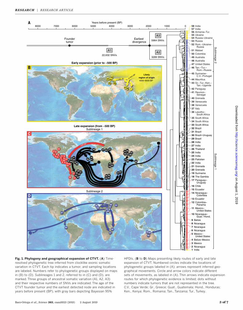
Poisson model to previously ascertained empi-rical observations (10) andwas estimated as 6.87 ×10⁻⁷ C>Tmutations per CpG site per year (8). Thetopology of the CTVT phylogenetic tree reveals along basal trunk (Fig. 1A), representing the chainof CTVT transmissions from its origin ~6220 yearsago [95%highest posterior density interval (HPDI)4148 to 8508 years ago] to the earliest detectednode ~1938 years ago (95% HPDI 993 to 3055years ago). This node splits a set of five tumorscollected in India from the remaining population(groups labeled 57 and 58; Fig. 1A). The secondand third most basal nodes (respectively ~1004years ago, 95% HPDI 497 to 1570 years ago, and~829 years ago, 95%HPDI 424 to 1310 years ago)separate 16 tumors from Eastern Europe and theBlack Sea region, and three tumors from NorthernIndia, from the remaining set, respectively (groupslabeled 54 to 56 and 1; Fig. 1A). Together withevidence that the founder dog shared ancestrywith ancient dog remains recovered in northeastSiberia and North America (10), the CTVT phylog-eny supports a model whereby CTVT originated~4000 to 8500 years ago in Central or NorthernAsia and remained within the area for the sub-sequent 2000 to 6000 years. Starting less than~2000 years ago, CTVT escaped from its foundingpopulation, perhaps due to contact between pre-viously isolated dog groups, and spread to severallocations in Asia and Europe (Fig. 1B).The more recent history of CTVT is marked
by rapid global expansion (11) (Fig. 1C and fig. S1).CTVT was introduced to the Americas with earlycolonial contact (~500 years ago, 95%HPDI 284 to888 years ago), probably initially toCentral America,and further into North and South America (redsublineage 1; Fig. 1, A and C). About 300 yearsago, this sublineage spread out of the Americasin an almost polytomous global sweep that broughtCTVT into Africa at least five times and reintro-duced the disease to Europe and Asia (black sub-lineage 1; Fig. 1, A and C). In parallel, a second
tumor sublineage spread out of Asia or Europeinto Australia and the Pacific (sublineage 2; Fig.1, A and D). This second sublineage is also de-tected in North America, and its tumors wereintroduced to Africa on at least two occasions. By~100 years ago, CTVTwas present in dog popula-tions worldwide, establishing local lineages thathave since remained largely in situ. The CTVTphylogeny thus suggests that dogs, together withtheir neoplastic parasites, were extensively trans-ported around theworld in the 15th to early 20thcenturies, probably by sea travel.
Mutational processes in CTVT
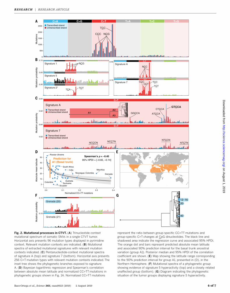
The CTVT mutational spectrum, a representationof the six substitution types together with theirimmediate 5′ and 3′ base contexts, is dominatedby C>Tmutations, as previously described (12, 13)(Fig. 2A). Applying Markov chain Monte Carlosampling on a Bayesian model of mutationalsignatures (8, 14), we extracted signatures of fivemutational processes from the CTVT mutationload. These include three signatures that closelyresemble COSMIC (15) signatures 1, 5, and 7 (Fig.2B). These signatures, which have previously beendescribed in CTVT (12), reflect endogenous muta-tional processes (signatures 1 and 5) and exposureto ultraviolet (UV) light (signature 7) (5). A fourthsignature displaying some similarity (cosine sim-ilarity 0.81) to COSMIC signature 2, which is as-sociated with activity of APOBEC enzymes (5), wasalso detected (labeled signature 2*, Fig. 2B).The fifth signature extracted from CTVT does
not resemble any previously described muta-tional pattern. This signature, which we designatesignature A, is characterized by C>T mutationsat NCC contexts (mutated nucleotide underlined)and shows substantial pentanucleotide sequencepreference forGTCCA (TGGACon the complemen-tary strand; Fig. 2, B and C, and fig. S2). Thisextended sequence preference ismarkedlymorepronounced than previously reported pentanu-
cleotide context biases, such as those associatedwith UV light or DNA polymerase epsilon defi-ciency (Fig. 2C) (16–18), and is not explained bythe sequence composition of the canine exome(fig. S3). It is possible that signature A’s causativemutagen is highly context-specific, or, alternatively,that this signature’s associated repair processes areineffective at certain sequence contexts (“repairshielding”) (19). In addition, signature A displaysstrong transcriptional strand bias, with moremutations of guanine on the untranscribed com-pared to the transcribed strand of genes, indicat-ing that its causative lesion is likely a guanineadduct subject to transcription-coupled repair(TCR). Notably, the guanine-directed transcrip-tional strand bias of signature A at TCC contextscounteracts the cytosine-directed transcriptionalstrand bias of signature 7 at TCC, such that nooverall transcriptional strand bias is observed atthis context in the CTVT mutational spectrum(Fig. 2A).Using the CTVT phylogenetic tree to isolate
subsets of mutations, we explored variation inmutational signature exposure across time andspace (figs. S4 and S5 and dataset S3). Notably,this revealed that signature A was highly activeprior to ~2000 years ago (causing ~35% of muta-tions in the basal trunk of the tree, branch A1)and persisted in parallel at lower levels in thetwo basal branches after the first node (~12 and~9%ofmutations in branches A2 and A3, respec-tively), but then abruptly vanished (Fig. 2C andfig. S5). Moreover, signature A is not detectablewithin the germ line of a global population of 495dogs (fig. S6). It is possible that signature A reflectsthe activity of an exogenous mutagen that wasexclusively present in the environment that CTVTinhabited before its escape from its founding pop-ulation. Alternatively, it is plausible that signatureAmay result froman endogenousDNA-damagingagent that occurred in CTVT cells early during thelineage’s history, but which ceased to accumulate
Baez-Ortega et al., Science 365, eaau9923 (2019) 2 August 2019 2 of 7
1Transmissible Cancer Group, Department of Veterinary Medicine, University of Cambridge, Cambridge, UK. 2Animal Management in Rural and Remote Indigenous Communities(AMRRIC), Darwin, Australia. 3World Vets, Gig Harbor, USA. 4Animal Shelter, Stichting Dierenbescherming Suriname, Paramaribo, Suriname. 5Sikkim Anti-Rabies and Animal HealthProgramme, Department of Animal Husbandry, Livestock, Fisheries and Veterinary Services, Government of Sikkim, India. 6Royal (Dick) School of Veterinary Studies and the RoslinInstitute, University of Edinburgh, Easter Bush Campus, Roslin EH25 9RG, UK. 7ConserLab, Animal Preventive Medicine Department, Faculty of Animal and Veterinary Sciences, Universityof Chile, Santiago, Chile. 8Corozal Veterinary Hospital, University of Panamá, Panama City, Republic of Panama. 9St. George's University, True Blue, Grenada. 10The Nakuru DistrictVeterinary Scheme Ltd, Nakuru, Kenya. 11Animal Medical Centre, Belize City, Belize. 12International Animal Welfare Training Institute, UC Davis School of Veterinary Medicine, Davis, CA,USA. 13Centro Universitário de Rio Preto (UNIRP), São José do Rio Preto, São Paulo, Brazil. 14Department of Clinical and Veterinary Surgery, São Paulo State University (UNESP), SãoPaulo, Brazil. 15Ladybrand Animal Clinic, Ladybrand, South Africa. 16Veterinary Clinic Sr. Dog’s, Guadalajara, Mexico. 17World Vets Latin America Veterinary Training Center, Granada,Nicaragua. 18National Veterinary Research Institute, Vom, Nigeria. 19Animal Clinic, Mombasa, Kenya. 20Intermunicipal Stray Animals Care Centre (DIKEPAZ), Perama, Greece. 21AnimalProtection Society of Samoa, Apia, Samoa. 22Faculty of Veterinary Science, University of Zulia, Maracaibo, Venezuela. 23Veterinary Clinic BIOCONTROL, Moscow, Russia. 24Faculty ofVeterinary Medicine, School of Health Sciences, University of Thessaly, Karditsa, Greece. 25Veterinary Clinic El Roble, Animal Healthcare Network, Faculty of Animal and VeterinarySciences, University of Chile, Santiago de Chile, Chile. 26OnevetGroup, Hospital Veterinário Berna, Lisboa, Portugal. 27Universidade Vila Velha, Vila Velha, Brazil. 28Veterinary ClinicZoovetservis, Kiev, Ukraine. 29École Inter-états des Sciences et Médecine Vétérinaires de Dakar, Dakar, Senegal. 30Department of Small Animal Medicine, Faculty of Veterinary Medicine,Utrecht University, Utrecht, Netherlands. 31Vetexpert Veterinary Group, Yerevan, Armenia. 32Veterinary Clinic Lopez Quintana, Maldonado, Uruguay. 33Clinique Veterinaire de Grand Fond,Saint Gilles les Bains, Reunion, France. 34Department of Veterinary Sciences, University of Messina, Messina, Italy. 35Facultad de Medicina Veterinaria y Zootecnia, Universidad Autónomadel Estado de México, Toluca, Mexico. 36School of Veterinary Medicine, Universidad de las Américas, Quito, Ecuador. 37Cancer Development and Innate Immune Evasion Lab,Champalimaud Center for the Unknown, Lisbon, Portugal. 38Touray & Meyer Vet Clinic, Serrekunda, The Gambia. 39Hillside Animal Hospital, St. Louis, MO, USA. 40The Kampala VeterinarySurgery, Kampala, Uganda. 41Asavet Veterinary Charities, Tucson, AZ, USA. 42Vets Beyond Borders, Bylakuppe, India. 43Faculty of Veterinary Medicine, Autonomous University ofYucatan, Merida, Mexico. 44Laboratorio de Patología Veterinaria, Universidad de Caldas, Manizales, Colombia. 45Interdisciplinary Centre of Research in Animal Health (CIISA), Faculty ofVeterinary Medicine, University of Lisbon, Lisboa, Portugal. 46Four Paws International, Vienna, Austria. 47Vets Beyond Borders, The Rocks, Australia. 48Help in Suffering, Jaipur, India.49Veterinary Clinic Dr José Rojas, Los Andes, Chile. 50Department of Biotechnology, Balochistan University of Information Technology, Engineering and Management Sciences, Quetta,Pakistan. 51Corozal Veterinary Clinic, Corozal Town, Belize. 52Veterinary Clinic Vetmaster, Ramenskoye, Russia. 53State Hospital of Veterinary Medicine, Dniprodzerzhynsk, Ukraine.54Jomo Kenyatta University of Agriculture and Technology, Juja, Kenya. 55Laboratory of Biomedicine and Regenerative Medicine, Department of Clinical Sciences, Faculty of Animal andVeterinary Sciences, University of Chile, Santiago, Chile 56Faculty of Veterinary and Agricultural Sciences, University of Melbourne, Melbourne, Australia. 57Animal Anti Cruelty League,Port Elizabeth, South Africa. 58Clinical Sciences Department, Faculty of Veterinary Medicine Bucharest, Bucharest, Romania. 59Department of Pathology, Faculty of Veterinary Medicine,Ankara University, Ankara, Turkey. 60Faculty of Veterinary Sciences, National University of Asuncion, San Lorenzo, Paraguay. 61Lilongwe Society for Protection and Care of Animals(LSPCA), Lilongwe, Malawi. 62Wellcome Sanger Institute, Hinxton, UK. 63Department of Cellular and Molecular Medicine, University of California, San Diego, La Jolla, CA, USA.*These authors contributed equally to this work.†Corresponding author. Email: [email protected]
Baez-Ortega et al., Science 365, eaau9923 (2019) 2 August 2019 3 of 7
~1,000 BP
Likelyregion of origin4000–8500 BP
~2,000 BP
~1,,0000000000000 BBBBBBBPPPPPPPPPPPPPPPPPPP ~2,00000 B
54
56
55
15758
56
54
53
51
50
49
47
4652
46
46
46
5252
4948
2223324
2627728
2825
31
303433
436
37
41
43
44
45
22
17
1716
13
6
55 4 43
7
15
~500 BP
1011
11 5
21014
12 45419
20021
394
338040 414
189
342
3303322
29
45 43
43343443
35
Early expansion (prior to ~500 BP)
Late expansion (from ~500 BP)Sublineage 1
Sublineage 2
B
C
D
A Years before present (BP)010002000300040005000600070008000
22,632 SNVs
A1
Foundertumor
Earliest divergence
IndiaNicaraguaMexicoBelize–Mexico
Mexico– United States
NicaraguaNicaraguaNicaraguaBelize
Nicaragua– Guat.–Hond.
Mexico– United States
Colombia– Panama
Ecuador
Nicaragua– Colombia
EcuadorChile
Paraguay– Uruguay
The GambiaSurinameGrenadaGrenadaIndiaPakistanIndiaIndiaThailandIndiaIndiaBrazilBrazil–UruguayBrazilBrazilSouth AfricaSouth AfricaSouth Africa
Lesotho– South Africa
ItalyVenezuelaVenezuelaGrenada
Reunion– Senegal
Paraguay
Gr.–Tur.–Ken.– Tan.–Uganda
Mauritius
Suriname– C.V.–Portugal
Tan.–Tur.– Rom.–Russia
United StatesAustraliaAustraliaColombiaMalawi
Rom.–Ukraine– Russia
RussiaRussia–UkraineUkraineArmenia–Tur.IndiaIndia
2
4
5678
1
3
9
10
11
1213
141516
17181920212223242526272829303132333435
3637383940
4142
4344
45
464748495051
52535455565758
Sublineage 2
Sublineage 1
3289 SNVs
A2
5964 SNVs
A3
Fig. 1. Phylogeny and geographical expansion of CTVT. (A) Time-resolved phylogenetic tree inferred from clocklike exonic somaticvariation in CTVT. Each tip indicates a tumor, and sampling locationsare labeled. Numbers refer to phylogenetic groups displayed on mapsin (B) to (D). Sublineages 1 and 2, referred to in (C) and (D), aremarked. Three groups of ancestral somatic variation (A1, A2, A3)and their respective numbers of SNVs are indicated. The age of theCTVT founder tumor and the earliest detected node are indicated inyears before present (BP), with gray bars depicting Bayesian 95%
HPDIs. (B to D) Maps presenting likely routes of early and lateexpansion of CTVT. Numbered circles indicate the locations ofphylogenetic groups labeled in (A); arrows represent inferred geo-graphical movements. Circle and arrow colors indicate differentsets of movements, as labeled in (A). Thin arrows indicate expansionroutes for which phylogenetic evidence is limited; dots withoutnumbers indicate tumors that are not represented in the tree.C.V., Cape Verde; Gr., Greece; Guat., Guatemala; Hond., Honduras;Ken., Kenya; Rom., Romania; Tan., Tanzania; Tur., Turkey.
Baez-Ortega et al., Science 365, eaau9923 (2019) 2 August 2019 4 of 7
A
B
C
D E
C>A C>G C>T T>A T>C T>G
TCC
NCGCCC
Grenada
Mauritius
0.15
Signature 1 NCG
Signature 50.05
0.30
Signature 2* TCTTCA
0.02
0.04C>T
GTCCACTCCA
ATCCAGGCCA
0.40 C>TC>
Signature ANCC
Transcribed strandUntranscribed strand
Mut
atio
n pr
obab
ility
C>T
0.02
0.04
NCCCN NCCTNNTCCN
NTCTN
0.15 C>T
Signature 7 TCC
TCT
CCCCCT
F
0
500
1000
1500
2000
2500
Mut
atio
ns
Transcribed strandUntranscribed strand
Transcribed strandUntranscribed strand
G0.10
Grenada (20)
0.10
Grenada (21)
12–1620
40
Normalized CC>TT mutations
Abs
olut
e m
ean
latit
ude
A2
A3A1
Signature 7
Signature A
Mut
atio
n pr
obab
ility
Mut
atio
n pr
obab
ility
0
10
20
30
40
50
0.0 0.1 0.2 0.3 0.4
Prediction for A1 (Basal trunk)
India
SurinameEcuador
Russia–Ukraine
South Africa
Colombia
Nicaragua−Colombia
Australia
Fig. 2. Mutational processes in CTVT. (A) Trinucleotide-contextmutational spectrum of somatic SNVs in a single CTVT tumor.Horizontal axis presents 96 mutation types displayed in pyrimidinecontext. Relevant mutation contexts are indicated. (B) Mutationalspectra of extracted mutational signatures with relevant mutationcontexts indicated. (C) Pentanucleotide-context mutational spectraof signature A (top) and signature 7 (bottom). Horizontal axis presents256 C>Tmutation types with relevant mutation contexts indicated. Theinset tree shows the phylogenetic branches exposed to signatureA. (D) Bayesian logarithmic regression and Spearman’s correlationbetween absolute mean latitude and normalized CC>TTmutations inphylogenetic groups shown in Fig. 1A. Normalized CC>TTmutations
represent the ratio between group-specific CC>TTmutations andgroup-specific C>T changes at CpG dinucleotides. The black line andshadowed area indicate the regression curve and associated 95% HPDI.The orange dot and bars represent predicted absolute mean latitudeand associated 90% prediction interval for the basal trunk ancestralvariation (group A1). Posterior median and 95% HPDI of the correlationcoefficient are shown. (E) Map showing the latitude range correspondingto the 90% prediction interval for group A1, presented in (D), in theNorthern Hemisphere. (F) Mutational spectra of a phylogenetic groupshowing evidence of signature 5 hyperactivity (top) and a closely relatedunaffected group (bottom). (G) Diagram indicating the phylogeneticsituation of the tumor groups displaying signature 5 hyperactivity.
from ~1000 years ago, perhaps as a result of acellular metabolic change. Although the natureof such a change is unknown, the replacement ofpossibly defectivemitochondrial DNA by horizon-tal transfer, which likely occurred in parallel inbranches A2 and A3 within the past ~1690 years(11), may have altered the metabolic environmentwithin CTVT cells.Although CTVT usually occurs within the in-
ternal genital tract, it may sometimes protrudefrom the genital orifice or spread to perineal skin,resulting in sporadic exposure to solar UV radia-tion (12, 13). The amount of UV radiation reach-ing Earth, however, varies substantially acrossglobal environments (20).We investigatedwhetherlatitude influenced the degree of UV exposurein CTVT tumors by estimating the contribution ofsignature 7 within subsets of mutations acquiredat known latitudes. Indeed, qualitative assessmentof mutational spectra of location-specific CTVTmutation subsets suggests extensive variation inUV exposure; for example, the mutational spec-tra of tumors collected in Mauritius show con-siderably more evidence of signature 7 comparedwith those of tumors collected in Russia (fig. S4).Using CC>TT dinucleotide mutations (21) as aproxy for signature 7 (fig. S7), we identified anonlinear association between latitude and UVexposure (Spearman’s correlation–0.40, 95%HPDI–0.65 to –0.14; Fig. 2D). By fitting CC>TT muta-tions observed in the basal trunk of the CTVTtree to this curve, we estimated the latitude of theCTVT founder population (Fig. 2, D and E) (8).Examining the contribution of signature 5 across
the CTVT lineage, we observed three independentphylogenetic groups of tumors that appear tohave acquired signature 5 hyperactivity pheno-types (groups labeled 12 to 16, 20, and 40; Fig. 2,F and G, and figs. S4 and S5). In one case, involv-ing tumors collected in several South andCentralAmerican countries (groups 12 to 16), the pheno-type has been maintained for ~150 years. Thisphenotype is likely to result from signature 5 andnot from the double-strandDNA repair deficiency–mediated COSMIC signature 3, which presentsa similarmutational profile (5, 22), as we failed toobserve the enrichment for indels that co-occurswith signature 3 (22, 23). It is possible, however,that these tumorswere exposed to another, as yetundescribed, mutational process. Signature 5 iswidespread in cancer and normal tissues andhas unknown etiology, although it may be partlyassociated with endogenously generated adductssubject to nucleotide excision repair (5, 9, 18). Weannotated nonsynonymous mutations occurringin the three groups’ respective clonal ancestors,providing a catalog of genes that may play arole in generation or suppression of signature 5(dataset S4).
CTVTmutations and gene expression
The prevalence of substitutionmutations in CTVTdecreases with increasing gene expression, likelyreflecting the activity of TCR operating on DNAdamage associated with signatures 7 and A, aswell as a signature 1 preference for genes withlower expression (16, 24, 25) (fig. S8, A andB).We
observed that exons have a higher substitutionprevalence than introns, possibly as a result ofsequence context (figs. S8A and S9). The preva-lence of indels is positively correlated with in-creasing gene expression, as has been observedin human cancers, andmay reflect transcription-associated damage (26) (fig. S8A).We assessed the contribution of TCR in two
temporally distinct subsets of mutations: thoseacquired before the earliest detectable node inthe phylogenetic tree (~8500 to 2000 years ago;branch A1 in Fig. 1A) and those acquired subse-quent to this node (~2000 years ago to the pres-ent). Although C>T mutations acquired at TCCcontexts in highly expressed genes in branch A1have little strand bias, likely because of the oppos-ing transcriptional strand preferences of sig-natures 7 and A at this context, those genes withvery low expression predominantly show thetranscriptional strand bias associated with sig-nature A (fig. S8C). Assuming that the transcrip-tional strand bias observed in these low-expressedgenes reflects earlier expression and subsequentsilencing of genes, this suggests that there mayhave been an early period in CTVT evolutionwhenthe lineage was exposed to signature A more in-tensely than it was to signature 7. Thismay reflectvariation in the climate or environment to whichCTVT was exposed early in its history.
Selection in CTVT
CTVT has a massive mutation burden, whichexceeds that observed in even the most highlymutated human cancer types (Fig. 3A). EachCTVTtumor carries on average 37,800 SNVs across itspredominantly diploid (12) exome (~2millionSNVsgenome-wide; table S2). Indeed, the tally of somaticmutations that have accumulated in CTVT sinceit departed its original host is comparable withthe number of germline variants that distinguishsome pairs of outbred dogs (fig. S10). Within theset of 546 tumors, 14,412 (~73%) protein-codinggenes carry at least one nonsynonymous muta-tion, and 5704 (~29%) havemutations predictedto cause protein truncation (Fig. 3B).We searched for evidence of positive selection
in CTVT. The drivermutations that initially causedCTVT, and promoted its transmissible pheno-type, will have occurred in the basal trunk ofthe CTVT tree. SETD2, CDKN2A,MYC [previouslydescribed (12, 27)], PTEN, and RB1, known cancergenes that frequently harbor driver mutations inhuman cancers (15), carry biallelic loss-of-functionor potential activatingmutations in the trunk andmay be early drivers of CTVT (Fig. 3C and tableS3). To search for late drivers, which may havebeen acquired in more recent parallel CTVT line-ages, we identified independent mutations thatoccurred repeatedly across the tree, andmeasuredthe normalized ratio of nonsynonymous to synon-ymous mutations (dN/dS) per gene after correct-ing for mutational biases and context effects (8).This approach only yielded two uncharacterizedgenes with dN/dS > 1 (q-value < 0.05), predictedto encode a neuroligin precursor and a round-about homolog (dataset S5). The potential forthese genes to act as late drivers in CTVT cannot
be assessed, and it is possible that local sequencestructures may result in higher-than-expected re-current mutation rates at these loci (28). Overall,we find little evidence that CTVT is continuing toadapt to its environment.Negative selection, which acts to remove dele-
terious mutations, is very weak in human cancers(17, 29, 30). Human cancers have short life spans,and their evolution is dominated by sweeps ofstrong positive selection, thus reducing the po-tential for negative selection to act (17). Given itslong life span, high mutation burden, and lackof ongoing positive selection, it is possible thatnegative selectionmay be amore dominant forcein CTVT evolution. Further, unlike in ordinarycancers, intertumor competition may offer moreopportunities for negative selection to manifestin CTVT, purging lineages less able to infect newhosts and spread through the host population.Indeed, negative selection has beendetected oper-ating on CTVTmitochondrial genomes (11). Ouranalysis of dN/dS in CTVT across all genes, how-ever, yielded dN/dS ≈ 1 for both missense andnonsensemutations, indicatingnear-neutral evolu-tion (Fig. 3D and dataset S5). Similarly, dN/dS didnot differ from neutrality in genes categorized byexpression level (Fig. 3D).Negative selection, actingboth onmissense andnonsensemutations, couldbe detected, however, in sets of genes with knownessential functions (Fig. 3D) and was particularlypronounced for nonsense mutations in essentialgenes occurring in haploid regions (dN/dS = 0.33,p < 10−4). A slight signal of negative selectionacting on nonsensemutations in haploid regions(dN/dS = 0.88, p = 0.027) is explained by 269 es-sential genes, as negative selectionwasnot detectedafter removal of these genes (Fig. 3D and datasetS5). These results imply that CTVT largely evolvesby neutral genetic drift. This may partly reflectfunctional obsolescenceofmanymammaliangenesin this relatively simple parasitic cancer, as wellas the buffering effect of CTVT’s largely diploidgenome (12). However, it is also likely that trans-mission bottlenecks between hosts render weakselection inefficient. This may be expected to leadto the progressive accumulation of deleteriousmu-tations in the population (Muller’s ratchet) (31),raising the possibility that CTVT may be declin-ing in fitness despite its global success.
Discussion
Studies of cancer evolution typically focus onhowmalignant clones alter during the first years,or perhaps decades, of their existence. We havetracked the evolution of a cancer over several thou-sand years, and compared themutational processesand selective forces that molded its genome withthose described in short-lived human cancers.Our results suggest that neutral genetic drift
may be the dominant evolutionary force operat-ing on cancer over the long term, in contrast tothe ongoing positive selection that is often ob-served in human cancers (7, 17). Thus, our resultssuggest that CTVTmay have optimized its adapta-tion to the transmissible cancer niche early inits history. Subsequently acquired advantageousmutations may have offered incremental change
Baez-Ortega et al., Science 365, eaau9923 (2019) 2 August 2019 5 of 7
of minimal benefit, such that they were insuffi-cient toovercome theneutral effects of drift.Notably,since the 1980s, CTVT has been routinely treatedwith vincristine, a cytotoxic microtubule inhibitor(32). Despite the strong selection pressure imposedby vincristine treatment, we find no evidence ofconvergent evolution of vincristine resistancemechanisms in CTVT at the level of point mu-tations or indels.The mechanisms whereby CTVT is tolerated
by the host immune system, despite its status asan allogeneic graft, are poorly understood (33, 34).Theweakness of negative selection beyond genesessential for cell viability implies that there arenegligible selective pressures imposed by immuno-editing of somatic neoepitopes at a genome-wide level. This is perhaps unsurprising, giventhe massive antigenic burden already presentedby allogeneic epitopes. These findings support evi-dence that CTVT largely circumvents the adaptiveimmune system, at least during its initial stages ofprogressive tumor growth, perhaps in part throughdown-regulation ofmajor histocompatibility com-plex molecules (13, 34–36).Our analyses reveal a mutational signature,
signature A, that occurred in the past but ceasedto be active from about 1000 years ago. A recentstudy (37) detected evidence for an excess of C>Tmutations at TCC contexts, themutation typemost
prevalent in signature A, accumulating in thehuman germ line between 15,000 and 2000 yearsago. If this humanmutation pulse is due to signa-ture A, it could indicate a shared environmentalexposure that was once widespread but has nowdisappeared. However, we find no evidence ofan excess of C>T mutations at GTCCA penta-nucleotides in the dog germ line, suggesting thatdogs were not systemically exposed to signatureA in their past. Further research will be requiredto elucidate the biological origin of signature Aand the mechanism of its pronounced penta-nucleotide sequence bias; however, this studyhighlights the potential for long-lived, widespreadclonal organisms to act as biomarkers for the ac-tivity of mutational processes.Genomic instability and ongoing positive se-
lection are often considered key hallmarks ofcarcinogenesis (38). CTVT does not have an in-trinsically highmutation rate (“genomic instability”),at least at the level of SNVs, and its vastmutationburden simply reflects the lineage’s age. We findno clear evidence for continued positive selectionbeyond initial truncal events. Thus, CTVT illus-trates that, once spawned and sufficiently well-adapted to its niche, neither hallmark is necessaryto sustain cancer over the long term.CTVT is a singular biological entity. It is the
oldest, most prolific, and most divergent cancer
lineage known in nature; it has spread through-out the globe and has seeded its tumors in manythousands of dogs. Here, we have traced thiscancer’s route through the steppes of Asia andEurope and as an unwelcome stowaway on globalvoyages. We have observed the patterns in itsmutational profiles reflecting the dynamics of itsexogenous and endogenous environment. Further,we have shown that CTVT largely evolves by neu-tral processes, and that the mutations that it con-tinues to acquire may pose a threat, rather thanan advantage, to its long-term fitness.
Materials and methods summary
The protein-coding genomes of 1051 CTVT andmatchedhost sampleswere capturedandsequencedon an IlluminaHiSeq2000 instrument. Germlineandsomatic variantswere identifiedusingabespokecomputational pipeline based on Platypus (39) andannotated with the Ensembl Variant Effect Predic-tor (40). A phylogenetic tree was inferred usingRAxML (41), and time of origin was estimatedwitha Poisson Bayesianmodel using information aboutclonal somatic variation from a case of direct CTVTtransmission (10). Probabilistic estimates of treebranch lengthsanddivergence timeswereobtainedusing BEAST (42). Mutational signatures and ex-posures were inferred from subsets of somaticvariants using the sigfit R package (14). Selection
Baez-Ortega et al., Science 365, eaau9923 (2019) 2 August 2019 6 of 7
Fig. 3. Selection in CTVT. (A) Somatic SNV prevalence across six humancancer types and CTVT. Dots represent individual tumors; red linesindicate median SNV prevalence. ALL, acute lymphoblastic leukemia.(B) Bars showing the percentage of protein-coding genes in theCTVT genome harboring ≥1 nonsynonymous somatic mutation (SNVor indel; 14,412 genes) and ≥1 somatic protein-truncating somaticmutation (5704 genes). (C) Diagram presenting the putative driverevents found in the set of basal trunk ancestral variants (group A1,
Fig. 1A). A description of each somatic alteration is shown next tothe corresponding gene symbol. (D) Exome-wide dN/dS ratios esti-mated for somatic SNVs in all protein-coding genes (left) and in setsof genes defined according to gene essentiality, copy number state,and expression level. Estimates of dN/dS are presented for missense(blue) and nonsense (orange) mutations in each gene group. Thedashed line indicates dN/dS = 1 (neutrality); error bars indicate95% confidence intervals.
was assessed by global and genewise estimatesof somatic dN/dS, obtained using the dNdScvR package (17).
REFERENCES AND NOTES
1. M. J. Metzger, S. P. Goff, A Sixth Modality of Infectious Disease:Contagious Cancer from Devils to Clams and Beyond. PLOS Pathog.12, (2016). doi: 10.1371/journal.ppat.1005904; pmid: 27788268
2. A. Strakova, E. P. Murchison, The cancer which survived:Insights from the genome of an 11000 year-old cancer.Curr. Opin. Genet. Dev. 30, 49–55 (2015). doi: 10.1016/j.gde.2015.03.005; pmid: 25867244
3. D. P. Blaine, A Domestic Treatise on the Diseases of Horses andDogs (T. Boosey, London, 1810).
4. A. Strakova, E. P. Murchison, The changing global distributionand prevalence of canine transmissible venereal tumour. BMCVet. Res. 10, 168 (2014). doi: 10.1186/s12917-014-0168-9;pmid: 25186078
5. L. B. Alexandrov et al., Signatures of mutational processes inhuman cancer. Nature 500, 415–421 (2013). doi: 10.1038/nature12477; pmid: 23945592
6. M. R. Stratton, P. J. Campbell, P. A. Futreal, The cancergenome. Nature 458, 719–724 (2009). doi: 10.1038/nature07943; pmid: 19360079
7. N. McGranahan, C. Swanton, Clonal heterogeneity and tumorevolution: Past, present, and the future. Cell 168, 613–628(2017). doi: 10.1016/j.cell.2017.01.018; pmid: 28187284
8. Materials and methods are available as supplementary materials.9. L. B. Alexandrov et al., Clock-like mutational processes in
10. M. Ní Leathlobhair et al., The evolutionary history of dogs inthe Americas. Science 361, 81–85 (2018). doi: 10.1126/science.aao4776; pmid: 29976825
11. A. Strakova et al., Mitochondrial genetic diversity, selection andrecombination in a canine transmissible cancer. eLife 5,(2016). doi: 10.7554/eLife.14552; pmid: 27185408
12. E. P. Murchison et al., Transmissible dog cancer genome revealsthe origin and history of an ancient cell lineage. Science 343,437–440 (2014). doi: 10.1126/science.1247167; pmid: 24458646
13. B. Decker et al., Comparison against 186 canid whole-genomesequences reveals survival strategies of an ancient clonallytransmissible canine tumor. Genome Res. 25, 1646–1655(2015). doi: 10.1101/gr.190314.115; pmid: 26232412
14. K. C. Gori, A. Baez-Ortega, sigfit: Flexible Bayesian inference ofmutational signatures. bioRxiv 372896 [Preprint]. 10 July 2018.https://doi.org/10.1101/372896
15. S. A. Forbes et al., COSMIC: Exploring the world’s knowledge ofsomatic mutations in human cancer. Nucleic Acids Res. 43,D805–D811 (2015). doi: 10.1093/nar/gku1075; pmid: 25355519
16. E. D. Pleasance et al., A comprehensive catalogue of somaticmutations from a human cancer genome. Nature 463, 191–196(2010). doi: 10.1038/nature08658; pmid: 20016485
17. I. Martincorena et al., Universal Patterns of Selection in Cancerand Somatic Tissues. Cell 171, 1029–1041.e21 (2017).doi: 10.1016/j.cell.2017.09.042; pmid: 29056346
18. L. B. Alexandrov et al., The repertoire of mutational signaturesin human cancer. bioRxiv 322859 [Preprint]. (15 May 2018).https://doi.org/10.1101/322859
19. S. S. Lange, K. M. Vasquez, HMGB1: The jack-of-all-tradesprotein is a master DNA repair mechanic. Mol. Carcinog. 48,571–580 (2009). doi: 10.1002/mc.20544; pmid: 19360789
20. R. L. McKenzie, P. J. Aucamp, A. F. Bais, L. O. Björn, M. Ilyas,Changes in biologically-active ultraviolet radiation reachingthe Earth’s surface. Photochem. Photobiol. Sci. 6, 218–231(2007). doi: 10.1039/B700017K; pmid: 17344959
21. G. P. Pfeifer, Y. H. You, A. Besaratinia, Mutations induced byultraviolet light. Mutat. Res. 571, 19–31 (2005). doi: 10.1016/j.mrfmmm.2004.06.057; pmid: 15748635
22. S. Nik-Zainal et al., Landscape of somatic mutations in 560breast cancer whole-genome sequences. Nature 534, 47–54(2016). doi: 10.1038/nature17676; pmid: 27135926
23. S. Morganella et al., The topography of mutational processes inbreast cancer genomes. Nat. Commun. 7, 11383 (2016).doi: 10.1038/ncomms11383; pmid: 27136393
24. N. J. Haradhvala et al., Mutational Strand Asymmetries inCancer Genomes Reveal Mechanisms of DNA Damage andRepair. Cell 164, 538–549 (2016). doi: 10.1016/j.cell.2015.12.050; pmid: 26806129
25. E. Letouzé et al., Mutational signatures reveal the dynamicinterplay of risk factors and cellular processes during livertumorigenesis. Nat. Commun. 8, 1315 (2017). doi: 10.1038/s41467-017-01358-x; pmid: 29101368
26. S. Jinks-Robertson, A. S. Bhagwat, Transcription-associatedmutagenesis. Annu. Rev. Genet. 48, 341–359 (2014).doi: 10.1146/annurev-genet-120213-092015; pmid: 25251854
27. N. Katzir et al., “Retroposon” insertion into the cellularoncogene c-myc in canine transmissible venereal tumor.Proc. Natl. Acad. Sci. U.S.A. 82, 1054–1058 (1985).doi: 10.1073/pnas.82.4.1054; pmid: 2983328
28. X. Zou et al., Short inverted repeats contribute tolocalized mutability in human somatic cells. Nucleic Acids Res.45, 11213–11221 (2017). doi: 10.1093/nar/gkx731;pmid: 28977645
29. D. Weghorn, S. Sunyaev, Bayesian inference of negative andpositive selection in human cancers. Nat. Genet. 49,1785–1788 (2017). doi: 10.1038/ng.3987; pmid: 29106416
30. J. Van den Eynden, S. Basu, E. Larsson, Somatic mutationpatterns in hemizygous genomic regions unveil purifyingselection during tumor evolution. PLOS Genet. 12, (2016).doi: 10.1371/journal.pgen.1006506; pmid: 28027311
31. H. J. Muller, The relation of recombination to mutationaladvance. Mutat. Res. 1, 2–9 (1964). doi: 10.1016/0027-5107(64)90047-8; pmid: 14195748
32. E. I. Amber, R. A. Henderson, J. B. Adeyanju, E. O. Gyang,Single-drug chemotherapy of canine transmissible venerealtumor with cyclophosphamide, methotrexate, or vincristine.J. Vet. Intern. Med. 4, 144–147 (1990). doi: 10.1111/j.1939-1676.1990.tb00887.x; pmid: 2366223
33. D. Frampton et al., Molecular Signatures of Regression of theCanine Transmissible Venereal Tumor. Cancer Cell 33,620–633.e6 (2018). doi: 10.1016/j.ccell.2018.03.003;pmid: 29634949
34. H. V. Siddle, J. Kaufman, Immunology of naturallytransmissible tumours. Immunology 144, 11–20 (2015).doi: 10.1111/imm.12377; pmid: 25187312
35. C. Murgia, J. K. Pritchard, S. Y. Kim, A. Fassati, R. A. Weiss,Clonal origin and evolution of a transmissible cancer. Cell 126,477–487 (2006). doi: 10.1016/j.cell.2006.05.051;pmid: 16901782
36. A. Fassati, N. A. Mitchison, Testing the theory of immuneselection in cancers that break the rules of transplantation.Cancer Immunol. Immunother. 59, 643–651 (2010).doi: 10.1007/s00262-009-0809-1; pmid: 20033157
37. K. Harris, J. K. Pritchard, Rapid evolution of the humanmutation spectrum. eLife 6, (2017). doi: 10.7554/eLife.24284;pmid: 28440220
38. P. C. Nowell, The clonal evolution of tumor cell populations.Science 194, 23–28 (1976). doi: 10.1126/science.959840;pmid: 959840
39. A. Rimmer et al., Integrating mapping-, assembly- andhaplotype-based approaches for calling variants in clinicalsequencing applications. Nat. Genet. 46, 912–918 (2014).doi: 10.1038/ng.3036; pmid: 25017105
40. W. McLaren et al., The Ensembl variant effect predictor.Genome Biol. 17, 122 (2016). doi: 10.1186/s13059-016-0974-4;pmid: 27268795
41. A. Stamatakis, RAxML version 8: A tool for phylogeneticanalysis and post-analysis of large phylogenies. Bioinformatics30, 1312–1313 (2014). doi: 10.1093/bioinformatics/btu033;pmid: 24451623
42. A. J. Drummond, M. A. Suchard, D. Xie, A. Rambaut, Bayesianphylogenetics with BEAUti and the BEAST 1.7. Mol. Biol. Evol.29, 1969–1973 (2012). doi: 10.1093/molbev/mss075;pmid: 22367748
43. A. Baez-Ortega, Somatic evolution and global expansion of anancient transmissible cancer lineage, University of CambridgeRepository (2019); doi: 10.17863/CAM.24962
ACKNOWLEDGMENTS
We acknowledge the Core Sequencing Facility, IT groups, andmembers of the Cancer Genome Project at the Wellcome
Sanger Institute. We thank the following individuals for usefulinformation and for their help obtaining samples for this project:I. Airikkala-Otter, J. Alzate-Ocampo, D. Argüello, J. I. Arias,C. L. Arnold, S. Barrass, E. Batrakova, R. Bortolotti Viéra, N. Brown,F. C. Casas, J. Cooper, A. C. Cotacachi, S. M. Cutter, J. de Vos,L. Dmytro, P. Farnham, A. Fassati, A. Fernandez-Riomalo, R. Gaitan,D. Hanzlíček, R. R. Huppes, J. M. Igundu, M. Jimenez-Coello,D. Kamstock, P. Kelly, T. Korytina, A. Kuznetsova, G. E. Lavalle,O. A. Lawal, T. Lerotholi, M. Lima-Maigua, J. Loayza-Feijoo,M. López-Bucheli, M. Maina, M. Mancero-Albuja, C. Marchiori Bueno,L. Martínez-López, A. Martínez-Meza, B. M. Masuruli, T. M. MorataRaposo, J. Mulholland, C. Murgia, A. Murison Swartz, F. Nargi,M. M. Onsare, E. Ortiz-Rodríguez, E. Peach, L. Pellegrini, G. Polton,F. Proaño-Pérez, J. C. Ramirez-Ante, C. Raw, C. Semedo,I. Stoikov, I. Swarisch, M. Tinucci Costa, E. Turitto, M. R. Vural,D. Walker, R. Weiss, K. Xie, M. Zandvliet, staff at Animal MedicalCentre Belize City (Belize), veterinary surgeons and staff at Helpin Suffering (Jaipur, India), staff at Hopkins Belize Humane Society(Belize), veterinary workers at Pet Centre (UVAS, Lahore, Pakistan),students from St. George's University (True Blue, Grenada, WestIndies) who assisted with sample collection, staff at Veterinary Clinic“El Roble” (Chile), staff and volunteers at World Vets (Gig Harbor,USA), and staff at the WVS International Training Centre in Ooty(India). We are grateful to the following organizations for helpfulinformation: American College of Veterinary Internal Medicine(ACVIM), Animal Balance, Animal Care Association (The Gambia),Animal Management in Rural and Remote Indigenous Communities(AMRRIC), Associação Bons Amigos de Cabo Verde, Humane Societyof Cozumel, Humane Society Veterinary Medical Association–RuralArea Veterinary Services (HSVMA–RAVS), Israel Veterinary MedicalAssociation, Italian Veterinary Oncology Society, Rural Vets SouthAfrica, Veterinary Cancer Society, Veterinary Society of SurgicalOncology (VSSO), VetPharma, Vets Beyond Borders, ViDAS andCoco’s Animal Welfare, The Spanky Project, VWB/VSF Canada,West Arnhem Land Dog Health Program (WALDHeP), World SmallAnimal Veterinary Association (WSAVA), МИР ВЕТЕРИНАРИИ(World Veterinary Medicine). Funding: This work was supported byWellcome (102942/Z/13/A) and by a Philip Leverhulme Prize fromthe Leverhulme Trust. A.St. was supported by a Postgraduate StudentAward from the Kennel Club Charitable Trust. Author contributions:E.P.M. designed and directed the project. A.B.-O. developed methodsand led computational data analysis. K.G. developed methods andassisted with computational analysis. A.St. collected samples,performed laboratory work, designed exome probes, oversawsequencing and provided conceptual advice. J.L.A., K.M.A., L.B.-I.,T.N.B., J.L.B., C.B., A.C.D., A.M.C., H.R.C., J.T.C., E.D., K.F.d.C.,A.B.d.N., A.P.d.V., L.D.K., E.M.D., A.R.E.H., I.A.F., M.F., E.F., S.N.F,F.G.-A., O.G., P.G.G., R.F.H.M., J.J.G.P.H., R.S.H., N.I., Y.K., C.K., D.K.,A.K., S.J.K., M.L.-P., M.L., A.M.L.Q., T.L., G.M., S.M.C., M.F.M.-L.,M.M., E.J.M., B.N., K.B.N., W.N., S.J.N., A.O.-P., F.P.-O., M.C.P., K.P.,R.J.P., J.F.R., J.R.G., H.S., S.K.S., O.S., A.G.S., A.E.S.-S., A.Sv., L.J.T.M.,I.T.N., C.G.T., E.M.T., M.G.v.d.W., B.A.V., S.A.V., O.W., A.S.W.-M. andS.A.E.W. provided clinical samples. Y.-M.K., M.N.L., and M.S. assistedwith analysis and contributed to interpretation of results. J.W.contributed to sample management and curation. M.R.S., L.B.A.,and I.M. provided technical advice and assisted with interpretation ofresults. A.B.-O. and E.P.M. wrote the manuscript and designed thefigures. All authors commented on the manuscript. Competinginterests: The authors declare no competing interests. Data andmaterials availability: Whole-exome sequence data have beendeposited in the European Nucleotide Archive (ENA; www.ebi.ac.uk/ena)under overarching accession number ERP109580. Variant callingdata and other data supporting analyses have been deposited in theUniversity of Cambridge Repository (www.repository.cam.ac.uk) (43).Custom algorithms employed for data processing and analysis areavailable in GitHub (https://github.com/baezortega/TCG2019).
SUPPLEMENTARY MATERIALS
science.sciencemag.org/content/365/6452/eaau9923/suppl/DC1Materials and MethodsFigs. S1 to S16Tables S1 to S4Datasets S1 to S6References (44–70)
22 August 2018; accepted 20 June 201910.1126/science.aau9923
Baez-Ortega et al., Science 365, eaau9923 (2019) 2 August 2019 7 of 7
Somatic evolution and global expansion of an ancient transmissible cancer lineage
MurchisonS. Wehrle-Martinez, Sophie A. E. Widdowson, Michael R. Stratton, Ludmil B. Alexandrov, Iñigo Martincorena and Elizabeth P.
AlvaroTorres, Elizabeth M. Tudor, Mirjam G. van der Wel, Bogdan A. Vitalaru, Sevil A. Vural, Oliver Walkinton, Jinhong Wang, Maximilian Stammnitz, Audrey E. Steenland-Smit, Alla Svitich, Lester J. Tapia Martínez, Ismail Thoya Ngoka, Cristian G.J. Pye, John F. Reece, Jose Rojas Gutierrez, Haleema Sadia, Sheila K. Schmeling, Olga Shamanova, Alan G. Sherlock, Leathlobhair, Sally J. Nixon, Antonio Ortega-Pacheco, Francisco Pedraza-Ordoñez, Maria C. Peleteiro, Katherine Polak, RuthMayra F. Martínez-López, Michael Meyer, Edward J. Migneco, Berna Nakanwagi, Karter B. Neal, Winifred Neunzig, Máire Ní Lanza-Perea, Mihran Lazyan, Adriana M. Lopez Quintana, Thibault Losfelt, Gabriele Marino, Simón Martínez Castañeda,Ignatenko, Yaghouba Kane, Cathy King, Debbie Koenig, Ada Krupa, Steven J. Kruzeniski, Young-Mi Kwon, Marta Glebova, Pagona G. Gouletsou, Rodrigo F. Häfelin Manrique, Joaquim J. G. P. Henriques, Rodrigo S. Horta, NataliaEspinoza Huerta, Ibikunle A. Faramade, Mohammed Fazil, Eleni Fotopoulou, Skye N. Fruean, Fanny Gallardo-Arrieta, Olga Davis, Karina F. de Castro, Andrigo B. de Nardi, Anna P. de Vos, Laura Delgadillo Keenan, Edward M. Donelan, Adela R.Jocelyn L. Bisson, Cristóbal Briceño, Artemio Castillo Domracheva, Anne M. Corrigan, Hugh R. Cran, Jane T. Crawford, Eric Adrian Baez-Ortega, Kevin Gori, Andrea Strakova, Janice L. Allen, Karen M. Allum, Leontine Bansse-Issa, Thinlay N. Bhutia,
, this issue p. eaau9923; see also p. 440Sciencemay reshape how we think about long-term cancer evolution.This suggests that the main driver of the lineage's evolution is neutral genetic drift. Understanding the influence of drift
signals.Perspective by Maley and Shibata). Most notably, both positive and negative selection show only weak or distant looked at the phylogenetic history of the cancer and describe several distinctive mutational patterns (see theal.
etthrough contact. It arose millennia ago and has been evolving independently from its hosts ever since. Baez-Ortega Canine transmissible venereal tumor is one of the few cancer lineages that is transferred among individuals