SOMATIC GENERATION OF IMMUNE DIVERSITY Nobel lecture, December 8, 1987 by SUSUMU TONEGAWA Center for Cancer Research and Department of Biology, Massachusetts Institute of Technology, Cambridge, Massachusetts, U.S.A. One day in the fall of 1970, I received an airmail letter from Renato Dulbecco who was travelling in Europe. At that time I was a postdoctoral fellow in his laboratory at the Salk Institute. The letter, written on stationery of the Hotel Hassler in Rome said: “Dear Susumu, I don’t know what arrangements you have made for after your departure from La Jolla at the end of the year but I would like to mention to you another possibility. The Institute of Immunology in Basel, Switzerland will start oper- ating in a month. They already have an excellent collection of immunologists, but have not yet built an adequate background in molecular biology. I talked about you to Niels Jerne, the Director, and they are interested in having you there … There are many immunologically interesting phenomena obtained with crude RNA preparations but they are unreliable because RNA is not characterized. In general, it seems the best system for understanding develop- ment at a molecular level and you may like to get into such a field. If you are interested, write to Niels K. Jerne, Basel Institute for Immunology, 487 Gren- zacherstrasse...” Thanks partly to this remarkably prophetic letter and partly to the U.S. immigration law that prevented me from remaining in the U.S.A., in February 1971 I found myself in this cozy Swiss town almost completely surrounded by immunologists. For someone who had had no formal training in immunology whatsoever and had never even visited Switzerland, it was a rather drastic change. Indeed, the first twelve months at the Base1 Institute were not easy. After I arrived in Basel I initially attempted to continue the project of my days in Dulbecco’s laboratory, namely, the transcriptional control of the simian virus 40 genes. However, I soon realized that this was not a subject that aroused great interest in an Institute almost entirely staffed by immunologists nor one that allowed me to take full advantage of my many talented colleagues. I therefore decided to learn immunology by talking to them, reading papers, and asking questions. An immunologist, Ita Askonas, and a geneticist, Charlie 381
Transcript
SOMATIC GENERATION OF IMMUNEDIVERSITY
Nobel lecture, December 8, 1987
by
SUSUMU TONEGAWA
Center for Cancer Research and Department of Biology, MassachusettsInstitute of Technology, Cambridge, Massachusetts, U.S.A.
One day in the fall of 1970, I received an airmail letter from Renato Dulbeccowho was travelling in Europe. At that time I was a postdoctoral fellow in hislaboratory at the Salk Institute. The letter, written on stationery of the HotelHassler in Rome said:
“Dear Susumu,I don’t know what arrangements you have made for after your departure
from La Jolla at the end of the year but I would like to mention to you anotherpossibility. The Institute of Immunology in Basel, Switzerland will start oper-ating in a month. They already have an excellent collection of immunologists,but have not yet built an adequate background in molecular biology. I talkedabout you to Niels Jerne, the Director, and they are interested in having youthere … There are many immunologically interesting phenomena obtainedwith crude RNA preparations but they are unreliable because RNA is notcharacterized. In general, it seems the best system for understanding develop-ment at a molecular level and you may like to get into such a field. If you areinterested, write to Niels K. Jerne, Basel Institute for Immunology, 487 Gren-zacherstrasse...”
Thanks partly to this remarkably prophetic letter and partly to the U.S.immigration law that prevented me from remaining in the U.S.A., in February1971 I found myself in this cozy Swiss town almost completely surrounded byimmunologists. For someone who had had no formal training in immunologywhatsoever and had never even visited Switzerland, it was a rather drasticchange. Indeed, the first twelve months at the Base1 Institute were not easy.
After I arrived in Basel I initially attempted to continue the project of mydays in Dulbecco’s laboratory, namely, the transcriptional control of the simianvirus 40 genes. However, I soon realized that this was not a subject that arousedgreat interest in an Institute almost entirely staffed by immunologists nor onethat allowed me to take full advantage of my many talented colleagues. Itherefore decided to learn immunology by talking to them, reading papers, andasking questions. An immunologist, Ita Askonas, and a geneticist, Charlie
381
382 Physiology or Medicine 1987
Steinberg, became my tutors and were most helpful to me in getting into a newfield. It was during this process that I was introduced’ to the problem of theorigin of antibody diversity.
The problemImmunologists agreed that an individual vertebrate synthesizes many millionsof structurally different forms of antibody molecules even before it encountersan antigen. Moreover, Gerald Edelman and Rodney Porter had shown that atypical antibody molecule is composed of two identical light chains and twoidentical heavy chains (1,2). It had also been found that each of these two typesof chain exhibits great sequence variability in the amino terminal regionbetween one antibody molecule and the next and little sequence variability inthe carboxyl terminal regions (3). These two regions were then referred to asthe variable, or V, and the constant, or C, regions. However, immunologistsand geneticists were divided for many years into two schools of thought withrespect to the issue of whether the genetic diversity required for the synthesis ofthese proteins is generated during evolution and is carried in the germline orduring development in which case it would be present in somatic but notgermline cells. One school of thought held that the germline must include aseparate gene for every polypeptide that ultimately appears in an antibodymolecule (4). In this germline theory, antibody or immunoglobulin genes areexpressed in exactly the same way as those for any other protein, and no specialgene-processing mechanisms are needed. On the other hand, the model re-quires an enormous number of immunoglobulin genes inherited from theparents. While the four chain structure of an immunoglobulin molecule allowsdiversity to be generated by chain paring, the number of genes required forboth light and heavy chains is still very large. One major difficulty for germlinetheories of antibody diversity was the observation that all antibody polypeptidechains of a given type share a common genetic marker (allotype) that segre-gates as a single Mendelian gene. If there were many thousands of light andheavy chain genes, how could the same genetic marker in all of these geneshave been maintained?
The second theory supposed that there are only a limited number of anti-body genes in the germline, and that these genes somehow diversify as theantibody-forming B lymphocytes emerge from their stem cells. In other words,the diversification of antibody gene sequences takes place in specialized somat-ic, or body, cells rather than being carried from generation to generation by thegerm cells (5-7). One attraction of this latter theory is that it relieves the hostof the need to commit a disproportionately large fraction of the inherited genesto code for antibodies, but the theory demands an unprecedented mechanismfor diversifying the inherited genes somatically.
Arguments for and against these contrasting ideas were made both vocallyand in written form for many years. However, all of these arguments werebased on the interpretation of amino acid sequences of immunoglobulin poly-peptide chains or on the generally accepted principles of evolution and gene-tics. No direct evidence for either view had been obtained. This was because no
Somatic Generation of Immune Diversity 383
technique was available that would allow an analysis of the line structure ofspecific genes from higher organisms.
Gene countingIn the early seventies the technology for purifying a specific eukaryotic mRNAwas just becoming available. Furthermore, a method to determine the numberof copies of a specific gene by kinetic analysis of nucleic acid hybridization hadalready been established (8,9). These technical developments led some scien-tists, including myself, to think that one can experimentally determine thenumber of immunoglobulin genes contained in a germline genome and therebydecide which of the two major theories of antibody diversity is correct. Thevalidity of this approach is based in part on the fact that the V region of a givenchain type, while being different, exhibits a high degree of amino acid sequencehomology. It was therefore thought that mRNA coding for a specific immunoglo-bulin polypeptide chain would hybridize not only with its own gene but alsowith many other immunoglobulin genes, if they existed in a germline genome.
I thus obtained mouse myeloma cells and put my effort to purifying immuno-globulin mRNA and carrying out the hybridization studies. However, theinitial studies focusing on the mouse χ light chain and heavy chain genes gaveambiguous results. The difficulty was primarily due to uncertainty about thepurity of the mRNA used as the hybridization probe as well as a lack ofknowledge on the extent to which a probe will hybridize with. the related butnot identical genes, and the precise effect of sequence differences on hybridiza-tion kinetics. Thus, it turned out to be nearly impossible to make a convincinginterpretation of the data obtained in these early studies in relation to the issueof the evolutionary versus somatic generation of antibody diversity.
One subsequent series of experiments which I carried out on genes coding forthe mouse h light chains, however, was very encouraging (10). Using a mRNApreparation that was more than 95% pure, I could show that the mouse λ lightchain gene is reiterated no more than the ß globin gene. The latter gene hadbeen shown to be essentially unique. Fortunately, Weigert, Cohn and theircoworkers had identified at least eight different region sequences amongBALB/c-derived myelomas (11). Since these V regions were highly homolo-gous, differing by only one, two or three amino acid residues, it was very likelythat the corresponding genes would crosshybridize extensively if they existedseparately in the germline genome. Furthermore, statistical analysis of λ lightchain-secreting myelomas strongly suggested that a BALB/c mouse has thecapacity to synthesize many more than the eight different regions identified.Thus, the number of mouse λ genes determined experimentally (no morethan a few) was far smaller than the number of different regions (at leasteight, most probably many more) detected in proteins. On the basis of theseresults I-was convinced that a somatic diversification occurs in this genesystem.
384 Physiology or Medicine I987
RearrangementIn the meantime I became aware that some immunologists had been speculat-ing that immunoglobulin polypeptide chains may be encoded by two separateDNA segments, one each for the V and C regions. Drawing an analogy fromthe elegant Campbell model (12) on the integration and excision of a phage λgenome, Dreyer and Bennett had further suggested that one of many “Vgenes” may be excised out from the original chromosomal position and joinedwith the single “C gene” in an immunoglobulin-producing B cell ( 13). Thismodel successfully explained the maintenance of the common genetic marker inall immunoglobulin polypeptide chains of a given type by postulating a singleC gene for that chain type. Although a somatic recombination between the “Vand C genes” is an inherent aspect of the model it is clearly a version of thegermline theory of antibody diversity because the model assumed that thegermline genome carries many “V genes”, one for every V region that anorganism can synthesize.
When the Dreyer and Bennett model was published in 1965, it was notwidelyaccepted by biologists. This is understable because the model was built ontwo hypotheses, both of which violated the then current dogmas of biology. Thesewere the principles of one gene encoding one polypeptide chain and of theconstancy of the genome during ontogeny and cell differentiation. My personalreaction to the model when I learned of it in the early nineteen seventies wasalso that of skepticism. However, at the same time I thought that the modelmight be testable if one were to use restriction enzymes. While in Dulbecco’slaboratory I had heard of Daniel Nathan’s breakthrough in the analysis of theSV40 genome by an application of the then newly discovered restrictionenzymes (14). As one who used to struggle to define the transcriptional units ofthis DNA virus I was keenly aware of the power of these enzymes for theanalysis of DNA structure. However, an extension of the restriction enzymeanalysis from a viral genome of 5 x l03 base pairs to the 2 x 10⁹ base pairgenome of an eucaryote as complex as a mouse, required the use of anadditional trick for the detection of a specific DNA fragment in a vast array ofirrelevant fragments. An obvious solution seemed to lie in the combination ofan electrophoretic separation of enzyme-digested DNA and the sensitive tech-nique of nucleic acid hybridization. I discussed with Charlie Steinberg the needfor developing a method that allows an in situ detection of a specific DNAsequence among the electrophoretically fractionated DNA fragments, but wereally could not come up with a good idea worthy of exploring. As we all nowknow, a very simple and elegant method ideal for this purpose was laterdeveloped by Edward Southern ( 15).
A few weeks passed by before I accidentally saw in one of the Institute’s coldrooms a huge plexiglass tray in which someone was fractionating serum pro-teins by starch gel electrophoresis. I thought one may be able to fractionate asufficient amount of digested DNA in a gel of such dimensions, so that the DNAeluted from gel slices could be used for liquid phase hybridization. A quickcalculation indicated that the experiment was feasible. Nobumichi Hozumi,a postdoctoral fellow in my laboratory, and I therefore decided to give
Somatic Generation of Immune Diversity 385
it a try although we were keenly aware of the intense labor required by this typeof experiment. As hybridization probes we used purified χ or λ light chainmRNA (V+C-probe) and its 3’-half fragment (C-probe) that had been iodinat-ed to a high specific activity. The rationale of the experiment was as follows:First, if an immunoglobulin polypeptide chain is encoded by two “genes” Vand C in the germline genome, it is highly probable that treatment with arestriction enzyme will separate these DNA sequences into fragments of dis-tinct size, germline genome, it is highly probable that treatment with a restric-tion enzyme will separate these DNA sequences into fragments of distinct size,thus allowing their electrophoretic separation. Second, if a somatic rearrange-ment joins the V and C “genes ” it is also highly probable that the myelomaDNA digested with the same restriction enzyme will contain a DNA fragmentcarrying both V and C “genes”.
The results obtained were clear cut: To our pleasant surprise the patterns ofhybridization of the embryo (a substitute of germline) DNA and a x-myelomaDNA were not only drastically different but also consistent with the occurrenceof separate V and C “genes” and a joined V plus C gene, respectively (16). Wewere of course aware of the alternative interpretations of the results, such as afortuitious modification of the enzyme cleavage sites in one of the two types ofDNA. However, we considered these alternative explanations of the resultsunlikely because they all required multiple fortuitious events. Our confidencewas fortified soon afterwards as the development of Southern -blot techniquesallowed us to carry out more extensive analyses using a variety of restrictionenzymes and myeloma cells.
Joining of gene segmentsWhile the experiments with restriction enzymes were informative, details of therearrangement were difficult to come by with this approach. Forunately, re-combinant DNA technology was just becoming available and was the idealmeans for this purpose. Debates on the possible hazards of this type of researchwere flaring initially in the United States and shortly afterwards in Europeancountries. In order to make sure that our research would not become a target ofcontroversy, Charlie and I got in touch with Werner Arber at the University ofBase1 who was coordinating recombinant DNA research activities in Switzer-land. A small informal working group was set up by the local researchersinterested in this technique. The consensus of the group, which was supportedby most of the other Swissresearchers, was that we should all follow the practicesand guidelines being adopted in the United States. We met about once a monthand exchanged information regarding both ethical and practical aspects of thetechnology.
On the basis of the previous experiments attempting to count immunoglobu-lin genes, I thought that it would be wise to start with the mouse λ light chainsystem, the simplest of all chain types that had been studied. Our goal was toclone the and “genes” in the germline state from embryonic cells as wellas the rearranged V plus C “genes” from a λ myeloma, and to determine therelationship between these genomic DNA clones by electronmicroscopy and
386 Physiology or Medicine 1987
DNA sequencing. No precedent existed at that time for cloning “unique”eucaryotic genes. We therefore had to devise a few tricks as we attempted toclone the first immunoglobulin gene. For instance, our available probe at thattime was again 95 % pure mRNA rather than a cDNA clone. This situationmade the screening of a large number of DNA clones difficult because of thehigh background. To avoid this problem we pre-enriched the λ gene-containinggenomic DNA fragments as much as possible using preparative R-loop forma-tion (17,18), so that the DNA library constructed would have the clone ofinterest at a high frequency.
Starting with the embryonic DNA we could isolate a clone that clearlyhybridized specifically with the λ mRNA (18). When an electronmicroscopist,Christine Brack, h h d . t joined us from the Biozentrum of the Universityof Basel, examined the mixture of this clone and λ mRNA that had beenannealed under an appropriate condition, she found a beautiful R-loop fromwhich about a half of the mRNA strand protruded. This and additionalanalysis convinced us that we had cloned a “gene” to which no C “gene” wascontiguously attached, thus confirming at the DNA clone level that the Vand C “genes” are indeed separate in the germline genome. A subsequentDNA sequencing study carried out in collaboration with Allan Maxam andWalter Gilbert of Harvard University revealed that this DNA clone corre-sponded to the V “gene” for the λ2 subtype ( 19).
In the meantime Minoru Hirama, another postdoctoral fellow, succeded inpreparing λ and χ cDNA clones. Once these probes became available isolationof the genomic clones became much easier. My assistant Rita Schuller and Iisolated a number of genomic DNA clones from λ and χ chain-synthesizingmyelomas as well as from embryos (20,21). Analysis of these DNA clones byelectromicroscopy, by restriction enzyme mapping and by DNA sequencing,not only confirmed the somatic rearrangement of immunoglobulin genes butalso revealed some striking features of their arrangement and rearrangement(Fig. 1). These can be summarized as follows:1. Although the V and C “genes” are rearranged and are much closer to each
other in myeloma cells than in embryo cells, they are not contiguous and areseparated by a few kilobases of DNA sequence that does not participate incoding of the polypeptide chain. This untranslated DNA sequence presentwithin the rearranged, complete immunoglobulin gene was unanticipatedand was also among the first demonstrations of an intron in eucaryotic genes
(22).2. The V “gene” found in the germline genome is about 13 codons short when
it is compared to the length of the conventionally defined V region. Themissing codons were found in a short stretch of DNA referred to as a J (orjoining) gene segment that is located many kilobases away from the incom-plete V “gene” (referred to as a V gene segment) and a few kilobasesupstream of the C “gene” (also referred to as a C gene segment). Inmyeloma cells the rearrangement event attaches the J gene segment to the Vgene segment and thereby creates a complete V region “gene” (20,23).
3. The signal peptide is encoded in yet another DNA segment referred to as the
Somatic Generation of Immune Diversity 387
TRANSCRIPTION
Figure 1. The basic scheme for rearrangement and expression of an immunoglobulin light chaingene. At top is an arrangement of the gene segments on a germline grnome. Somatic rearrangementlinks the V and J gene segment and generates a complete light chain gene shown just below thegermline genomc. The entire gene containing the leader exon (L), the V region exon (V and J), theC region exon (C), and the introns present between these exons are transcribed into a premRNA inthe nuclei of the B cell. The premRNA is processed by RNA splicing as it is transported from thenuclei to the cytoplasm. The resulting mRNA, devoid of introns, is translated in the endoplasmicreticulum into a nascent polypeptide chain from which a mature λ light chain is generated aftercleavage of the signal peptide.
L (or leader) exon that is separated from the V gene segment by a shortintron (19,23).
Finding that the “gene” was split into two gene segments, and inthe germline genome was completely unexpected. But as soon as this discoverywas made its implication for the somatic generation of antibody diversity wasobvious. If the germline genome carries multiple copies of different V and Jgene segments the number of complete V “genes” that can be generated by
388 Physiology or Medicine I987
random joinings between these two types of gene segments would be muchgreater than the total number of the inherited gene segments. Thus, contrary tothe Dreyer and Bennett original concept, DNA rearrangement could provide amajor means for the somatic diversification of antibody molecules. The aminoacid sequence data of the χ light and heavy chains were consistent with theview that the germline genome carries multiple different V and J gene seg-ments (24,25). Indeed, the nucleotide sequence analysis of the mouse K chaingene complex carried out both in my laboratory and in Phillip Leder’s labora-tory at The United States National Institutes of Health confirmed that agermline genome contains multiple V and J gene segments and that these genesegments are joined in different combinations in each myeloma cell (20,26).Four different Jx gene segments were found several kilobases upstream of the gene segment. The exact number of gene segments is unknown even today,but it is estimated to be two to three hundred (27).
Heavy chain genesInasmuch as an immunoglobulin heavy chain is also composed of V and Cregions, it was reasonable to expect that its gene also would undergo the type ofDNA rearrangement discribed for the light chain genes. This supposition wasconfirmed by Leroy Hood and his coworkers at California Institute of Technol-
Figure 2. Organization of the immunoglobulin heavy chain gene family. At top, middle, and bottomare organization in a germline genome, in a genome of B cells synthesizing a µ class heavy chain,and in a genome of a plasma cell synthesizing a g class heavy chain, respectively. A mouse haploidgenome carries several hundred different V gene segments, about a dozen D gene segments, four J
gene segments, and one copy of C gene segment for each of the eight different classes or subclassesof immunoglobulin heavy chains. In a virgin B cell one copy each of the D, and J gene segmentpools have been linked up and the joined V D J DNA sequence is transcribed into a premRNAtogether with the gene segment. In different B cells of the same organism a different set of D,
and J gene segments are usually hooked up and expressed. As the virgin B cell differentiateseither to a plasma cell or to a memory B cell (see Fig. 5) the second type of somatic recombinationcalled “switch recombination” often occurs between a region located upstream of the genesegment and another region located upstream of the gene segment. As shown at the bottom,the switch recombination replaces the gene segment with the gene segment without changingthe V D J exon. Filled circles designate transcriptional promotors present at the upstream of everyV gene segment. The open circle designates the transcriptional enhancer (102, 103) which togetherwith the promotor activate the rearranged heavy chain gene for a high level expression,
Somatic Generation 0f Immune Diversity 389
ogy and by ourselves (Fig. 2) (28,29). As in Κ genes four J gene segmentswere found several kilobases upstream of the C gene segments coding for the Cregion of the µ class heavy chain. Multiple V gene segments were also identi-fied.
While these features of the organization of heavy chain genes are essentiallythe same as those of the light chain genes, one observation made during thesestudies suggested that the somatic assembly of gene segments plays an evenmore prominent role in the diversification of heavy chains than of light chains.It was found that from one or two to a dozen amino acid codons that arepresent in the V-J junction region of the assembled gene are not found in eitherof the apparently corresponding germline V or J gene segments (30,31). Thissuggested that a third type of short gene segment referred to as D (or diversity)might participate in the somatic assembly of a heavy chain gene. Indeed,Hitoshi Sakano and Yoshi Kurosawa, two postdoctoral fellows in my laborato-ry, soon discovered about a dozen D gene segments (32,33) which weresubsequently mapped in a region upstream of the J cluster in the germlinegenome (34,35). Thus, the construction of a complete heavy chain V “gene”requires two DNA recombinational events, one joining a V with a D genesegment and the other the same D with a J gene segment.
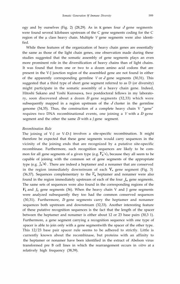
Recombination RuleThe joining of V-J or V-D-J involves a site-specific recombination. It mighttherefore be expected that these gene segments would carry sequences in thevicinity of the joining ends that are recognized by a putative site-specificrecombinase. Furthermore, such recognition sequences are likely to be com-mon for all gene segments of a given type (e.g. because they all seem to becapable of joining with the common set of gene segments of the appropriatetype (e.g. . There are indeed a heptamer and a nonamer that are conservedin the region immediately downstream of each gene segment (Fig. 3)(36,37). Sequences complementary to the heptamer and nonamer were alsofound in the region immediately upstream of each of the four gene segments.The same sets of sequences were also found in the corresponding regions of the
and gene segments (36). When the heavy chain V and J gene segmentswere analyzed subsequently they too had the common conserved sequences(30,31). Furthermore, D gene segments carry the heptamer and nonamersequences both upstream and downstream (32,33). Another interesting featureof these putative recognition sequences is the fact that the length of the spacerbetween the heptamer and nonamer is either about 12 or 23 base pairs (30,3 1).Furthermore, a gene segment carrying a recognition sequence with one type ofspacer is able to join only with a gene segmentwith the spacer of the other type.This 12/23 base pair spacer rule seems to be adhered to strictly. Little iscurrently known about the recombinase, but proteins with an affinity tothe heptamer or nonamer have been identified in the extract of Abelson virustransformed pre B cell lines in which the rearrangement occurs in vitro at arelatively high frequency (38,39).
Physiology or Medicine 1987
Figure 3. Putative recognition sequences for the rearrangement of immunoglobulin and T cellreceptor genes. The conserved heptamer and nonamer sequences and the length of the spacerbetween these sequences are schematically illustrated for immunoglobulin (Panel a) and for T cellreceptor (Panel b) gene families. The sequences shown on top are consensus sequences. Individualsequences may deviate from these consensus sequences by a few nucleotides.
Diversity generated at the joinsWhen the deduced amino acid sequence of a germline gene segment wascompared with the determined amino acid sequences of those χ chains that areencoded in part by that gene segment, it was noticed that the 5’ end of the gene segment is not prefixed but rather shifts toward upstream or downstreamby several base pairs in different joining events (36,37). This flexibility in theprecise site of the joining was subsequently found to be a characteristic of thejoining ends of other gene segments rather than of just JX -gene segments (31).It applies even when the same pair of gene segments were joined in different B
Somatic Generation of Immune Diversity 391
cell precursors, such that the completed V “genes” are likely to have slightlydifferent codons in the junction regions.
The V-D and D-J junctions exhibit diversity of yet another type. We foundthat up to a dozen base pairs of essentially random sequence are inserted inthese junctions apparently without a template during the breakage and reunionof the recombining gene segments (32,33). While the precise mechanism is yetunknown, the terminal deoxynucleotide transferase which is found in early Blymphatic nuclei or an enzyme with similar characteristics is thought to play arole in this phenomenon, (40).
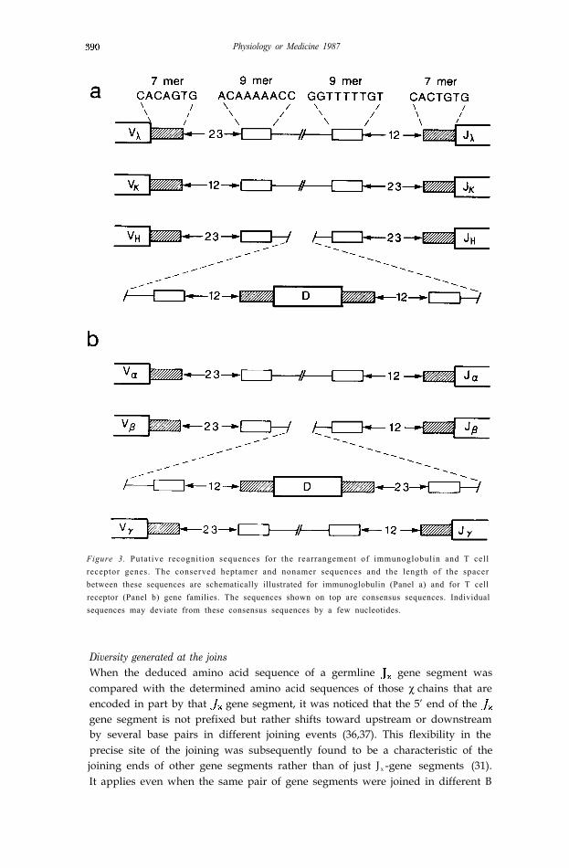
The part of the V region affected by the above two diversification mecha-nisms is limited. But this does not mean that they do not play a significant rolein the determination of antibody specificity. On the contrary, the junctionsencode the most variable two of the six loops of polypeptides that make up theantigen binding region of the antibody molecule (Fig. 4). Furthermore,specific cases are known where the affinity of an antibody to a defined antigen isdrastically altered by a slight change in one junctional sequence (41). Thus, thejunctional variation is also a potent somatic generator of antibody diversity.
Figure 4. Space-filling, stereo image of an antibody combining site. Atomic coordinates of mouseimmunoglobulin MOPC 603 (104) were used to produce the picture. The heavy chain variabledomain is color-coded dark grey, the light chain variable domain light grey. The hypervariableregions [except the VH third hypervariable region) are blue, the heavy chain segment coded for bythe D gene is red, and the heavy and light chain segments coded for by the J genes are yellow. TheD segment corresponds virtually exactly to the third heavy chain hypervariable region; hypervari-able regions were defined as in Novotney et al. (105) except for the heavy chain second hypervaria-ble region, which is marked as defined by Kabat et al . (25). The antigen of this particularimmunoglobulin, phosphoryl choline, binds into the cavity in the middle of the picture in betweenthe VH and VL domains, making contacts to amino acid residues belonging to the VH and Jsegments of the heavy chain and the VL segment of the light chain. Importance of the D segment iswell illustrated in the two crystallographic structures of antibodies which bind the protein antigenlysozyme (106, 107). There, the contact area contributed by the D segment amount to 50% and24%, respectively, of the total heavy chain contact area. This image was computer-generated byJiri Novotny using the program SPHERE of Robert Bruccoleri.
392
Somatic mutationWhen F. Macfarlane Burnet proposed the clonal selection theory he recognizedthe need for some kind of random genetic process in order to generate anti-bodies able to bind specifically to the vast variety of antigens (42). He consid-ered somatic mutations as the most plausible mechanism. Subsequently, thisidea was adopted and forcefully presented by many including Joshua Lederberg,Niels Kaj Jerne and Melvin Cohn (5,6,7).
The amino acid sequence data accumulated by Martin Weigert in MelvinCohn’s laboratory at the Salk Institute provided an excellent opportunity toexamine directly the role of somatic mutations in antibody diversity (7,11).They had analyzed the λ1 light chains derived from eighteen myelomas. All themice were of an inbred strain BALB/c and should thus have been geneticallyidentical. They found that twelve of the regions were identical but that theother six differed both from the majority sequence and from one another byonly one, two, or three amino acid residues. They proposed that BALB/c micemay carry only one germline “gene” which codes for the majority se-quence, and that all the other regions observed are encoded by somaticmutants of this single “gene” that arose in B cell development. As I alreadymentioned in an earlier section our gene-counting experiment by hybridizationkinetics suggested that the germline BALB/c genome carries no more than afew “genes”. This number was reduced to one when we reevaluated thecopy number by the more reliable Southern blotting method (20). The finalproof of somatic mutation in came when we cloned and sequenced the solegermline gene segment and the rearranged λ1 genes expressed in a myeloma(23). As Weigert and Cohn guessed the nucleotide sequence of the germline gene segment corresponded to the major amino acid sequence, while the λ1
gene expressed in the myeloma had been altered by single base changes.Since this work several subsets of χ light and heavy chains and their germline
V gene segments have been analyzed by cloning and sequencing (43-46).These results have all confirmed that somatic mutations further amplify thediversity encoded in the germline genome. Particularly revealing was theanalysis carried out by Patricia J. Gearhart, Leroy Hood and their coworkersfor the regions associated with the binding of phosphorylclorine (PC). Theydemonstrated the single base changes can be extensive and yet are restricted tothe joined VDJ sequences and the immediately adjacent regions (47,48).
Developmental control of rearrangement and hypermutationWhy have two extraordinary somatic genetic mechanisms, recombination andhypermutation, evolved in the immune system in order to carry out whatappears to be one task, namely to diversify antibodies?
I believe that the answer may be the differential roles of these two geneticmechanisms. Thanks to the efforts of several independent groups of cellularand molecular immunologists a general picture is emerging that describes therelationship between the stages of B cell development and the occurance ofsomatic recombination or mutation (Fig. 5) (49-55). Somatic recombina-tions contributing to diversity are initiated first for the heavy chain and then for
Somatic Generation of Immune Diversity 393
Figure 5. Differentiation of B cells. Note that the receptors present on the memory cells and theantibody molecules secreted by the plasma cells of the secondary response have a tighter fit to theantigen than the receptors on the ancestral virgin B cells or the antibodies secreted by the plasmacells of the primary response. See text for the full explanation.
the light chain during the differentiation of progenitor cells, and the completionof somatic recombination is accompanied by the appearance of virgin B cells(56-58). These B cells form clones each of which is composed of cells bearinghomogeneous IgM molecules as surface receptors. Thus, somatic recombina-tion is completed prior to any possible interaction of a B cell with antigens.
When an antigen enters the lymphatic system for the first time, it will bescreened by these virgin B cells. The small fraction of these B cells that happento have sufficient affinity for the antigenic determinants in question will re-spond and follow either of two pathways: they will produce the primaryantibody response, or they will contribute to the generation of memory B cells.
394 Physiology or Medicine 1987
In the former pathway, the selected B cells will proliferate and differentiate intoantibody-secreting plasma cells. During this process, the C region of the heavychain can switch from µ to another class, but mutation is rare in either theheavy or the light chain V region. Consequently, the antibodies secreted byplasma cells in the primary response would largely have the same V regions asthe immunoglobulin receptors on the virgin B cells from which they derive.
By contrast, immunoglobulin remains in the cell surface receptor formduring the other pathway taken by the antigen-activated virgin B cells, namelythe generation of memory B cells. ‘During this process the hypermutationapparatus appears to be most active, and the rate of the mutation approaches10-3 base substitution per cell per generation. Antigen selects, in a stepwisefashion, better and better fitting mutants so that the immunoglobulins on thesurface of memory B cells achieve a substantially higher affinity than theimmunoglobulins on the ancestral virgin B cells. Switch recombination alsooccurs frequently during this process too. When the same antigen as the onethat elicited the primary response reenters the body, the memory B cells areselectively propagated and differentiate into plasma cells. This is the so calledsecondary antibody response, which, therefore, consists of high affinity anti-bodies of “mature” isotype, and these antibodies show extensive somatic muta-tion in their V regions. Somatic mutations appear to cease after memory cellsare generated, and little or no further mutation takes place during the secon-dary antibody response.
This scheme of B cell differentiation can be rephrased as follows. An organ-ism is prepared for infection with pathogens bearing virtually any antigens witha large variety of resting B cells. These B cells bear unique immunoglobulinreceptors encoded by one copy each of complete light and heavy chain genesthat have been constructed by a random or quasi-random assembly of theinherited gene segments. Since the assembly occurs independent of antigens,and since the inherited gene segments are not usually selected during evolutionfor precise lit to most antigens, the antibody secreted by the plasma cellsderived directly from the selected resting virgin B cells during a primaryantibody response usually have a relatively low affinity. By contrast, thefrequent single base changes that occur during the generation of memory Bcells provide the organism with a great variety of finely altered immunoglobu-lin receptors from which only those with the best fit to the antigen in questionwill be selected. Since the plasma cells generated during the secondary anti-body response are mostly direct descendants of these memory B cells, havingno further alterations in the antigencombining sites, these antibodies usuallyexhibit a much higher affinity for the antigen than do the primary antibodies. Thisexplains the long known phenomenon of affinity maturation of antibodiesduring the course of repeated immunizations (59).
Thus, somatic creation of antibody genes can be viewed as a two step process.In the first step, blocks of gene segments are employed to build, in an antigen-independent fashion, a set of genes coding for antibodies of great diversity butwith low affinity. In the second step, once the antigen is defined, a smallselected set of B cells bearing low affinity antibodies as cell surface receptors
Somatic Generation of Immune Diversity 395
undergo somatic mutations with the result that a fraction of them develop ahigher affinity to that antigen and can be selected for further expansion. Thisprocess improves the ability of the immune system to detect a low concentra-tion of antigens. One wonders what happens to those cells in which mutationdid not improve affinity. A recent study suggests that at least some of these cellsmaybe set aside for selection by different antigens (54). Thus, somatic mutationmay also contribute to the repertoire of receptors specific for antigens notpreviously introduced into an immune system.
T cell receptorsAs the mystery of the genetic origin of antibody diversity was unravelled atleast in its basic outlines, it seemed natural to extend our research to “the otherhalf” of the lymphoid system, namely T cells. Although WC often discussed theidea of research on the nature of antigen recognition by T cells in the laboratoryin the late seventies while I was still in Basel, the real work did not start untilthe early eighties in my new laboratory at M.I.T. Although T cells were knownto recognize and distinguish antigens as precisely as B cells, nothing was knownabout the biochemical nature of the molecules responsible for this task, namelyT cell receptors (TCR). This lack of information was in stark contrast to thewealth of information about antibodies. Much debate took place among cellu-lar immunologists on the nature of these molecules. Some argued that T cellreceptors are just another class of immunoglobulins. Others thought T cellreceptors would have to be quite different from immunoglobulins. Indeedstudies carried out in the late seventies had shown that the way in which a Tcell recognizes an antigen is quite different from the way a B cell does: The Tcell reacts to antigens on a cell surface, and the T cell receptor simultaneouslyrecognizes both an antigen and a determinant present on a glycoproteinencoded by a gene in the major histocompatibility complex (MHC) (60-62).This discovery raised another issue: Does a T cell recognize two determinantswith one receptor or does it have two receptors, one for antigen and the otherfor a MHC product?
The receptor protein was first detected in 1983 in cxpcriments carried out bythree independent groups of scientists headed by James P. Allison, Ellis L.Reinherz, and Phillipa Marrack and John Kappler (63-65). They preparedantibodies that bind to a protein on the T cell surface. Since these proteinswere similar but exhibited clonally distributed structural diversity, they werethought to be a good candidate for the receptor. Furthermore, the antibodiesthey prepared were T cell clone-specific, and they could show that theseantibodies blocked activation of the T cell clone in a clone-specific fashion. Thereceptor identified by these experiments was composed of two polypeptidesubunits, designated a and b, that are held together by a disulfide bond. Thesestudies were critical in the sense that the receptor was finally identified, itsoverall structure defined, and its predicted structural variability confirmed.However, the paucity of the protein on the T cell surface and the absence of thesecreted form of the receptor made it very difficult to obtain further informationabout the structure of this molecule, especially its amino acid sequence.
396 Physiology or Medicine 1987
α and ß genesIn the meantime, molecular biologists were attempting to identify the genescoding for the T cell receptor. This turned out to be a much more difficultendeavor than the cloning of immunoglobulin genes. Although T cell lines andhybridomas expressing a homogeneous receptor were becoming available,these cells were more difficult to grow than myelomas, and the amount of thereceptor made was at least two orders of magnitude less than the amount ofimmunoglobulin produced by a myeloma cell. In 1984, Mark Davis and hiscoworkers at Stanford University and Tak Mak and his coworkers at theUniversity of Toronto independently made a breakthrough (66-68). Theirexperimental strategy depended on two assumptions. First that mRNAs codingfor the α and β polypeptide chains are present in a T cell hybridoma or T celltumor but are absent in B cell tumors. And second that the α and β chain genesare rearranged in T cells in a manner similar to the immunoglobulin genes in Bcells. Thus, they made a library from the fraction of T cell-cDNA that did nothybridize with B cell-derived mRNA and tested each T cell-specific cDNAclone for rearrangement of the corresponding gene in T cells. As the source of Tcells, Davis’ group used a hybridoma obtained by fusing a mouse helper T cellrecognizing antigen plus self class II MHC molecules with a T cell tumor whileMak’s group used a human T cell tumor. The two groups came up with onecommon class of cDNA clones that satisfied the above criteria. The nucleotidesequence showed that the corresponding polypeptide chain is significantly(30-35%) homologous to immunoglobulin chains. Furthermore, the cDNAclones contained sequences homologous to V and C regions in the correctorientation. Thus, it seemed certain that the gene represented by this class ofcDNA clones codes for one of the two subunits of the T cell receptor. That thisgene encodes the β subunit was soon confirmed by determination of the partialamino acid sequence of the human β chain (69).
In my laboratory at M.I.T. Haruo Saito and I collaborated with DavidKranz and Herman Eisen to isolate both α and β cDNA clones from anothertype of T cell, namely a cytotoxic T cell clone specific for class I MHCmolecule. In 1984, using a modified subtractive cDNA library method, weidentified two classes of cDNA clones that also satisfied the criteria for a T cellreceptor gene (70). One class of these clones represented the β subunit. Takentogether with the earlier finding by Davis’ group this demonstrated that thetwo major classes of T cells, helper T cells and cytotoxic T cells, employ thesame set of genes at least for the β subunit. The same conclusion was drawnsubsequently for the α subunit. This point is significant because the two typesof T cell are specific for two distinct subclasses of MHC gene products. Thus,the same T cell receptor genes mediate recognition of both class I and class IIMHC.
The polypeptide chains encoded by the other class of rearranging T cell-specific cDNAs isolated by Saito and myself was also homologous to immunog-lobulin chains by 30-35%. These cDNA clones were, however, clearly distinctfrom ß cDNA clones because the polypeptide chains encoded by the two sets ofcDNAs were homologous by only 30-35% not only in the V regions but also in
Somatic Generation of Immune Diversity 397
the C regions. Since only two subunits, α and β, were known for the T cellreceptor, we initially proposed that this second class of cDNAs represented theα gene (70). However, even before the work was published a question aroseabout the assignment of this gene as encoding the α chain. The putative αcDNAs do not carry codons for N-linked glycosylation sites, while unpublishedstudies from Charlie Janeway’s laboratory at Yale University and from JimAllison’s at the University of California, Berkeley, indicated that both α and βsubunits of at least some T cells carry N-linked carbohydrates. While it wasstill possible that the apparent discrepancy in glycosylation could be explainedby differences in the type of T cell or in mouse strains used, continued screeningof our subtracted cDNA library yielded within a few weeks a third class ofclones whose genes also rearrange specifically in T cells (71). This gene notonly was as homologous to immunoglobulin genes as the first two classes of Tcell-specific genes, but also had two potential sites for N-linked glycosylation,and therefore was a better candidate for the a gene. This proposition was soonconfirmed by comparing its nucleotide sequence with the partial amino acidsequence of the human subunit (72). Furthermore, the a gene was alsocloned at about the same time’ from a helper hybridoma (73).
Once cDNA encoding the α and β chains were identified, it was straightfor-ward to determine the organization of the corresponding genes in genomicDNA. These studies demonstrated that both α and β genes are organized in thegermline genome and rearranged in T cells in a way remarkably similar to theimmunoglobulin genes (74-77). Thus, the organism inherits the genetic infor-mation for these polypeptide chains as and gene segments or and
gene segments, and a random assembly of these gene segments occursexclusively during T cell development to generate a diversity comparable tothat of immunoglobulins for receptors expressed on the surface of mature Tcells. Even the presumed recognition sequences for the site specific recombin-ase, the so called heptamers and nonamers with a 12 or 23 base pair spacer,seem to be common for both the immunoglobulin and T cell receptor genes(Fig. 3).
The complete primary structure of a T cell receptor can be deduced from thenucleotide sequences of the α and β cDNA clones. Its comparison with theprimary structure of an immunoglobulin molecule suggests that the externalpart of the receptor is composed of four compact, immunoglobulin-like globulardomains associated in two non-covalently bound pairs and andfurther stabilized by an interchain disulfide bond between the C domain andthe transmembrane region. This extracellular part of the receptor is anchoredon the membrane lipid bilayer through two transmembrane peptides, one eachfrom the α and β chains (Fig. 6) (70).
Determining the structure and organization of genes encoding the T cellreceptors settled the issue of their relationship with immunoglobulins andaccounted for the genetic origin of their diversity. However, these studies didnot illuminate the mechanism by which these receptors can accomplish thedual recognition of an antigen and a MHC determinant. This last issue isparticularly tantalizing because recent studies using a technique for injecting T
398 Physiology or Medicine 1987
Figure 6. Diagram showing the subunit structures of T cell receptors αβ (panel a) and γδ (panel b)as deduced from the nucleotide sequences of cDNA clones. The αβ receptor is from an alloreactivecytotoxic mouse T cell clone, 2C and the γδ receptor from a mouse thymocyte hybridoma, KN6,prepared by Osami Kanagawa of Lilly Research Laboratories, La Jolla, California. Intra- and
inter-chain disulfide bonds are indicated. The receptors are thought to be anchored on themembrane lipid bilayer by transmembrane peptides. The invariant CD3 complex associated withthe heterodimers are not shown.
cell receptor genes into cloned, functional T cells confirmed that the α βheterodimer alone is sufficient to mediate this dual specificity (78). In order tounderstand how the heterodimer simultaneously recognizes the two determi-nants much more information is needed as to the structure of the receptor andits compound ligand. It seems that the ultimate solution has to come from X-raycrystallographic analysis of the receptor protein.
A new T cell receptor, Since it was established that the third T cell-specific rearranging gene discov-ered was for the α subunit, the second one became an orphan. This gene is soclosely related to the other two genes, however, that it seems certain that itmust have some role in recognition by T cells. Nevertheless, previous immuno-chemical studies did not reveal any polypeptide chain that might be consideredas a candidate for the protein product of this gene. The γ gene is also assembledsomatically from I’, J, and C gene segments and shares a number of characteris-tics with the a and β genes as well as with immunoglobulin genes (79,80).
A number of possibilities were considered initially as firsthand roles for theputative γ chain. For instance, it was thought that the γ chain may be a subunitfor a second T cell receptor coexpressed with the αβ heterodimer. This hypoth-
Somatic Generation of Immune Diversity 399
esis is in line with the two receptor model of dual recognition of antigen andMHC by T cells. Another possibility proposed was that there may be a switchin the subunits of the T cell receptor during T cell development. A model wasproposed in which a T cell receptor composed initially of a γδ heterodimerswitches to an αβ heterodimer as T cells differentiate in the thymus (81,82).This model, which seemed to be supported by the time course kinetics ofappearance of α−, β−, and γ-specific RNA in developing hymocytes, was anattempt to explain an apparent dilemma in the intrathymic selection of the Tcell repertoire (for instance see ref. 83).
Subsequent studies carried out in my and several other laboratories, howev-er, revealed a number of features of the y gene and its expression which are notnecessarily consistent with these hypotheses. First, the γ gene is not rearrangedin some T cell clones or hybridomas. Furthermore, even in many of those Tcells in which this gene is rearranged, the joining of the V and J gene segmentsdoes not allow the J region codons to be translated in phase with the V regioncodons (84-86). Thus, the γ gene product does not seem to be universallyexpressed in conventional, αβ receptor-positive cytotoxic and helper T cells.Second, the γ polypeptide chain is expressed on the surface of a small (less than0.5%) subset of peripheral T cells as a component of a heterodimer referred toas γδ (87-89). The majority of these T cells exhibit none of the CD4 or CD8glycoproteins of conventional αβ receptor T cells on their surface, and thereforebelong to a distinct cell population. Third, like the αβ heterodimer, the γδheterodimer is associated relatively tightly with another glycoprotein, CD3(87). The invariant CD3 protein complex contains a subunit that seems to playa critical role in the transmission of the signal received by the variable hetero-dimer into the cell (90). Thus, the similarly between the γδ− and ab-receptorheterodimer includes both their structure and transmembrane signal transmis-sion. Fourth, ψδ−bearing cells are relatively abundant in the CD4-, CD8-
fraction of fetal and adult thymocytes (91-94). For instance, thymocytes of 16-day old fetal mice, which are mostly double negative cells (i.e. CD4 - and CDB-),are a relatively rich source of γδ-bearing cells. Since double negativethymocyte populations contain precursors for mature, functional, Tcells (95), a natural question that arises is whether γδ-bearing thymocytes areprecursors for αβ-bearing T cells. Fifth, another major site of expression of they&receptor is epidermal tissues. It was recently found by two groups that thistissue contains Thy-l+ (another cell surface marker shared by all types of Tcel ls ) , a n d C D 8-cells bearing (96,97). Unlikeconventional T cells, these cells look more like dendritic cells and are thereforereferred to as dendritic epidermal cells (DEC). Finally, the δ gene also under-goes rearrangement. D, J, and C gene segments for δ polypeptide chains haverecently been mapped within the a gene family between and genesegments (98). The nested configuration of α and δ gene segments is intriguingand provokes curiosity about the possible relationship of gene organizationwith the regulation of the rearrangement and expression of the two types ofgenes. Another intriguing question is whether and to what extent the pool of and gene segments overlap.
400 Physiology or Medicine I987
Despite the rapid progress made in the characterization of the y and δ genesand their products, the most intriguing problem, namely the physiological roleof the γδ receptor-bearing cells is currently unknown. One can only speculateon this matter using the currently available information. As to the ligand of thisnew receptor, it is probably correct to emphasize the fact that the receptorshares with the immunoglobulin B cell and the αβ T cell receptors the samegenetic basis for somatic diversification. It therefore is likely that the ligand inquestion will also exhibit structural diversity. In this respect it is interestingthat recent studies by us and others suggest that at least part of the ligand isencoded in MHC (99,100). The effector function of the cells bearing γ δreceptors has not yet been defined with certainty, but recent studies usinghuman and mouse γδ cell clones suggest that many of these cells have cytotoxiccapacity (89,100,101). The finding of a new type of T cell with an apparentlydistinct set of T cell receptors in epidermal tissues stimulates one’s imagina-tion. It may be that occurrence of this type of T cell is not restricted to the outerepithelial tissues but extends to all epithelial layers including the lining ofvarious internal organs (C. Janeway, pers. commun.). If so, these cells mayhave evolved to protect the part of the body that is most vulnerable to infection,namely its external and internal epithelial surface that are in direct contactwith the environment. However, the prominence of γδ cells in the thymussuggests an intrathymic role of these cells as well. An exciting possibility thathas not been ruled out is a role for these cells in the intrathymic selection ofappropriate αβ T cells.
Concluding remarksUse of restriction enzymes and recombinant DNA methods allowed resolutionof a long standing and central issue in immunology, the genetic origin ofantibody diversity. It turned out that an organism does not inherit even a singlecomplete gene for antibody polypeptide chains. Rather, the genetic informationis transmitted in germline as no more than several hundred gene segments.Through a series of specialized somatic recombinations occuring specificallyduring the differentiation of B lymphocytes, these gene segments are assembledinto tens of thousands of complete genes. Somatic hypermutation occurring inthese assembled genes further diversifies antibody polypeptide chains, so that Bcells displaying immunoglobulin receptors having a better lit to a given antigencan be selected in a later phase of B cell differentiation. Thus, in the immunesystem, organisms have exploited two major processes for modification ofDNA, recombination and mutation, as means to diversify somatically thelimited amount of inherited genetic information in order to cope with the vastlydiverse antigen universe.
Why has somatic diversification been necessary in the evolution of theimmune system? Microorganisms and substances produced by them are theprimary source of biologically relevent antigens against which vertebrates needto produce antibodies for survival. Since the generation time of microorganismsis several orders of magnitude shorter than that of vertebrates, the former canproduce genetic variants much faster than the latter. Thus, if genetic alterations
Somatic Generation of Immune Diversity 401
in the germline genome were to be the only source of antibody diversity,vertebrates would be unable to deal effectively with the rapidly changing worldof antigens. Somatic diversification allows the individual organism to generatea virtually limitless number of lymphocyte variants. Like organisms in anecosystem, these lymphocytes are subject to selection by antigens and the fittestwill survive. Thus, as Jerne and Burnet were aware, the individual immunesystem can be conceived of as a kind of Darwinian microcosm.
The molecular biological approach played an even more fundamental role inthe analysis of the T cell receptor in that very little structural informationexisted prior to the cloning of the receptor genes. It was demonstrated that thepolypeptide chains composing the receptor protein are encoded by genes thatshare a common ancestor with the immunoglobulin genes. Like immunoglobu-lins, T cell receptors are diversified by somatic recombination, but unlikeimmunoglobulins, these receptor proteins have not been observed to undergo further diversification by somatic mutation. The reason for this difference isunknown, but it seems likely that the explanation will be as follows. First,unlike immunoglobulins, T cell receptors function exclusively as cell surfacereceptors which are specialized for interacting with cellbound antigens. Sinceboth ligand and receptor are distributed in two dimensional space when a Tcell interacts with an antigen-presenting cell, and as T cells have mechanismsfor transiently adhering to other cells, these receptor-ligand interactions occurunder conditions of high local concentration. Thus, improvement of the affinitybeyond the one accomplished by somatic rearrangement may not be necessaryin T cell recognition.
Second, the ligand consists in part of an essentially invariant component, selfMHC. As the T cell receptor is selected for self MHC recognition, both duringdevelopment and during immunization, the extreme variability availabe viasomatic mutation may not only be unnecessary but even disadvantageous.Third, and probably more important, T cells appear to be selected early indevelopment for self tolerance, the inability to recognize self antigen. Somaticmutation during antigenic stimulation, as occurs in B lymphocytes, could leadto the development of autoreactivity. While autoreactive B cells depend uponthe additional presence of autoreactive helper T cells to generate autoimmun-ity, this is not true of autoreactive T cells, which can directly produce disease.Thus, Ehrlich’s famous concept of “honor autotoxins”, originally developed forantibodies, is probably critical only for T cells. It will be interesting to examineautoreactive T cell receptors for any evidence of post-thymic somatic diversifi-cation.
Finally, it is interesting to notice that during the fifteen years in which I havestudied the immune system, the role of molecular genetics in immunologicalresearch has altered radically. When I started investigating the problem ofantibody diversity, there was abundant information about the structure andfunction of antibody molecules, while virtually nothing was known about theirgenes. By contrast, in the most recent study on T cell recognition no geneproduct was known at all when the rearranging gene, γ was discovered. Fromthe structure of the gene and its rearranging behavior, it was deduced to be a
402 Physiology or Medicine 1987
receptor gene, and this discovery has led directly to new insights into T celldevelopment and T cell biology. This short history of research in one area,lymphocyte receptors, is yet another witness to the power of DNA technology,and to the ability of this approach not only to explain known biologicalphenomena, but also to contribute to the discovery of new biological systems.
AcknowledgementThe work summarized in this article is the result of collaboration with manycolleagues, students and technical assistants. I wish to extend my sincerethanks to everyone of them. I also extend my thanks to Hoffman LaRocheCompany which so generously supported my work in Basel. My special thanksare extended to Charles A. Janeway Jr., Nancy Hopkins and Yohtaroh Taka-gaki for many useful comments on the manuscript, to Jiri Novotny for prepar-ing Figure 4, and to my secretary, Eleanor Lahey Basel, for her tirelessdevotion.
REFERENCES
1.2.3.
4.5.6.7.
8.
9.10.11.
12.13.14.15.16.
17.
18.
19.
20.
21.
Porter, R.R., Science l80: 713-716 (1973).Edelman, G.M., Science l80: 830-840 (1973).Hilschmann, N. and Craig, L.C., Proc. Natl. Acad. Sci. USA 53: 1403-1409(1965).Hood, I,., and Talmage D.W. Science 168: 325-334 (1970).Lederberg, J. Science 129: 1649-1653 (1959).Jerne, N.K. Eur. J. Immuno. 1: l-14 (1971).Cohn, M. Blomberg. B., Geckeler, W., Raschka, W., Riblet, R. and Weigert, M.in “The Immune System: Genes, Receptors, Signals” (E.E. Sercarz, A.R. Wil-liamson and C.F. Fox. eds, PP 89 Academic Press, New York and London (1974).Gelderman, A.H., Rake, A.V., and Britten, R.J. Proc. Natl. Acad. Sci. USA68: 172-176 (1971).Bishop, J.O. Biochem. J. 126: 171-185 (1971).Tonegawa, S., Proc. Natl. Acad. Sci. USA, 73: 203-207, (1976).Weigert, M., Cesari, M., Yonkovich, S.J. and Cohn, M. Nature 228: 1045-1047 (1970).(1970).Campbell, A.,Advan. Genet. 11: 101 (1962).Dryer, W.J., and Bennett, J., Proc. Natl. Acad. Sci. USA 54, 864-869 (1965).Danna,J.J., Sack Jr.. G.H., and Nathans, D. J. Mol. Biol. 78: 363 (1973).Southern, E., J, Mol. Biol. 98: 503 (1975).Hozumi, N., and Tonegawa, S., Proc. Natl. Acad. Sci., USA, 73: 3628-3632,(1976).Thomas, M., White, R.L. and Davis, R.N. Proc. Natl. Acad. Sci. USA 73: 2294-2298 (1976).Tonegawa. S.. Brack, C., Hozumi, N. and Schuller, R., Proc. Natl. Acad. Sci.USA. 74: 3518-3522 (1977).Tonegawa, S.. Maxam. A.M. Tizard, R., Bernard, O., and Gilbert. W., Proc.Natl. Acad. Sci. USA, 75: 1485-1489 (1978).Brack, C. Hirama. M . Lenhard-Schuller, R., and Tonegawa, S. Cell, 15: l-14(1978).Lenhard-Schuller, R.. Hohn. B.. Brack, C., Hirama, XI., and Tonegawa, S., Proc.Natl. Acad. Sci. USA, 75: 4709-4713, (1978).
Somatic Generation of Immune Diversity 403
22. Brack, C. and Tonegawa, S., Proc. Natl. Acad. Sci. USA, 74: 5652-5656, (1977).23. Bernard, O., Hozumi, N., and Tonegawa, S., Cell, 15: 1133-1144 (1978).24. Weigert, M., Gatmaitan, L., Loh, E., Schilling, J., and Hood, L., Nature 2 7 6 ,
785-789 (1978).25. Kabat, E.A., WU, T.T.. Bilofsky, H., Reid-Miller, M., and Perry. H., U.S. Dept.
of Health and Human Services Publication (1983).26. Seidman, J.G., Leder, A., Nau, M., Norman, B., and Leder, P., Science 202: 11-16
(1978).27. Cory, S., Tyler, B.M., Adams, J.M. J. Mol. Appl. Genet. 1: 103-116 (1981).28. Davis, M., Calame, K., Early, P., l.ivant, D., Joho, R.. Weisman, L, and Hood, L.
gawa, S., Proc. Natl. Acad. Sci. USA, 77: 2138-2142 (1980).30. Early, P., Huang, H., Davis, M., Calame. K., and Hood, L., Cell 19: 981-092
(1980).31. Sakano, H., Rlaki, R., Kurasawa, Y., Roeder, W., and Tonegawa. S., Nature
286: 676-683 (1980).32. Sakano. H., Kurosawa, Y., Weigert. M., and Tonegawa, S., Nature. 290: 562-565
(1981).33. Kurosawa, Y., von Boehmer, H., Haas, W., Sakano, H., Traunecker, A., and
Tonegawa, S., Nature 290: 565-570 (1981).34. Kurosawa, Y., and Tonegawa, S.,J. Exp. Med. 155: 201-218 (1982).35. Wood, C., and Tonegawa, S., Proc. Natl. Acad. Sci. USA 80: 3030-3034 (1983).36. Sakano, H., Huppi, K., Heinrich. G., and Tonegawa, S., Nature 280, 288-294
(1979).37. Max. E.E., Seidman, J.G., and Leder, P., Proc. Natl. Acad. Sci. USA 76: 3450-
3454 ( t 979).38. Halligan, B.D., and Desiderio, S.V. Proc. Natl. Acad. Sci. USA 84: 7019-7023
(1987).39. Aguitera, R.J., Akira, S., Okagaki, K. and Sakano, H. Cell (in press)40. Alt, F., and Baltimore, D., Proc. Natl. Acad. Sci. USA 79: 4118-4I22 (1982).41. Azuma, T., lgras, V., Reilly, E., and Eisen, H.N.. Proc. Natl. Acad. Sci. USA
81: 6139-6143 (1984).42. Burnet, F.M. The Clonal Selection Theory of Acquired Immunity. London:
Cambridge Univ. (1959).43. Bothwell. A.L.M., Paskind, M., Reth, M., lmanishi-Kari. T., Rawesky, K., and
Baltimore, D., Cell 24: 624 (1981).44. Crews, S, Griffin, J., Huang, H., Calame, K., and Hood, L.. Cell 25: 59-66
(1981).45. Givol, D., Zakut, R., Effron, K.. Rechavi, G., Ram, D., and Cohen, J.B., Nature 292: 4 2 6 - 4 3 0 ( 1 9 8 1 ) .46. Selsing, E., and Storb, U., Nucl. Acids Res. 9: 5725-5735 (1981).47. Gearhart, P.J., Johnson, N.D., Douglas, R., and Hood, L. Nature 291: 29-33
(3981).48. Kim, S., Davis, M., Sinn, E., Patten, P., and Hood, I.., Cell 27: 573-580 (1981).49, Askonas, B.A. and Williamson, A.R. Eur. J. lmmunol. 2: 487-493 (1972).50. Berek, C., Griffiths, G.M., and Milstein, C. Nature 316: 412-418 (1985).51. McKean, D., Hüppi, K., Bell, M., Standt: I,., Gerhard, W., and Weigert, M. Proc.
Natl. Acad. Sci. USA 81: 3180-3184 (1984).52. Okumura, K., Julius, M.H., Tsu, T., Herzenberg, L.A., and Herzenberg, L.A.
Eur. J, lmmunol. 6: 467-472 (1976).53. Sablitzberg, M., Kocks, C., and Rajewsky, K. EMBOJ. 4: 345-350 (1985).54. Sickevitz, M., Kocks, C., Rajewsky, K., and Dildrop, K. Cell 48: 757-770 (1987).53. Wysocki, L.J.. Manser, T., and Gefter, M.L. Proc. Natl. Acad. Sci. USA 83: 1847-
1851 (1986).
404 Physiology or Medicine 1987
56. Maki, R., Kearney, J., Paige, C., and Tonegawa, S., Science, 209: 1366-1369(1980).
57. Perry, R.P., Kelley, D.E., Coleclough, C., and Kearney, J.F., Proc. Natl. Acad.Sci. USA 78: 247-251 (1981).