HUMAN DNA POLYMERASE P MUTATIONS ALLOWING EFFICIENT ABASIC SITE BYPASS Sonja Gieseking, Konrad Bergen, Francesca Di Pasquale, Kay Diederichs, Wolfram Welte, and Andreas Marx* From Departments of Chemistry and Biology, Konstanz Research School Chemical Biology, University of Konstanz, 78464 Konstanz, Germany Address correspondence to: Andreas Marx, Department of Chemistry, Universitat Konstanz, Universitatsstral3e 10, D-78464 Konstanz, Germany, Fax: (+49) 7531-88-5140, E-mail: The DNA of every cell in the human body gets damaged more than 50,000 times a day. The most frequent damages are abasic sites. This kind of damage blocks proceeding DNA synthesis by several DNA polymerases that are involved in DNA replication and repair. The mechanistic basis for the incapability of these DNA polymerases to bypass abasic sites is not clarified. To gain insights into the mechanistic basis we intended to identify amino acid residues that govern for the pausing of DNA polymerase P when incorporating a nucleotide opposite to abasic sites. Human DNA polymerase p was chosen since it is a well characterized DNA polymerase and serves as model enzyme for studies of DNA polymerase mechanisms. Moreover, it acts as the main gap filling enzyme in base excision repair and human tumor studies suggest a link between DNA polymerase p and cancer. In this study we employed high throughput screening of a library of more than 11,000 human DNA polymerase p variants. We identified two mutants that have increased ability to incorporate a nucleotide opposite to an abasic site. We found that the substitutions Glu232Lys and Thr233I1e promote incorporation opposite the lesion. Besides this feature the variants have an increased activity and a lower fidelity when processing non-damaged DNA. The mutations described in this work are located in well characterized regions but have not been reported before. A crystallographic structure of one of the mutants was obtained providing structural insights. The DNA of every cell in the human body gets damaged more than 50,000 times a day. I The relation between DNA damage and repair has a significant effect on various cancers, neurological aberrations, and the process of premature aging. 2 DNA polymerases are key enzymes which function in maintaining the integrity of the encoded genetic information in DNA replication, DNA repair, DNA recombination and the bypassing of damages in DNA. Therefore they are central to the aforementioned interplay.3 The most frequent DNA damage observed under physiological conditions are abasic sites resulting from spontaneous hydrolysis of the bond that connects the sugar and the nucleobase in DNA.4 Guanine and adenine nucleobase residues are cleaved most efficiently resulting in the abasic sugar moiety (AP, Fig. 1 A) with the loss of the genetic information stored in the nucleobase. 5 Since these lesions are devoid of the genetic information they are potentially mutagenic. The bulk of this damage is removed by base excision repair (BER) pathway, which uses the sister strand to guide incorporation of the right nucleotide in places of the lesion. 6 ,7 DNA polymerase p acts as the main gap filling enzyme in BER.8 The enzyme governs for selecting the right nucleotide complementary to the undamaged templating nucleotide. 9 DNA polymerases have been grouped in seven different families named A, B, C, D, X, Y and reverse transcriptases. The grouping depends on sequence homology and structural similarity. 10 DNA polymerase p belongs to the X-family. The single polypeptide is the smallest eukaryotic DNA polymerase (39 kDa), containing 335 amino acid residues. II It is folded into discrete domains and subdomains. I2 The enzyme consists of an amino- terminal lyase domain (8 kDa), which is connected to the DNA polymerase domain (31 kDa) by a short protease-sensitive segment. 13 The important role of DNA polymerase p in the maintenance of genome integrity is emphasized by the finding that approximately 30% of all tumors studied to date harbour mutations in the DNA polymerase p gene. 14 The tumor-associated
Transcript
HUMAN DNA POLYMERASE P MUTATIONS ALLOWING EFFICIENT ABASIC SITE BYPASS
Sonja Gieseking, Konrad Bergen, Francesca Di Pasquale, Kay Diederichs, Wolfram Welte, and Andreas Marx*
From Departments of Chemistry and Biology, Konstanz Research School Chemical Biology, University of Konstanz, 78464 Konstanz, Germany
Address correspondence to: Andreas Marx, Department of Chemistry, Universitat Konstanz, Universitatsstral3e 10, D-78464 Konstanz, Germany, Fax: (+49) 7531-88-5140, E-mail: andreas.marx@uni-konst~mz-,~
The DNA of every cell in the human body gets damaged more than 50,000 times a day. The most frequent damages are abasic sites. This kind of damage blocks proceeding DNA synthesis by several DNA polymerases that are involved in DNA replication and repair. The mechanistic basis for the incapability of these DNA polymerases to bypass abasic sites is not clarified. To gain insights into the mechanistic basis we intended to identify amino acid residues that govern for the pausing of DNA polymerase P when incorporating a nucleotide opposite to abasic sites. Human DNA polymerase p was chosen since it is a well characterized DNA polymerase and serves as model enzyme for studies of DNA polymerase mechanisms. Moreover, it acts as the main gap filling enzyme in base excision repair and human tumor studies suggest a link between DNA polymerase p and cancer. In this study we employed high throughput screening of a library of more than 11,000 human DNA polymerase p variants. We identified two mutants that have increased ability to incorporate a nucleotide opposite to an abasic site. We found that the substitutions Glu232Lys and Thr233I1e promote incorporation opposite the lesion. Besides this feature the variants have an increased activity and a lower fidelity when processing non-damaged DNA. The mutations described in this work are located in well characterized regions but have not been reported before. A crystallographic structure of one of the mutants was obtained providing structural insights.
The DNA of every cell in the human body gets damaged more than 50,000 times a day. I The relation between DNA damage and repair has a significant effect on various cancers, neurological aberrations, and the process of premature aging.2
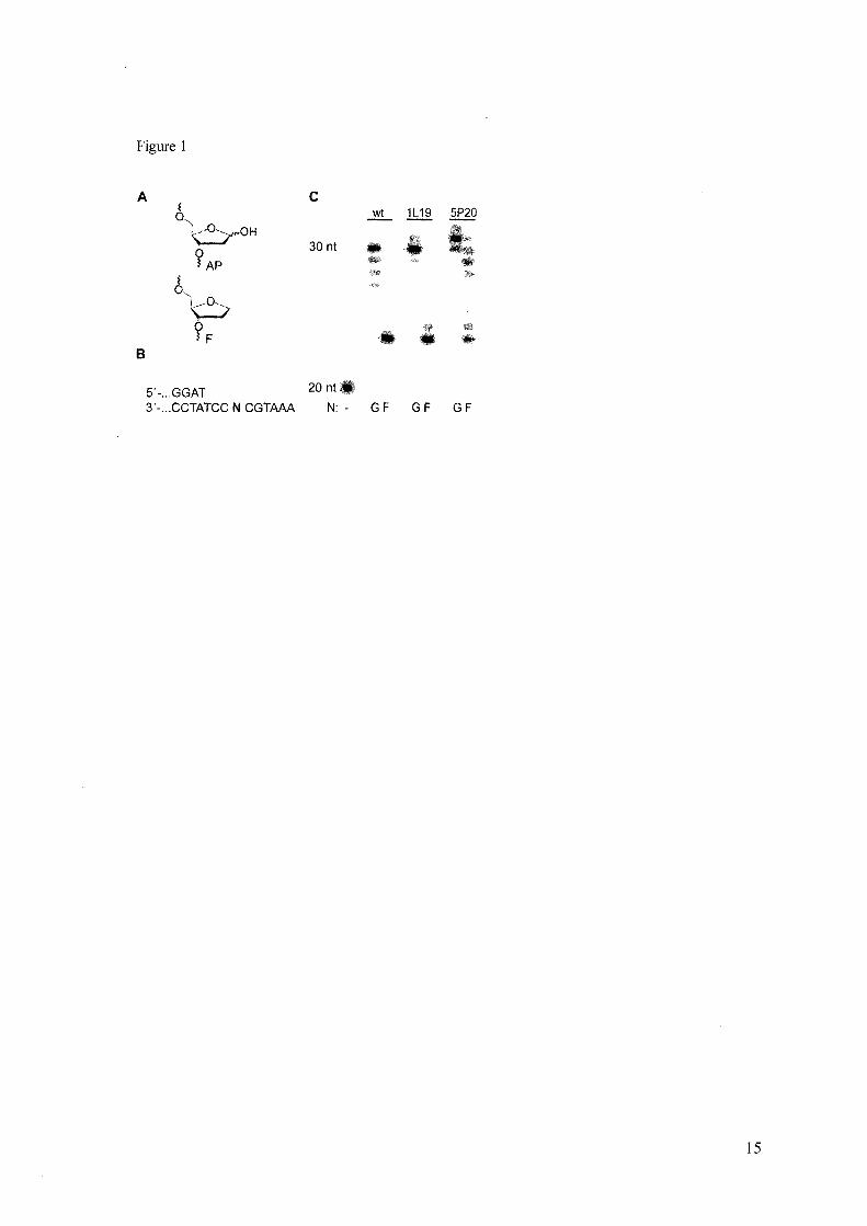
DNA polymerases are key enzymes which function in maintaining the integrity of the encoded genetic information in DNA replication, DNA repair, DNA recombination and the bypassing of damages in DNA. Therefore they are central to the aforementioned interplay.3 The most frequent DNA damage observed under physiological conditions are abasic sites resulting from spontaneous hydrolysis of the bond that connects the sugar and the nucleobase in DNA.4 Guanine and adenine nucleobase residues are cleaved most efficiently resulting in the abasic sugar moiety (AP, Fig. 1 A) with the loss of the genetic information stored in the nucleobase.5
Since these lesions are devoid of the genetic information they are potentially mutagenic. The bulk of this damage is removed by base excision repair (BER) pathway, which uses the sister strand to guide incorporation of the right nucleotide in places of the lesion.6
,7 DNA polymerase p acts as the main gap filling enzyme in BER.8 The enzyme governs for selecting the right nucleotide complementary to the undamaged templating nucleotide.9
DNA polymerases have been grouped in seven different families named A, B, C, D, X, Y and reverse transcriptases. The grouping depends on sequence homology and structural similarity. 10 DNA polymerase p belongs to the X-family. The single polypeptide is the smallest eukaryotic DNA polymerase (39 kDa), containing 335 amino acid residues. II It is folded into discrete domains and subdomains. I2 The enzyme consists of an aminoterminal lyase domain (8 kDa), which is connected to the DNA polymerase domain (31 kDa) by a short protease-sensitive segment. 13
The important role of DNA polymerase p in the maintenance of genome integrity is emphasized by the finding that approximately 30% of all tumors studied to date harbour mutations in the DNA polymerase p gene. 14 The tumor-associated
mutations are found in the lyase as well as in the DNA polymerase domain. The variants show altered enzyme properties such as fidelity and lyase function. 14,15,16,17,18,19,20
On the other side DNA polymerase P serves as a very useful model for studying basic mechanisms of DNA polymerase function such as activity and fidelity. The enzyme has been studied extensively in a functional and structural context in the past. 13
Several crystal structures are available including those of the enzyme in ternary complex with various substrates.9,21,22,23,24
Lesions like abasic sites that remain undetected by the repair systems or are formed during the Sphase remain great challenge to the replication machinery. Abasic sites block proceeding DNA synthesis by several DNA polymerases including those that are involved in DNA replication and BER such as DNA polymerase p.25 Stalling of DNA replication might be essential to allow repair processes to occur that ensure faithful DNA repair without introducing mutations. However, about a decade ago, novel specialized DNA polymerases were identified to be capable in bypassing DNA lesion.26 These enzymes are found in a variety of organisms and e.g. are involved in the suppression of skin cancer in humans. The translesion synthesis pathways are believed to occur after chromosomal replication thus, outside S_phase.27
,28
The mechanistic basis of the proficiency of some DNA polymerases to bypass certain lesions and the failure of other DNA polymerase is still under investigation. In order to identifY amino acid residues that govern for the pausing of DNA polymerase p when incorporating a nucleotide opposite to an abasic sites we generated and screened a library of more than 11,000 human DNA polymerase p variants randomly mutated using error prone PCR. By using a high throughput screening system we selected mutants which have an increased propensity to bypass abasic sites in comparison to the wild-type enzyme. Besides this feature the variants have an increased activity and a somewhat lower fidelity when processing nondamaged DNA. The mutations described in this work are located in well characterized regions but have not been reported before. A crystallographic structure of one of the mutants was obtained providing structural insights.
Experimental Procedures
Library Construction - The human DNA polymerase p gene (NM_002690) was purchased codon optimized for expression in E. coli from Geneart AG, Regensburg. Noteworthy, codon optimization resulted in changes within the gene sequence without affecting the amino acid sequence (see supporting information). The gene was cloned into the plasmid pGDRI I using the restriction enzymes BamHI and Sail. The insert was sequenced to confirm that it does not contain mutations. Random mutations were inserted by error-prone PCR. PCR-reactions were performed using 5 units of Taq DNA polymerase (Fermentas) in the reaction buffer (10 mM TrisHCI (pH 8.3), 50 mM KCI, 7 mM MgClz, 0.05 mM MnCh, 1 mM dCTP/dTTP, 0.2 mM dATP/dGTP) , 350 pglf.ll plasmid pGDRI I (hDNApoIP), and 200 nM of each primer (forward: 5'-CAC CAT CAC CAT CAC CAT ACG GAT CCG ATG-3'; reverse: 5'GCT AAT TAA GCT TGG CTG CAG GTC GAC TTA-3') in a 50 f.ll total volume. 20 cycles ofPCR (95 QC for I min, 63 QC for I min, 72 QC for 2.5 min) were conducted. The PCR-product was isolated by electrophoresis in a 0.8% agarose gel and purified (MinEluteTM Gel Extraction Kit, QIAGEN). After digestion with BamHI and San the restriction fragment was ligated with the digested and dephosphorylated (Antarctic Phosphatase, NEB) plasmid pGDRI I by using T4 DNA Ligase (Fermentas) 16h at 16 QC. The resulting plasmids were transformed into E. coli XL I 0 Gold. Clones were picked from LB agar plates and separately grown overnight in 384 well plates containing 150 f.ll LB medium with 100 f.lg/ml carbenicillin. hDNApolP mutants were parallel expressed in 1 ml cultures in 96 well plates by inducing during exponential growing phase (OD6oo = 0,6) with 1 mM IPTG for 4 hours. The cells were pelleted by centrifugation (40 min, 4000 rpm, 4 QC) and Iysated in 300 f.lllysis buffer (50 mM TrisHCI (pH 8.0), 300 mM NaCI, O. I mg/ml lysozyme). After incubating on ice for 45 min 900 f.ll storage buffer (20 mM TrisHCI (pH 8.0), 100 mM NaCI, I mM DTT, 0.1 mM EDTA) was added followed by another 45 min on ice and centrifugation (40 min, 4000 rpm, 4 QC). The lysate was directly used for screening. Screening - Reaction mixtures for library screening contained 2 mM MnC\z, 200 f.lM of each dNTP, 100 f.lg/ml BSA, 150 nM primer
2
(P20thioAG: 5'-CGT TGG TCC TGA AGG AGG *A*T-3', *= position of phosphorothiolate) and 150 nM template (T90A: 5'-CCG TCA GCT GTG CCG TCG CGC AGC ACG CGC CGC CGT GGA CAG AGG ACT GCA GAA AAT CAA CCT A TC CTC CTT CAG GAC CAA CGT ACA GAG-3' or T90F 5'-CCG TCA GCT GTG CCG TCG CGC AGC ACG CGC CGC CGT GGA CAG AGG ACT GCA GAA AA T CAF CCT ATC CTC CTT CAG GAC CAA CGT ACA GAG-3'; F=abasic site analogue, respectively) in buffer (50 mM TrisHCI (pH 7.8), 70 mM KCI, 1 mM DTT, 100 J.!g/ml BSA, 5% [v/v] glycerol) in 20 J.!I total volume. Reaction mixtures were dispensed in 384 well plates using an automated liquid handling device (Hamilton Microlab Star) followed by addition of 5 J.!I lysate solution. As positive control the DNA polymerase p wt, as negative control the catalytic inactive DNA polymerase p D256A29 in analogously treated cultures of E. coli XL 1 0 gold cells were used. After incubation at 37 QC for 30 min the reaction was stopped by adding 45 J.!I/well SYBR® Green I containing solution (50 mM TrisHCI (pH 7.5), 10 mM EDTA, 4x SYBR® Green J). Fluorescence intensities were quantified using a plate reader (Polarstar Optima, BMG Labtechnologies GmbH) with excitation at 485 nm and emission at 520 nm. Purification of DNA Polymerase f3 - The wt and mutants of the DNA polymerase p were expressed and purified as described.30 The purity of the proteins was >95% as controlled by SDS-PAGE. The concentrations were determined by the Bradford assay. Primer Extension Studies - Reactions contained primer (P20: 5'-CGT TGG TCC TGA AGG AGG AT-3'or P23: 5'-CGT TGG TCC TGA AGG AGG ATA GG-3'), template (T33G: 5'-AAA TGC GCC TAT CCT CCT TCA GGA CCA ACG TAC-3'or T33F: 5'-AAA TGC FCC TAT CCT CCT TCA GGA CCA ACG TAC-3'; F=abasic site analogue), and purified enzyme in storage buffer (20 mM TrisHCI (pH 8.0), 100 mM NaCI, I mM DTT, I mM EDTA, 50% [v/v] glycerol), 200 J.!g/ml BSA, 200 J.!M dNTPs (N) or single nucleotide dATP (A), dCTP (C), dGTP (G) or dTTP (T) and Ix reaction buffer (50 mM TrisHCI (pH 8.0), 20 mM NaCI, 20 mM KCI, 10 mM MgCh, 2 mM DTT, 1% [v/v] glycerol) in a final volume of 20 J.!l. The concentrations of the respective enzyme and the primer template complexes are given in the figure legends. The
reaction mixtures were incubated for the times indicated in the corresponding figures legends at 37 QC and subsequently quenched using PAGE loading solution (80% [v/v] formamide, 20 mM EDT A, 0.025% [w/v] bromophenol blue, 0.025% lw/v] xylene cyanol). The samples were denatured at 95 QC for 5 min analyzed by 12% PAGE containing 8 M urea. Visualization was performed using phosphorimaging. Kinetics - The rate of single turnover, single nucleotide incorporation was determined using the different primer/33mer template DNA duplexes using rapid quench technology by using a KinTek RQF-3 rapid quench flow apparatus (KinTek Corp.). Reactions equal or longer 1 min were quenched manually. Reaction mixtures contained 200 nM primer (P23 or P24); 200 nM template (T33G or T33F), 2 J.!M purified enzyme in storage buffer, 400 J.!g/ml BSA, Ix reaction buffer and different concentrations of dNTP as indicated in the figure legends. The reaction was initiated by mixing equal volumes (15 J.!I each) of the enzyme reaction mixture with a solution containing dNTP in Ix reaction buffer. For reaction times ranging from 0.05 to 10 s the rapid quench instrument was used. The reactions were quenched using 0.3 M EDT A prior to mixing with a PAGE loading solution (80% [v/v] formamide, 0.025% lw/v] bromophenol blue, 0.025% [w/v] xylene cyanol). For reaction times longer than 10 s a manual quench was performed. Samples of the quenched reactions were denatured at 95 QC for 5 min and analyzed by 12% PAGE containing 8 M urea. Quantification was performed using phosphorimaging. The product bands were quantified using the BioRad Quantity One software. For pre-steady state analysis, experimental data were fit by nonlinear regression using the program GraphPad Prism4. The data were fit to a one phase exponential equation [primer + I] = A*(I - exp(-kobs*t)). The observed catalytic rates (I<obs) were plotted against the used dNTP concentration. These data were fitted to a hyperbolic equation to determine the Kd of the incoming nucleotide. The incorporation efficiency is given by kpollKd. The kinetic data results from multiple independently conducted experiments. DNA Binding Assay - The DNA binding affinity was measured using a gel mobility shift assay.31,32 P23 and T33G or T33F were annealed as described above. Up to twelve protein concentrations ranging from 0.125 nM
3
up to 5 JlM, expected to bracket the dissociation constant (Kd), were incubated with 0.1 nM primer template complex in buffer containing 10 mM Tris, pH 7.5, 6 mM MgCh, 100 mM NaCI, 10% glycerol and 0.1 % Nonidet p_40.30 After incubation for 10 minutes at 20°C the samples were loaded onto a nondenaturing 8% polyacrylamide gel (in 7.5 mM Tris borate, 2 mM MgCh, 0.1 mM EDTAi3 which run at 300 V for 1 h. After loading, voltage was reduced to 150V and the gel was run for 1.5 h. Quantification was performed using phosphorimaging. The product bands were quantified using the BioRad Quantity One software. The dissociation constant Kd was derived by fitting the experimental data using nonlinear regression with the program GraphPad Prism4 using the equation: Y = [(m! *X)] / (Kd+ X)+c, where m] is a scaling factor and c is the apparent minimum Y value as described.30
DNA Polymerase Activity Determination - For activity determination primer extension reactions were carried out with varying concentrations of DNA polymerase p wt and mutants. The reaction contained 150 nM primer (P23), 150 nM template (T33GCG), 200 JlM dNTP, 200 Jlg/ml BSA and Ix reaction buffer (50 mM TrisHCI (pH 8.0), 20 mM NaCI, 20 mM KCI, 10 mM MgCh, 2 mM DTT, 1% [v/v] glycerol). Reactions were performed at 37 QC for 10 min, subsequently quenched using PAGE loading solution. Products were separated on a 12% denaturing PAGE. Phosphorimaging was carried out and the obtained intensities for each band were transformed into dNTP conversion. Activity was measured by quantifying the intensity of each band produced by the respective DNA polymerase with a phosphorimager. From this quantification, the amount of incorporated nucleotides was calculated. The total amount of incorporated nucleotides for each reaction equals the sum of incorporated nucleotide of each band. The presented results are from measurements that were independently repeated at least three times. Crystallization - The enzyme mutant was expressed in E. coli BL21 AI carrying the CDS in a pET21 b vector. The recombinant enzyme was purified as described earlier34 with slight modification. Protein concentration was determined by
absorbance at 280 nm (£=23,380 M-! cm-I) and Bradford assay. Enzyme solutions were concentrated up to 20 mg/ml (VivaSpin) and stored at -80QC. Crystals of the binary complex were obtained as described by Pelletier et al.24
against a reservoir solution containing 100 mM MES (pH 6.3), 10% PEG 3350 and 100 mM NaCl in hanging drop vapour diffusion plates (Nextal). Data was collected at PXI (PSI, Villigen, CH) at the PILA TUS 6M detector. Data integration and reduction were performed using XDS.35,36 Crystals had similar lattice constants as PDB ID 9ICW24, and structure determination was therefore initiated by rigid body refinement of 9ICW, using the PHENIX suite.3? As model for refinement PDB ID 9ICW24 for the binary complex was chosen. Subsequent refinement and model building was performed using the PHENIX suite and Cooe8
Molecular graphics were prepared in pyMol.39
Results
Screening - To evolve human DNA polymerase p variants with increased ability to bypass an abasic site we first randomized its gene by error-prone PCR. A library of 11,520 entities was generated by picking colonies. The proteins were expressed and Iysated in multi well plates. For screening of activity we first employed a screen based on primer extension using an undamaged template in 384-well plates.4o,4! Enzymes were screened directly after lysis without further purification steps. As controls we used colonies expressing the wild-type enzyme and the catalytic inactive DNA polymerase p mutant D256A29. DNA polymerase activity was monitored through termination by addition of EDT A and quantification of synthesized double stranded DNA through staining with SYBR® Green 1. Active mutants (39% of all screened entities) were pooled and subsequently tested for ability to bypass an abasic site using a similar screen as described above with the difference that a template that contains the abasic site analogue F (Fig. 1 A) was employed. We identified two DNA polymerase p mutants that exhibited promising properties. We refer to these enzymes as 1 L 19 and 5P20 referring to their location in the library. Sequencing revealed that each enzyme has four mutations (1 L19: Ser44Arg, Phe99Leu, Glu123Lys, Thr233I1e; 5P20: Ser2Gly, Phe99Leu, Glu232Lys, VaI269Met). These mutants were subsequently purified to
4
homogeneity and were subjected to further characterization. Primer Extension and Abasic Site Bypass by Wt and Mutants - In order to compare the ability to bypass F the purified wild-type (wt) and mutated enzymes were subsequently used in different primer-extension studies under identical reaction conditions. In a running start experiment, employing all four dNTPs the DNA polymerases extended the radioactively labelled primer strand by three nucleotides before approaching the abasic site in the template strand (Fig. 1 B). The wt enzyme only sluggishly incorporates a nucleotide opposite the abasic site as shown by a faint band at the respective position after PAGE analysis (Fig. 1 C). The synthesis of longer products resulting from bypass of the abasic site could not be detected. Nevertheless, when an undamaged template was used full-length extension was observed. In the undamaged template a G was chosen instead of the abasic site since it has been shown that G is the predominant source of abasic sites under physiological conditions.s
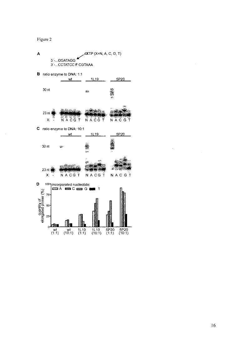
Next, the mutants were investigated. Both mutant enzymes show pausing opposite the abasic site as well however, they were able to synthesize longer products resulting from bypass of the abasic site contrasting the finding for the wt enzyme. Next we investigated which nucleotide is preferentially incorporated opposite the abasic site. Thus the primer template complex was designed in a way that the 3' -end of the primer strand terminates opposite the template (Fig. 2 A). Two different set of experiments were conducted. In one series the enzyme concentration was equal to the concentration of the primer template complex (Fig. 2B). In the second series the enzyme concentration was increased to be 10-fold higher than the primer template complex in order to ensure that all primer template complexes are bound to the enzyme (Fig. 2C). In all single nucleotide incorporation experiments it became clear that the mutants incorporate a nucleotide opposite an abasic site more preferentially than the wild-type enzyme regardless the ration of the enzyme to the primer template complex (Fig. 2BD). Kinetics of Wt and Mutant Enzymes - For quantification of the aforementioned results we measured nucleotide incorporation under presteady-state conditions using a quench flow device. Using this method the incorporations
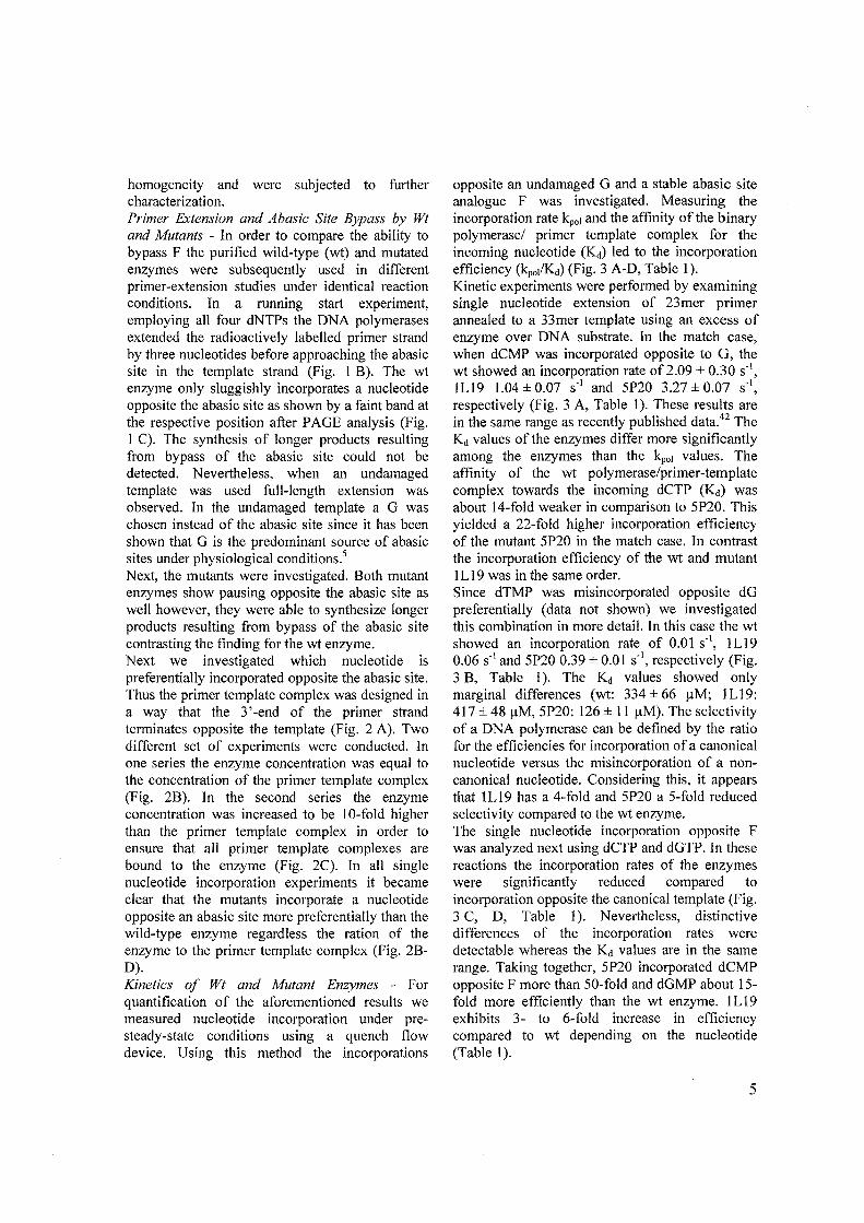
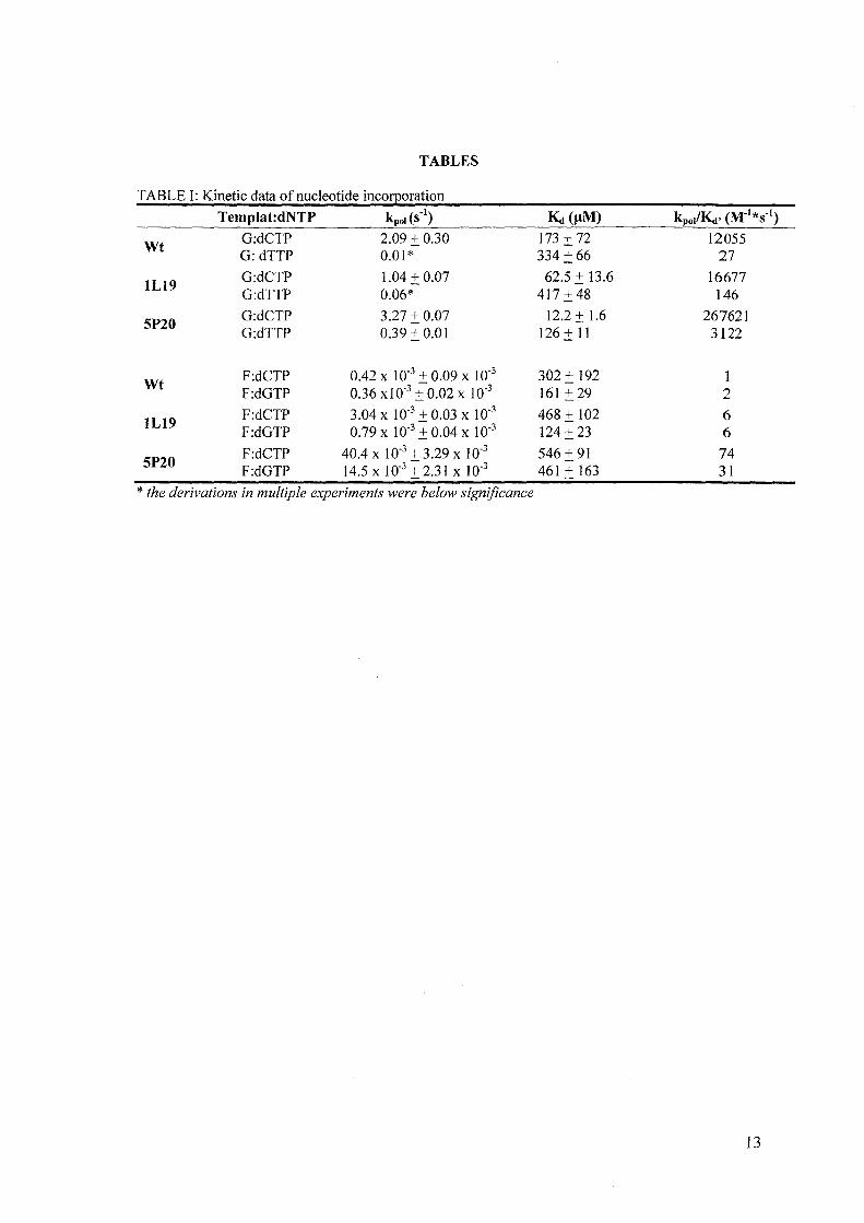
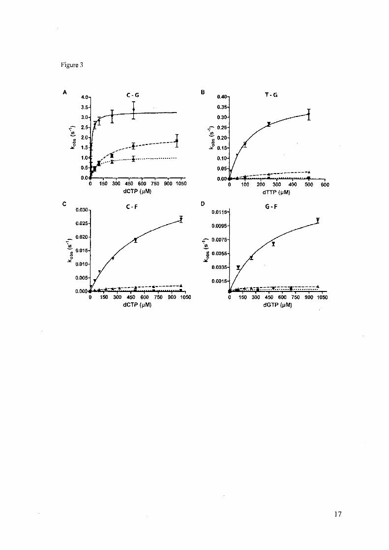
opposite an undamaged G and a stable abasic site analogue F was investigated. Measuring the incorporation rate kpol and the affinity of the binary polymerase/ primer template complex for the incoming nucleotide (Kd) led to the incorporation efficiency (kpolKd) (Fig. 3 A-D, Table 1). Kinetic experiments were performed by examining single nucleotide extension of 23mer primer annealed to a 33mer template using an excess of enzyme over DNA substrate. In the match case, when dCMP was incorporated opposite to G, the wt showed an incorporation rate of 2.09 ± 0.30 S·I,
ILl9 1.04 ± 0.07 s·' and SP20 3.27 ± 0.07 s", respectively (Fig. 3 A, Table I). These results are in the same range as recently published data.42 The Kd values of the enzymes differ more significantly among the enzymes than the kpol values. The affinity of the wt polymerase/primer-template complex towards the incoming dCTP (Kd) was about 14-fold weaker in comparison to SP20. This yielded a 22-fold higher incorporation efficiency of the mutant SP20 in the match case. In contrast the incorporation efficiency of the wt and mutant I Ll9 was in the same order. Since dTMP was misincorporated opposite dG preferentially (data not shown) we investigated this combination in more detail. In this case the wt showed an incorporation rate of 0.01 s", ILl9 0.06 S·I and SP20 0.39 ± 0.Q1 S·I, respectively (Fig. 3 B, Table I). The Kd values showed only marginal differences (wt: 334 ± 66 f!M; 1 Ll9: 417 ± 48 f!M, SP20: 126 ± 11 f!M). The selectivity of a DNA polymerase can be defined by the ratio for the efficiencies for incorporation of a canonical nucleotide versus the misincorporation of a noncanonical nucleotide. Considering this, it appears that lL19 has a 4-fold and SP20 a S-fold reduced selectivity compared to the wt enzyme. The single nucleotide incorporation opposite F was analyzed next using dCTP and dGTP. In these reactions the incorporation rates of the enzymes were significantly reduced compared to incorporation opposite the canonical template (Fig. 3 C, D, Table 1). Nevertheless, distinctive differences of the incorporation rates were detectable whereas the Kd values are in the same range. Taking together, SP20 incorporated dCMP opposite F more than SO-fold and dGMP about ISfold more efficiently than the wt enzyme. 1 L 19 exhibits 3- to 6-fold increase in efficiency compared to wt depending on the nucleotide (Table 1).
5
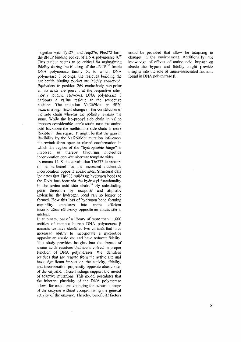
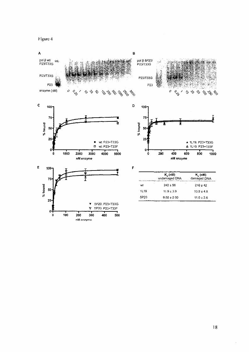
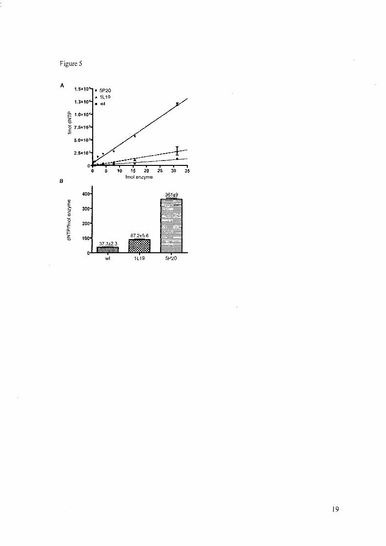
Affinity of Wt and Mutants for DNA - To determine whether the mutant enzymes had an altered affinity for the primer template complexes we performed a gel mobility shift assay (Fig. 4) using approaches described
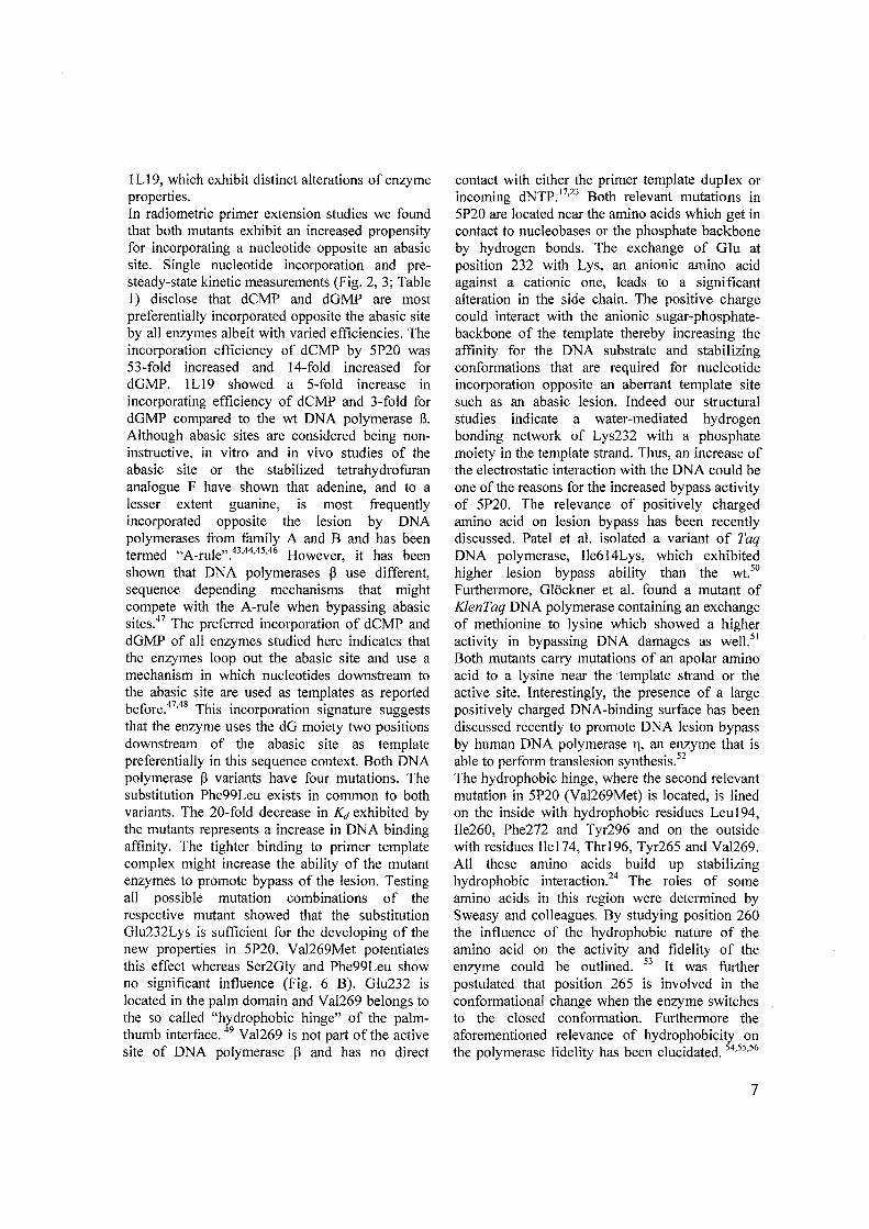
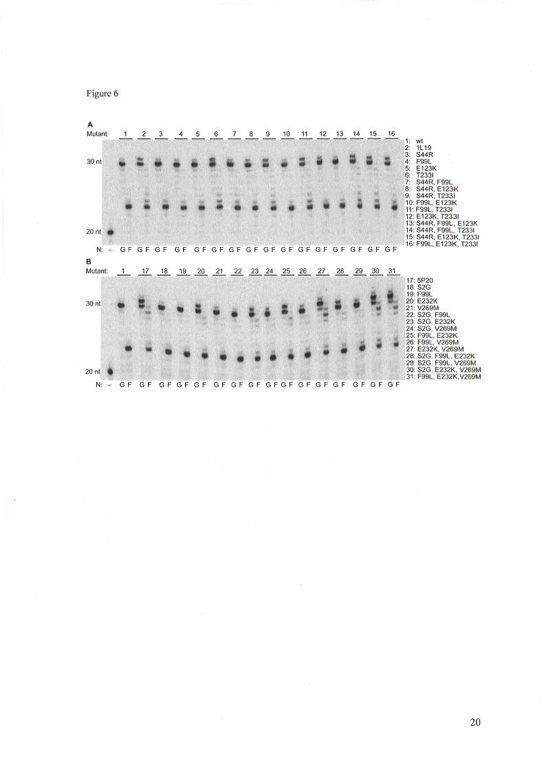
I· 30-33 Th ear ler. e Kd values for undamaged and damaged DNA of wt enzyme were similar (242 ± 56 nM and 2 I 0 ± 42 nM, respectively). The Kd values of the mutant enzymes were similar as well for undamaged and damaged template (lL19: 11.9 ± 3.9 nM and 10.9 ± 4.8 nM, 5P20: 9.5 ± 2.0 nM and 11.0 ± 2.5 nM). Thus, the mutants appear to interact with the primer template complex with the same affinity that is approximately 20-fold higher that the wt enzyme. Specific Activity of Wt and Mutants - Next we in~estigated the specific activity of the enzymes uSlllg an assay that detects nucleotide incorporation and extension. Thus, in primer extension the amount of dNTPs that is incorporated within 10 min depending on the enzyme concentration was investigated. We found that the wt enzyme exhibits a specific activity of 37.3±2.3 dNTP/fmol enzyme while the activity of the mutant ILl9 was threefold (87.2±5.6) (Fig. 5 A) and the mutant 5P20 (360±9) tenfold increased, respectively (Fig. 5 B). Contribution of Mutations to Abasic Site Bypass Proficiency - in order to determine whether all mutations of 1 Ll9 and 5P20 are required to render the enzymes proficient in bypassing an active site every single mutation as well as combinations of d.oubl~ and triple mutations were generated by sIte-dIrected-mutagenesis. All mutants were subsequently purified to homogeneity and were utilized in primer extension reactions employing identical reactions conditions. An unmodified (T33A) and a damaged (T33F) template containing an abasic site were used in combination with a 20 nt primer (P20) (Fig. 6). We found that all variants of the 1 L 19 mutant which contain the mutation Thr233lie showed the highest proficiency for bypassing an abasic site (Fig. 6 A). Interestingly, when comparing all mutants derived from the 5P20 scaffold it turned out that a single mutation (Glu232Lys) was sufficient for more bypass of the abasic site. However, the combination of this mutation with Val269Met results into a more efficient translesion enzyme. Interestingly, the Val269Met mutation alone is not
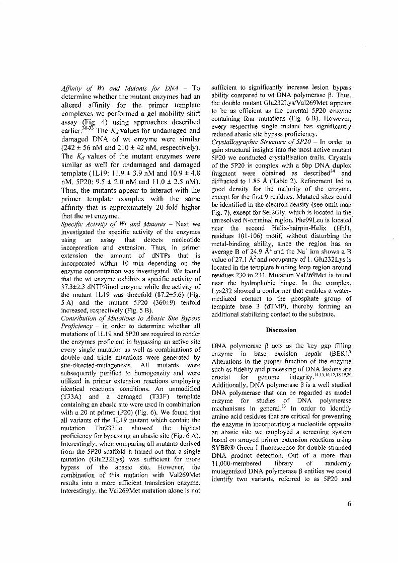
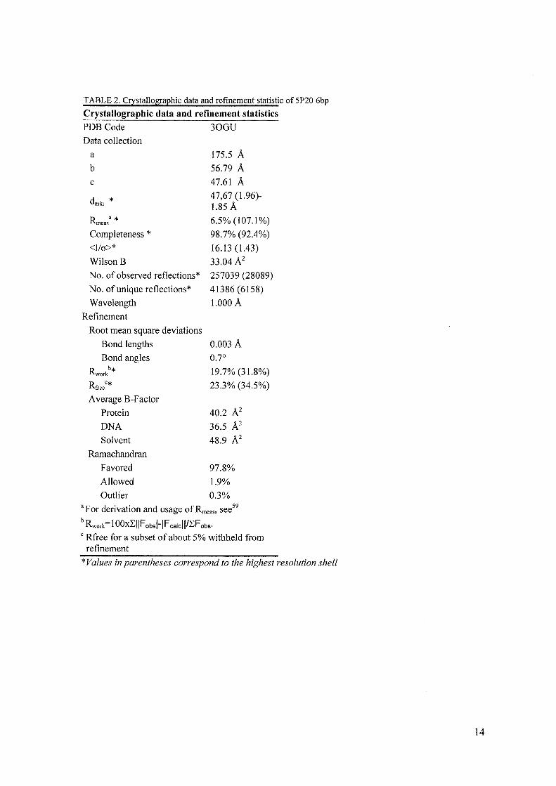
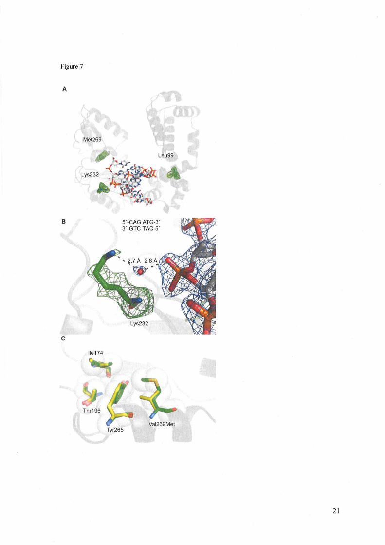
sufficient to significantly increase lesion bypass ability compared to wt DNA polymerase ~. Thus, the double mutant Glu232LyslVal269Met appears to be as efficient as the parental 5P20 enzyme containing four mutations (Fig. 6 B). However, ev.ery respective single mutant has significantly reduced abasic site bypass proficiency. Crystallographic Structure of 5P20 - In order to gain structural insights into the most active mutant 5P20 we conducted crystallisation trails. Crystals of the 5P20 in complex with a 6bp DNA duplex fragment were obtained as described24 and diffracted to 1.85 A (Table 2). Refinement led to good density for the majority of the enzyme, except for the first 9 residues. Mutated sites could b~ identified in the electron density (see omit map FIg. 7), except for Ser2Gly, which is located in the unresolved N-terminal region. Phe99Leu is located ne~r the second Helix-hairpin-Helix (HhH, resIdues 10 I-I 06) motif, without disturbing the metal-binding ability, since the region has an average B of 24.9 A2 and the Na+ ion shows a B value of27.I A 2 and occupancy of I. Glu232Lys is located in the template binding loop region around residues 230 to 234. Mutation Val269Met is found near the hydrophobic hinge. In the complex, Lys232 showed a conformer that enables a watermediated contact to the phosphate group of template base 3 (dTMP), thereby forming an additional stabilizing contact to the substrate.
Discussion
DNA polymerase ~ acts as the key gap filling enzyme in base excision repair (BER).8 Alterations in the proper function of the enzyme such as fidelity and processing of DNA lesions are crucial for genome integrity. 14,15,16,17,18,19,20
Additionally, DNA polymerase ~ is a well studied DNA polymerase that can be regarded as model enzyme for studies of DNA polymerase
h ·· 13 mec anIsms III general. In order to identity amino acid residues that are critical for preventing the enzyme in incorporating a nucleotide opposite an abasic site we employed a screening system based on arrayed primer extension reactions using SYBR® Green I fluorescence for double stranded DNA product detection. Out of a more than Il,OOO-membered library of randomly mutagenized DNA polymerase ~ entities we could identifY two variants, referred to as 5P20 and
6
I LI 9, which exhibit distinct alterations of enzyme properties. In radiometric primer extension studies we found that both mutants exhibit an increased propensity for incorporating a nucleotide opposite an abasic site. Single nucleotide incorporation and presteady-state kinetic measurements (Fig. 2, 3; Table I) disclose that dCMP and dGMP are most preferentially incorporated opposite the abasic site by all enzymes albeit with varied efficiencies. The incorporation efficiency of dCMP by 5P20 was 53-fold increased and 14-fold increased for dGMP. lLI9 showed a 5-fold increase in incorporating efficiency of dCMP and 3-fold for dGMP compared to the wt DNA polymerase 13. Although abasic sites are considered being noninstructive, in vitro and in vivo studies of the abasic site or the stabilized tetrahydrofuran analogue F have shown that adenine, and to a lesser extent guanine, is most frequently incorporated opposite the lesion by DNA polymerases from family A and B and has been termed "A_rule".43,44,45,46 However, it has been shown that DNA polymerases ~ use different, sequence depending mechanisms that might compete with the A-rule when bypassing abasic sites.47 The preferred incorporation of dCMP and dGMP of all enzymes studied here indicates that the enzymes loop out the abasic site and use a mechanism in which nucleotides downstream to the abasic site are used as templates as reported before.47,48 This incorporation signature suggests that the enzyme uses the dG moiety two positions downstream of the abasic site as template preferentially in this sequence context. Both DNA polymerase ~ variants have four mutations. The substitution Phe99Leu exists in common to both variants. The 20-fold decrease in Kd exhibited by the mutants represents a increase in DNA binding affinity. The tighter binding to primer template complex might increase the ability of the mutant enzymes to promote bypass of the lesion. Testing all possible mutation combinations of the respective mutant showed that the substitution Glu232Lys is sufficient for the developing of the new properties in 5P20. Val269Met potentiates this effect whereas Ser2GIy and Phe99Leu show no significant influence (Fig. 6 B). Glu232 is located in the palm domain and Val269 belongs to the so called "hydrophobic hinge" of the palmthumb interface. 49 Val269 is not part of the active site of DNA polymerase ~ and has no direct
contact with either the primer template duplex or incoming dNTP. l7,23 Both relevant mutations in 5P20 are located near the amino acids which get in contact to nucleobases or the phosphate backbone by hydrogen bonds. The exchange of GIu at position 232 with Lys, an anionic amino acid against a cationic one, leads to a significant alteration in the side chain. The positive charge could interact with the anionic sugar-phosphatebackbone of the template thereby increasing the affinity for the DNA substrate and stabilizing conformations that are required for nucleotide incorporation opposite an aberrant template site such as an abasic lesion. Indeed our structural studies indicate a water-mediated hydrogen bonding network of Lys232 with a phosphate moiety in the template strand. Thus, an increase of the electrostatic interaction with the DNA could be one of the reasons for the increased bypass activity of 5P20. The relevance of positively charged amino acid on lesion bypass has been recently discussed. Patel et al. isolated a variant of Taq DNA polymerase, IIe614Lys,which exhibited higher lesion bypass ability than the wt. 50 Furthermore, G16ckner et al. found a mutant of KlenTaq DNA polymerase containing an exchange of methionine to lysine which showed a higher activity in bypassing DNA damages as well.5l
Both mutants carry mutations of an apolar amino acid to a lysine near the template strand or the active site. Interestingly, the presence of a large positively chaTged DNA-binding surface has been discussed recently to promote DNA lesion bypass by human DNA polymerase Tt, an enzyme that is able to perform translesion synthesis.52
The hydrophobic hinge, where the second relevant mutation in 5P20 (VaI269Met) is located, is lined on the inside with hydrophobic residues Leu194, IIe260, Phe272 and Tyr296 and on the outside with residues IIe174, Thr196, Tyr265 and Va1269. All these amino acids build up stabilizing hydrophobic interaction.24 The roles of some amino acids in this region were determined by Sweasy and colleagues. By studying position 260 the influence of the hydrophobic nature of the amino acid on the activity and fidelity of the enzyme could be outlined. 53 It was further postulated that position 265 is involved in the conformational change when the enzyme switches to the closed conformation. Furthermore the aforementioned relevance of hydrophobicity on the polymerase fidelity has been elucidated. 54,55,56
7
Together with Tyr271 and Asp276, Phe272 form the dNTP binding pocket of DNA polymerase 8.49
This residue seems to be critical for maintaining fidelity during the binding of the dNTP.57 Inside DNA polymerase family X, to which DNA polymerase P belongs, the residues building the nucleotide binding pocket are highly conserved. Equivalent to position 269 exclusively non-polar amino acids are present at the respective sites, mostly leucine. However, DNA polymerase p harbours a valine residue at the respective position. The mutation Val269Met in 5P20 induces a significant change of the constitution of the side chain whereas the polarity remains the same. While the iso-propyl side chain in valine imposes considerable steric strain near the amino acid backbone the methionine side chain is more flexible in this regard. It might be that the gain in flexibility by the Val269Met mutation influences the switch form open to closed conformation in which the region of the "hydrophobic hinge" is involved in thereby favouring nucleotide incorporation opposite aberrant template sides. In mutant lLl9 the substitution Thr233Ile appears to be sufficient for the increased nucleotide incorporation opposite abasic sites. Structural data indicates that Thr233 builds up hydrogen bonds to the DNA backbone via the hydroxyl functionality in the amino acid side chain.58 By substituting polar threonine by nonpolar and aliphatic isoleucine the hydrogen bond can no longer be formed. How this loss of hydrogen bond forming capability translates into more efficient incorporation efficiency opposite an abasic site is unclear. In summary, out of a library of more than 11,000 entities of random human DNA polymerase P mutants we have identified two variants that have increased ability to incorporate a nucleotide opposite an abasic site and have reduced fidelity. This study provides insights into the impact of amino acids residues that are involved in proper function of DNA polymerases. We identified residues that are remote from the active site and have significant impact on the activity, fidelity, and incorporation propensity opposite abasic sites of the enyzme. These findings support the model of adaptive mutations. This model postulates that the inherent plasticity of the DNA polymerase aIlows for mutations changing the substrate scope of the enzyme without compromising the general activity of the enzyme. Thereby, beneficial factors
could be provided that allow for adapting to changes in the environment. Additionally, the knowledge of effects of amino acid impact on abasic site bypass and fidelity might provide insights into the role of tumor-associated mutants found in DNA polymerase p.
8
REFERENCES
1 Saul, R. L., and Ames, B. N. (1986) Basic Life Sei. 38,529-535 2 Kulkarni, A., and Wilson, D. M. (2008) Am. J. Hum. Genet. 82, 539-566 3 Huebscher, U., Maga, G., and Spadari, S. (2002) Annu. Rev. Biochem. 71, 133-163 4 Lindahl, T. (1993) Nature 362,709-715 5 Loeb, L. A. and Preston, B. D. (1986) Annu. Rev. Genet. 20, 201-230 6 Lindahl, T., and Wood, R. D. (1999) Science 286, 1897-1905 7 Nilsen, H., and Krokan, H. E. (2001) Carcinogenesis 22, 987-998 8 Seeberg, E., Eide, L., and Bjoras, M. T. (1995) Trends Biochem. Sei. 20,391-397 9 Beard, W. A., Shock, D. D., Batra, V. K., Pedersen, L. C., and Wilson, S. H. (2009) J. Bioi. Chem.
284, 31680-31689 10 Filee, J., Forterre, P., Sen-Lin, T., and Laurent, J. (2002)J. Mol. Evol. 54,763-73 11 Idriss, H. T., AI-Assar, 0., and Wilson, S. H. (2002) 1nt. J. Biochem. Cell. Bioi. 34, 321-324 12 Kumar, A., Widen, S. G., Williams, K. R., Kedar, P., Karpel, R. L., and Wilson, S. H. (1990) J
Bioi. Chem. 265, 2124-2131 13 Beard, W. A., and Wilson, S. H. (2006) Chem. Rev. 106,361-382 14 Dalal, S., Chikova, A., Jaeger J., and Sweasy, J. B. (2008) Nucleic Acids Res. 36,411-422 15 Starcevic, D., Dalal, S., and Sweasy, J. B. (2004) Cell Cycle 3,998-1001 16 Lang, T., Maitra, M., Starcevic, D., Li, S. X., and Sweasy, 1. B. (2004) Proc. Natl. Acad. Sei.
Us. A., 101,6074-6079 17 Dalal, S., Hile, S., Eckert, K. A., Sun, K. W., Starcevic, D., and Sweasy, J. B. (2005) Biochemistry
44, 15664-15673 18 Sweasy, J. B., Lang, T., Starcevic, D., Sun, K. W., Lai, C. c., Dimaio, D., and Dalal, S. (2005)
Proc. Natl. Acad. Sei. U S. A. 102, 14350Q4355 19 Sweasy,1. B., Lauper, J. M., and Eckert, K. A. (2006) Radiat. Res. 166,693-714 20 Lang, T., Dalal, S., Chikova, A., DiMaio, D., and Sweasy, J. B. (2007) Mol. Cell Bioi. 27,5587-
5596 21 Batra, V. K., Beard, W. A., Shock, D. D., Pedersen, L. C., and Wilson, S. H. (2008) Mol. Cell 30,
315-324 22 Krahn, J. M., Beard, W. A, Miller, H., Grollman, A. P., and Wilson, S. H. (2003) Structure 11,
121-127 23 Sawaya, M. R., Prasad, R., Wilson, S. H., Kraut, J., and Pelletier, H. (1997) Biochemistry 36,
11205-11215 24 Pelletier, H., Sawaya, M. R., Wolfle, W., Wilson, S. H., and Kraut, J. (1996) Biochemistry 35,
12742-12761 25 Sobol, R. W., Horton, J. K., Kuhn, R., Gu, H., Singhal, R. K., Prasad, R., Rajewsky, K., and
Wilson, S. H. (1996) Nature 379, 183-186 26 Satya, P., Johnson, R. E., and Prakash, L. (2005) Annu. Rev. Biochem. 74,317-353 27 Karras, G. I., and Jentsch, S. (2010) Cell 141, 255-267 28 Daigaku, Y., Davies, A. A., and Ulrich, H. D. (2010) Nature 465, 951-955 29 Menge, K. L., Hostomsky, Z., Nodes, B. R., Hudson, G. 0., Rahmati, S., Moomaw, E. W.,
Almassy, R. J., and Hostomska, Z. (1995) Biochemistry 34, 15934-15942 30 Kosa, J. L., and Sweasy, J. B. (l999)J. Biol. Chem. 274,3851-3858 31 Minnick, D. T., Astatke, M., Joyce, C. M., and Kunkel, T. A. (1996) J. Biol. Chem. 271, 24954-
24961 32 Casas-Finet, J. R., Kumar, A., Karpel, R. L., and Wilson, S. H. (1992) Biochemistry 31, 10272-
10280 33 Astake, M., Grindley, N. D. F., and Joyce, C. M. (1995)J. BioI. Chem. 270,1945-1954 34 Abbotts, 1., SenGupta, D. N., Zmudzka, B., Widen, S. G., Notario, V., and Wilson, S. H. (1988)
Biochemistry 27, 901-909 35 Kabsch, W. (201O)ActaCryst., D66, 125-132 36 Kabsch, W. (2010), Acta Cryst., D66, 133-144
9
37 Adams, P. D., Grosse-Kunstleve, R. W., Hung, L. W., Ioerger, T. R., McCoy, A. J., Moriarty, N. W., Read, R. J., Sacchettini, J. C., Sauter, N. K., and Terwilliger, T. C. (2002) Acta Crystallogr., Sect. D: BioI. Crystallogr. 58, 1948-1954
38 Emsley, P., and Cowtan, K. (2004) Acta Crystallogr., Sect. D: Bio!. Crystallogr. 60,2126-2132 39 DeLano, W. L. (2002). The PyMOL Molecular Graphics System. Palo Alto, CA, USA. 40 Summerer, D., Rudinger, N. Z., Detmer, I., and Marx, A. (2005) Angew. Chem., Int. Ed 44,4712-
4715 41 Strerath, M., Gloeckner, C., Liu, D., Schnur, A., and Marx, A. (2007) ChemBioChem 8, 395-401 42 Ahn, J., Werneburg, B. G., and Tsai, M. D. (1997) Biochemistry 36, 1100-1107 43 . Sagher D., Strauss B. (1983) Biochemistry 22,4518-4526 44 Schaaper R.M., Kunkel T.A., Loeb L.A. (1983) Proc. Natl. Acad. Sci. USA 80,487-491 45 Strauss B.S. (2002) DNA Repair 1, 125-135 46 Obeid, S., Blatter, N., Kranaster, R., Schnur, A., Welte, W., Diederichs, K., Marx, A. (2010)
EMBOJ. 29,1738-1747 47 Efrati, E., Tocco, G., Eritja, R., Wilson, S. H., and Goodman, M. F. (1997)J. BioI. Chem. 272,
2559-2569 48 Efrati, E., Tocco, G., Eritja, R., Wilson, S. H., and Goodman, M. F. (1999) J. BioI. Chem.
274,15920-15926 49 Sawaya, M. R, Prasad, R., Wilson, S. H., Kraut, J., and Pelletier, H. (1997) Biochemistry 36,
11205-11215 50 Patel, P. H., Kawate, H., Adman, E., Ashbach, M., and Loeb, L. A. (2001) J. BioI. Chem. 276,
5044-5051 51 Gloeckner, C., Sauter, K. B., and Marx, A. (2007) Angew. Chem., Int. Ed. 46, 3115-3117 52 Biertlimpfel, C., Zhao, Y., Kondo, Y., Ramon-Maiques, S., Gregory, M., Lee, J. Y., Masutani, C.,
Lehmann, A. R., Hanaoka, F., and Yang, W. (2010) Nature, 465, 1044-1048 53 Starcevic, D., Dalal, S., and Sweasy, J. B. (2005) Biochemistry 44,3775-3884 54 Clairmont, C. A., Narayanan, L., Sun, K. W., Glazer, P. M., and Sweasy, J. B. (1999) Proc. Nat!.
Acad. Sci. U. S. A. 96,9580-9585 55 Shah, A. M., Li, S. X., Anderson, K. S., and Sweasy, J. B. (2001) J. BioI. Chem. 276, 10824-
10831 56 Opresko, P. L., Sweasy, 1. B., and Eckert, K. A. (1998) Biochemistry 37, 111-119 57 Li, S. x., Vaccaro, J. A., and Sweasy, 1. B. (1999) Biochemistry 38,4800--4808 58 Pelletier, H., Sawaya, M.R., Kumar, A., Wilson, S. H., and Kraut, J. (1994) Science 264, 1891-
1903 59 Diederichs, K., and Karplus, P. A. (1997) Nature Struct. BioI. 4,269-275
FOOTNOTES
Acknowledgments - We thank the Deutsche Forschungsgemeinschaft (DFG) for generous funding within SPP 1170 and the Swiss Light Source synchrotron site for support.
10
FIGURE LEGENDS
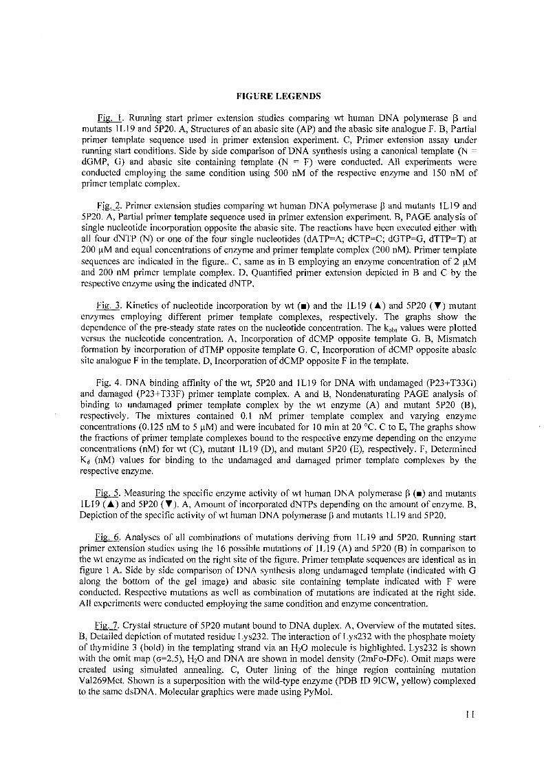
.EigJ. Running start primer extension studies comparing wt human DNA polymerase 13 and mutants ILl 9 and 5P20. A, Structures of an abasic site (AP) and the abasic site analogue F. B, Partial primer template sequence used in primer extension experiment. C, Primer extension assay under running start conditions. Side by side comparison of DNA synthesis using a canonical template (N = dGMP, G) and abasic site containing template (N = F) were conducted. All experiments were conducted employing the same condition using 500 nM of the respective enzyme and ISO nM of primer template complex.
Fig. 2. Primer extension studies comparing wt human DNA polymerase ~ and mutants ILl 9 and 5P20. A, Partial primer template sequence used in primer extension experiment. B, PAGE analysis of single nucleotide incorporation opposite the abasic site. The reactions have been executed either with all four dNTP (N) or one of the four single nucleotides (dA TP=A; dCTP=C; dGTP=G, dTTP=T) at 200 /lM and equal concentrations of enzyme and primer template complex (200 nM). Primer template sequences are indicated in the figure .. C, same as in B employing an enzyme concentration of 2 ~lM and 200 nM primer template complex. D, Quantified primer extension depicted in Band C by the respective enzyme using the indicated dNTP.
Fig. 3. Kinetics of nucleotide incorporation by wt (_) and the ILl9 (~) and 5P20 (T) mutant enzymes employing different primer template complexes, respectively. The graphs show the dependence of the pre-steady state rates on the nucleotide concentration. The kobs values were plotted versus the nucleotide concentration. A, Incorporation of dCMP opposite template G. B, Mismatch formation by incorporation of dTMP opposite template G. C, Incorporation of dCMP opposite abasic site analogue F in the template. D, Incorporation of dCMP opposite F in the template.
Fig. 4. DNA binding affinity of the wt, 5P20 and ILl 9 for DNA with undamaged (P23+ T33G) and damaged (P23+T33F) primer template complex. A and B, Nondenaturating PAGE analysis of binding to undamaged primer template complex by the wt enzyme (A) and mutant 5P20 (B), respectively. The mixtures contained 0.1 nM primer template complex and varying enzyme concentrations (0.125 nM to 5 /lM) and were incubated for 10 min at 20 QC. C to E, The graphs show the fractions of primer template complexes bound to the respective enzyme depending on the enzyme concentrations (nM) for wt (C), mutant ILI9 (D), and mutant 5P20 (E), respectively. F, Determined Kd (nM) values for binding to the undamaged and damaged primer template complexes by the respective enzyme.
Fig. 5. Measuring the specific enzyme activity of wt human DNA polymerase ~ (_) and mutants 1 L 19 (~) and 5P20 (T). A, Amount of incorporated dNTPs depending on the amount of enzyme. B, Depiction of the specific activity of wt human DNA polymerase ~ and mutants I L I 9 and 5P20.
Fig. 6. Analyses of all combinations of mutations deriving from I L 19 and 5P20. Running start primer extension studies using the 16 possible mutations of IL19 (A) and 5P20 (B) in comparison to the wt enzyme as indicated on the right site of the figure. Primer template sequences are identical as in figure I A. Side by side comparison of DNA synthesis along undamaged template (indicated with G along the bottom of the gel image) and abasic site containing template indicated with F were conducted. Respective mutations as well as combination of mutations are indicated at the right side. All experiments were conducted employing the same condition and enzyme concentration.
Fig. 7. Crystal structure of 5P20 mutant bound to DNA duplex. A, Overview ofthe mutated sites. B, Detailed depiction of mutated residue Lys232. The interaction of Lys232 with the phosphate moiety of thymidine 3 (bold) in the templating strand via an H20 molecule is highlighted. Lys232 is shown with the omit map (cr=2.5), H20 and DNA are shown in model density (2mFo-DFc). Omit maps were created using simulated annealing. C, Outer lining of the hinge region containing mutation Va1269Met. Shown is a superposition with the wild-type enzyme (PDB ID 9ICW, yellow) complexed to the same dsDNA. Molecular graphics were made using PyMol.