57
Sonodynamic Therapy Literature Seminar 2018. 12. 13 Kazuki Takahashi (M1) 1

Sonodynamic Therapy
Literature Seminar
2018. 12. 13
Kazuki Takahashi (M1)
1
Index
1. Introduction• About ultrasound• Various medical uses of ultrasound• Safety range of frequency and intensity
2. Sonodynamic therapy for cancer cells• Photodynamic therapy & sonodynamic therapy• Example of drugs used for sonodynamic therapy• Mechanism & Drug design
2
Index
1. Introduction• About ultrasound• Various medical uses of ultrasound• Safety range of frequency and intensity
2. Sonodynamic therapy for cancer cells• Photodynamic therapy & sonodynamic therapy• Example of drugs used for sonodynamic therapy• Mechanism & Drug design
3
About ultrasound
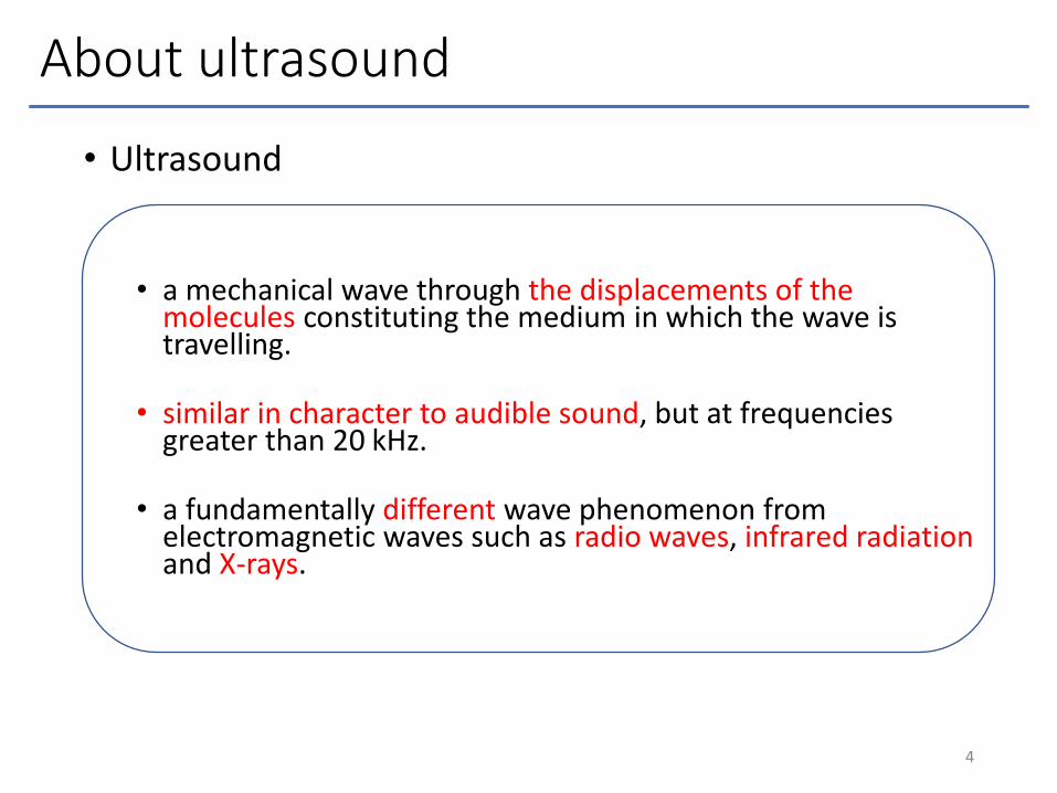
• Ultrasound
• a mechanical wave through the displacements of the molecules constituting the medium in which the wave is travelling.
• similar in character to audible sound, but at frequencies greater than 20 kHz.
• a fundamentally different wave phenomenon from electromagnetic waves such as radio waves, infrared radiation and X-rays.
4
Various medical uses of ultrasound
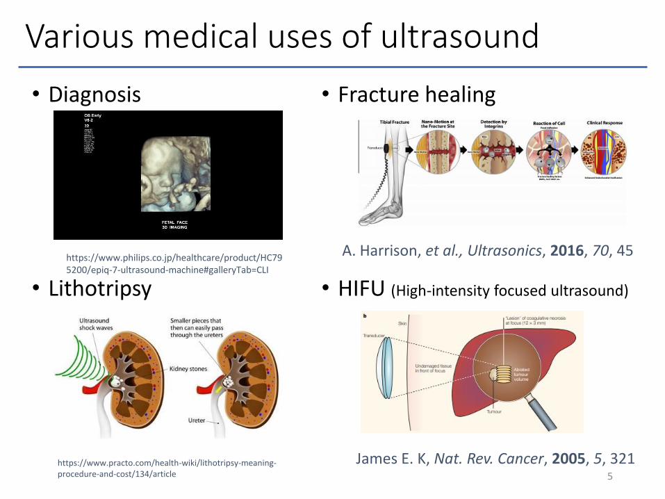
• Diagnosis
• Lithotripsy
• Fracture healing
• HIFU (High-intensity focused ultrasound)
5
https://www.philips.co.jp/healthcare/product/HC795200/epiq-7-ultrasound-machine#galleryTab=CLI
A. Harrison, et al., Ultrasonics, 2016, 70, 45
https://www.practo.com/health-wiki/lithotripsy-meaning-procedure-and-cost/134/article
James E. K, Nat. Rev. Cancer, 2005, 5, 321
Practical range of frequency
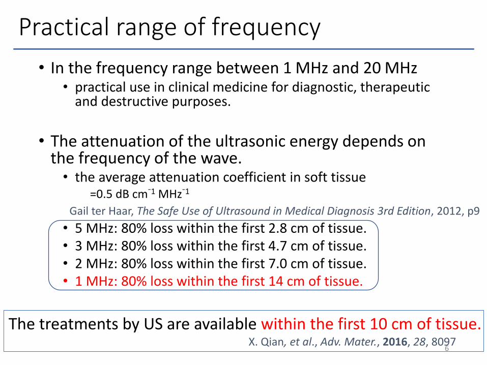
• In the frequency range between 1 MHz and 20 MHz• practical use in clinical medicine for diagnostic, therapeutic
and destructive purposes.
• The attenuation of the ultrasonic energy depends on the frequency of the wave.• the average attenuation coefficient in soft tissue
=0.5 dB cm⁻1 MHz⁻1
• 5 MHz: 80% loss within the first 2.8 cm of tissue. • 3 MHz: 80% loss within the first 4.7 cm of tissue. • 2 MHz: 80% loss within the first 7.0 cm of tissue. • 1 MHz: 80% loss within the first 14 cm of tissue.
6
Gail ter Haar, The Safe Use of Ultrasound in Medical Diagnosis 3rd Edition, 2012, p9
X. Qian, et al., Adv. Mater., 2016, 28, 8097
The treatments by US are available within the first 10 cm of tissue.

US intensity
7Atsumi Ohota, 理学療法の歩み, 2006, 17, 14
• High intensity of US makes burn and cell death.• Low intensity is not effective.• US intensity is recommended
between 0.1~5 MHz for treatment.

Hydroxyl radical produced by cavitation
8Sabina Z., et al., Proceedings, 2018, 2, 188
• Ultrasonic irradiation of liquids causes acoustic cavitations, i.e., the formation, growth and implosive collapse of bubbles.
• Such cavitation generates local sites of high temperature andpressure for short periods of time.
• The extreme temperature conditions generated by a collapsing bubble can also lead to the formation of radical chemical species.
Örjan J., et al., Sustainable and energy efficient leaching of tungsten (W) by ultrasound controlled cavitation, 2018, p8
Sonoluminescence produced by cavitation
9
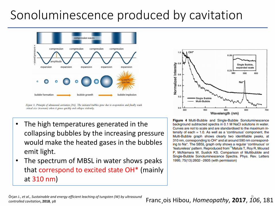
• The high temperatures generated in the collapsing bubbles by the increasing pressure would make the heated gases in the bubbles emit light.
• The spectrum of MBSL in water shows peaks that correspond to excited state OH* (mainly at 310 nm)
Örjan J., et al., Sustainable and energy efficient leaching of tungsten (W) by ultrasound controlled cavitation, 2018, p8 Franc¸ois Hibou, Homeopathy, 2017, 106, 181
Short summary
• US is used for Diagnosis, Fracture healing, Lithotripsy, HIFU, and so on.
• US is able to penetrate into deep tissues (ten centimeter).
• Around 1 MHz of US frequency is recommended for practical use into deep tissues .
• 0.1~5 W/cm2 of US intensity is also recommended.
• Cavitation in water generates hydroxy radical andother radical species, and makes sonoluminescence.
10
Index
1. Introduction• About ultrasound• Various medical uses of ultrasound• Safety range of frequency and intensity
2. Sonodynamic therapy for cancer cells• Photodynamic therapy & sonodynamic therapy• Example of drugs used for sonodynamic therapy• Mechanism & Drug design
11
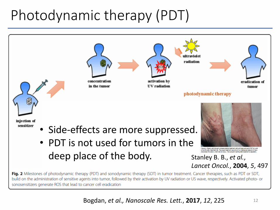
Photodynamic therapy (PDT)
12Bogdan, et al., Nanoscale Res. Lett., 2017, 12, 225
• Side-effects are more suppressed.• PDT is not used for tumors in the
deep place of the body. Stanley B. B., et al., Lancet Oncol., 2004, 5, 497
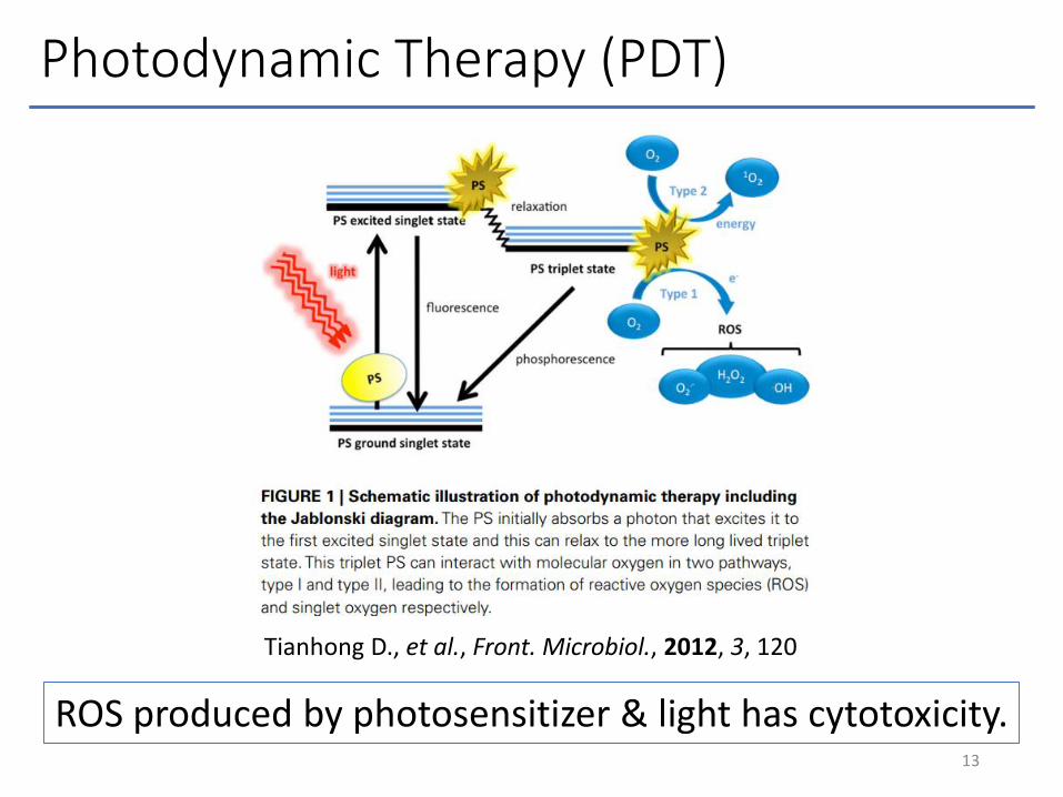
Photodynamic Therapy (PDT)
13
Tianhong D., et al., Front. Microbiol., 2012, 3, 120
ROS produced by photosensitizer & light has cytotoxicity.
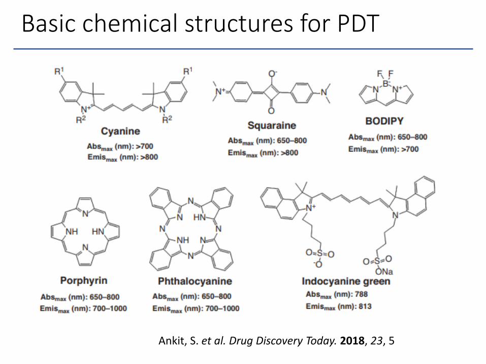
Basic chemical structures for PDT
Ankit, S. et al. Drug Discovery Today. 2018, 23, 5
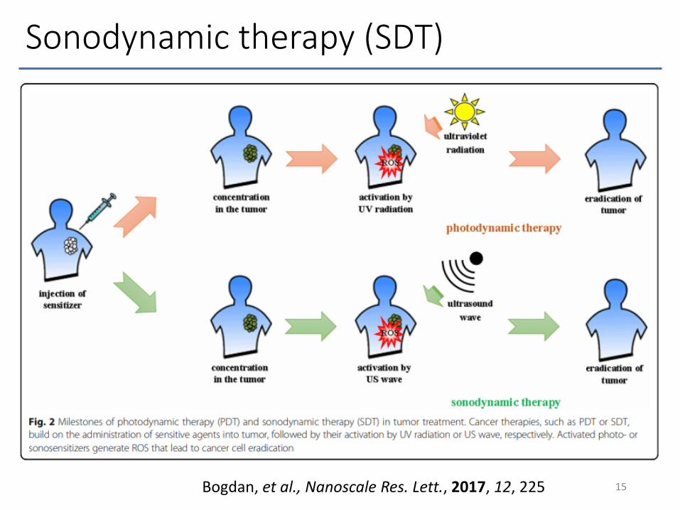
Sonodynamic therapy (SDT)
15Bogdan, et al., Nanoscale Res. Lett., 2017, 12, 225

Sonodynamic therapy (SDT)
16Bogdan, et al., Nanoscale Res. Lett., 2017, 12, 225
• Deeper penetration• Non-invasive treatment• Good repeatability

First example of drugs used for SDT
17Yumita, N et al., Jpn. J. Cancer Res., 1989, 80, 219

First example of drugs used for SDT
18
6015 30 45
Exposure duration (s)
Un
stai
ned
fra
ctio
n
△: 50 mg/mL Hp alone〇: US alone■: 50 mg/mL Hp + US
Yumita, N et al., Jpn. J. Cancer Res., 1989, 80, 219

Other examples of drugs used for SDT
19
Porphyrin compound sensitizers
Xanthone compound sensitizers non-steroid anti-inflammatory agent sensitizers
Other sound-sensitizers
Liu R., et al., Photodiagnosis and Photodynamic Therapy, 2017, 19, 159

Mechanism of SDT (in vitro)
• Investigating the US-responsiveness of a variety of metal-porphyrin complexes, freebase porphyrin and Fe(III), Zn(II) and Pd(II) porphyrin.
• Analyzing their ROS generation under US exposure and related bio-effects.
20F. Giuntini, et al., Free Radical Biology and Medicine, 2018, 121, 190

Sono-induced ROS detected by EPR
• Porphyrin 3 has the highest hydroxyl radical generation efficiency.
• Porphyrin 4 generated most singlet oxygens.21F. Giuntini, et al., Free Radical Biology and Medicine, 2018, 121, 190

Sonoluminescence
• SL emitted both in water (blue) and in the presence of porphyrins 3 (black) and 4 (red).
• The hydroxyl radical emission observed in the presence of porphyrin 3.
22
• Sonoluminescence emission spectra
F. Giuntini, et al., Free Radical Biology and Medicine, 2018, 121, 190
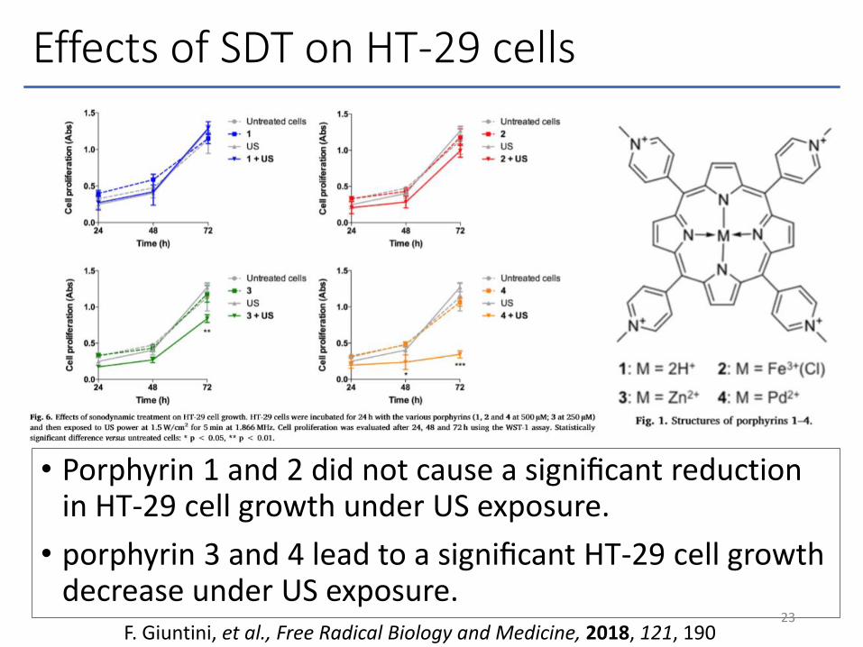
Effects of SDT on HT-29 cells
• Porphyrin 1 and 2 did not cause a significant reduction in HT-29 cell growth under US exposure.
• porphyrin 3 and 4 lead to a significant HT-29 cell growth decrease under US exposure.
23F. Giuntini, et al., Free Radical Biology and Medicine, 2018, 121, 190
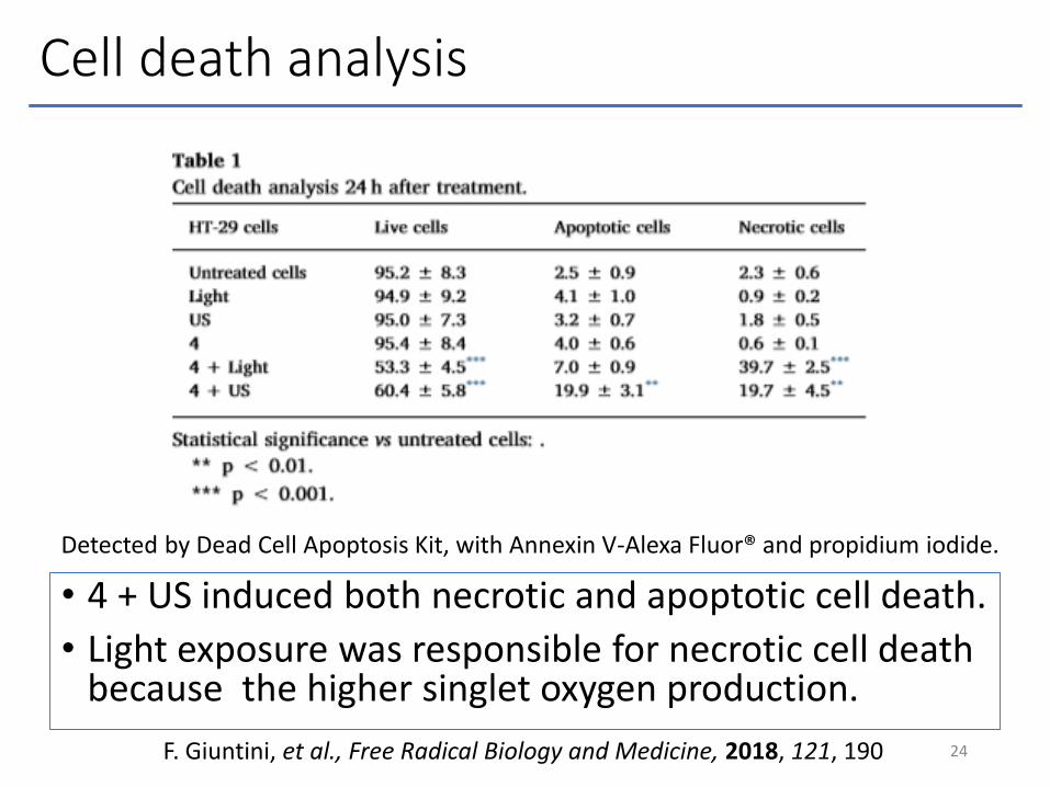
Cell death analysis
• 4 + US induced both necrotic and apoptotic cell death.
• Light exposure was responsible for necrotic cell death because the higher singlet oxygen production.
24F. Giuntini, et al., Free Radical Biology and Medicine, 2018, 121, 190
Detected by Dead Cell Apoptosis Kit, with Annexin V-Alexa Fluor® and propidium iodide.
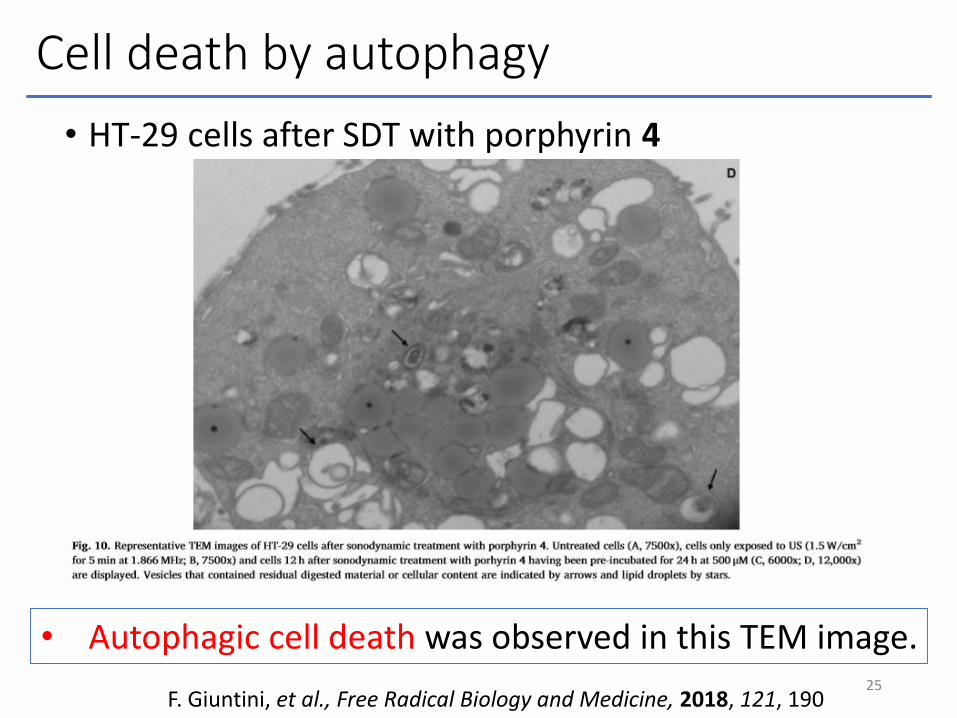
Cell death by autophagy
• HT-29 cells after SDT with porphyrin 4
25
• Autophagic cell death was observed in this TEM image.
F. Giuntini, et al., Free Radical Biology and Medicine, 2018, 121, 190

Disscussion
• Metal ions affect the ability of porphyrins to undergo intersystem crossing (ISC) and can also influence the lifetimes of the resulting excited triplets.
• Porphyrin activation by US can be mediated by photo-activation via sonoluminescence.
• SDT of porphyrin 4 is not effective than PDT.
26
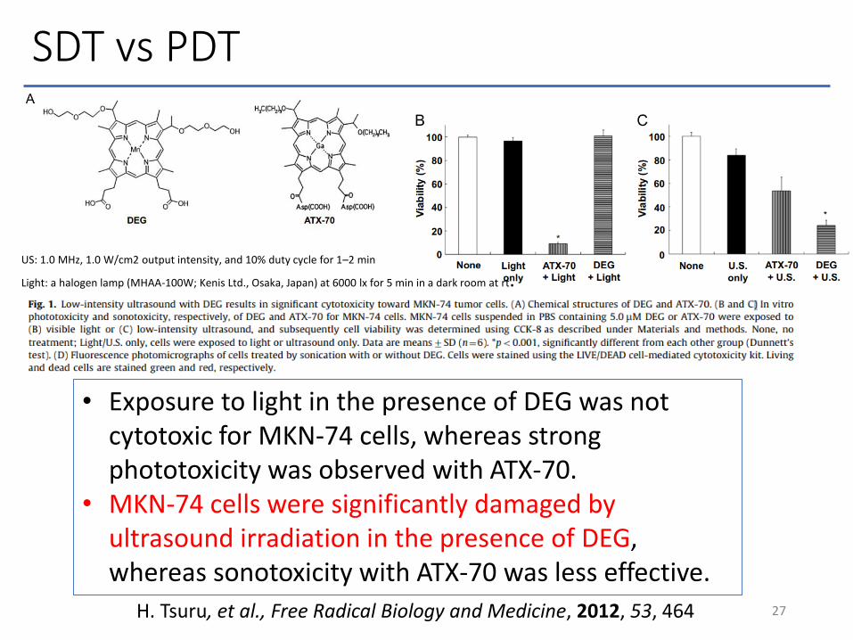
SDT vs PDT
27H. Tsuru, et al., Free Radical Biology and Medicine, 2012, 53, 464
• Exposure to light in the presence of DEG was not cytotoxic for MKN-74 cells, whereas strong phototoxicity was observed with ATX-70.
• MKN-74 cells were significantly damaged by ultrasound irradiation in the presence of DEG, whereas sonotoxicity with ATX-70 was less effective.
US: 1.0 MHz, 1.0 W/cm2 output intensity, and 10% duty cycle for 1–2 min
Light: a halogen lamp (MHAA-100W; Kenis Ltd., Osaka, Japan) at 6000 lx for 5 min in a dark room at rt.
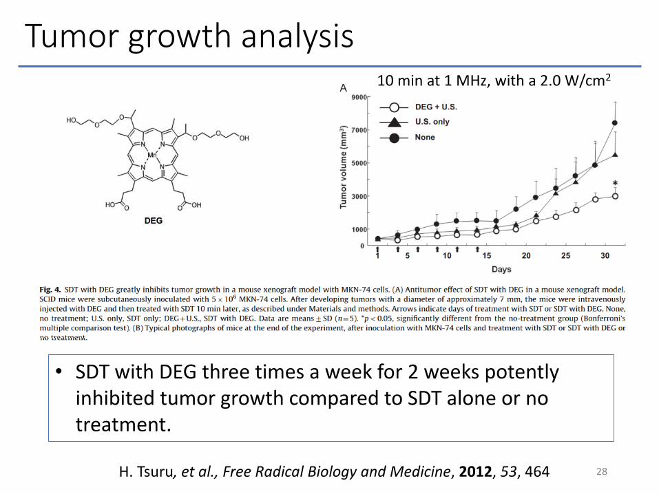
Tumor growth analysis
28H. Tsuru, et al., Free Radical Biology and Medicine, 2012, 53, 464
• SDT with DEG three times a week for 2 weeks potently inhibited tumor growth compared to SDT alone or no treatment.
10 min at 1 MHz, with a 2.0 W/cm2
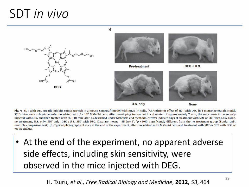
SDT in vivo
29H. Tsuru, et al., Free Radical Biology and Medicine, 2012, 53, 464
• At the end of the experiment, no apparent adverse side effects, including skin sensitivity, were observed in the mice injected with DEG.
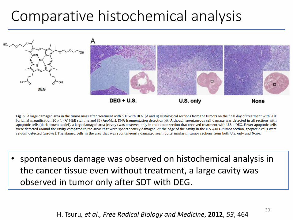
Comparative histochemical analysis
30H. Tsuru, et al., Free Radical Biology and Medicine, 2012, 53, 464
• spontaneous damage was observed on histochemical analysis in the cancer tissue even without treatment, a large cavity was observed in tumor only after SDT with DEG.

Antibody-drug conjugate (ADC)
• Antibody can carry Drug to the target cells selectively.
• ADC is expected suppressing the side-effect to normal cells.
31

ADC for Sonodynamic therapy
• How to synthesize ADC (DAR is no data.)
32Abe H., et al., Anticancer Research, 2002, 22, 1575
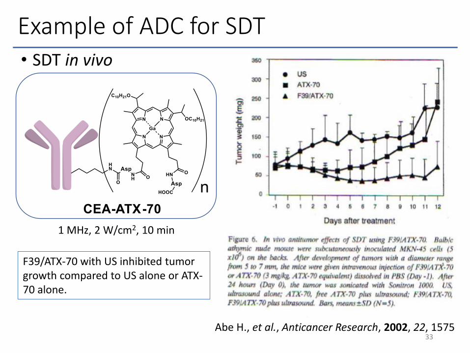
Example of ADC for SDT• SDT in vivo
33Abe H., et al., Anticancer Research, 2002, 22, 1575
1 MHz, 2 W/cm2, 10 min
F39/ATX-70 with US inhibited tumor growth compared to US alone or ATX-70 alone.

Summary
34
• US frequency is recommended for use around 1 MHz, US intensity is recommended at 1~5 W/cm2.
• SDT is a non-invasive treatment for tumors located in the deep places of the body.
• Sonosensitizers are activated by sonoluminescence.
• ADC for SDT is few reported, but reported to beeffective to tumor.
• SDT and sonochemistry is a developing study, so it will be developed more rapidly in the future.
Appendix
35
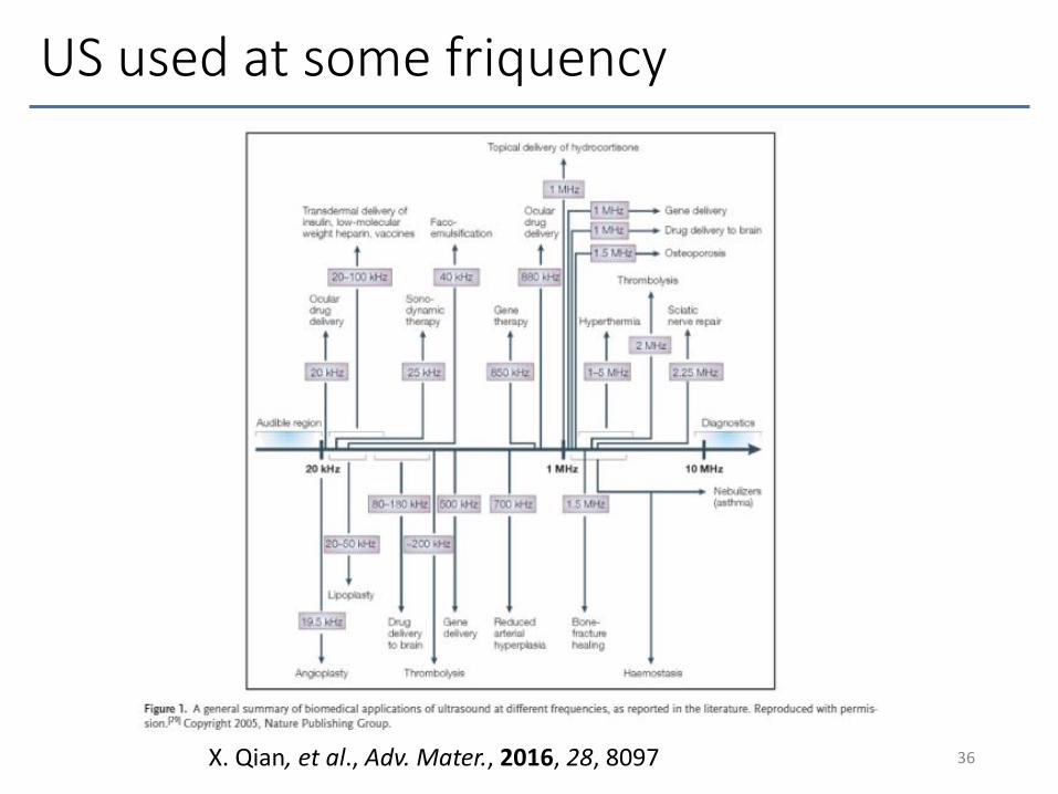
US used at some friquency
36X. Qian, et al., Adv. Mater., 2016, 28, 8097
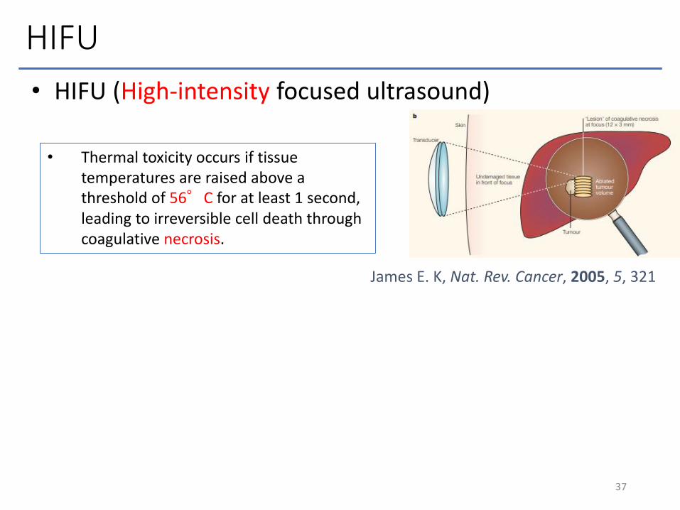
HIFU
• HIFU (High-intensity focused ultrasound)
37
• Thermal toxicity occurs if tissue temperatures are raised above a threshold of 56°C for at least 1 second, leading to irreversible cell death through coagulative necrosis.
James E. K, Nat. Rev. Cancer, 2005, 5, 321
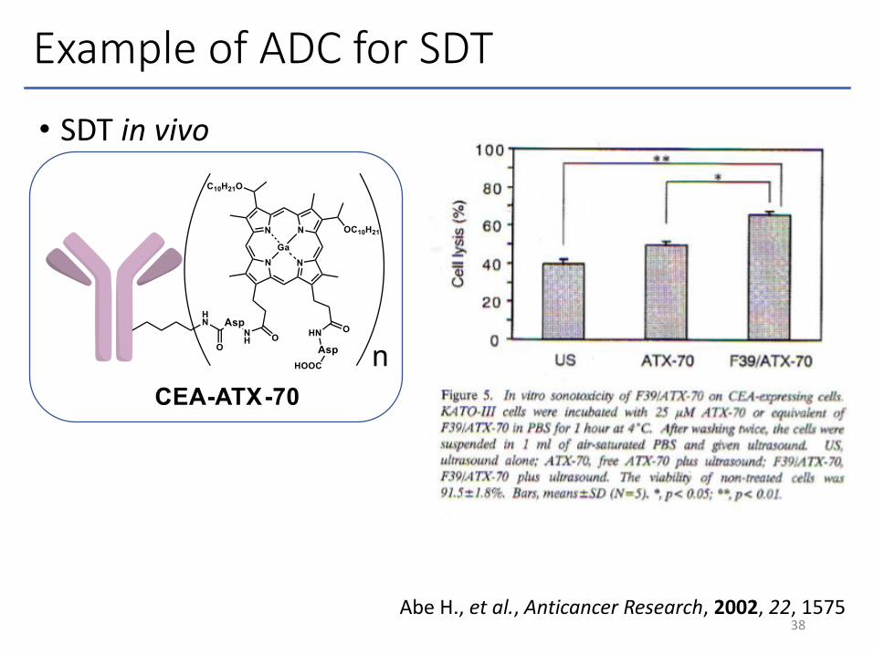
Example of ADC for SDT
• SDT in vivo
38Abe H., et al., Anticancer Research, 2002, 22, 1575
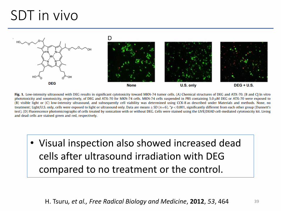
SDT in vivo
39H. Tsuru, et al., Free Radical Biology and Medicine, 2012, 53, 464
• Visual inspection also showed increased dead cells after ultrasound irradiation with DEG compared to no treatment or the control.
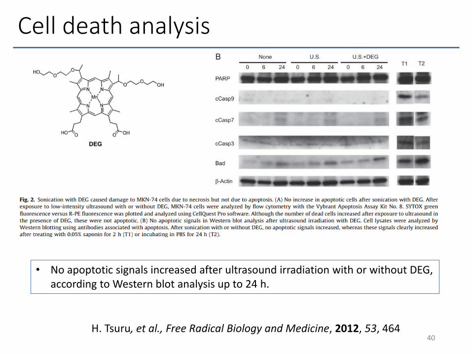
Cell death analysis
40H. Tsuru, et al., Free Radical Biology and Medicine, 2012, 53, 464
• No apoptotic signals increased after ultrasound irradiation with or without DEG, according to Western blot analysis up to 24 h.

Cell death analysis
41H. Tsuru, et al., Free Radical Biology and Medicine, 2012, 53, 464

Photo-induced ROS detected by EPR
• All porphyrins showed comparable singlet oxygen generation efficiency.
• Light irradiation is more efficient than US exposure for excitation.
42
F. Giuntini, et al., Free Radical Biology and Medicine, 2018, 121, 190
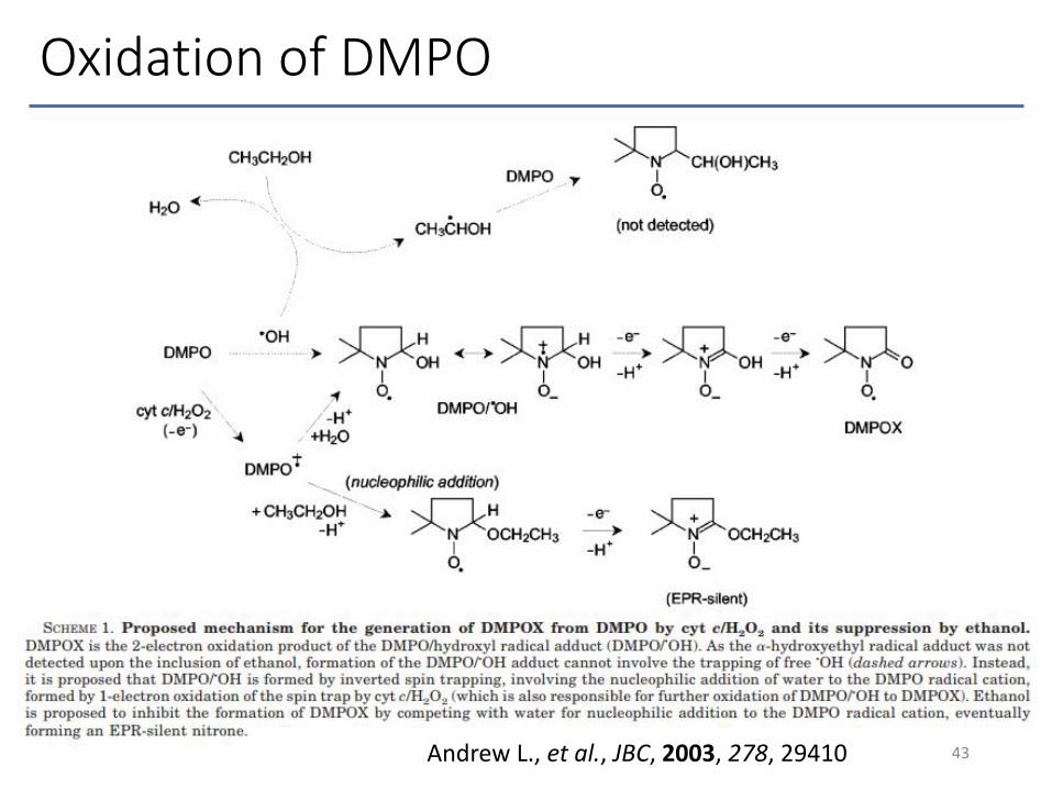
Oxidation of DMPO
43Andrew L., et al., JBC, 2003, 278, 29410

Effects of PDT on HT-29 cells
• All porphyrins induced a significant reduction in cell proliferation under light irradiation.
44F. Giuntini, et al., Free Radical Biology and Medicine, 2018, 121, 190

ROS production after SDT
• Porphyrin 3 and 4 showed ROS emission after US irradiation.
• Porphyrin 4 showed the highest US-responsiveness.
45F. Giuntini, et al., Free Radical Biology and Medicine, 2018, 121, 190
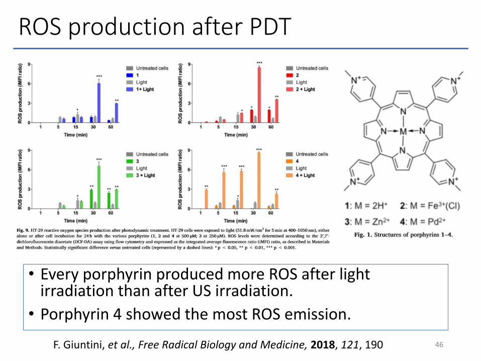
ROS production after PDT
• Every porphyrin produced more ROS after light irradiation than after US irradiation.
• Porphyrin 4 showed the most ROS emission.
46F. Giuntini, et al., Free Radical Biology and Medicine, 2018, 121, 190

Cell death analysis
47
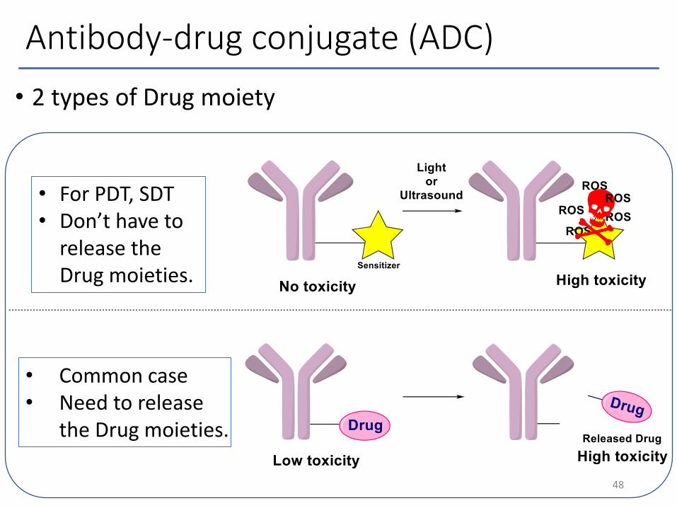
Antibody-drug conjugate (ADC)
• 2 types of Drug moiety
48
• For PDT, SDT• Don’t have to
release the Drug moieties.
• Common case• Need to release
the Drug moieties.

Enzyme & Photo cleavable linker
• Enzyme-cleavable linkerJessica R. M., et al.,
The AAPS Journal, 2015, 17, 339
• Photo-cleavable linkerRoger R. N., et al.,
ACS Cent. Sci., 2017, 3, 329
49
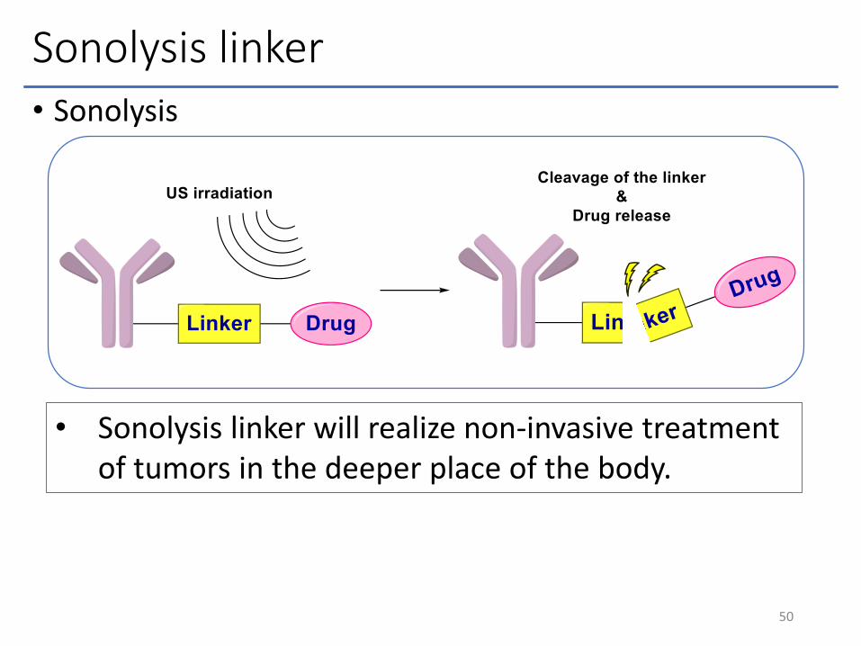
Sonolysis linker• Sonolysis
50
• Sonolysis linker will realize non-invasive treatment of tumors in the deeper place of the body.
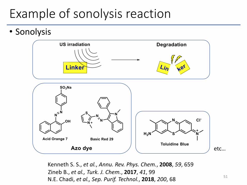
Example of sonolysis reaction• Sonolysis
51
Kenneth S. S., et al., Annu. Rev. Phys. Chem., 2008, 59, 659
etc…
Zineb B., et al., Turk. J. Chem., 2017, 41, 99 N.E. Chadi, et al., Sep. Purif. Technol., 2018, 200, 68
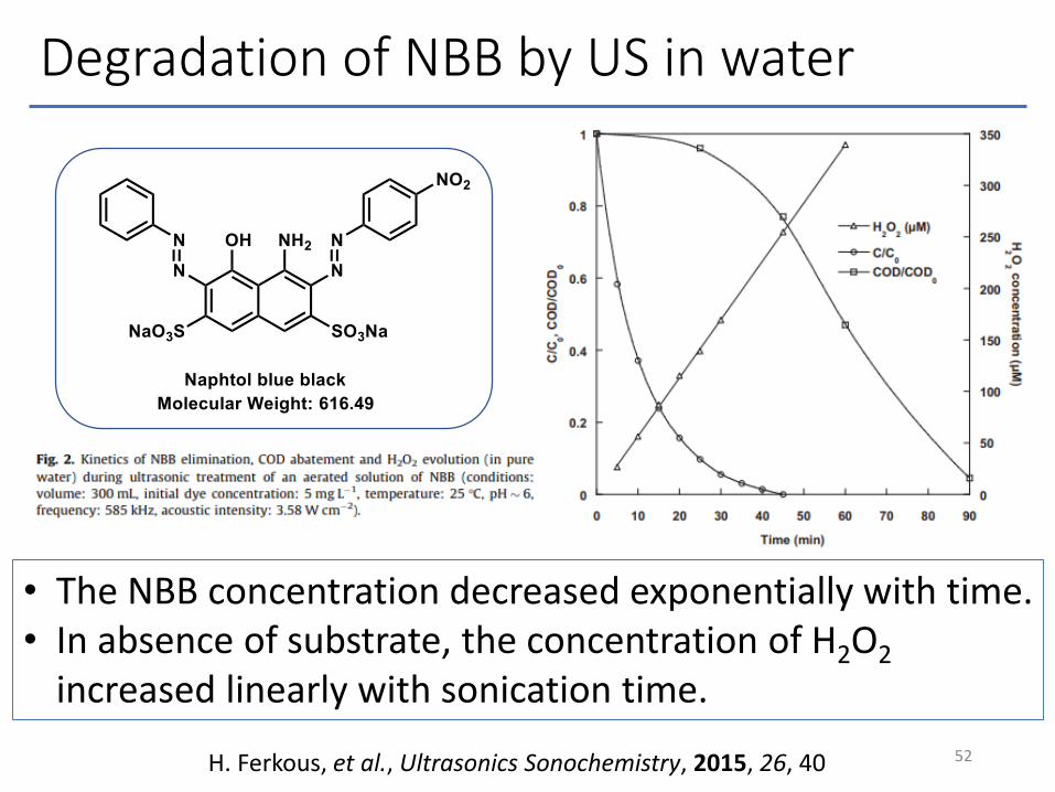
Degradation of NBB by US in water
52H. Ferkous, et al., Ultrasonics Sonochemistry, 2015, 26, 40
• The NBB concentration decreased exponentially with time.• In absence of substrate, the concentration of H2O2
increased linearly with sonication time.
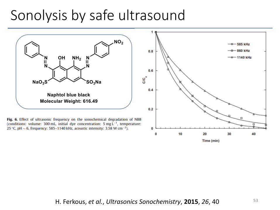
Sonolysis by safe ultrasound
53H. Ferkous, et al., Ultrasonics Sonochemistry, 2015, 26, 40

Sonolysis by safe ultrasound
54H. Ferkous, et al., Ultrasonics Sonochemistry, 2015, 26, 40

N2 inhibits sonolysis reaction competitively.
• N2 is reacted in water by US
55
J. Yao et al., Ultrason. Sonochem., 2018, 42, 42
• N-N bond is cleavable in water under US exposure.
Supeno, et al., Ultrason. Sonochem., 2000, 7, 109
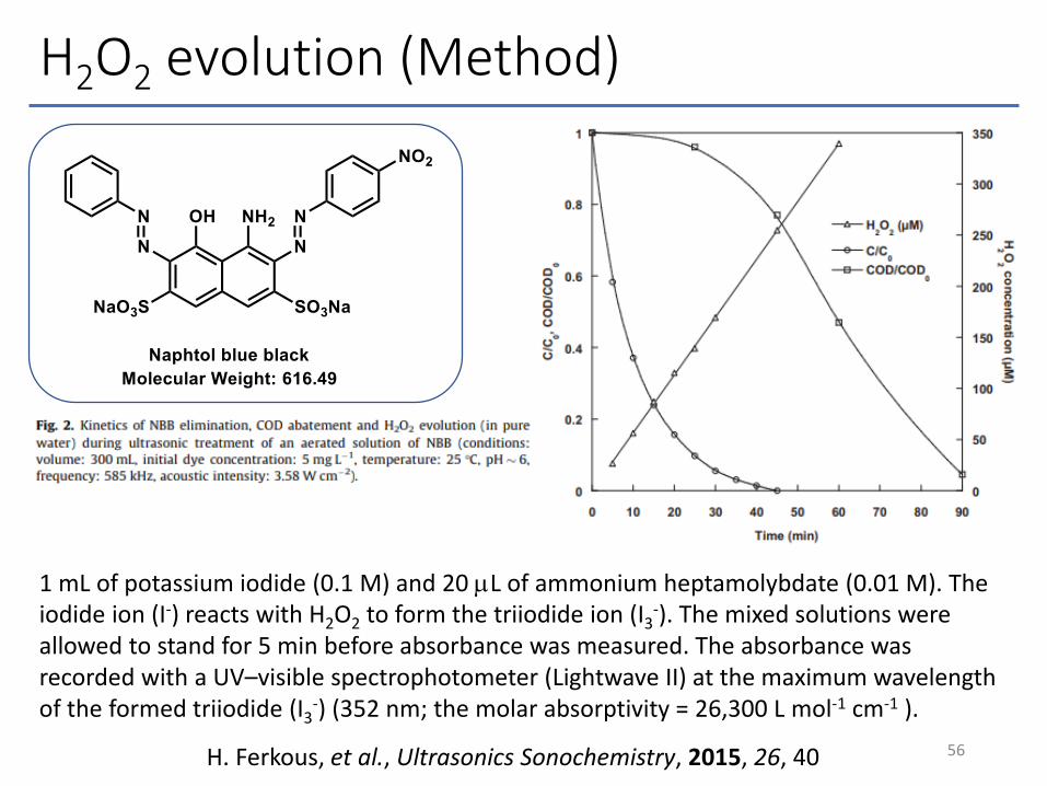
H2O2 evolution (Method)
56H. Ferkous, et al., Ultrasonics Sonochemistry, 2015, 26, 40
1 mL of potassium iodide (0.1 M) and 20 mL of ammonium heptamolybdate (0.01 M). The iodide ion (I-) reacts with H2O2 to form the triiodide ion (I3
-). The mixed solutions were allowed to stand for 5 min before absorbance was measured. The absorbance was recorded with a UV–visible spectrophotometer (Lightwave II) at the maximum wavelength of the formed triiodide (I3
-) (352 nm; the molar absorptivity = 26,300 L mol-1 cm-1 ).
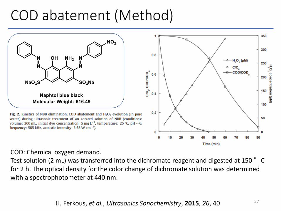
COD abatement (Method)
57H. Ferkous, et al., Ultrasonics Sonochemistry, 2015, 26, 40
COD: Chemical oxygen demand.Test solution (2 mL) was transferred into the dichromate reagent and digested at 150 °C for 2 h. The optical density for the color change of dichromate solution was determined with a spectrophotometer at 440 nm.


![RENDEZVOUS · 2019-06-24 · RENDEZVOUS 園田学園女子大学・園田学園女子大学短期大学部図書館報 No.46 [発行日] 令和元年6 月30 日 発行所 園田学園女子大学・園田学園女子大学短期大学部図書館](https://static.documents.pub/doc/80x56/5f7a67fd10aa8f61156a552e/rendezvous-2019-06-24-rendezvous-oecoefoecoecoeefe.jpg)


