ORIGINAL RESEARCH PEDIATRICS Sonographic Development of the Pericallosal Vascularization in the First and Early Second Trimester of Pregnancy X B. De Keersmaecker, X H. Pottel, X G. Naulaers, and X L. De Catte ABSTRACT BACKGROUND AND PURPOSE: Anomalies of the corpus callosum are rare. Routine scanning in midtrimester of the pregnancy often fails to identify defective development. The purpose of the study was to identify the pericallosal artery and all its main branching arteries during early gestation from the first trimester onward, to measure the length of the pericallosal artery during its development, and to establish a normal vascular map for each week of development. MATERIALS AND METHODS: We performed a single-center prospective, longitudinal clinical study in 15 patients between 11 and 22 weeks of gestation. The origin and course of the different blood vessels were identified. RESULTS: There was a linear association among gestational age, the biparietal diameter, and the length of the pericallosal artery. The curvature of the developing pericallosal artery increases linearly with the gestational age and biparietal diameter, and 4 variations of branching of the callosomarginal artery were observed. CONCLUSIONS: The pericallosal artery and its branches can be identified and measured from 11 weeks on, and the pericallosal artery takes its characteristic course. A defective course or an abnormal biometry of the pericallosal artery could be an early sonographic marker of abnormal development of the corpus callosum. ABBREVIATIONS: BPD biparietal diameter; CC corpus callosum; CMA marginal callosal artery; L1 and L2 the anterior and distal part of the pericallosal artery to the highest point T he development of the corpus callosum (CC) starts with the formation of the genu during the eleventh week of gesta- tion and progresses in an anterior-to-posterior direction with the development of the body and splenium. 1 Finally, the most anterior part, the rostrum, is formed. 2,3 More recent neuro- imaging studies have shown callosal connections originating more centrally in the hippocampal primordium near and su- perior to the anterior commissure. 4 The expansion of the lobes makes the anterior border of the CC move progressively for- ward to coincide with the enhanced anterior curvature of the cingulate gyrus. 3 Finally, the rostrum and the genu connect the frontal lobes, the body of the CC joins the posterior part of the frontal lobes and the parietal lobes, and the splenium unites the temporal and occipital lobes. Traditionally in the second trimester of pregnancy, the fetal brain is examined in 3 axial planes. 5,6 Absence of the cavum septi pellucidi, an interruption of the cerebral falx, and absence of a transverse hypoechoic communication between the 2 frontal hemispheres are indirect sonographic signs of absence of the CC. Because anomalies of the CC are rare (0.3%– 0.7% to 2%– 3%), their detection in a nonselected population remains diffi- cult. 7,8 Furthermore, routine axial scanning planes fail to identify defective development of the CC before midgestation. 5,6,9 How- ever, direct and complete visualization of the CC and pericallosal arteries can be established in the sagittal plane from 18 weeks on, 10 though the fetal position, maternal obesity, and oligohydramnios may limit an optimal view in a sagittal plane. High-resolution transvaginal sonography probes allow exam- ining the central nervous system and diagnosing pathologic con- ditions at aneuploidy screening at 11–14 weeks. 11-15 Nevertheless, in a retrospective analysis of 45,000 pregnancies scanned be- tween the eleventh and thirteenth week of gestation, none of the 10 cases of agenesis of the corpus callosum were either suspected or diagnosed. 16 Received August 15, 2017; accepted after revision October 30. From the Department of Fetal Medicine (B.D.K.), Universitaire Ziekenhuizen Leu- ven, Leuven, Belgium; Department of Obstetrics and Gynaecology (B.D.K.), AZ Groeninge, Kortrijk, Belgium; Department of Public Health and Primary Care (H.P.), Katholieke Universiteit Leuven, Leuven, Belgium; and Departments of Women and Child (G.N.) and Fetal Medicine (L.D.C.), University Hospitals Leuven, Leuven, Belgium. Please address correspondence to Luc De Catte, MD, University Hospitals Leuven, Department of Fetal Medicine, Leuven, BE; e-mail: [email protected]Indicates open access to non-subscribers at www.ajnr.org http://dx.doi.org/10.3174/ajnr.A5562 AJNR Am J Neuroradiol ●:● ● 2018 www.ajnr.org 1 Published February 22, 2018 as 10.3174/ajnr.A5562 Copyright 2018 by American Society of Neuroradiology.
Transcript
ORIGINAL RESEARCHPEDIATRICS
Sonographic Development of the Pericallosal Vascularizationin the First and Early Second Trimester of Pregnancy
X B. De Keersmaecker, X H. Pottel, X G. Naulaers, and X L. De Catte
ABSTRACT
BACKGROUND AND PURPOSE: Anomalies of the corpus callosum are rare. Routine scanning in midtrimester of the pregnancy often failsto identify defective development. The purpose of the study was to identify the pericallosal artery and all its main branching arteriesduring early gestation from the first trimester onward, to measure the length of the pericallosal artery during its development, and toestablish a normal vascular map for each week of development.
MATERIALS AND METHODS: We performed a single-center prospective, longitudinal clinical study in 15 patients between 11 and 22 weeksof gestation. The origin and course of the different blood vessels were identified.
RESULTS: There was a linear association among gestational age, the biparietal diameter, and the length of the pericallosal artery. Thecurvature of the developing pericallosal artery increases linearly with the gestational age and biparietal diameter, and 4 variations ofbranching of the callosomarginal artery were observed.
CONCLUSIONS: The pericallosal artery and its branches can be identified and measured from 11 weeks on, and the pericallosal artery takesits characteristic course. A defective course or an abnormal biometry of the pericallosal artery could be an early sonographic marker ofabnormal development of the corpus callosum.
ABBREVIATIONS: BPD � biparietal diameter; CC � corpus callosum; CMA � marginal callosal artery; L1 and L2 � the anterior and distal part of the pericallosalartery to the highest point
The development of the corpus callosum (CC) starts with the
formation of the genu during the eleventh week of gesta-
tion and progresses in an anterior-to-posterior direction with
the development of the body and splenium.1 Finally, the most
anterior part, the rostrum, is formed.2,3 More recent neuro-
imaging studies have shown callosal connections originating
more centrally in the hippocampal primordium near and su-
perior to the anterior commissure.4 The expansion of the lobes
makes the anterior border of the CC move progressively for-
ward to coincide with the enhanced anterior curvature of the
cingulate gyrus.3 Finally, the rostrum and the genu connect the
frontal lobes, the body of the CC joins the posterior part of
the frontal lobes and the parietal lobes, and the splenium
unites the temporal and occipital lobes.
Traditionally in the second trimester of pregnancy, the fetal
brain is examined in 3 axial planes.5,6 Absence of the cavum septi
pellucidi, an interruption of the cerebral falx, and absence of a
transverse hypoechoic communication between the 2 frontal
hemispheres are indirect sonographic signs of absence of the CC.
Because anomalies of the CC are rare (0.3%– 0.7% to 2%–
3%), their detection in a nonselected population remains diffi-
cult.7,8 Furthermore, routine axial scanning planes fail to identify
defective development of the CC before midgestation.5,6,9 How-
ever, direct and complete visualization of the CC and pericallosal
arteries can be established in the sagittal plane from 18 weeks on,10
though the fetal position, maternal obesity, and oligohydramnios
ining the central nervous system and diagnosing pathologic con-
ditions at aneuploidy screening at 11–14 weeks.11-15 Nevertheless,
in a retrospective analysis of �45,000 pregnancies scanned be-
tween the eleventh and thirteenth week of gestation, none of the
10 cases of agenesis of the corpus callosum were either suspected
or diagnosed.16
Received August 15, 2017; accepted after revision October 30.
From the Department of Fetal Medicine (B.D.K.), Universitaire Ziekenhuizen Leu-ven, Leuven, Belgium; Department of Obstetrics and Gynaecology (B.D.K.), AZGroeninge, Kortrijk, Belgium; Department of Public Health and Primary Care (H.P.),Katholieke Universiteit Leuven, Leuven, Belgium; and Departments of Women andChild (G.N.) and Fetal Medicine (L.D.C.), University Hospitals Leuven, Leuven,Belgium.
Please address correspondence to Luc De Catte, MD, University Hospitals Leuven,Department of Fetal Medicine, Leuven, BE; e-mail: [email protected]
Indicates open access to non-subscribers at www.ajnr.org
http://dx.doi.org/10.3174/ajnr.A5562
AJNR Am J Neuroradiol ●:● ● 2018 www.ajnr.org 1
Published February 22, 2018 as 10.3174/ajnr.A5562
Copyright 2018 by American Society of Neuroradiology.
The CC is lined by the pericallosal arteries, which branch dis-
tally from the anterior cerebral artery. These vessels are divided
into 5 segments as presented in Fig 1.
Power Doppler flow demonstrates the normal distribution of
the pericallosal artery and its variant branching at the twentieth-
week sonographic examination and more recently even at the end
of the first trimester.17
The sensitivity of screening for fetal CC agenesis using in-
direct sonographic signs such as an abnormally shaped or ab-
sent cavum septi pellucidi (or ventriculomegaly/colpocephaly
from midgestation onward) is poor5,6 and is not applicable in
the first trimester. Furthermore, dysgenesis of the CC could
escape detection because most of the aforementioned signs are
lacking.17
AimIn the absence of specific screening tools for and direct visualiza-
tion of the developing CC before midgestation, we hypothesized
that the progressive development of the pericallosal vasculariza-
tion precedes callosal development and might therefore act as a
marker for the early callosal development.
The blood supply of the corpus callosum is ensured by 2 arte-
rial systems. The carotid system supplies the pericallosal artery. A
part of the splenium is supplied by the vertebrobasilar system by
its terminal branches. These systems give rise to perforating
arteries that ensure the intrinsic vascularization of the corpus
callosum, creating a system of regular stitches around the fi-
bers of the corpus callosum.41,42,43 The formation of the cor-
pus callosum is associated with medial and upward rotation of
the cingulate gyrus, with consequent formation of the cingu-
late sulcus. When the CC does not form, the cingulate gyri do
not rotate and are small due to hypoplasia of the cingulum, and
the medial hemisphere sulci radiate to the third ventricle. An
abnormal pattern might be an early indirect sonographic
marker.
Therefore, dysgenesis of the corpus callosum could be re-
flected by a misshapen or abnormal course of the pericallosal
arteries and their branches.18-20 With this study, we aimed to
document the normal longitudinal development and variants
of the pericallosal vasculature from 11 to 22 weeks of gestation
using power Doppler flow and high-frequency sonography
probes.
MATERIALS AND METHODSWe performed a single-center prospective, clinical study in-
cluding 15 patients referred for sonographic examination at
11–13 weeks of gestation. Patients were eligible in case of a
viable singleton pregnancy with a low first-trimester aneu-
ploidy risk (�1/1000), no subsequent chromosomal abnor-
malities or growth restriction, and no sonographic evidence
of fetal anomalies. Gestational age was determined by an early
dating scan.21
Patients younger than 18 years of age or with multiple preg-
nancies were excluded. Eligible patients were invited for a
weekly or biweekly follow-up scan by a single Fetal Medicine
Foundation– certified operator up to 22 weeks of gestation, the
FIG 1. A1 is the segment originating from the internal carotid arteryand extending to the anterior communicating artery. A2 extends fromthe anterior communicating artery to a region between the rostrumand genu. The A3 segment courses around the genu to the rostral partof the body. A4 and A5 segments are the continuation of the perical-losal artery.
FIG 2. The callosomarginal artery is the largest branch of the perical-losal artery. The main branches are the frontopolar artery (A.Fronto-polaris), the anterior internal frontal artery, the middle internal frontalartery, the posterior internal frontal artery, and the paracentral artery.They may arise from the pericallosal artery or the CMA.
FIG 3. N-PA indicates the distance between the frontonasal junctionand the origin of the pericallosal artery; L-PA, the length of the peri-callosal artery: a straight line connecting the most anterior and pos-terior part of this artery; HP, the highest point of the curvature of thepericallosal artery perpendicular to L-PA line; L1, the anterior part ofthe L-PA distance to the HP; L2, the posterior part of the L-PA dis-tance to the HP; ACA, anterior cerebral artery.
2 De Keersmaecker ● 2018 www.ajnr.org
time at which the fully developed corpus callosum could be
identified by sagittal scanning of the fetal brain.
All patients underwent a second-trimester sonography by an-
other sonographer who demonstrated a normal corpus callosum
in a midsagittal view.
Transabdominal sonography was performed with a Voluson E8
Expert, (GE Healthcare, Milwaukee, Wisconsin) with a transab-
dominal RAB 4–8 D transducer (GE Healthcare).
A complete fetal biometry and a first-trimester aneuploidy
screening were performed. The midsagittal plane used for iden-
tifying the nasal bone and nuchal translucency served as a tem-
plate for the high-definition power color Doppler investigation of
the pericallosal region in accordance with the as low as reasonably
achievable principles. Settings were the following: armonics-high,
speckle reduction imaging II 3, frequency mid, wall motion filter
low, pulse rate frequency 0.6 kHz, persistence high).
The thermal and mechanical indices were kept below 1 for
safety reasons according to the recommendations of the Bio-
Effects and Safety Committee of the International Society of Ul-
trasound in Obstetrics and Gynecology.22 The origin and the
course of the frontopolar artery, the ramus anterior, the ramus
medianus, the ramus posterior, the callosomarginal artery, the
paracentral artery, and the precunealis were identified (Fig 2).
Corresponding images and clips were digitally stored.
To define the natural course of the pericallosal arteries in re-
lation to the fetal head, we measured the distance between the
frontonasal junction and the origin of the pericallosal artery in
a sagittal plane. The length of the pericallosal artery was mea-
sured by drawing a straight line connecting the most anterior
to most posterior part of this artery as visualized by color
Doppler flow at 94 different time points. The mean and the
fifth and ninety-fifth percentiles were calculated for the length
FIG 4. The different branches of the pericallosal arteries at week 12, week 14, week 16 and week 18, respectively.
Number of observations in relation to gestational age. Meanlength of the pericallosal artery (millimeter) in relation togestational age (�SD)
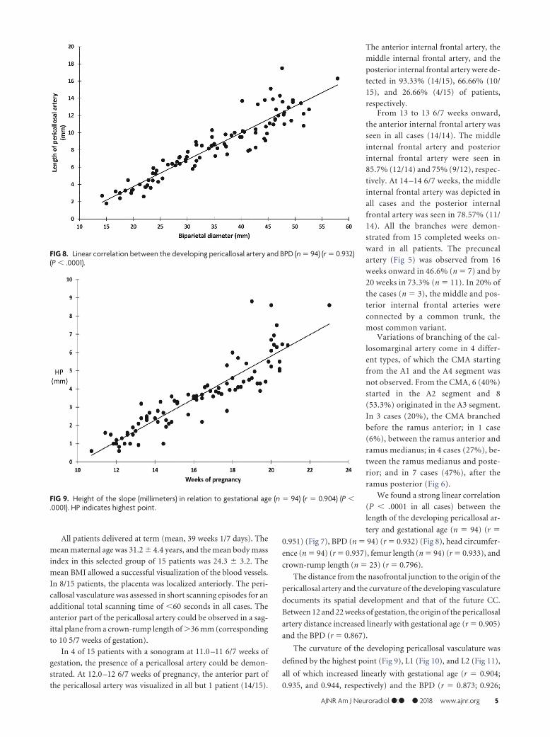
The distance from the nasofrontal junction to the origin of the
pericallosal artery and the curvature of the developing vasculature
documents its spatial development and that of the future CC.
Between 12 and 22 weeks of gestation, the origin of the pericallosal
artery distance increased linearly with gestational age (r � 0.905)
and the BPD (r � 0.867).
The curvature of the developing pericallosal vasculature was
defined by the highest point (Fig 9), L1 (Fig 10), and L2 (Fig 11),
all of which increased linearly with gestational age (r � 0.904;
0.935, and 0.944, respectively) and the BPD (r � 0.873; 0.926;
FIG 8. Linear correlation between the developing pericallosal artery and BPD (n � 94) (r � 0.932)(P � .0001).
FIG 9. Height of the slope (millimeters) in relation to gestational age (n � 94) (r � 0.904) (P �.0001). HP indicates highest point.
AJNR Am J Neuroradiol ●:● ● 2018 www.ajnr.org 5
0.913). The highest point/L1 ratio, representing the slope of the an-
terior part of the pericallosal artery and hence the developing CC,
decreased slightly throughout the investigated timeframe.
We measured the distances between the different developing
branches across time. The frontopolar artery and the ramus ante-
rior, the ramus anterior and the ramus medianus, the ramus
medianus and the ramus posterior, the ramus posterior and the
callosomarginal artery, the callosomarginal artery and the para-
central artery, and the paracentral artery
and precuneal artery illustrate the pro-
portional growth of the pericallosal
blood vessels. Intraobserver variations
of the length of the pericallosal artery
and the distances between the origins of
the different branches were not statisti-
cally significant.
DISCUSSIONAgenesis and dysgenesis of the corpus
callosum are the more frequent central
nervous system malformations associ-
ated with variable prognosis. In associa-
tion with chromosomal abnormalities,
genetic syndromes, and central nervous
system and non-CNS abnormalities, the
prognosis is invariably poor.24 Isolated
complete agenesis, however, seems to
have a better prognosis than a partial or
hypoplastic corpus callosum.19 A recent
meta-analysis of 27 studies on the out-
come shows a higher proportion of
chromosomal anomalies, more gross
and fine motor control affection, and a
higher percentage of epilepsy in the par-
tial agenesis group compared with the
complete agenesis of the CC group.40
Today, diagnosis relies on a midtrimes-
ter sonographic examination potentially
revealing �1of the associated signs such
as mild ventriculomegaly, colpocephaly,
absent cavum septi pellucidi, upward
displacement of the third ventricle, tear-
drop configuration of the lateral ventri-
cles, or cystic dilation of the third ventri-
cle.25,26 However, these signs might be
subtle or missing.27,28 The presence of a
normal CC and its biometry has been
assessed by transabdominal and trans-
vaginal 2D and 3D sonography and with
fetal MR imaging from 18 weeks on-
ward.10,17,29-33
Indirect appreciation of the develop-
ing CC will be seen by demonstration of
the pericallosal artery and its branches.
Recently, color Doppler mapping docu-
mented the modified arterial vascular
supply with loss of the semicircular loop
in CC agenesis (Fig 12).25 In a partial agenesis of the CC, the
paracentral artery follows the anterior part of the CC but loses its
normal course when the CC vanishes. At this level, the artery
moves in an upward and posteriorly oblique direction (Fig
13).19,20 The corpus callosum formation is associated with a me-
dial and upward rotation of the cingulate gyrus, with consequent
formation of the cingulate sulcus. In cases of an absent CC, the
cingulate gyri do not rotate and are small due to hypoplasia. In
FIG 10. Increasing slope of the pericallosal artery (millimeters) in relation to gestational age. (n �94) (r � 0.935) (P � .0001).
FIG 11. Decreasing slope of the pericallosal artery (millimeters) in relation to gestational age (n �94) (r � 0.944) (P � .0001).
6 De Keersmaecker ● 2018 www.ajnr.org
cases of partial agenesis, we expect therefore a shorter length of the
pericallosal artery as well as a different branching pattern and
course. Knowledge of the development and variations in the dif-
ferent branches may enhance the diagnosis of partial agenesis of
the corpus callosum.
In the first trimester, the midsagittal plane of the fetal head
allows investigating the nasal bone and the nuchal translucency as
screening markers for Down syndrome and the intracranial trans-
lucency for the detection of open neural tube defects.34,35 Adding
power color Doppler flow in fetuses at rest for short time intervals,
respecting thermal index and mechanic index, shows the devel-
oping pericallosal vasculature and its variants36-38 in the first tri-
mester either with 2D or 3D sonography.
Most first-trimester studies document either the presence or
the course and/or length of the pericallosal artery only. In a cross-
sectional study including 80 patients attending for first-trimester
aneuploidy screening, chorionic villus sampling, or amniocente-
sis, a reference range of the length of the pericallosal artery was
provided from 14 weeks onward in relation to BPD and gesta-
tional age.36 In agreement with Pati et al,36 we also detected a high
linear correlation (�0.9) between the length of the pericallosal
artery and gestational age and the BPD, respectively. However, in
that study, the developing vascular map was not analyzed. Con-
turso et al38 viewed the pericallosal arteries in healthy fetuses at
11–13 weeks of gestation in 70 cases using 3D technology in the
first trimester of pregnancy.
Diaz-Guerrero et al37 evaluated 150 fetuses between 11 and
14 weeks and failed to visualize the pericallosal artery in only 6
cases. Subsequently, 2 of these 6 cases were diagnosed with
agenesis of the corpus callosum in association with a chromo-
somal abnormality. In the 4 other fetuses, the pericallosal ar-
tery was not seen due to the fetal position and excessive fetal
movement.37 However, in addition to the biometry of the peri-
callosal artery, we favor evaluating the morphology of the vas-
culature of the pericallosal artery and its branches because it
might enhance the diagnosis of complete agenesis as well as
dysgenesis of the CC. This evaluation has already been de-
scribed in the second trimester of pregnancy.20 Therefore,
detailed knowledge of the arterial supply of the corpus callo-
sum might distinguish normal variants from deteriorated vas-
cularization associated with abnormal development of the
CC.17,39
Limitations of our study are the small number of healthy
subjects, therefore the lack of an unhealthy case, and an aver-
age body mass index of 24, which does not always represent the
general population.
CONCLUSIONSIn a population of healthy fetuses, the pericallosal artery and its
branches can be consistently identified and measured from 11
weeks on. A defective course or an abnormal biometry of the
pericallosal artery could be an early sonographic marker for iden-tifying abnormal development of the corpus callosum. Further
prospective evaluation of the vascularization and biometry of thepericallosal artery in the late first trimes-
ter is needed for proof of this concept.
ACKNOWLEDGMENTSWe sincerely thank the patients for the
participation in this study.
REFERENCES1. Rakic P, Yakovlev P. Development of the
corpus callosum and cavum septi inman. J Comp Neurol 1968;132:45–72CrossRef Medline
2. Hansen PE, Ballesteros MC, Soila K, et al.MR imaging of the developing brain, 1:prenatal development. Radiographics1993;13:21–36 CrossRef Medline
3. Bull J. The corpus callosum. Clin Ra-diol 1967;18:2–18 CrossRef Medline
4. Raybaud C. The corpus callosum, theother great forebrain commisures andthe septum pellucidum: anatomy, de-velopment and malformation. Neurora-diology 2010;S2:447–77 CrossRefMedline
FIG 12. Pericallosal artery and branches in agenesis of the corpuscallosum.
FIG 13. Aberrant pattern of the pericallosal artery and its branches in partial agenesis of thecorpus callosum.
5. Pilu G, Hobbins J. Sonography of the fetal cerebrospinal anomalies.Prenat Diagn 2002;22:321–30 CrossRef Medline
6. Reece EB, Goldstein I. Three-level view of fetal brain imaging in theprenatal diagnosis of congenital anomalies. J Matern Fetal Med1999;8:249 –52 Medline
7. Dobyns W. Absence makes the search growth longer. Am J HumGenet 1996;58:7–16 Medline
8. Salomon L, Alfirevic Z, Berghella V, et al; ISUOG Clinical StandardsCommittee. Practice guidelines for performance of the routinemid-trimester fetal ultrasound scan. Ultrasound Obstet Gynecol2011;37:116 –26 CrossRef Medline
9. Rossi A, Prefumo F. Accuracy of ultrasonography at 11–14 weeks ofgestation for detection of fetal structural anomalies: a systematicreview. Am J Obstet Gynecol 2013;122:1160 – 67 CrossRef Medline
10. Achiron R, Achiron A. Development of the human corpus callosum:a high-resolution, cross-sectional sonographic study. UltrasoundObstet Gynecol 2001;18:343– 47 CrossRef Medline
11. Fong K, Toi A, Salem S, et al. Detection of fetal structural abnormal-ities with US during early pregnancy. Radiographics 2004;24:157–74CrossRef Medline
12. Sepulveda W, Dezerega V, Be C. First-trimester sonographic diag-nosis of holoprosencephaly: value of the “butterfly” sign. J Ultra-sound Med 2004;23:761– 65; quiz 766 – 67 CrossRef Medline
13. Sepulveda W, Lutz I, Be C. Holoprosencephaly at 9 weeks and 6 daysin a triploid fetus: two-and 3-dimensional sonographic findings. JUltrasound Med 2007;26:411–14 CrossRef Medline
14. van Zahlen-Sprock R, van Vugt J, van Geyn H. First and early secondtrimester diagnosis of anomalies of the central nervous system.J Ultrasound Med 1995;145:603–10 Medline
15. Engels A, Joyeux L, Brantner C, et al. Sonographic detection of cen-tral nervous system defects in the first trimester of pregnancy. Pre-nat Diagn 2016;36:266 –73 CrossRef Medline
16. Syngelaki A, Chelemen T, Dagklis T, et al. Challenges in the diagno-sis of fetal non-chromosomal abnormalities at 11–13 weeks. PrenatDiagn 2011;31:90 –102 CrossRef Medline
17. Pashaj S, Merz E. Prenatal demonstration of normal variants of thepericallosal artery by 3D ultrasound. Ultraschall Med 2014;35:129 –36 CrossRef Medline
18. Shinar S, Har-Toov J, Lerman-Sagie T, et al. A thick corpus callosumin the second trimester can be transient and of uncertain signifi-cance. Ultrasound Obstet Gynecol 2016;48:452– 457 CrossRef Medline
19. Ghi T, Carletti A, Contro E, et al. Prenatal diagnosis and outcome ofpartial agenesis and hypoplasia of the corpus callosum. UltrasoundObstet Gynecol 2010;35:35– 41 CrossRef Medline
20. Volpe P, Paladini D, Resta M, et al. Characteristics, associations andoutcome of partial agenesis of the corpus callosum in the fetus. Ul-trasound Obstet Gynecol 2006;27:509 –16 CrossRef Medline
21. Pexters A, Daemen A, Bottomley C, et al. New crown-rump lengthcurve based on over 3500 pregnancies. Ultrasound Obstet Gynecol2010;35:650 –55 CrossRef Medline
22. Salvesen K, Lees C, Abramowicz J, et al. Safe use of Doppler ultra-sound during the 11 to 13 � 6-week scan: is it possible? UltrasoundObstet Gynecol 2011;37:625–28 CrossRef Medline
23. Fisher CM. The circulus of Willis: anatomical variations. Vas Dis1965;2:99 –105
24. Pisani F, Maria Edgarda B, Giovanni P, et al. Prenatal diagnosis ofagenesis of corpus callosum: what is the neurodevelopmental out-come? Pediatr Int 2006;48:298 –304 CrossRef Medline
25. Pilu G, Sandri F, Perolo A, et al. Sonographic fetal agenesis of the
corpus callosum: a survey of 35 cases. Ultrasound Obstet Gynecol1993;3:318 –29 CrossRef Medline
26. Gupta J, Lilford R. Assessment and management of fetal agenesis ofcorpus callosum. Prenat Diagn 1995;15:302–12 Medline
27. Paladini D, Pastore G, Cavallaro A, et al. Agenesis of the fetal corpuscallosum: sonographic signs change with gestational age. Ultra-sound Obstet Gynecol 2013;42:687–90 CrossRef Medline
28. Malinger G, Lev D, Oren M, et al. Non-visualization of the cavumsepti pellucidi is not synonymous with agenesis of the corpus callo-sum. Ultrasound Obstet Gynecol 2012;40:165–70 CrossRef Medline
29. Bennett G, Bromley B, Benacerraf BR. Agenesis of the corpuscallosum: prenatal detection usually is not possible before 22 weeksof gestation. Radiology 1996;199:447–50 CrossRef Medline
30. Chasen S, Birnholz J, Gurewitsch E, et al. Antenatal growth of thecorpus callosum. Am J Obstet Gynecol 1997;176:S66 CrossRef
31. Harreld J, Bhore R, Chason D, et al. Corpus callosum length by ges-tational age as evaluated by fetal MR imaging. AJNR Am J Neurora-diol 2011;32:490 –94 CrossRef Medline
32. Malinger G, Zakut H. The corpus callosum: normal fetal develop-ment as shown by transvaginal sonography. AJR Am J Roentgenol1993;161:1041– 43 CrossRef Medline
33. Araujo Junior E, Visentainer M, Simioni C, et al. Reference values forthe length and area of the fetal corpus callosum on 3-dimensionalsonography using the transfrontal view. J Ultrasound Med 2012;31:205–12 CrossRef Medline
34. Nicolaides KH. Screening for fetal aneuploidies at 11 to 13 weeks.Prenat Diagn 2011;31:7–15 CrossRef Medline
35. Chaoui R, Benoit B, Mitkowska-Wosniak H, et al. Assessment ofintracranial translucency (IT) in the detection of spina bifida at 11to 13 week scan. Ultrasound Obstetric Gynecology 2009;34:249 –52CrossRef Medline
36. Pati M, Cani C, Bertucci E, et al. Early visualization and measure-ment of the pericallosal artery: an indirect sign of corpus callosumdevelopment. J Ultrasound Med 2012;31:231–37 CrossRef Medline
37. Díaz-Guerrero L, Guigni-Chalbaud G, Soso-Olavarría A. Assessmentof pericallosal arteries by color Doppler ultrasonography at 11–14weeks: an early marker of fetal corpus callosum development innormal fetuses and agenesis in cases with chromosomal anomalies.Fetal Diagn Ther 2013;34:85– 89 CrossRef Medline
38. Conturso R, Contro E, Bellusi F, et al. Demonstration of the perical-losal artery at 11–13 weeks of gestation using 3D ultrasound. FetalDiagn Ther 2015;37:305– 09 CrossRef Medline
39. Cavalcanti D, Albuquerque F, Silva B, et al. The anatomy of the cal-losomarginal artery: applications to microsurgery and endovascu-lar surgery. Neurosurgery 2010;66:602–10 CrossRef Medline
40. D’Antonio F, Pagani G, Familiari A, et al. Outcomes associated withisolated agenesis of the corpus callosum: a meta-analysis. Pediatrics2016;138. pii: e20160445 CrossRef Medline
41. Kakou M, Velut S, Destrieux C. Arterial and venous vascularizationof the corpus callosum [in French]. Neurochirurgie 1998;44(1 suppl):31–37 Medline
42. Wahl M, Strominger Z, Jeremy R, et al. Variability of homotopic andheterotypic callosal connectivity in partial agenesis of the corpuscallosum: a 3T diffusion tensor imaging and Q-ball tractographystudy. AJNR Am J Neuroradiol 2009;30:382– 89 CrossRef Medline
43. Nakata Y, Barkovich A, Wahl M, et al. Diffusion abnormalities andreduced volume of the ventral cingulum bundle in genesis of thecorpus callosum: a 3T imaging study. AJNR Am J Neuroradiol 2009;30:1142– 48 CrossRef Medline