Page 1 Update on Ectopic Pregnancy Mary C. Frates, MD, FACR Background Products of conception implanted outside of the endometrial cavity 1.5 to 2.0% pregnancies Complications of EP are the leading cause of pregnancy related deaths during the first trimester in the U.S. Barnhart KT. Ectopic Pregnancy NEJM 2009 Ectopic Pregnancy >95% occur in fallopian tube 2% 8% 90% Cervical 0.1% Ovary 0.1% C Section ???? Ectopic Pregnancy Risk Factors: Tubal scarring (PID, prev EP) IUD Assisted fertilization 25% of pregnancies occuring in pts w/ IUD or TL are ectopic 50% of pts with EP have no known risk factor Casey hydrosalpinx Stammen hydrosalpinx.jpg Ectopic Pregnancy Classic presentation: pain, vaginal bleeding, adnexal mass Positive pregnancy test Ultrasound
Transcript
Page 1
Update on Ectopic Pregnancy
Mary C. Frates, MD, FACR
Background
Products of conception implanted outside of the endometrial cavity
1.5 to 2.0% pregnancies
Complications of EP are the leading cause of pregnancy related deaths during the first trimester in the U.S.
Barnhart KT. Ectopic Pregnancy NEJM 2009
Ectopic Pregnancy
>95% occur in fallopian tube
2%
8%
90%
Cervical 0.1%
Ovary 0.1%
C Section
????
Ectopic Pregnancy
Risk Factors:
Tubal scarring (PID, prev EP)
IUD
Assisted fertilization
25% of pregnancies occuring in pts w/ IUD or TL are ectopic
50% of pts with EP have no known risk factor
Casey hydrosalpinx Stammen hydrosalpinx.jpg
Ectopic Pregnancy
Classic presentation: pain, vaginal bleeding, adnexal mass
Positive pregnancy test
Ultrasound
Page 2
Pregnancy Test
Trophoblastic tissue makes hCG 8 days after conception
Normal pregnancy: sac typically seen by TVS with hCG of 1000 mIU/ml
17/51 (33%) patients with hCG > 2000, not treated for EP, had IUPs at follow-up*
*Mehta et al, Radiology 1997; 205:569-573
Pregnancy Test (BWH data)
hCG within 24 hours of US (225 EPs)
Range 7 – 107,949 mIU/ml
Average 3256 mIU/ml
significantly higher with +FH in EP
20,980 vs 1,901 (no FH)
77% had hCG <3000, 7% had hCG >10,000
Pregnancy Test
BWH cautionary case
hCG over 4000
Nothing in uterus, nothing in adnexa
followup …………..Nml IUP
Do NOT dx and treat (for EP) a stable patient until certain
+ hCG and no IUP: PUL
Pregnancy of Unknown Location
only 3 choices:
very early IUP
SAB / chemical
EP
Uterus
IUP
round, echogenic rim, contains YS, pole, FH
located within decidua
Don’t be misled by fluid in the endometrial cavity
Donlon nml sac intradecidual Akasapu early gestational sac mimics pseudosac Park pos preg test uncertain dates tiny
fluid cllection cant tell if IUP
Small gestational sac
? Probable?
Page 3
Wolfe 6 wk sac w subchorionic hematoma
Pseudosacs johnson champion Walsh Gomez hcg 70 pseudosac in uterus adnexa neg.avi
No gestational sac seen
US of the endometrium
Endometrial thickness can predict presence of IUP
Is it any good?
Moschos et al, 2008: no IUP had an endometrium <8 mm
4 EP’s had endometrium >25mm
US of the endometrium
Trilaminar pattern more frequent in ectopic pregnancy
Is it any good?
sens 21%; spec 93%; ppv 50%
Col-Madendag et al, Arch Gyn Obst: 2010
Hanson outside US no IUP live EP missed same day Cor Rt ov.jpg
30 yo on Clomid
7.5 weeks by LMP
Read as negative
Hanson outside US no IUP live EP missed same day Cor Rt ov.jpg
Repeat that afternoon
at BWH
Page 4
Adnexa
EP better diagnosed by presence of an adnexal mass rather than by absence of an IUP
earlier identification of a mass allows earlier treatment
Bern R thick tube old surgery.jpg
Prior EP
4.7 wks w/Pain
Followup- Normal IUP
Adnexa
Tubal ring (Gestational sac)
echogenic ring, anechoic center
25% of patients with EP**
ring + YS (8%)
ring + YS + cardiac activity (7%)
**Study of 231 EPs @BWH
Frates et al JUM 2014; 33:697
Robles live 8 wk EP no pain just vag bleeding cor YS.jpg
Monahan heterotopic 6wks
Bernstein tiny EP L sag.jpg
Page 5
Adnexa
Complex mass
poorly defined borders
55% EPs present with this**
careful search may reveal a central ring or YS
think hematosalpinx
**Study of 231 EPs @BWH
Frates et al JUM 2014; 33:697
Jones small solid mass
5.7 weeks
b Cranmer EP in wall of tube
Adnexa
Meta-analysis of 10 studies
Most appropriate criteria for making diagnosis of EP:
ANY noncystic extraovarian adnexal mass
Brown, Doubilet J Ultrasound Med 1994;13:259-266
Adnexa
Noncystic adnexal mass:
specificity 98.9%
sensitivity 84.4%
pos predictive value 96.3%
neg predictive value 94.8%
Brown, Doubilet J Ultrasound Med 1994;13:259-266
Page 6
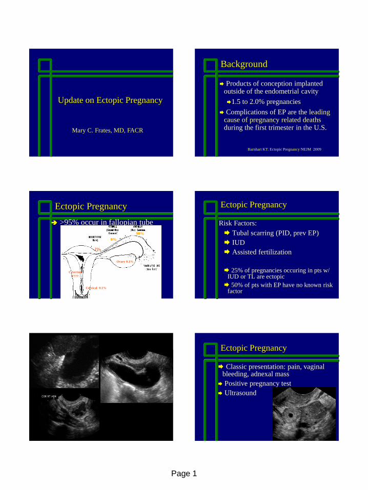
Adnexa
Noncystic nonovarian mass
specificity 99.9%
sensitivity 90.9%
pos predictive value 93.5%
neg predictive value 99.8%
Condous et al Human Reproduction 2005:1404-1409
Mcelaney Rt EP stuck to ROv cor.
Wolfson Lt hematosalpinx EP cor.jpg
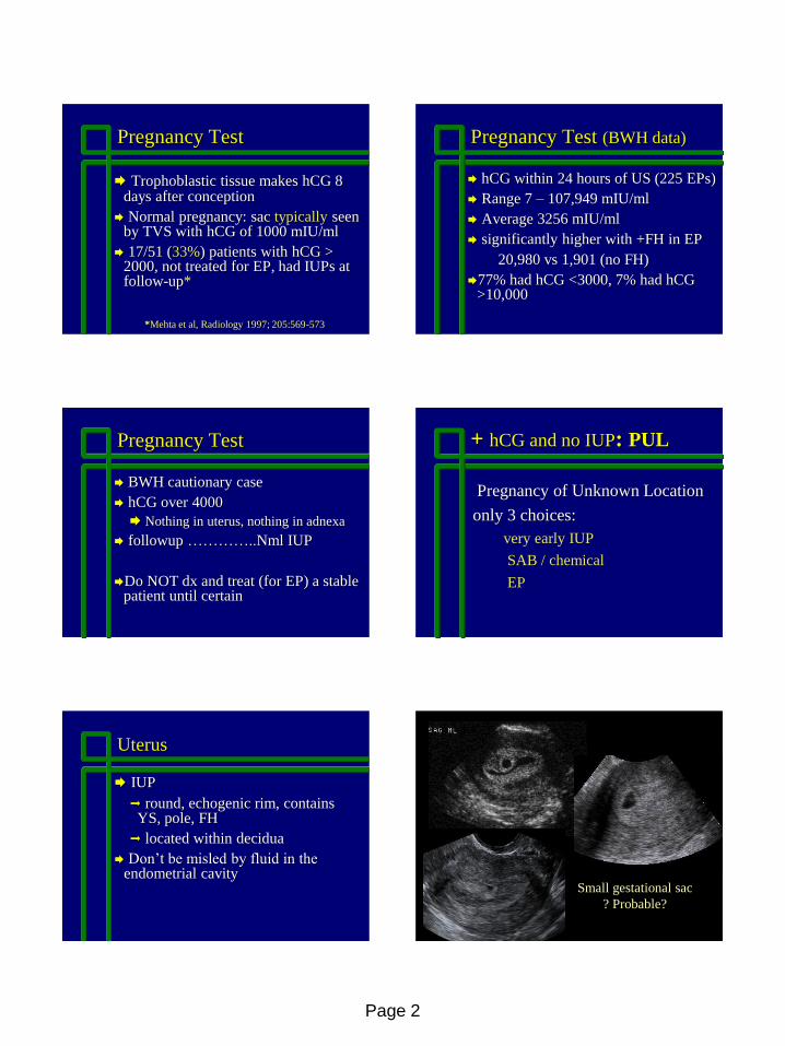
Things to consider….
Can the mass be separated from the ovary?
What is the echotexture of the mass?
Walsh EP moves away from ovary
Movement of Mass Resnick CL inseparable from ovary
No Movement of Mass
Page 7
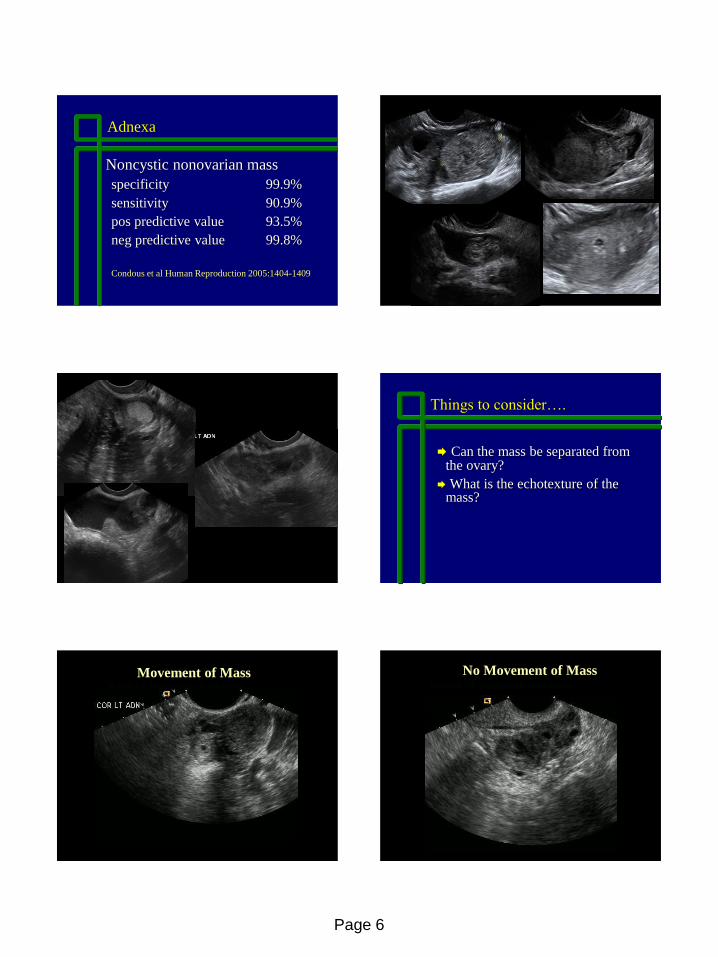
Movement of Adnexal Mass
21/23 patients with EP showed movement of mass with palpation
6/49 patients without EP showed movement of mass with palpation
NPV = 96.1%
PPV = 77.8%
Blaivas et al JUM 2005; 24:599-603
McNeil 6.7 IVF Rt EP.jpg
McNeil 6.7 IVF Rt EP.jpg
Rojas heterotopic EP CL Persistent pain, everything OK at OSH
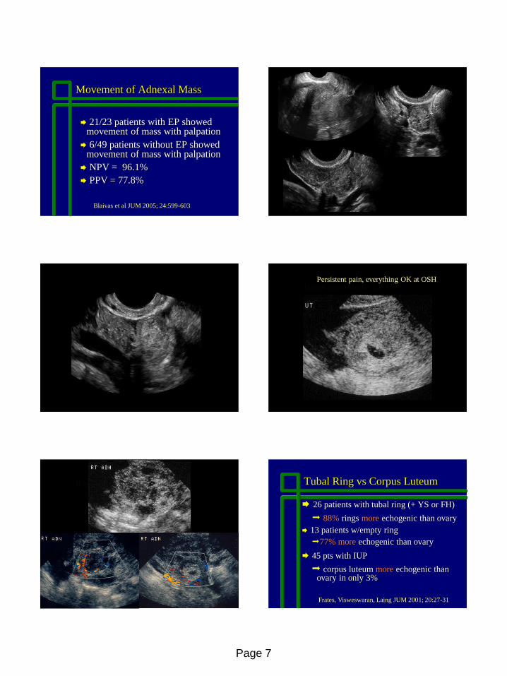
Rojas Ep and CL Tubal Ring vs Corpus Luteum
26 patients with tubal ring (+ YS or FH)
88% rings more echogenic than ovary
13 patients w/empty ring
77% more echogenic than ovary
45 pts with IUP
corpus luteum more echogenic than ovary in only 3%
Frates, Visweswaran, Laing JUM 2001; 20:27-31
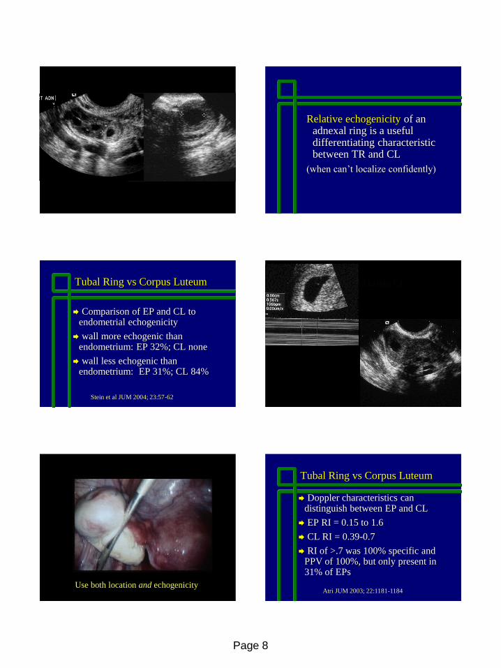
Page 8
Gibson pretty echogenic TR
Relative echogenicity of an adnexal ring is a useful differentiating characteristic between TR and CL
(when can’t localize confidently)
Tubal Ring vs Corpus Luteum
Comparison of EP and CL to endometrial echogenicity
wall more echogenic than endometrium: EP 32%; CL none
wall less echogenic than endometrium: EP 31%; CL 84%
Stein et al JUM 2004; 23:57-62
Nemrow IUP and bright CL
Nemrow intraop of CL
Use both location and echogenicity
Tubal Ring vs Corpus Luteum
Doppler characteristics can distinguish between EP and CL
EP RI = 0.15 to 1.6
CL RI = 0.39-0.7
RI of >.7 was 100% specific and PPV of 100%, but only present in 31% of EPs
Atri JUM 2003; 22:1181-1184
Page 9
Free Fluid: is it reliable?
anechoic vs echogenic
echogenic fluid correlates with hemoperitoneum
suggests high risk for EP
Nyberg et al Radiology 1991
Echogenic Fluid
185 pts to OR for EP
125 pts echogenic fluid- 98%+ blood
30 anechoic fluid- 0% blood
30 no fluid- 0% blood
Echogenic fluid correlates with hemoperitoneum
Sens 100%, Spec 95%, PPV 98%
Sickler et al, JUM 1998 17;431-435
Free Fluid: is it reliable?
38/523 PUL patients with isolated free fluid
42% of 38 had EP
22% of those with moderate fluid
73% of those with large fluid
pts with isolated CDS fluid are at moderate risk for EP; risk increases if echogenic or large
Dart et al; Am J Emerg Med 2002; 20:1-4
Anglim hemoperitoneum 5.4wks
5.4 weeks solid dates
hCG = 35
Negative Exam
EP not seen: very early GA, high BMI, fibroids, inexperience, ovarian pathology
5% in the BWH series
Stable patient: followup hCG and US
Unstable patient: to the OR
Nee live R ov EP cor
Nee Live Rt ovarian EP
Page 10
Nee ovarian ectopic
Nee Live Rt ovarian EP
Ovarian Ectopic
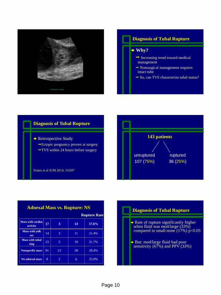
Diagnosis of Tubal Rupture
Why?
Increasing trend toward medical
management
Nonsurgical management requires intact tube
So, can TVS characterize tubal status?
Retrospective Study
Ectopic pregnancy proven at surgery
TVS within 24 hours before surgery
Frates et al JUM 2014; 33:697
Diagnosis of Tubal Rupture
143 patients
unruptured
107 (75%)
ruptured
36 (25%)
Adnexal Mass vs. Rupture: NS
Mass with cardiac
activity 17 3 14 17.6%
Mass with yolk
sac 14 3 11 21.4%
Mass with tubal
ring 23 5 18 21.7%
Nonspecific mass 81 23 58 28.4%
No adnexal mass 8 2 6 25.0%
Rupture Rate
Diagnosis of Tubal Rupture
Rate of rupture significantly higher when fluid was mod/large (33%) compared to small-none (17%) p<0.05
But: mod/large fluid had poor sensitivity (67%) and PPV (33%)
Page 11
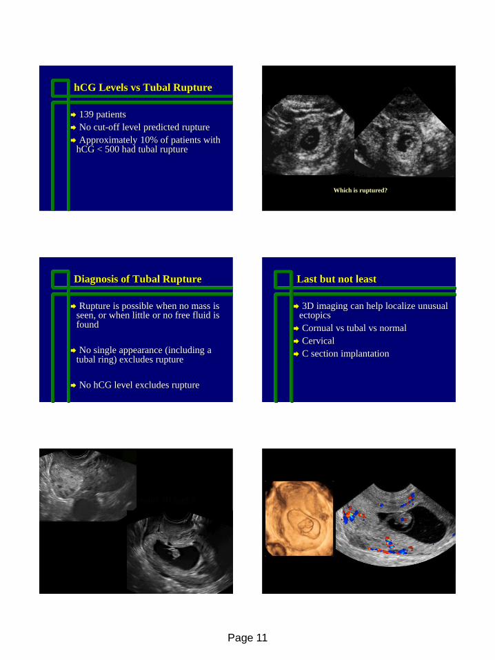
hCG Levels vs Tubal Rupture
139 patients
No cut-off level predicted rupture
Approximately 10% of patients with hCG < 500 had tubal rupture
Unruptured TR Vidockler rutured Blackett
Which is ruptured?
Diagnosis of Tubal Rupture
Rupture is possible when no mass is seen, or when little or no free fluid is found
No single appearance (including a tubal ring) excludes rupture
No hCG level excludes rupture
Last but not least
3D imaging can help localize unusual ectopics
Cornual vs tubal vs normal
Cervical
C section implantation
Jodoin C section ectopic 3D.jpgEP 6.5wks.avi Jodoin C section ectopic 3D.jpg
Page 12
Ackerman 6.5 wks failed 3D turned 90 degrees shows csection site clearly
5.5 weeks ? C –section implantation
Ackerman 6.5 wks failed 3D turned 90 degrees shows csection site clearly
6.5 weeks ? C –section implantation
Truong 7.7 wk live cornual color.jpg
7.7 weeks Cornual Implantation
Truong 7.7 wk live cornual color.jpg
Pantazelos failed cornual EP 6.5wks.avi
6.5 weeks Cornual Implantation
Castillo left isthmic EP close location to ut COR.jpgEP 6.5wks.avi
Page 13
Conclusion
Transvaginal sonography continues to be the optimal method for the evaluation of ectopic pregnancy. Early dx allows less invasive treatment options.