+ All Categories
Home > Documents > Sonography of a Hemorrhagic Cerebral Contusion

Sonography of a Hemorrhagic Cerebral Contusion

Date post: 04-Jan-2017
Category:
Upload: duongdang
View: 227 times
Download: 1 times
Share this document with a friend
2
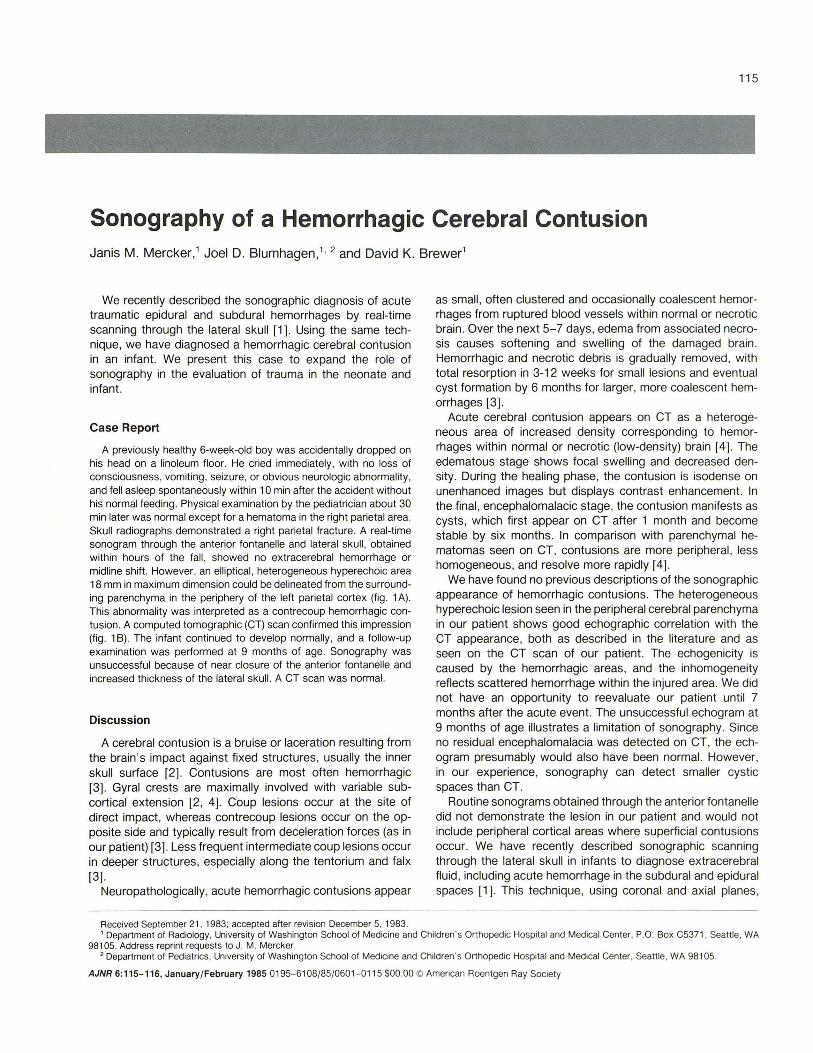
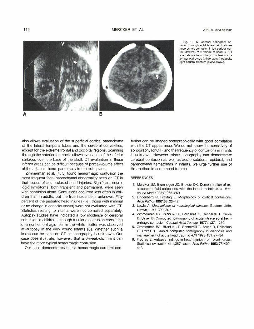
115 Sonography of a Hemorrhagic Cerebral Contusion Janis M. Mercker,' Joel D. Blumhagen ," 2 and David K. Brewer' We recently described the sonographic diagnosis of acute traumatic epidural and subdural hemorrhages by real-time scanning through the lateral skull [1]. Using the same tech- nique, we have diagnosed a hemorrhagic cerebral contusion in an infant. We present this case to expand the role of sonography in the evaluation of trauma in the neonate and infant. Case Report A previously healthy 6-week-old boy was accidentally dropped on his head on a linoleum floor. He cried immediately, with no loss of consciousness, vomiting, seizure, or obvious neurologic abnormality, and fell asleep spontaneously within 10 min after the accident without his normal feeding . Physical examination by the pediatrician about 30 min later was normal except for a hematoma in the right parietal area. Skull radiographs demonstrated a right parietal fracture. A real-time sonogram through the anterior fontanelle and lateral skull , obtained within hours of the fall , showed no extracerebral hemorrhage or midline shift. However, an elliptical, heterogeneous hyperechoic area 18 mm in ma ximum dimension could be delineated from the surround- ing parenchyma in the periphery of the left parietal cortex (fig. 1 A) . This abnormality was interpreted as a contrecoup hemorrhagic con- tusion. A computed tomographic (CT) scan confirmed this impression (fig . 1 B) . The infant continued to develop normally, and a follow-up examination was performed at 9 months of age. Sonography was unsuccessful because of near closure of the anterior fontanelle and increased thickness of the lateral skull. A CT scan was normal. Discussion A cerebral contusion is a bruise or laceration resulting from the brain's impact against fi xed structures, usually the inner skull surface [2]. Contusions are most often hemorrhagic [3]. Gyral crests are maximally involved with variable sub- cortical extension [2, 4]. Coup lesions occur at the site of direct impact, whereas contrecoup lesions occur on the op- posite side and typically result from deceleration forces (as in our patient) [3]. Less frequent intermediate coup lesions occur in deeper structures, especially along the tentorium and fal x [3]. Neuropathologically, acute hemorrhagic contusions appear Received September 21. 1983; accepted after revision December 5, 1983. as small, often clustered and oc casionally coalescent hemor- rhages from ruptured blood vessels within normal or necrotic brain . Over the next 5- 7 days, edema from associated necro- sis causes softening and swelling of th e damaged brai n. Hemorrhagic and necrotic debris is gradually removed , wi th total resorption in 3-12 weeks for small lesions and eventual cyst formation by 6 months for larger, more coalescent hem- orrhages [3] . Acute cerebral contusion appears on CT as a heterog e- neous area of increased density corresponding to hemor- rhages within normal or necrotic (low-density) brain [4]. The edematous stage shows focal swelling and decreased den- sity. During the healing phase, t he contusion is isodense on unenhanced images but displays contrast enhancement. In the final , encephalomalacic stage, the contusion manifests as cysts, which first appear on CT after 1 month and bec ome stable by si x months. In comparison with parenchymal he- matomas seen on CT , contusions are more peripheral, less homogeneous, and resolve more rapidly [4] . We have found no previous descriptions of the sonographic appearance of hemorrhagic contusions. The heterogeneous hyperechoic lesion seen in the peripheral cerebral parenchyma in our patient shows good echographic correlation with the CT appearance, both as described in the literature and as seen on the CT scan of our patient. The echogenicity is caused by the hemorrhagic areas, and the inhomogeneity reflects scattered hemorrhage within the inju re d area. We did not have an opportunity to reevaluate our patient un t il 7 months after the acute event. The unsucce ssful echogram at 9 months of age illustrates a limitation of sonography. Since no residual encephalomalacia was detect ed on CT , th e ech - ogram presumably would also have been normal. However, in our experience, sonography can detect small er cystic spaces than CT . Routine sonograms obtained through th e anterior fontanell e did not demonstrate the lesion in our patient and would not include peripheral cortical areas where superficial contusions occur. We have recently described sonographic sc anning through t he lateral skull in infants to diagnose extracere bral fluid, including acute hemorrhage in the subdural and ep idural spaces [1 ]. Th is technique, using coronal and axial planes , 1 Department of Radi ology. Uni versity of Washington School of Medicine and Children's Orthopedic Hospital and Medical Center. P.O. Box C5371 , Sea ttle . WA 98105. Address reprin t requests to J. M. Mercker. 2 Department of Pedi atrics, Universi ty of Washington School of Medicine and Children 'S Orthopedic Hospital and Medical Center. Seattle, WA 98105. AJNR 6: 115-116, January/February 1985 0195-6 108/85/ 0601-0115 $00.00 © American Roentgen Ray Society
Transcript

115

Sonography of a Hemorrhagic Cerebral Contusion Janis M. Mercker,' Joel D. Blumhagen," 2 and David K. Brewer'

We recently described the sonographic diagnosis of acute traumatic epidural and subdural hemorrhages by real-time scanning through the lateral skull [1]. Using the same tech­nique, we have diagnosed a hemorrhagic cerebral contusion in an infant. We present this case to expand the role of sonography in the evaluation of trauma in the neonate and infant.

Case Report

A previously healthy 6-week-old boy was accidentally dropped on his head on a linoleum floor. He cried immediately, with no loss of consciousness, vomiting, seizure, or obvious neurologic abnormality , and fell asleep spontaneously within 10 min after the accident without his normal feeding . Physical examination by the pediatrician about 30 min later was normal except for a hematoma in the right parietal area. Skull radiographs demonstrated a right parietal fracture. A real-time sonogram through the anterior fontanelle and lateral skull , obtained within hours of the fall , showed no extracerebral hemorrhage or midline shift. However, an elliptical , heterogeneous hyperechoic area 18 mm in maximum dimension could be delineated from the surround­ing parenchyma in the periphery of the left parietal cortex (fig. 1 A) . This abnormality was interpreted as a contrecoup hemorrhagic con­tusion. A computed tomographic (CT) scan confirmed this impression (fig . 1 B). The infant continued to develop normally, and a follow-up examination was performed at 9 months of age. Sonography was unsuccessful because of near closure of the anterior fontanelle and increased thickness of the lateral skull. A CT scan was normal.

Discussion

A cerebral contusion is a bruise or laceration resulting from the brain 's impact against fi xed structures, usually the inner skull surface [2] . Contusions are most often hemorrhagic [3]. Gyral crests are maximally involved with variable sub­cortical extension [2 , 4] . Coup lesions occur at the site of direct impact, whereas contrecoup lesions occur on the op­posite side and typically result from deceleration forces (as in our patient) [3]. Less frequent intermediate coup lesions occur in deeper structures , especially along the tentorium and falx [3] .

Neuropathologically, acute hemorrhagic contusions appear

Received September 21. 1983; accepted after revision December 5, 1983.

as small , often clustered and occasionally coalescent hemor­rhages from ruptured blood vessels within normal or necrotic brain . Over the next 5-7 days, edema from associated necro­sis causes softening and swelling of the damaged brain. Hemorrhagic and necrotic debris is gradually removed , with total resorption in 3-12 weeks for small lesions and eventual cyst formation by 6 months for larger, more coalescent hem­orrhages [3] .

Acute cerebral contusion appears on CT as a heteroge­neous area of increased density corresponding to hemor­rhages within normal or necrotic (low-density) brain [4]. The edematous stage shows focal swelling and decreased den­sity. During the healing phase, the contusion is isodense on unenhanced images but displays contrast enhancement. In the final , encephalomalacic stage, the contusion manifests as cysts, which first appear on CT after 1 month and become stable by six months. In comparison with parenchymal he­matomas seen on CT, contusions are more peripheral, less homogeneous, and resolve more rapidly [4] .

We have found no previous descriptions of the sonographic appearance of hemorrhagic contusions. The heterogeneous hyperechoic lesion seen in the peripheral cerebral parenchyma in our patient shows good echographic correlation with the CT appearance, both as described in the literature and as seen on the CT scan of our patient. The echogenicity is caused by the hemorrhagic areas, and the inhomogeneity reflects scattered hemorrhage within the injured area. We did not have an opportunity to reevaluate our patient until 7 months after the acute event. The unsuccessful echogram at 9 months of age illustrates a limitation of sonography. Since no residual encephalomalacia was detected on CT, the ech­ogram presumably would also have been normal. However, in our experience, sonography can detect smaller cystic spaces than CT.

Routine sonograms obtained through the anterior fontanelle did not demonstrate the lesion in our patient and would not include peripheral cortical areas where superficial contusions occur. We have recently described sonographic scanning through the lateral skull in infants to diagnose extracerebral fluid , including acute hemorrhage in the subdural and epidural spaces [1 ]. This technique, using coronal and axial planes ,

1 Department of Radiology. University of Washington School of Medicine and Children's Orthopedic Hospital and Medical Center. P.O. Box C5371 , Seattle . WA 98105. Address reprint requests to J. M. Mercker.

2 Department of Pediatrics, Universi ty of Washington School of Medicine and Children 'S Orthopedic Hospital and Medical Center. Seattle , WA 98105.

AJNR 6:115-116, January/ February 1985 0195-6108/85/0601-0115 $00.00 © American Roentgen Ray Society

116 MERCKER ET AL AJNR :6, Jan/Feb 1985

A B

also allows evaluation of the superficial cortical parenchyma of the lateral temporal lobes and the cerebral convexities, except for the extreme frontal and occipital regions. Scanning through the anterior fontanelle allows evaluation of the inferior surfaces over the base of the skull . CT evaluation in these inferior areas can be difficult because of partial-volume effect of the adjacent bone , particularly in the axial plane.

Zimmerman et al. [4 , 5] found hemorrhagic contusion the most frequent focal parenchymal abnormality seen on CT in their series of acute closed head injuries. Significant neuro­logic symptoms, both transient and permanent, were seen with contusion alone. Contusions occurred less often in chil­dren than in adults, but the true incidence is unknown. Fifty percent of the pediatric head injuries (i .e., those with minimal or no change in consciousness) were not evaluated with CT. Statistics relating to infants were not compiled separately. Autopsy studies have indicated a low incidence of cerebral contusion in children , although a unique contusion consisting of a non hemorrhagic tear in the white matter was observed at autopsy in the very young infants [6]. Whether such a lesion can be seen on CT or sonography is unknown. Our case does illustrate, however, that a 6-week-old infant can have the more typical hemorrhagic contusion.

Our case demonstrates that a hemorrhagic cerebral con-

Fig . 1.-A, Coronal sonogram ob­tained through right lateral skull shows hyperechoic contusion in left parietal cor­tex (arrows ). V = vertex of head. B, CT scan shows hemorrhagic contusion in a left parietal gyrus (white arrow ) opposite right parietal fracture (black arrow ).

tusion can be imaged sonographically with good correlation with the CT appearance. We do not know the sensitivity of sonography (or CT), and the frequency of contusions in infants is unknown. However, since sonography can demonstrate cerebral contusion as well as acute subdural, epidural , and parenchymal hematomas in infants, we urge further use of this method in acute head trauma.

REFERENCES

1. Mercker JM, Blumhagen JD, Brewer DK. Demonstration of ex­tracerebral fluid collections with the lateral technique. J Ultra­sound Med 1983;2 :265-269

2. Lindenberg R, Freytag E. Morphology of cortical contusions. Arch Patho/1957;63:23-42

3. Lewis A. Mechanisms of neurological disease. Boston: Little, Brown, 1976:300-307

4. Zimmerman RA, Bilaniuk L T, Dolinskas C, Genneralli T, Bruce D, Uzzell B. Computed tomography of acute intracerebral hem­orrhagic contusion . Comput Axial Tomogr 1977;1: 271-280

5. Zimmerman RA, Bilaniuk L T, Genneralli T, Bruce D, Dolinskas C, Uzzell B. Cranial computed tomography in diagnosis and management of acute head trauma. AJR 1978;131 :27-34

6. Freytag E. Autopsy findings in head injuries from blunt forces . Statistical evaluation of 1,367 cases. Arch Patho/1953;75 :402-413


Recommended