30
Sparse Paradigm Free Mapping: an inverse problem in fMRI Ian Dryden School of Mathematical Sciences Joint work with Cesar Caballero Gaudes,
| Date post: | 24-Dec-2015 |
| Category: |
Documents |
| Upload: | eleanor-chambers |
| View: | 214 times |
| Download: | 0 times |

Sparse Paradigm Free Mapping: an inverse problem in fMRI
Ian Dryden School of Mathematical Sciences Joint work with Cesar Caballero Gaudes, Natalia Petridou, Susan Francis, Penny Gowland, Li Bai
Statistical Challenges in Neuroscience, University of Warwick, September 3, 2014

Plan
1. Introduction2. Paradigm Free Mapping (PFM)
Ridge regression and tests3. Sparse Paradigm Free Mapping (SPFM) Dantzig selector4. Resting state networks5. Conclusions

1. Introduction : fMRI Ultra-high field 7T scanner at SPMMR Centre, Nottingham

BOLD fMRI: Haemodynamic Response

Traditional approach to fMRI• A study is carried out according to a GIVEN
PARADIGM – i.e. a design where it is known when stimuli are applied, usually repeatedly.
• Carry out linear regression using the general linear model (GLM), taking care to set up an appropriate design matrix which takes into account the haemodynamic response function of the BOLD signal.
• Widely used software available: SPM (UCL)

Considering a linear time-invariant model, the measured fMRI BOLD signal is the convolution of the HRF h(t) with an underlying neural-related signal s(t), plus instrumental and physiological fluctuations and noise ε(t)
Discrete (vectorial) Model
y Hs
Estimate of s(t)
Deconvolution of s(t)

Linear model with H being the Toeplitz convolution matrix defined from
the HRF shape
At each voxel we have the centered response y, and we know the design matrix H. We have zero mean, correlated errors
.
y length T, s length T, H T x T matrix.
We want to estimate the stimulus s
An inverse problem
y Hs

Ridge regression (Hoerl and Kennard, 1970) with correlated errors. Estimate stimuli s by minimizing:
Lasso (Tibshirani, 1995). Estimate stimulis by minimizing
Lasso gives a sparse estimator – lots of zeroes – solution similar in practice to the Dantzig Selector (Candes and Tao, 2007)

Linear estimation of the neural-related signal via ridge regression with correlated temporal errors
Choice of weight in the penalty?

Finite sample BIC criterion to choose AR(p) orderxyz
3D-neighbouring region (C )
Noise statistical characterization is estimated from a set of B baseline time points and a 3D-region of L voxels around each voxel time series prior to Ridge Regression estimation.
Σ
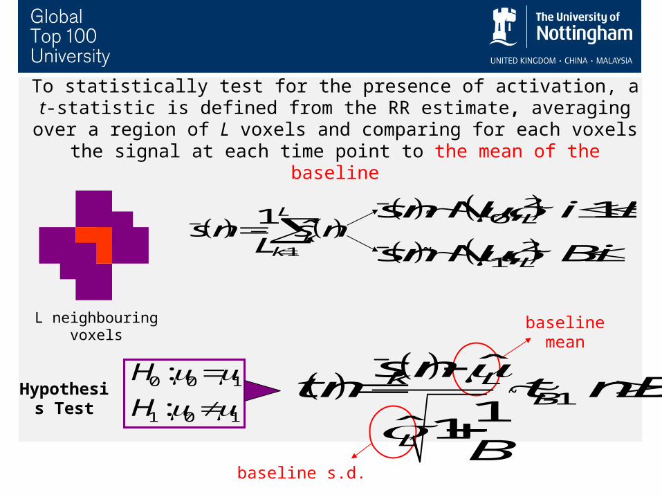
Hypothesis Test
L neighbouring voxels
1
ˆ
1ˆ 1
k LB
L
s nt n t n B
B
0 0 1
1 0 1
:
:
H
H
baseline mean
baseline s.d.
1
1ˆ
L
kk
s n s nL
20
21
, 1
,
L
L
s n N i B
s n N B i
To statistically test for the presence of activation, a t-statistic is defined from the RR estimate, averaging over a region of L voxels and comparing for each voxels the signal at each time point to the
mean of the baseline
~
~
~

Control for Multiple Hypothesis Testing:
BH- FDR
False Discovery Rate for spatial correction of each T-map

Temporal Activation
Maps
Ridge Regressionestimate of
s
TemporalT-statistic
Spatial and Temporal
Preprocessing
Noise Model
AR estimation
FDRCorrection
In contrast: the GLM approach assumes the stimulus timing is known

Experiment
Paradigm: Visually cued (VC) and self-paced (SP) finger tapping with dominant hand
EMG measurements were recorded at both hands (right/left extensor and right flexor) to capture any hand movement
Tap at will
384s
TAP
140s
VC TAP
180s
VC S
PSP
684s
+Baselines
+Rest 1 Rest 2
++
0s
Five subjects were scanned at 7T using a 16-channel head coil during a motor paradigm (visually-cued or self-paced finger tapping with dominant hand)

Data were corrected for Motion and linear and quadratic trends with AFNI (NIMH/NIH) and physiological fluctuations with RETROICOR (Glover et al.,
2000)
Proposed methodology to obtain a time course of thresholded T-maps Ridge Regression deconvolution + Spatio-temporal T-statistic + FDR
correction Spatial clustering: Minimum cluster size of 5 voxels Activation Time Series (ATS): # of active voxels Activation T-mapsValidation: Traditional GLM analysis with onset equal to first time point of cluster in ATS.
Bush and Cisler (2013, MRI) report strong classification performance of PFM in comparative studies.

TR 2s

No information about the paradigm (except # of baselines) was employed
for data analysis
EMG-fMRI Activation Time Series
Electromyography (EMG) measures muscle activity in left extensor and right flexor (muscles)
Activation Time Series: (ATS) Total number of active voxels
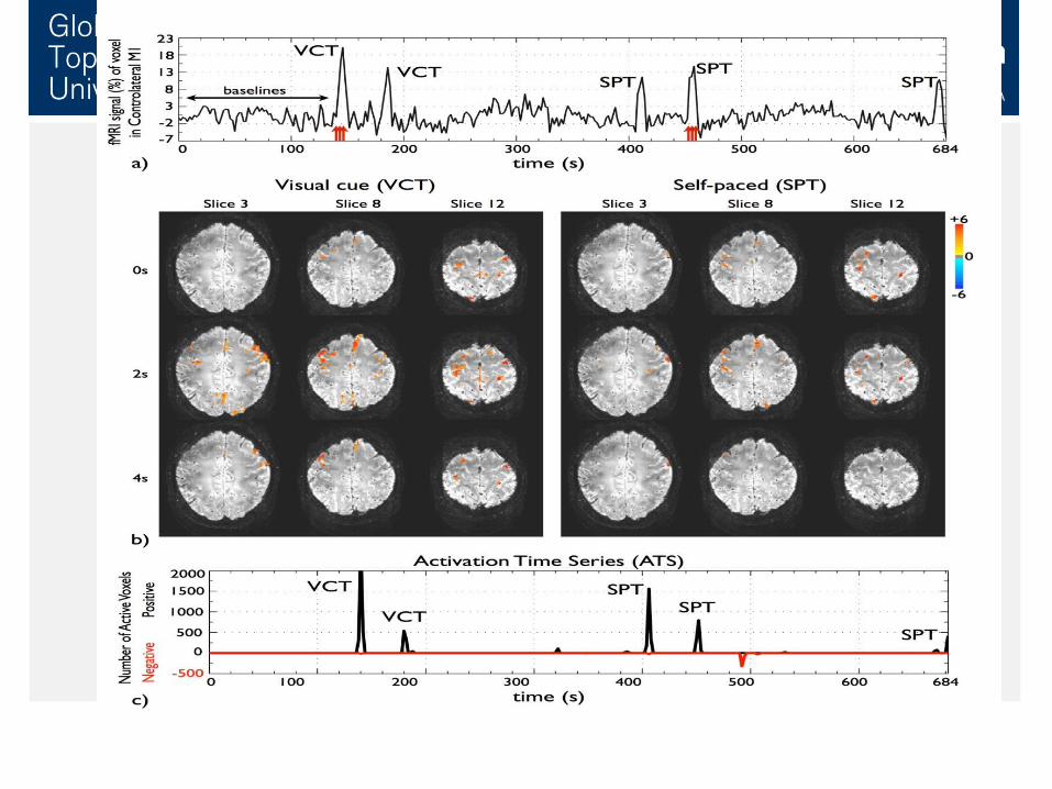

Brain activity is detected in:- Supplementary motor area
(SMA) and cingulate gyrus: initiation and self-control of motor movements.
- Primary motor cortex: motor execution
- Primary somatosensory cortex (S1): sense of touch.
- Primary and secondary visual cortex
Paradigm Free Mapping for Single Trial BOLD fMRI analysis
Results

Plan1. Introduction2. Paradigm Free Mapping (PFM)
Ridge regression and tests3. Sparse Paradigm Free Mapping (SPFM) Dantzig selector4. Resting state networks5. Conclusions

Sparsedeconvolution
of HRF
StatisticalModel
Selection
Spatial and Temporal
Preprocessing
Dantzig Selector algorithm
1ˆ min
ss s T
m m DSH y H s
subject to
The regularization parameter l controls the sparseness of the estimate, setting a maximum correlation between the model and the residuals (L∞-norm)
Combination of homotopy-type algorithms and model selection criteria
2
1ˆLnarg minDS p
p
pp y Hs K
N N
AIC: K= 2
BIC: K= Ln(N)
Temporal Activation
Maps
3. Sparse Paradigm Free Mapping – No need for the baseline!
( Candes and Tao, Annals of Statistics, 2007).
( Zou et al., Annals of Statistics, 2007).


Simulation

4. Resting state activity
A CB
BA C
Spontaneous patterns of cortical activations detected during resting state with BOLD fMRI
Paradigm Free Mapping (Dantzig Selector + BIC)
GLM (OLS + AR(0), F-test, p < 0.05, FDR corrected)
BA C

>6-6<
n2
n3 n4
tapE
F
A B
C D
n1
n1 sensorimotor networkn2 visual networkn3 episodic memory, self-referential processing, default-mode networkn4 working memory, dorsal attention network Maps show significant seed-voxel correlations

PFM and Sparse PFM are based on the deconvolution of the HRF with Ridge Regression or Sparse Regression algorithms (Dantzig Selector).
Key issues for the inverse problem: form of penalty, choice of penalty weight.
Other approaches: ICA, change points, hidden process models, …
5. ConclusionsThere is a need for a paradigm-free fMRI analysis technique to detect brain activations automatically without prior information of activation onset.

• Multi-disciplinary team – statistics/mathematics, physics, computer science, signal processing.
• Special thanks:
Cesar Caballero Gaudes
BCBL, San Sebastian, Spain
European Union funded PhD
• - and co-authors Natalia Petridou (Utrecht), Penny Gowland, Susan Francis (both SPMMRC and Physics), Li Bai (CS).

Caballero Gaudes, C., Petridou, N., Dryden, I.L., Bai, L., Francis, S.T. and Gowland, P.A. (2011). Detection and characterization of single-trial fMRI BOLD responses: Paradigm Free Mapping. Human Brain Mapping. 32, 1400–1418.
Caballero Gaudes, Petridou, N., Francis, S., Dryden, I.L. and Gowland, P. (2013). Paradigm Free Mapping with sparse regression automatically detects single-trial fMRI BOLD responses. Human Brain Mapping. 34, 501-518.
Petridou, N., Caballero Gaudes, C., Dryden, I.L., Francis, S.T. and Gowland, P.A. (2013). Periods of rest in fMRI contain transient events which are related to slowly fluctuating spontaneous activity. Human Brain Mapping. 34, 1319-1329.

Thank you!


