- 264 - SPATIAL DATABASE CREATION AND MAINTENANCE FOR PIERCE’S DISEASE AND GLASSY-WINGED SHARPSHOOTER IN CALIFORNIA Project Leader: Maggi Kelly Dept. of Environmental Science, Policy, & Management University of California Berkeley, CA 94720 Reporting Period: The results reported here are from work conducted from July 1, 2004 to October 1, 2004. INTRODUCTION Whether tracking invasive species, assessing water quality, or monitoring the spread of disease, comprehensive data collection is a key component of scientific inquiry and sustainable natural resource management. Geographic Information Systems (GIS) allow us to unite in one structure spatially referenced data with other information, affording new insights in relationships between variables at multiple scales (original proposal contains full references), as well as assisting in collaborative efforts at natural resource management and multi-disciplinary problem solving. Such is the case with Pierce’s Disease, where disparate datasets on PD location and GWSS trap data could, if available in a Geographical Information System (GIS) format with other spatially referenced data “layers” such as crops, hydrography, climate, and roads, aid in management of the disease, as well as in epidemiological research. Several agencies and individuals have recognized the need for such a geospatial database for PD research and management. Indeed, the University of California Agriculture and Natural Resources “Report of the Pierce’s Disease Research and Emergency Response Task Force(http://danr.ucop.edu/news/speeches/executivesummary.html) lists the following recommendations: Support is needed for a coordinated, statewide monitoring, trapping and reporting program involving governmental agencies, the agriculture and nursery industries and UC. The objective is to locate populations of GWSS and BGSS, track the incidence and distribution of Pierce's disease and carry out emergency response programs to slow the spread of PD and its vectors. CDFA or UC should manage a GIS to store, display, manipulate and overlay information collected by statewide monitoring and tracking programs. This data should be available to decision makers, growers and scientists. We propose to develop a statewide database for PD and GWSS, maintaining the data with the best QA/QC methods, and full metadata (for data ownership tracking), maintained in a GIS format. We also propose to build a mechanism for researcher access to the database via the web, so that data can be downloaded for research purposes, and uploaded to the collection. We are not linking this effort with any analytical proposal, but aim to create the best possible, accessible database for others to use in research. These two components: (1) GIS database storage and maintenance and (2) Internet accessibility, when combined, are called “webGIS”, and although not yet widely used in natural resource management, such systems are a promising option for entering and storing heterogeneous datasets, indexed by location, and making them widely available in a visual, dynamic, and interactive format. We use as our model the Sudden Oak Death monitoring project (please see the website at: http://kellylab.berkeley.edu/SODmonitoring) created by the Project Leader M. Kelly and housed at UC Berkeley. The multi-scale data provide by the database structure described here, and specifically the access to the data, will contribute to finding a solution to PD by allowing researchers to use PD and GWSS data in concert with other spatial data “layers” such as climate, crops, and roads. In this way epidemiological hypotheses about distribution and spread at several scales – from vineyard to county to regional - can be formed. In addition, the data will aid in disease management, as researchers can see the spatial effect of different management options such as vine removal. We are committed to collaborate with relevant researchers in this pursuit, and understand that there are already existing groups collecting such data. It is not our wish to supercede those efforts, but to lend our expertise to the data collection, storage, and distribution dynamic in support of Pierce’s Disease science. OBJECTIVES The objectives and priorities for this project are as follows: 1. Create spatially referenced database of PD occurrence from field data; 2. Create spatially referenced database of GWSS trap data; 3. Maintain these data with other relevant spatial data for researchers use; and 4. Develop a web-based tool for researchers to submit data to the database, and for researchers to access existing data. Possibly, we will also develop a tool for the public to report presence of GWSS. RESULTS Funding for this project arrived at UC Berkeley on October 11, 2004, so we have no specific data analysis to report. I have a Staff Research Associate – Dave Shaari – who will work half time on this project, and I am in the process of locating an
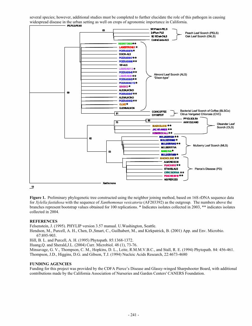
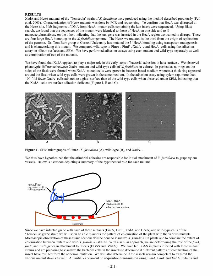
Transcript
- 264 -
SPATIAL DATABASE CREATION AND MAINTENANCE FOR PIERCE’S DISEASE AND GLASSY-WINGED SHARPSHOOTER IN CALIFORNIA
Project Leader: Maggi Kelly Dept. of Environmental Science, Policy, & Management University of California Berkeley, CA 94720 Reporting Period: The results reported here are from work conducted from July 1, 2004 to October 1, 2004. INTRODUCTION Whether tracking invasive species, assessing water quality, or monitoring the spread of disease, comprehensive data collection is a key component of scientific inquiry and sustainable natural resource management. Geographic Information Systems (GIS) allow us to unite in one structure spatially referenced data with other information, affording new insights in relationships between variables at multiple scales (original proposal contains full references), as well as assisting in collaborative efforts at natural resource management and multi-disciplinary problem solving. Such is the case with Pierce’s Disease, where disparate datasets on PD location and GWSS trap data could, if available in a Geographical Information System (GIS) format with other spatially referenced data “layers” such as crops, hydrography, climate, and roads, aid in management of the disease, as well as in epidemiological research. Several agencies and individuals have recognized the need for such a geospatial database for PD research and management. Indeed, the University of California Agriculture and Natural Resources “Report of the Pierce’s Disease Research and Emergency Response Task Force(http://danr.ucop.edu/news/speeches/executivesummary.html) lists the following recommendations: Support is needed for a coordinated, statewide monitoring, trapping and reporting program involving governmental agencies, the agriculture and nursery industries and UC. The objective is to locate populations of GWSS and BGSS, track the incidence and distribution of Pierce's disease and carry out emergency response programs to slow the spread of PD and its vectors. CDFA or UC should manage a GIS to store, display, manipulate and overlay information collected by statewide monitoring and tracking programs. This data should be available to decision makers, growers and scientists. We propose to develop a statewide database for PD and GWSS, maintaining the data with the best QA/QC methods, and full metadata (for data ownership tracking), maintained in a GIS format. We also propose to build a mechanism for researcher access to the database via the web, so that data can be downloaded for research purposes, and uploaded to the collection. We are not linking this effort with any analytical proposal, but aim to create the best possible, accessible database for others to use in research. These two components: (1) GIS database storage and maintenance and (2) Internet accessibility, when combined, are called “webGIS”, and although not yet widely used in natural resource management, such systems are a promising option for entering and storing heterogeneous datasets, indexed by location, and making them widely available in a visual, dynamic, and interactive format. We use as our model the Sudden Oak Death monitoring project (please see the website at: http://kellylab.berkeley.edu/SODmonitoring) created by the Project Leader M. Kelly and housed at UC Berkeley. The multi-scale data provide by the database structure described here, and specifically the access to the data, will contribute to finding a solution to PD by allowing researchers to use PD and GWSS data in concert with other spatial data “layers” such as climate, crops, and roads. In this way epidemiological hypotheses about distribution and spread at several scales – from vineyard to county to regional - can be formed. In addition, the data will aid in disease management, as researchers can see the spatial effect of different management options such as vine removal. We are committed to collaborate with relevant researchers in this pursuit, and understand that there are already existing groups collecting such data. It is not our wish to supercede those efforts, but to lend our expertise to the data collection, storage, and distribution dynamic in support of Pierce’s Disease science. OBJECTIVES The objectives and priorities for this project are as follows: 1. Create spatially referenced database of PD occurrence from field data; 2. Create spatially referenced database of GWSS trap data; 3. Maintain these data with other relevant spatial data for researchers use; and 4. Develop a web-based tool for researchers to submit data to the database, and for researchers to access existing data.
Possibly, we will also develop a tool for the public to report presence of GWSS. RESULTS Funding for this project arrived at UC Berkeley on October 11, 2004, so we have no specific data analysis to report. I have a Staff Research Associate – Dave Shaari – who will work half time on this project, and I am in the process of locating an
- 263 -
experiments. We intend for the risk to be very small, and the knowledge gained to be of great benefit in the practical control of PD in the southern San Joaquin and elsewhere in California. We would be happy to work collaboratively with other researchers and cooperators on various aspects of this research. REFERENCES 1. Feil, H., Feil, W. S., Purcell, A. H. (2003) Effects of date of inoculation on the within-plant movement of Xylella
fastidiosa and persistence of Pierce’s Disease within field grapevines. Phytopath. 93: 244-251. 2. Hashim, J., Hill, B. L. (2003) Monitoring and control measures for Pierce’s disease in Kern County and
Epidemiological assessments of Pierce’s Disease. Pp. 95-98 In CDFA (ed.), Proceedings of Pierce’s Disease Research Symposium 2003, Coronado, CA.
3. Hill, B. L., Purcell, A. H. (1995). Multiplication and movement of Xylella fastidiosa within grape and four other plants. Phytopath. 85: 1368-1372.
4. Hill, B. L., Purcell, A. H. (1997). Populations of Xylella fastidiosa in plants required for transmission by an efficient vector. Phytopath. 87: 1197-1201.
FUNDING AGENCIES Funding for these projects was provided by the University of California Pierce’s Disease Grant Program and the CDFA Pierce’s Disease and Glassy-winged Sharpshooter Board.
- 262 -
multiply to relatively high (easily detectable population sizes) before acquisition becomes efficient (4). Because it multiplies and spreads faster, we hypothesize that bacteria become available for acquisition in an infected grapevine of a susceptible variety earlier in the season than in a vine of a tolerant variety. Putting these two parts of the hypothesis together can explain why the varietal differences in disease rate were observed. In the most susceptible varieties inoculations occurring later in the growing season can result in infections that survive the winter to become chronic. Because of the faster bacterial multiplication and spread there is still enough time in the growing season to reach a threshold for survival. At the same time, the bacteria multiply in previously infected vines fast enough to become available for acquisition by GWSS earlier in the season. The timing of these two processes results in an overlap, that is a window of opportunity when GWSS can acquire Xf from an infected vine, transmit the acquired bacteria to a new vine, and the new infection has enough time to progress to chronic infection and disease. That window of time would close during the seasen, but vine to vine transmissions would still be occurring. However those later season transmissions, after the window of opportunity has ended, would be cured over the winter. So vine to vine transmission occurring within the window would become chronic, and vine to vine transmission occurring after the window would be winter-cured. Conversely in the tolerant varieties infections must occur earlier in the season in order to have enough time, at the slower rate of multiplication and spread, to progress to chronic disease. At the same time bacteria from previously infected vines also multiply and spread slowly and do not become available for vector acquisition until later in the season. The result is that there is no overlap, no window of opportunity where GWSS can acquire Xf from an infected vine, transmit to a new vine, and have the newly infected vine progress to chronic disease. In this case all of the vine to vine transmissions occur too late in the season, and the result is that all the vine to vine infections are cured over the winter. One question is why do epidemics that are vectored by GWSS result in vine to vine disease spread in susceptible varieties whereas no vine to vine disease spread seems to occur when the traditional native California sharpshooter vector species are transmitting the bacterium? The answer may be related to the feeding and inoculation locations of GWSS vs. other vectors. The GWSS will feed (and therefore inoculate vines) at the base of the canes, but the native vectors all feed almost exclusively at the tip of the cane. Inoculations at the tip of the cane probably require more time to move to an over-wintering refuge, so an early season inoculation is necessary for the infection to survive the winter and become chronic disease. Thus the window for vine to vine transmission leading to chronic disease would not exist. In this case only the early season primary spread from sources outside the vineyard would result in chronic disease ,and because vine to vine transmission cannot begin until mid-season, these infections would be winter-cured. If this hypothesis is correct, there are a number of possible consequences and conclusions that could improve PD management and control in areas where GWSS is present. • The risk to growers of tolerant varieties is far less than has been previously assumed. • There is a critical window of time somewhere in mid-season when susceptible vines need to be protected from vine to
vine spread of PD. Chemical vineyard treatments early and late in the season, that is before and after this window, may be less effective than has previously been assumed.
• Economically important rates of secondary spread of PD may only happen in susceptible varieties and when large populations of GWSS are involved. Low but persistent populations of GWSS in Kern County do not appear to have resulted in appreciable losses from of vine to vine spread.
• Better targeted and timed chemical treatments could result in lower costs and be more compatible with other IPM programs.
• Late season vineyard surveys and rouging of infected vines is an important and cost effective management tool. • The GWSS monitoring programs could be tailored to critical parts of the season, thereby possibly reducing the overall
cost of these programs. • The GWSS population treatment thresholds could be based on better epidemiological information, again possibly
reducing overall PD management costs.
Because of the beneficial implications for PD management, it is important to experimentally test this hypothesis. We will be proposing to conduct experiments over the next two years to test the components of this hypothesis. The best experimental protocol would involve experiments conducted in two adjacent working vineyards, one tolerant and one susceptible variety. Ideally the experimental site would be in southern San Joaquin valley with climatological conditions representative of the viticulture areas of Kern or Tulare counties. One experiment would involve inoculations of both varieties vines at intervals throughout the growing season to establish the probability curves for the over-winter survival of Xf as a function of time of inoculation. The hypothesis predicts that the probability curves would be significantly different. Another experiment, for year two, would involve acquisition of Xf by GWSS at intervals throughout the season from vines of both varieties that were inoculated the previous year. This would establish the probability curves for the acquisition of Xf by GWSS as a function of time. The hypothesis predicts that these probability curves would also be significantly different. Other components of the experiments would look for differences between the varieties in the rate of multiplication and spread of Xf in the vines. Again the hypothesis would predict differences. It is critically important to everyone involved that these experiments do not create any new local PD problems or outbreaks. We have considered extensive safeguards in the design of these
- 261 -
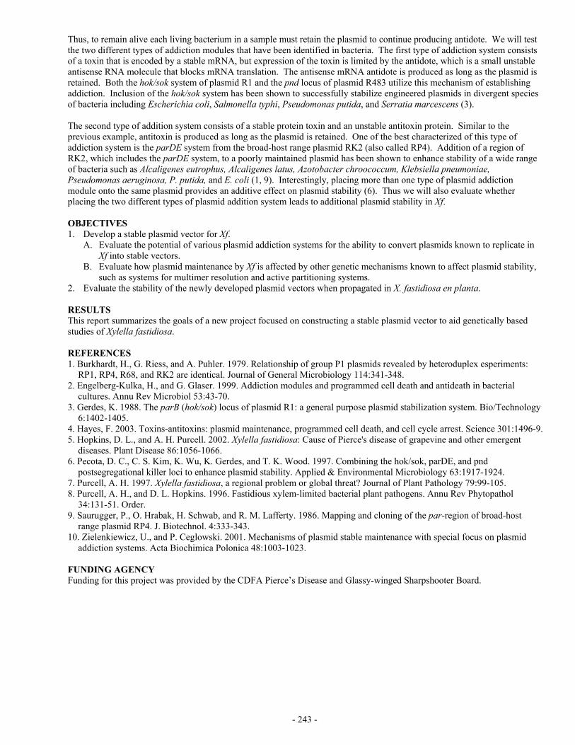
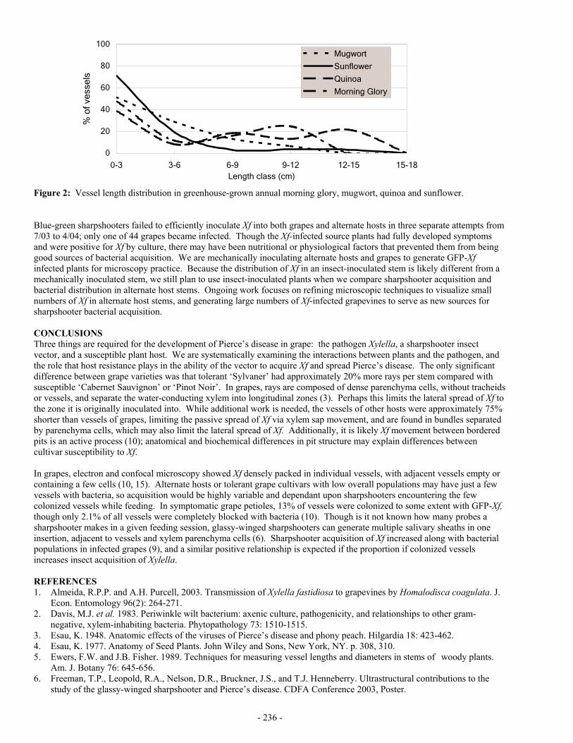
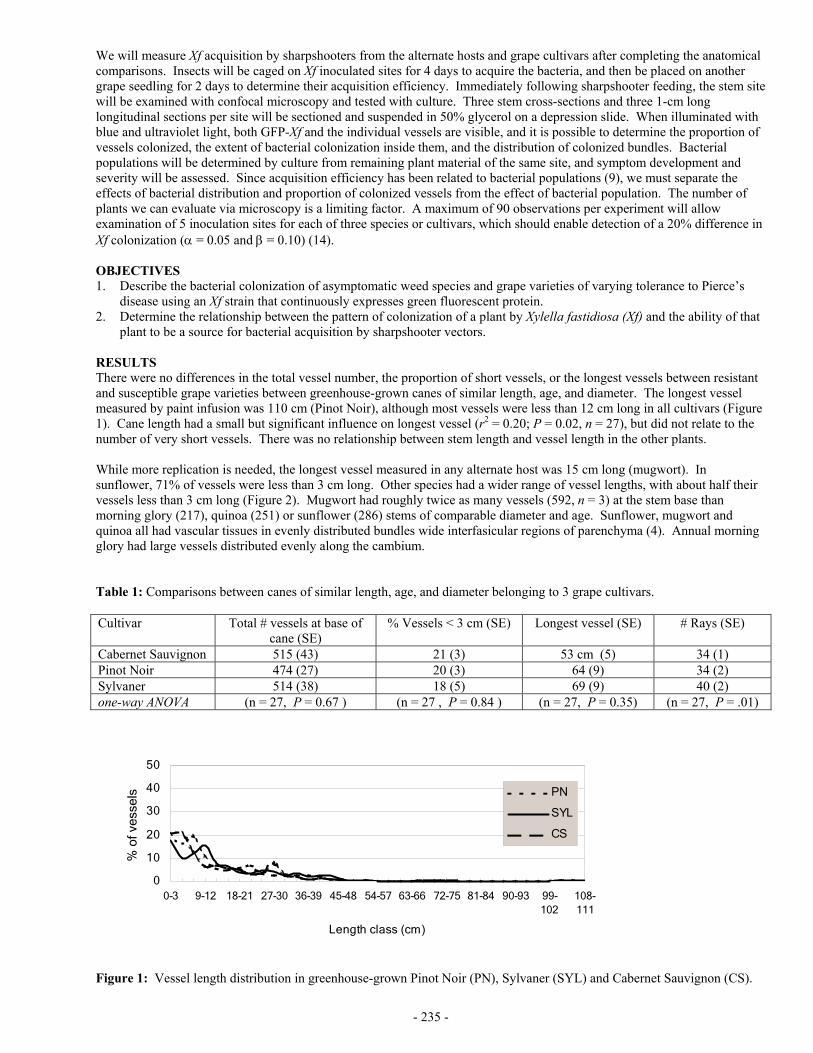
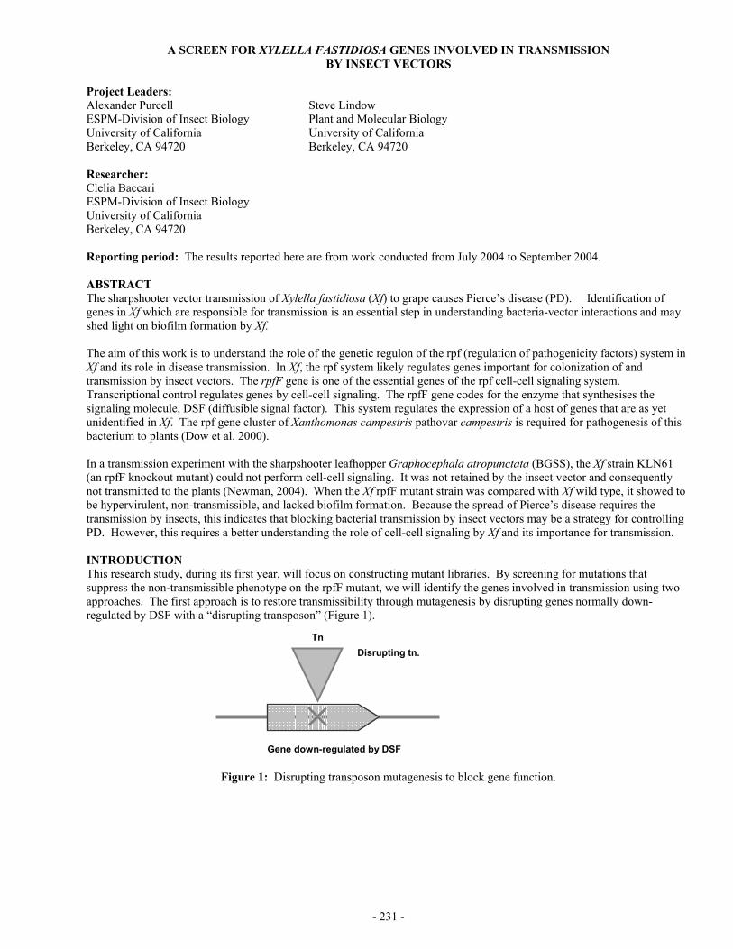
RESULTS AND CONCLUSIONS Vineyards were monitored by visually inspecting each vine for PD symptoms, and by collecting and testing (by ELISA) samples from symptomatic vines (2). Thus far in October 2004 all but 2 of the General Beale vineyards have been completed, but much of the other areas of Kern County are still in progress. The results thus far in the General Beale area indicate that the dramatic decrease in the number of infected vines is continuing. From 2002 to 2003 the number of infected vines decreased by 85%, and from 2003 to 2004 the decrease was an additional 68%. Following the survey of these vineyards in 2001 and 2002 the vines found to have confirmed Xf infections were removed. The continued decline of Xf infection in this area demonstrates that effective PD control can be obtained with a combination of GWSS control, monitoring for infected vines, and removal of infected vines. These projects have demonstrated that vineyard disease monitoring and vine removal is cost effective. Throughout the county as part of this project vines found to be infected with Xf were removed at the end of that season. As a result the surveys in 2003 and 2004 are identifying vines that are newly infected. The rate of infection in all areas of Kern county outside the General Beale and Northern areas is very low, an overall rate throughout the county of less than one new infection per 10,000 vines. By contrast in the General Beale area some of the vineyards developed very high levels of disease within a 2 to 3 year period, peaking in 2002. Several vineyards were entirely lost. Before the arrival of GWSS, primary spread of Xf from sources outside the vineyard accounted for most or all of the PD in California. The rates of new infections in Kern county may be the result of both primary spread and secondary spread, that is vine to vine spread. The low rates of new infections outside the epidemic area is consistent with primary spread, but the rapid rates of infection in many vineyards within the General Beale area is consistent with secondary, vine to vine spread. Perhaps the most startling epidemiological discovery of this project so far was that in 2002, 99% of the PD infected vines in the General Beale area were in Redglobe and Crimson vineyards, the 2 most susceptible of the 6 varieties surveyed. The following year, 2003, these same vineyards accounted for 97% of the diseased vines. These two varieties comprised only 18% of the acreage surveyed in the General Beale area. There were dramatic instances where Redglobe and Flame Seedless were growing in adjacent vineyards, and the susceptible Redglobe vineyards were heavily impacted or totally lost, whereas the more tolerant Flame Seedless vines growing just a few feet away were almost unaffected. The rate of infection in vineyards in General Beale of varieties other than Red Globe and Crimson in any of the three years was less than 14 infected vines out of 337,693 vines surveyed. In the worst epidemic area in Kern County the infection rate in varieties other than Redglobe and Crimson was essentially negligible. The Crimson loss in the General Beale area involved only one vineyard, and these vines were less than three years old. Younger vines are more susceptible to PD than older vines, and it is possible that the losses in the Crimson vineyard were primarily related to their more vulnerable age, rather than a varietal susceptibility. Older Crimson vines may not have been so heavily impacted. We have developed a new hypothesis that would explain what might be causing this varietal difference. It is based on the timing of when in the season GWSS can acquire Xf, when in the season GWSS transmits Xf to new vines, and the phenomenon of over-winter curing of Xf infections. Over-winter curing of PD has been demonstrated to occur in many areas of California, including the San Joaquin Valley. Populations of Xf in grapevines are reduced during the winter dormant season. It has been experimentally demonstrated that if a vine is infected early in the season, the bacterium has enough time left in the growing season to multiply to high enough population levels and spread into areas of the vine where some of the bacterial cells find a refuge and can survive the winter dormancy. The vine then becomes chronically infected and usually eventually dies. Conversely, if a vine becomes infected later in the season, all the bacteria in the vine die over the winter, and the vine is free of disease the following year (1). Also pruning may play some role in over-winter curing. Vines that are inoculated late in the season when there is insufficient time for bacteria to move beyond the inoculated cane would, of course, lose the infection when that cane is pruned. However the bacteria in an un-pruned cane may die over-winter anyway. Our new hypothesis is predicated on the finding that Xf multiplies and spreads faster within a susceptible plant than it does in a more tolerant plant (3). It would reasonably follow that the bacterium would also multiply and spread more rapidly in the more susceptible grapevine varieties of Redglobe or Crimson than it would in the more tolerant varieties such as Flame Seedless or Thompson. The first part of our hypothesis is about when in the season a grapevine must become inoculated in order for the bacterium to survive the first winter dormancy in the plant thereby progressing to chronic Pierce’s disease. We hypothesize that the tolerant varieties have to become infected with Xf earlier in the season than susceptible varieties in order for the bacterium to have enough time left in the growing season to multiply and spread sufficiently in the vine to be able to survive the winter dormancy period. In general it has been demonstrated that vines must be inoculated before some critical time in the season if the bacterium is to survive the winter (1). However the existence of differences among varieties regarding that critical necessary time of inoculation has not yet been experimentally demonstrated. The second part of our hypothesis is about when in the growing season the bacterial cells, having over-wintered in a previously infected plant, multiply and spread from their winter refuge into the new growth and achieve population numbers great enough to be efficiently acquired by an insect vector, in this case GWSS. This growth and movement of the bacterium following winter dormancy has to happen before vine to vine spread can begin to occur. It is not possible to detect Xf in the new growth of an infected plant until sometime about mid-season, and it ha been demonstrated that the bacterium must
- 260 -
EPIDEMIOLOGICAL ASSESSMENTS OF PIERCE’S DISEASE, AND MONITORING AND CONTROL MEASURES FOR PIERCE’S DISEASE IN KERN COUNTY
Project Leaders: Barry L. Hill Calif. Dept. of Food and Agriculture Pierce’s Disease Control Program Sacramento, CA 95814
Jennifer Hashim UC Cooperative Extension Bakersfield, CA 93307
Reporting period: The results reported here are from work conducted from July 2004 to October 2004. ABSTRACT Vineyards in the 7 grape production areas of Kern County’s area wide management project were surveyed for PD again in 2004. Incidence of PD in the highly affected areas (General Beale and North) peaked in 2002, and declined dramatically in both 2003 and 2004. Treatments to reduce GWSS and to identify and remove PD infected vines each year were associated with these dramatic reductions. Survey and epidemiological data is being processed at CAMFER, a GIS-based research institute at U.C. Berkeley. More than 98% of the vines infected with Xylella fastidiosa in the recent epidemic in the General Beale area of Kern County were of the two most susceptible varieties: 6 Red Globe and 2 Crimson vineyards. Thirty-two other nearby or contiguous vineyards of four less susceptible varieties were almost unaffected. A hypothetical mechanism for this varietal difference is proposed. INTRODUCTION These two projects have complimentary objectives and methods, and were thus pursued and are being reported here cooperatively. This combination of people and resources has resulted in synergistic efficiency and maximum utilization of resources. The cooperative area-wide pest management project for the control of GWSS has defined 7 distinct grape growing areas in Kern County. The PD epidemic that peaked in 2002 only affected two of these, the General Beale and the adjacent Northern area. These were also the only areas where the populations of GWSS exploded in 2000 and 2001 to extremely high populations not seen elsewhere in the county. Insect control measures begun in winter 2001-2002 brought the GWSS populations down dramatically. During this time the population dynamics and control methods for controlling GWSS were studied extensively with effective results. However our understanding of how to control the disease (goal of project 1) and the epidemiology of PD when the causal bacterium is transmitted by GWSS (goal of project 2) had been based on limited actual field data. These two projects began in 2002 as 5 year projects to obtain extensive data about the incidence and control of the disease. This disease information would compliment the insect information to enable understanding of the dynamics of the epidemic and methods to control other potential outbreaks. A total of 216 vineyards with 4060 acres and 2,015,698 vines were surveyed, about 4.6% of the vineyard acres in Kern County. There have been two recent major California epidemics of PD that have been vectored by GWSS: General Beale in Kern County and Temecula in Riverside County. However data about each of these was not obtained until the epidemic was well underway or had already peaked. Because the other five viticulture areas of Kern County did not yet have such high numbers of GWSS, it was thought that disease and insect data from those would provide baseline information in the event that another epidemic such as the General Beale and Northern outbreak might occur, and such an epidemic could be studied from the beginning. Among the other 5 viticulture areas, 4 (Central, South A, South B, and West) have had low numbers of GWSS present since sometime before 2000, and GWSS was discovered in the 5th (Hwy 65-Delano) after 2000. Thus this extensive project to monitor the PD disease incidence in these areas was intended to provide both an understanding of the effect of low populations of GWSS on the incidence of PD, as well as a complete epidemic profile over time if another one should occur in this county. OBJECTIVES Project 1: Epidemiological assessments of Pierce’s Disease. (BLH) 1. Evaluate the importance of epidemiological factors such as GWSS population size, vine age, cultivar susceptibility,
control practices, and GWSS control treatments in vineyards and nearby GWSS hosts or habitat. 2. Make all the epidemiological data obtained available in a commonly acceptable GIS format for analysis by other
qualified researchers and epidemiologists. Project 2: Monitoring and Control Measures For Pierce’s Disease In Kern County. (JH) 1. Determine changes in the incidence of PD over time in seven distinct grape-growing areas in Kern County. 2. Develop PD monitoring and management techniques and strategies for use by growers to reduce risk and damage.
Update and provide educational materials to assist vineyard managers, pest control advisors, other researchers and government agencies involved in advising growers in the area-wide pest management of the GWSS project.
- 259 -
Table 2. The mean (±SD) ELISA readings and the percentages of Hippodamia convergens scoring positive for the presence of chicken egg white or non fat dry milk for up to 35 days after marking. H. convergens were scored positive for the presence of each marker if the ELISA value exceeded the mean negative control value by 3 standard deviations.
1/The retention of nonfat milk by contact application was not investigated for H. convergens. REFERENCES Blackmer, J. L., J.R. Hagler, G. Simmons and L. Cañas. 2004. Comparative dispersal of Homalodisca coagulata and
Homalodisca liturata. Environ. Entomol. 33: 88-99. Blua, M.J. and D. Morgan. 2003. Dispersion of Homalodisca coagulata a vector of Xylella fastidiosa, into vineyards in
southern California. J. Econ. Entomol. 96: 1369-1374. Hagler, J. R. 1997a. Field retention of a novel mark-release-recapture method. Environ. Entomol. 26: 1079-1086. Hagler, J. R. 1997b. Protein marking insects for mark-release-recapture studies. Trends Entomol. 1: 105-115. Hagler, J. R., and C. G. Jackson. 1998. An immunomarking technique for labeling minute parasitoids. Environ. Entomol. 27:
1010-1016. Hagler, J.R. and C.G. Jackson. 2001. Methods for marking insects: Current techniques and future prospects. Annual Rev.
Entomol. 46: 511-543. Hagler, J.R. and S.E. Naranjo. 2004. A multiple ELISA system for simultaneously monitoring intercrop movement and
feeding activity of mass-released predators. Internat. J. Pest Manage. 50: 100-207. Hagler, J., S. Machtley, and J. Leggett. 2002. Parasitoid mark-release-recapture techniques: II. Development and application
of a protein marking technique for Eretmocerus spp., parasitoids of Bemisia argentifolii. Biocont. Sci. Technol. 12: 661-675.
Varela, L. G., R. J. Smith, and P. A. Phillips. 2001. Pierce’s Disease, 20 pp. University of California, Agriculture and Natural Resources Publication 21600.
- 258 -
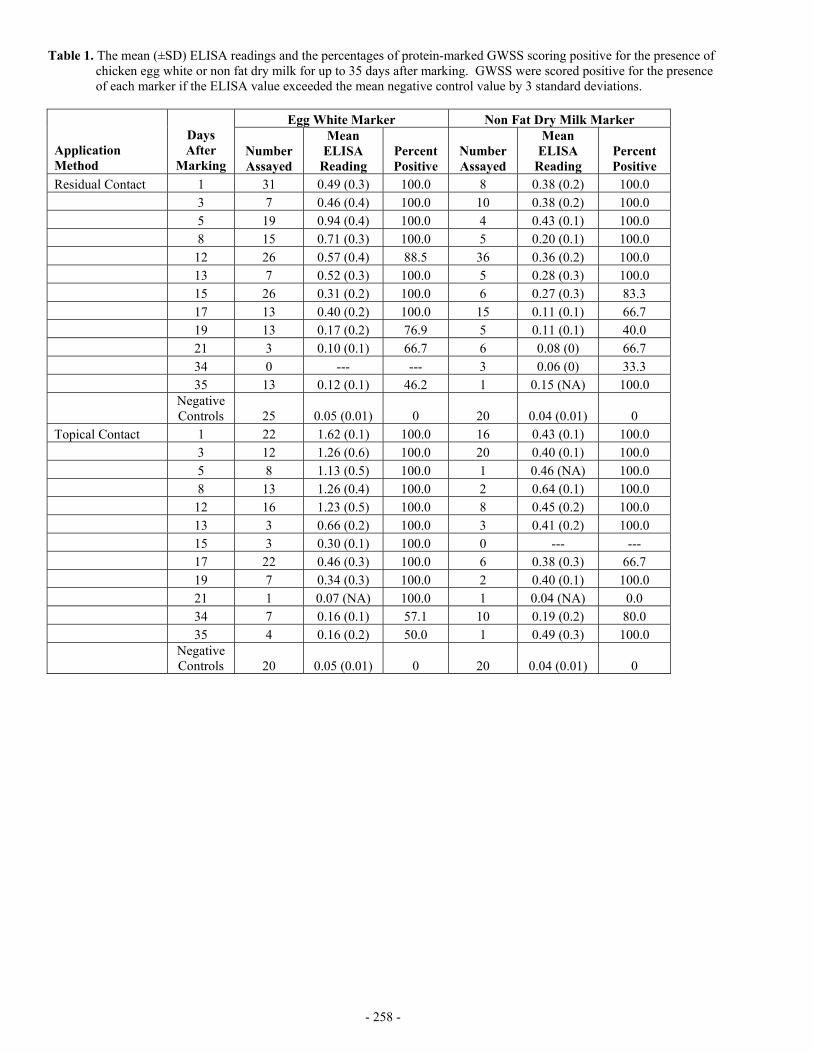
Table 1. The mean (±SD) ELISA readings and the percentages of protein-marked GWSS scoring positive for the presence of chicken egg white or non fat dry milk for up to 35 days after marking. GWSS were scored positive for the presence of each marker if the ELISA value exceeded the mean negative control value by 3 standard deviations.
OBJECTIVES The overall objectives of our research are to: 1. Quantify GWSS and natural enemy dispersal patterns in a complex landscape and 2. Determine which factors influence their dispersal. To accomplish these objectives we must first develop a mark-capture
protein marking technique and quantify the protein marking retention intervals for the targeted insects. Field application of better mark-capture techniques will enhance our understanding of the area-wide dispersal patterns of GWSS and its natural enemies.
RESULTS Direct Contact Marking Method Dozens of nylon-meshed sleeve cages (66 X 70-cm, 18-cm dia.) were placed on randomly selected citrus branches located at the Agricultural Operations Research Station in Riverside, CA. Adult GWSS and H. convergens were then introduced into each cage and sprayed with a 5.0% solution of non-fat dry milk (NFDM) or chicken egg whites (All Whites™). A single cage from each marking treatment was randomly selected on 12 different sampling dates for up to 35 days after marking. All of the surviving GWSS and H. convergens in the randomly selected cages were assayed by an anti-NFDM or an anti-egg white ELISA to detect the presence of each respective protein mark. Residual Contact Marking Method Randomly selected citrus branches located at the Agricultural Operations Research Station in Riverside, CA were sprayed with a 5.0% solution of NFDM or chicken egg whites. The branches were allowed to dry for several hours, and then nylon-meshed sleeve cages were placed on the branches. Adult GWSS and H. convergens were then introduced into each cage. The sampling scheme was the same as the one described above. All of the surviving GWSSs and H. convergens in the randomly selected cages were assayed by an anti-NFDM or an anti-egg white ELISA to detect for the presence of each respective protein marker. The ELISA results for the protein marked GWSS are given in Table 1. Data indicate that both marking procedures, regardless of the type of protein marker used, were retained well on GWSS. As expected, the topical marking procedure yielded higher ELISA values and had longer retention than the residual contact marking method. Generally, the markers were retained on 100% of the GWSS for ≈ 2 and 3 weeks by the residual and topical marking procedures, respectively. The ELISA results for the protein-marked H. convergens are given in Table 2. H. convergens ELISA reactions were very similar to the reactions yielded by GWSS. CONCLUSIONS In the first phase of our research described here, we showed that protein markers can be retained on insects several weeks after marking in the field. This marking technique provides the necessary tool to distinguish GWSS and its natural enemies so that studies of dispersal, migration, longevity, and density can be conducted. Additionally, different protein markers can be used to identify insects released at different times, in different areas, or in different crops. Next, we will use this technique to investigate the landscape-level movement of GWSS (nymphs and adults) and its natural enemies. We propose to use the mark-capture system to simultaneously quantify the intercrop dispersal of GWSS and its natural enemies. Specifically, we will spray large areas (e.g., field plots, whole trees, bushes, etc.) with inexpensive proteins using conventional spray equipment. In turn, insects that are hit by the protein solutions or that eat or walk on plant material containing protein residues will obtain enough protein to be detected by protein-specific ELISAs. Because the two marking ELISAs (chicken egg whites and NFDM) do not cross-react, we can apply the materials to two different host plants in close proximity to one another. Then, insects can be collected using temporal and spatial sampling schemes and analyzed for the presence of each respective protein marker to determine not only the insect’s point of origin but the timing and extent to which portions of the population move among different plant species. FUNDING AGENCIES Funding for this project was provided by the University of California Pierce’s Disease Grant Program and the USDA Agricultural Research Service.
- 256 -
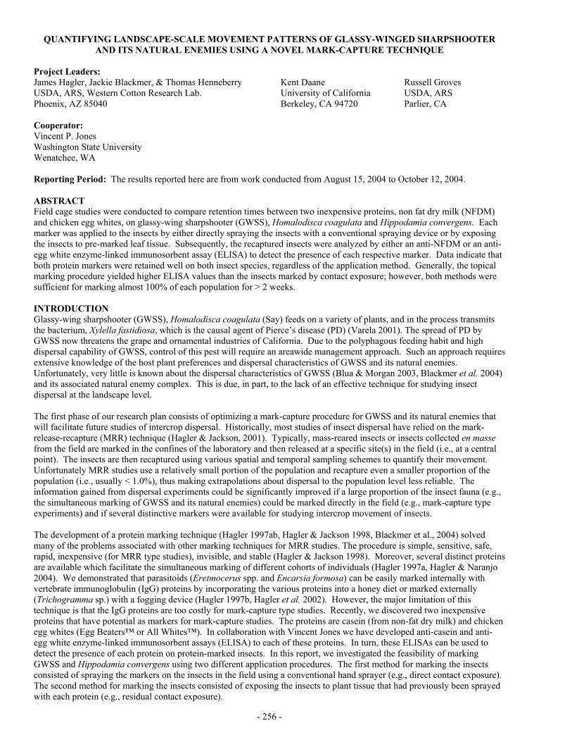
QUANTIFYING LANDSCAPE-SCALE MOVEMENT PATTERNS OF GLASSY-WINGED SHARPSHOOTER AND ITS NATURAL ENEMIES USING A NOVEL MARK-CAPTURE TECHNIQUE
Project Leaders: James Hagler, Jackie Blackmer, & Thomas Henneberry USDA, ARS, Western Cotton Research Lab. Phoenix, AZ 85040
Kent Daane University of California Berkeley, CA 94720
Russell Groves USDA, ARS Parlier, CA
Cooperator: Vincent P. Jones Washington State University Wenatchee, WA Reporting Period: The results reported here are from work conducted from August 15, 2004 to October 12, 2004. ABSTRACT Field cage studies were conducted to compare retention times between two inexpensive proteins, non fat dry milk (NFDM) and chicken egg whites, on glassy-wing sharpshooter (GWSS), Homalodisca coagulata and Hippodamia convergens. Each marker was applied to the insects by either directly spraying the insects with a conventional spraying device or by exposing the insects to pre-marked leaf tissue. Subsequently, the recaptured insects were analyzed by either an anti-NFDM or an anti-egg white enzyme-linked immunosorbent assay (ELISA) to detect the presence of each respective marker. Data indicate that both protein markers were retained well on both insect species, regardless of the application method. Generally, the topical marking procedure yielded higher ELISA values than the insects marked by contact exposure; however, both methods were sufficient for marking almost 100% of each population for > 2 weeks. INTRODUCTION Glassy-wing sharpshooter (GWSS), Homalodisca coagulata (Say) feeds on a variety of plants, and in the process transmits the bacterium, Xylella fastidiosa, which is the causal agent of Pierce’s disease (PD) (Varela 2001). The spread of PD by GWSS now threatens the grape and ornamental industries of California. Due to the polyphagous feeding habit and high dispersal capability of GWSS, control of this pest will require an areawide management approach. Such an approach requires extensive knowledge of the host plant preferences and dispersal characteristics of GWSS and its natural enemies. Unfortunately, very little is known about the dispersal characteristics of GWSS (Blua & Morgan 2003, Blackmer et al. 2004) and its associated natural enemy complex. This is due, in part, to the lack of an effective technique for studying insect dispersal at the landscape level. The first phase of our research plan consists of optimizing a mark-capture procedure for GWSS and its natural enemies that will facilitate future studies of intercrop dispersal. Historically, most studies of insect dispersal have relied on the mark-release-recapture (MRR) technique (Hagler & Jackson, 2001). Typically, mass-reared insects or insects collected en masse from the field are marked in the confines of the laboratory and then released at a specific site(s) in the field (i.e., at a central point). The insects are then recaptured using various spatial and temporal sampling schemes to quantify their movement. Unfortunately MRR studies use a relatively small portion of the population and recapture even a smaller proportion of the population (i.e., usually < 1.0%), thus making extrapolations about dispersal to the population level less reliable. The information gained from dispersal experiments could be significantly improved if a large proportion of the insect fauna (e.g., the simultaneous marking of GWSS and its natural enemies) could be marked directly in the field (e.g., mark-capture type experiments) and if several distinctive markers were available for studying intercrop movement of insects. The development of a protein marking technique (Hagler 1997ab, Hagler & Jackson 1998, Blackmer et al., 2004) solved many of the problems associated with other marking techniques for MRR studies. The procedure is simple, sensitive, safe, rapid, inexpensive (for MRR type studies), invisible, and stable (Hagler & Jackson 1998). Moreover, several distinct proteins are available which facilitate the simultaneous marking of different cohorts of individuals (Hagler 1997a, Hagler & Naranjo 2004). We demonstrated that parasitoids (Eretmocerus spp. and Encarsia formosa) can be easily marked internally with vertebrate immunoglobulin (IgG) proteins by incorporating the various proteins into a honey diet or marked externally (Trichogramma sp.) with a fogging device (Hagler 1997b, Hagler et al. 2002). However, the major limitation of this technique is that the IgG proteins are too costly for mark-capture type studies. Recently, we discovered two inexpensive proteins that have potential as markers for mark-capture studies. The proteins are casein (from non-fat dry milk) and chicken egg whites (Egg Beaters™ or All Whites™). In collaboration with Vincent Jones we have developed anti-casein and anti-egg white enzyme-linked immunosorbent assays (ELISA) to each of these proteins. In turn, these ELISAs can be used to detect the presence of each protein on protein-marked insects. In this report, we investigated the feasibility of marking GWSS and Hippodamia convergens using two different application procedures. The first method for marking the insects consisted of spraying the markers on the insects in the field using a conventional hand sprayer (e.g., direct contact exposure). The second method for marking the insects consisted of exposing the insects to plant tissue that had previously been sprayed with each protein (e.g., residual contact exposure).
- 255 -
but with most positives falling in the 0.2—0.6 range (Figure 3). However, a few individuals proved to be highly positive for Xf with A490 readings >1.0, and in one case >2.4 (Figure 3).
CONCLUSION The data generated thus far is interesting from the standpoint of the large differences in the number of infected GWSS adults in Riverside compared to Redlands or Piru. As the new summer generation of adults ages, one would expect to find increasing proportions positive for Xf as they experience a greater diversity of host plants. This appears to be the case in the Riverside insects, but not for the insects from the other 2 locations. Ongoing collections will help to determine if the location difference is real. REFERENCES Almeida, R.P.P., and A.H. Purcell. 2003. Transmission of Xylella fastidiosa to grapevines by Homalodisca coagulata
(Hemiptera: Cicadellidae). J. Econ. Entomol. 96:264-271. Anderson, R.M. 1981. Population dynamics of indirectly transmitted disease agents: The vector component. Pp. 13-43 in
Vectors of Disease Agents (eds. J.J. McKelvey, Jr., B.F. Eldridge, and K. Maramorosch), Praeger Scientific, New York. Naranjo, S.E., S.J. Castle, and N.C. Toscano. 2003. Sampling, seasonal abundance, and comparative dispersal of glassy-
winged sharpshooters in citrus and grapes: Sampling progress report. Pp 196-199 in Proceedings of the Pierce’s Disease Research Symposium, December 8-11, 2003, San Diego, CA.
FUNDING AGENCIES Funding for this project was provided by the University of California Pierce’s Disease Grant Program.
Figure 3. Histogram of Absorbance490 readings of GWSS adults collected in Riverside between August and October 2004.
- 254 -
0
5
10
15
20
25
20-Aug 7-Sep 24-Sep 7-Oct
No. TestedNo. Infected
0
5
10
15
20
25
Piru Redlands Riverside
No. TestedNo. Infected
The almost complete absence of information regarding the degree of Xf incidence in GWSS populations has helped fuel much speculation about the future of the GWSS/PD crisis in California. In reality, there is very little that we understand regarding mechanisms of acquisition and inoculation of Xf by GWSS adults, either in the controlled conditions of the laboratory and greenhouse, or in the more challenging setting of their natural habitat. While the laboratory approach can provide essential answers to questions regarding the rate of acquisition and efficiency of transmission, it ultimately reflects the conditions imposed by the researcher. For example, the type and age of the acquisition source plant, the isolate of Xf used and period of time that the acquisition source plant has been infected, as well as the source of the experimental GWSS individuals and the conditions under which they are provided access to the Xf source plant are all variables controlled by the researcher. A dual approach that balances the findings from the laboratory with monitoring information from the field will improve our understanding of how epidemics of Xf occur in vineyards and elsewhere. A compilation of data from many sources has contributed to a good understanding of the distribution of GWSS populations within California and the relative intensities of regional infestations. What is now needed is to determine what proportion of individuals within these populations is infected with Xf while also identifying the factors that determine a given level of infectivity. I propose that the approaches and methods to be utilized will address a critical deficiency in our understanding of Xf epidemiology, i.e. the proportion of the vector population infected and infectious with the pathogen. OBJECTIVES 1. Monitor GWSS adults from citrus and other sources year-round to determine the proportion positive for X. fastidiosa
using ELISA, PCR, and media culturing techniques. 2. Perform transmission experiments on a portion of the field-collected adults using grapevine seedlings to determine the
seasonal transmission rate. 3. Quantify the titer of X. fastidiosa in GWSS adults that transmitted X. fastidiosa to grape seedlings using quantitative
ELISA and RT-PCR, and determine the relationship between transmission rate and titer in the vector. RESULTS As a new project that began July 2004, progress is being made on gathering the materials for carrying out transmission experiments and detection and quantification of Xf in field-collected GWSS. A propagation chamber has been assembled that will enable production of experimental grapevines having homogeneous genotypes to be used in the transmission studies. Lateral branch shoots consisting of 4-5 leaves are being cut from certified disease-free parental grapevines (var. Chardonnay) and placed in propagation media until roots are generated. These are transplanted to 4” pots and allowed a minimum of 3-4 weeks to establish before being used in transmission experiments. Ventilated corsage cages will enclose each grapevine plant and provide full access to the entire plants by GWSS adults. A single adult per plant will be confined 3 days for inoculation access followed by recovery and freezing (-80°C) for PCR and ELISA analysis, or for immediate plating to PD 3 media preceded by surface sterilization. An essential component of each of these approaches will be the availability of clean GWSS that are presently being reared. Experimental grapevines will be held a minimum of 2 months to allow for symptom development and then scored. Xylem fluid will be collected from each plant for ELISA/PCR analysis as an independent evaluation to compare with the visual assessments. Experimental and analytical results will be collated to determine which analytical procedure provides the closest agreement with transmission test results.
Field collections of GWSS adults that commenced in August 2004 have so far been made in Piru, Redlands, and Riverside. A sub-sample of 24 adults collected from each of these locations in early October 2004 was processed for ELISA detection of Xf. More than 50% of the Riverside adults were positive for Xf (= absorbance490 values > A490 mean + 4 standard deviations for the GWSS clean control insects) compared to 4% for Redlands and 0 for Piru insects (Figure 1). A progressive increase in the number of Xf-positive insects (Figure 2) occurred between 20 August 2004 (5/24) and 7 October (13/24) in accordance with trends observed from previous years (Naranjo et al. 2003). The distribution of positive A490 readings was quite wide,
Figure 2. Number of infected GWSS adults out of the number tested for 4 collections from Riverside.
Figure 1. Number of infected GWSS adults from 3 locations collected early October 2004.
- 253 -
MONITORING THE SEASONAL INCIDENCE OF XYLELLA FASTIDIOSA IN GLASSY-WINGED SHARPSHOOTER POPULATIONS
Project Leader: Steve Castle USDA, ARS Phoenix, AZ 85040
Cooperators: Nilima Prabhaker University of California Riverside, CA 92521
Nick Toscano University of California Riverside, CA 92521
Reporting Period: The results reported here are from work conducted from July 2004 to October 2004. ABSTRACT The seasonal incidence of Xylella fastidiosa in GWSS populations will be examined using a combination of analytical and experimental techniques. Collections of live GWSS adults will be made at various locations in southern California throughout the year at regular intervals. Live insects will be confined individually to grapevine plants (var. Chardonnay) to determine what proportion from the field transmit Xf. Following a 3 day inoculation access period, each test insect will be processed accordingly for detection of Xf by PCR, ELISA, and/or culturing techniques. By examining sufficient numbers of insects from the field and comparing transmission test results to analytical results, the relative efficiencies of each technique at identifying infected or infectious insects will be determined. Moreover, the seasonal occurrence of infectious insects will be determined and may provide guidance for when to be most vigilant for protecting against primary spread of Xf into vineyards. INTRODUCTION The rate of Xylella fastidiosa Wells transmission in the natural environment is a fundamental component of the epidemiology of Xf, but one that is thus far poorly defined. As a xylem-limited bacterial pathogen of plants, Xf is dependent upon xylophagous leafhoppers for movement from one host to another. The rate that such movement occurs is determined by a large number of factors and interactions among plant hosts, vectors, and bacterial pathogen within the context of variable environmental conditions. Although the inherent complexity of vector-borne diseases defies whole-system approaches to epidemiological studies, specific parameters can be studied towards an overall understanding of vector-borne epidemiology. In the case of Xf, the number of leafhoppers feeding upon Xf-infected plants, the proportion of those that attain Xf through feeding, and the proportion of those that visit and ultimately inoculate uninfected host plants plays a critical role in the spatial and temporal dynamics of Pierce’s Disease (PD) and other Xf-caused diseases. By investigating the proportion of glassy-winged sharpshooters (GWSS, Homalodisca coagulata [Say]) in the natural environment infected with Xf (i.e. positive for presence of Xf) and determining the proportion of those that are infectious (i.e. positive for transmission of Xf) (Anderson 1981), greater understanding of the relationship between GWSS densities and Xf incidence in vineyards or other plant stands will be obtained. Measurement of GWSS infectivity and infectiousness may prove invaluable in addressing the issue of whether or not there is an upper threshold of GWSS numbers that can be tolerated in a given region. Information already available indicates that GWSS is relatively inefficient as a vector of Xf in a laboratory setting (Almeida and Purcell 2003). However, large numbers of highly mobile vectors such as GWSS can easily make up the difference lost to poor transmission efficiency, especially if a large proportion in the natural environment is infectious with Xf. Regional control efforts made over the past few years in areas such as Temecula and the General Beale Road study area in Kern County have proven very effective at reducing local GWSS populations. However, the question of how many of the remaining GWSS in these regions are infectious is still unanswered. Until some measurement is completed of the proportion of GWSS populations that are infected, and more importantly infectious, our understanding of the relative risks posed by variable densities of GWSS throughout California will be limited. More importantly, policy decisions that process information on relative risks posed by GWSS infestations in particular regions will be compromised without data that describes what proportion of a GWSS population is actually causing new infections in a vineyard or in the urban landscape. Better epidemiological information will contribute to improved basic knowledge and understanding and to more sound policy. The California grape industry remains at the greatest risk of Xf movement and transmission by reason of large acreages spread throughout the state and because of the severity of PD. Primary spread of Xf into a vineyard occurs when a cicadellid vector such as GWSS acquires the bacterium from a host outside and subsequently transmits to a grapevine within the vineyard. An infected grapevine can then serve (after an unknown latent period) as a source of secondary spread from infected to susceptible grapevines. Because so little is known about the movement of GWSS in the field and when they become infective with Xf, it is unknown whether most grapevine infections occur as a result of primary or secondary spread of Xf. What is certain, however, is that secondary spread will not occur until a primary infection has occurred, i.e. at least one grapevine has become infected with Xf. This is a critical event that poses a high level of risk to the vineyard because of the establishment of a Xf source within rather than outside of the vineyard. It is therefore important that all appropriate measures be undertaken to prevent that first critical infection. Towards this goal, it will be most helpful to know the temporal pattern of Xf incidence within GWSS populations so that maximum protection can be applied at the most vulnerable times.
- 252 -
12. Hopkins DL (1977) Diseases caused by leafhopper-borne, rickettsia-like bacteria. Annua. Rev. Phytopathol. 17:277-294 13. Lockey C, Ott E, Long Z (1998) Real-time fluorescence detection of a single DNA molecule. Biotechnol. 24:744-746 14. Minsavage GV, Hopkins DL, Leite RMVBC, Stall RE (1993) Comparison of PCR amplification of DNA and ELISA for
the detection of Xylella fastidiosa in plant extracts. Phytopathol. 83:1399 15. Perring TP, Farrar CA, Blua MJ (2001) Proximity to citrus influences Pierce's disease in Temecula Valley vineyards.
Calif. Agric. 55:13-18 16. Pooler MR, Hartung JS (1995) Specific PCR detection and identification of Xylella fastidiosa strains causing citrus
variegated chlorosis. Curr. Microbiol. 31:377-381 17. Purcell AH (1997) Xylella fastidiosa, a regional problem or global threat? J. Plant Pathol. 79:99-105 18. Redak RA, Prucell AH, Lopes JRS, Blua MJ, Mizell III RF, Andersen PC (2003) The biology of xylem sap-feeding
insect vectors of Xylella fastidiosa and their relation to disease epidemiology. Annua. Rev. Entomol. In press 19. Sherald JL, Lei JD (1991) Evaluation of rapid ELISA test kit for detection of Xylella fastidiosa in landscape trees. Plant
Dis. 75:200-203 20. Smart CD, Hendson M, Guilhabert MR, Saunders S, Friebertshauser G, Purcell AH, Kirkpatrick BC (1998) Seasonal
detection of Xylella fastidiosa in grapevines with culture, ELISA and PCR. Phytopath. 88:S83 21. Sorensen JT, Gill RJ (1996) A range extension of Homalodisca coagulata (Say) (Hemiptera: Clypeorrhyncha:
Cicadellidae) to southern California. Pan-Pacific Entomol. 72:160-161 FUNDING AGENCIES Funding for this project was provided by the University of California Pierce’s Disease Grant Program and the USDA Animal and Plant Health Inspection Service.
- 251 -
Table 3. Proportion of GWSS positive for Xf after outdoor exposure on a yellow sticky card.
Trial Mean proportion of GWSS positive for Xf a Day 0 Day 3 Day 6
aMeans in the same row followed by the same letter were not statistically different (trial 1 χ2=3.069, df=2, p=0.216, trial 2 χ2= 2.845, df=2, p= 0.241) CONCLUSIONS Our study was conducted to find a means of accelerating a series of steps required to conduct epidemiological studies involving GWSS spread of Xf, while maintaining a high degree of detection sensitivity. Epidemiological studies require the examination of a large numbers of samples; therefore, an efficient testing protocol is necessary. Through our investigation, we improved the efficiency of Xf detection by streamlining DNA extraction and implementing a QRT PCR-based detection system. The vacuum method was simple, requiring only that heads be removed, pinned into position, and covered with extraction buffer. While time efficiency is the most obvious advantage to using the vacuum extraction method, other advantages also exist which did not impact the studies reported here but may affect detection in field samples. First, no insect tissue is homogenized; it is likely that fewer PCR inhibitors are released to interfere with the PCR reaction and less non-template DNA would be extracted. These factors often hinder detection of pathogen DNA in low concentrations. Second, by flushing the content of the insect’s foregut the search for the presence of Xf is being concentrated in the area of the insect that will most likely contain the organism of interest. QRT-PCR is a sensitive detection technique that allows low concentrations of bacteria to be detected in environmental samples [13]. Our QRT-PCR detection system improved detect an order of magnitude, from 500 Xf cells (with traditional PCR[4]) to 50 Xf cells per insect sample. The implementation of such a system is well suited for the detection of pathogen DNA in an insect vector. A disadvantage of using a molecular technique like PCR for the detection of a pathogen in a host is that detection is based on the presence of pathogen DNA. Unfortunately this does not necessarily mean that the pathogen was alive at the time of collection; the presence of DNA confirms the presence of the pathogen in the host. While other techniques, such as culturing [2], determine the presence of live cells, the sensitivity of such a technique is lower than molecular techniques. The 5-10 d growth period required to see Xf colonies on a nutrient agar plate allows time for contaminants to overgrow the plate. Although specialized media are often used for growth, confirmation of bacterial identity is still needed. While morphological and colony growth characteristics are often used, genetically based identification is more reliable and discriminatory. The mean number of GWSS testing positive varied between trials and between experiments. This was most likely due to natural variation in the ability of GWSS to harbor Xf which may be a function of both the insect’s age and its exposure to other biotic and abiotic factor that influence the ability of the bacterium to colonize the foregut of GWSS. This does not compromise our objective which was to develop a detection protocol that could be used regardless of field conditions. REFERENCES 1. Almeida RPP, Purcell AH (2003) Biological traits of Xylella fastidiosa strains from grapes and almonds. Appl. Environ.
Microbiol. 69:7447-7452 2. Almeida RPP, Purcell AH (2003) Transmission of Xylella fastidiosa to grapevines by Homalodisca coagulata
(Hemiptera : Cicadellidae). J. Econ. Entomol. 96:264-271 3. Bextine B, Miller TA (2004) Comparison of whole-tissue and xylem fluid collection techniques to detect Xylella
fastidiosa in grapevine and oleander. Plant Dis. In press 4. Bextine B, Tuan SJ, Shaikh H, Blua MJ, Miller TA (2004) Evaluation of methods for extracting Xylella fastidiosa DNA
from the glassy-winged sharpshooter. J. Econ. Entomol.:In Press 5. Blua MJ, Morgan DJW (2003) Dispersion of Homalodisca coagulata (Cicadellidae: Homoptera), a vector of Xylella
fastidiosa, into vineyards in southern California. J. Econ. Entomol. In press 6. Brlansky RH, Davis CL, Timmer LW (1991) Xylem-limited bacteria in citrus from Argentina with symptoms of citrus
variegated chlorosis. Phytopath. 81:1210 7. Brlansky RH, Timmer LW, French WJ, McCoy RE (1983) Colonization of the sharpshooter vectors, Oncometopia
nigricans and Homalodisca coagulata by xylem-limited bacteria. Phytopath. 73:530 8. Chen J, Banks D, Jarret RL, Newman M, Chang CJ, Smith BJ (1999) Using 16S rDNA sequences to identify Xylella
fastidiosa strains. Syst. Appl. Microbiol. 23:349-354 10. Costa HS, Blua MS, Bethke JA, Redak RA (2000) Transmission of Xylella fastidiosa to oleander by the glassywinged
sharpshooter, Homalodisca coagulata. Hortsci. 35:1265-1267 11. Hoddle MS, Triapitsyn SV, Morgan DJW (2003) Distribution and plant association records for Homalodisca coagulata
(Hemiptera: Cicadellidae) in Florida. Flo. Entomol. 86:89-91
- 250 -
the slow release valve was opened and pressure was slowly returned to ambient. The vacuum application and release was repeated 3 times. In this way, the insect’s forgut and mouthparts were flushed out with PBS. The pinned heads were removed and DNA was extracted from the fluid using the DNeasy Tissue kit (Qiagen Inc.). QRT PCR was conducted as described earlier. To compare our vacuum extraction method to a more conventional maceration technique, heads from GWSS infected with Xf, as above, were either macerated in PBS buffer with a pellet pestle in a disposable 1.5mL microcentrifuge tube (Kontes Glass Company, Vineland, NJ) or vacuum extracted in PBS buffer. In further experiments insects were collected and immediately extracted (n=24) as previously described or stored at -4ºC for 10 d either submerged in mineral oil (n=24) or not (n=24). Finally, infectious GWSS were placed by hand on yellow sticky cards (Trécé Inc., Adair, OK). Yellow sticky cards were placed outside in a sunny location. GWSS were removed from the traps for DNA extraction at 0, 3, and 6 d after placement. DNA was extracted individually from GWSS heads using the vacuum technique and QRT-PCR was used for detection of Xf. DNA Extraction The vacuum extraction technique developed in this study improved the speed and efficiency of extraction. Extraction of DNA using traditional maceration with the Qiagen DNeasy tissue kit averaged 90 minutes for 24 samples. About 30-40 minutes of the extraction was preparing for and executing the maceration step of the procedure. Using the vacuum extraction technique we prepared 24 samples in an average of 15 min. The vacuum extraction technique neither improved nor compromised our ability to detect Xf in GWSS heads. No statistical differences were revealed between maceration-extracted and vacuum-extracted samples in any trial for either the number of positive samples or the relative amounts of Xf DNA measured (Table 1). However, in 5 of 6 trials mean positives and mean relative fluorescence levels were greater for macerated samples than vacuum-extracted samples (Table 1).
Table 1. Proportion of GWSS positive for Xf, and mean relative fluorescence using vacuum (VE) and maceration (MP) sample collection prior to DNA extraction (n=24).
aMeans in the same row followed by the same letter were not statistically different (χ2>6.6, df=1 , p > 0.359). bRelative fluorescence correlates to cell number. Means in the same row followed by the same letter were not statistically different (χ2<3, df=1, p<0.01).
Comparison of Sample Storage Methods On either collection date, there were no significant differences in mean number of GWSS testing positive for the presence of Xf that could be attributed to the method of storage following GWSS collection (trial 1 χ2=1.626, df=2, p=0.443; trial 2 χ2=2.4, df=2, p=0.3;) (Table 2).
Table 2. Comparison of Xf detection in GWSS following storage by three methods (n=24)
Storage method (n=24)a Trial Directly off Plant -4ºC (10 d) -4ºC in mineral oil (10 d)
1 0.875a 0.792a 0.917a 2 0.833a 0.750a 0.917a
aMeans in the same row followed by the same letter were not statistically different (trial 1 χ2=1.626 , df=2, p=0.443; trial 2 χ2=2.4, df=2, p=0.3).
Detection Capabilities Following Insect Trapping Exposure to the elements after capture on sticky cards had little effect on the ability to detect Xf in GWSS samples (Table. 3). Chi-square test for goodness of fit revealed no statistical differences among means from trial 1 (data taken 0, 3, and 6 days following capture, χ2=3.069, df=2, p=0.216), or trial 2 (data taken 0, 3, and 6 days following capture, χ2= 2.845, df=2, p= 0.241).
- 249 -
DEVELOPING A METHOD TO DETECT XYLELLA FASTIDIOSA IN THE GLASSY-WINGED SHARPSHOOTER
Project Leaders: Blake Bextine Dept. of Entomology University of California Riverside, CA 92521
Matthew J. Blua Dept. of Entomology University of California Riverside, CA 92521
Richard Redak Dept. of Entomology University of California Riverside, CA 92521
Reporting Period: The results reported here are from work conducted from September 2003 to September 2004. ABSTRACT A rapid and reproducible technique to detect Xylella fastidiosa (Xf) in the glassy-winged sharpshooter (GWSS) is important for epidemiological studies, and monitoring programs in support of Pierce’s disease management. Such a technique must be amenable to large samples sizes, while remaining sensitive enough to detect pathogen DNA in low amounts. In this study we have improved the speed of tissue extraction by developing a simple vacuum step that replaces labor and time-intensive tissue maceration, and is compatible with manufactured DNA extraction kits and a SYBR Green® based real-time (QRT) PCR system. No statistical differences in the ability to detect Xf were found among samples that were extracted using traditional maceration vs. our vacuum extraction method. Further experiments using our vacuum extraction methods detected no significant differences among samples immediately extracted, or stored for 10 d at -4ºC, dry or in mineral oil. In another experiment we placed Xf -fed GWSS on yellow sticky cards in a sunny location for 0 to 6 d. We found that there was no significant reduction in our detection capabilities for insects left on the cards. INTRODUCTION Grapevines infected with Xylella fastidiosa (Xf ), the bacterium that induces Pierce’s disease of grapevine [12], usually die within three to five years after infection due to the occlusion of xylem vessels [17]. The glassy-winged sharpshooter (GWSS) has recently become an important vector of Xf in California, spreading Xf to grapevines that traditionally had little or no Pierce’s disease [2, 17]. This vector can disperse widely [5], and has a large host range [18] resulting in alarming spread of Xf to new areas [11]. The presence of GWSS in new regions of California, greater incidences of Xf -induced diseases in several crops, including grapevine [15], almond [1], oleander [10], and the threat of citrus variegated chlorosis (not currently found in the US) has lead to great concern over the ecology of this pest/pathogen interaction. Over the past several years control programs have focused on reducing pathogen spread by managing vector populations [18]. Improvements of these strategies can be achieved through studies examining patterns of disease epidemiology [15, 20], and GWSS population densities and dispersion [5, 11, 21]. Most epidemiological studies of this system have involved Xf’s interaction with host plants [3, 6, 15, 20] or the population and behavioral ecology of the pest insect [5, 11]. Investigations of the interactions between Xf and insect vectors have largely been limited to laboratory and greenhouse studies [2, 4, 10]. Molecular protocols, such as PCR, to detect Xf in plants have been developed and are currently being used in epidemiological studies in other disease systems [8, 9, 14, 16, 19, 20]. Unfortunately, methods adapted to detect Xf in insects are inefficient. Detection methods designed for epidemiological studies, from collection of insect specimen to analysis of samples for the presence of Xf, need to be rapid, reproducible, inexpensive, and amenable to large sample sizes. We recently developed a DNA extraction protocol using the DNeasy tissue extraction kit (Qiagen Inc.) in conjunction with a SYBR Green® based real-time (QRT) PCR system to detect Xf in infectious GWSS [4]. Using this protocol, we reliably detected 50-500 Xf cells with GWSS background. This method used labor-intensive maceration of tissue to extract Xf from insect tissue where the bacterium resides in infectious insects [7]. The speed and efficiency of this method could be improved by simplifying this extraction step. OBJECTIVES Our overall goal is to develop a method of detecting Xf in infectious GWSS that would allow us to conduct epidemiological studies and optimize plant protection. To this end, the objectives for this study are to develop an efficient method to remove Xf cells from the foregut and mouthparts of GWSS for PCR based detection. RESULTS In this study we tested a vacuum extraction protocol for removal of Xf cells from GWSS foreguts for detection by QRT PCR. GWSS adults, collected from orange trees at the University of California, Riverside, were placed in rearing cages and allowed to feed for a 6 d acquisition access period on cuttings of Xf -infected grapevines that showed Pierce’s disease symptoms. GWSS heads were removed, and because they float, an insect pin was placed through the back of the insect head and forced through the frons, so that the tip of the pin protruded slightly. The pinned head was then placed in a microcentrifuge tube (one per tube) and 500µl phosphate buffered saline (PBS) was added to the tube so that the head was completely submerged. Tubes were loaded into a tube rack and placed in a glass vacuum desiccator. With the desiccator lid in place, vacuum was applied to 20 bars slowly, to keep buffer from being displaced from its tube, and held for 15 s. Then,
- 248 -
probing activities). In preliminary experiments, longer feeding durations did not influence the number of cells transmitted. Other data are too preliminary to present at this time. CONCLUSIONS We have the tools in place to determine transmission rates at the molecular level. Experiments are underway to determine the number of Xf cells that are transmitted under certain conditions. Until recently the molecular tools were not available to monitor the movement of single cells in the manner that QRT PCR allows. Almeida et al. [1]encountered difficulty in detecting levels of Xf in GWSS that can successfully inoculate a grapevine. That is, they found GWSS that were able to inoculate plants with Xf that did not test positive for the pathogen. The most reasonable explanation for these “false negatives” is that these GWSS harbored a titer of Xf that can cause infection in grapevines, but were below detection limits. Theoretically, one cell can cause a chronic infection; however, the probability is very low. We suspect the number of cells that are likely introduced into plants is greater than a single cell, but lower than the detection threshold of the method used by Almeida et al. [1], which is 102 cells. We need to embrace the molecular tools that are available to accomplish our objective. REFERENCES 1. Almeida RPP, Purcell AH (2003) Transmission of Xylella fastidiosa to grapevines by Homalodisca coagulata
(Hemiptera : Cicadellidae). J. Econ. Entomol. 96:264-271 2. Bextine B, Blua MJ, Miller TA (2004) A quantitative real-time PCR protocol and novel DNA extraction technique to
detect Xylella fastidiosa in glassy-winged sharpshooters. J. Econ. Entomol.:Submitted 3. Redak RA, Prucell AH, Lopes JRS, Blua MJ, Mizell III RF, Andersen PC (2003) The biology of xylem sap-feeding
insect vectors of Xylella fastidiosa and their relation to disease epidemiology. Ann. Rev. Entomol. In press FUNDING AGENCIES Funding for this project was provided by the CDFA Pierce’s Disease and Glassy-winged Sharpshooter Board.
- 247 -
QUANTITATIVE ASPECTS OF THE TRANSMISSION OF XYLELLA FASTIDIOSA BY THE GLASSY-WINGED SHARPSHOOTER
Project Leaders: Blake Bextine Dept. of Entomology University of California Riverside, CA 92521
Matthew Blua Dept. of Entomology University of California Riverside, CA 92521
T.A. Miller Dept. of Entomology University of California Riverside, CA 92521
Reporting Period: The results reported here are from work conducted from July 2004 to October 2004. ABSTRACT Transmission of Xylella fastidiosa (Xf) by the glassy-winged sharpshooters (GWSS) involves a series of events from acquisition of the bacterium to inoculation of Xf to a new host. While this process is often over-simplified, certain insect/pathogen interactions may be necessary to achieve a successful transmission event and the number of Xf cells acquired or inoculated may govern whether or not transmission will occur. In our preliminary studies, neither higher titers of Xf nor longer feeding periods by GWSS result in higher rates of transmission nor a greater number of bacteria transmitted. INTRODUCTION Solutions to Pierce’s disease (PD) are coming out of an understanding of basic biological aspects of the vector, the pathogen, their hosts, and especially the interactions among these three divergent organisms that culminate in a disease epidemic. The most important of these interactions is the transmission of the pathogen by the vector to a non-infected plant. Transmission is a product of vector acquisition of the pathogen from an infected plant, and inoculation of the pathogen into a non-infected plant. It is a complex process involving sharpshooter host finding and feeding behaviors, and probabilities that a critical titer of bacterium will be acquired from an infected host by a feeding sharpshooter, and once acquired, will be inoculated into an uninfected host. In addition, for an inoculation event to lead to infection, a critical titer of bacterium must be inoculated into plant tissue that supports reproduction and movement. Recent advancements in technology allow us to examine quantitative aspects of Xf transmission with high sensitivity, unlike traditional means. This includes two techniques we have mastered in our laboratories. First, we are currently using a quantitative real-time (QRT PCR) technique in conjunction with commercially available DNA extraction kits to detect and quantify low titers (currently ca 5 X 101 cells) of Xf in plant and insect tissue [2]. Second we have developed a low-cost method to rapidly extract DNA from GWSS and plant tissue in 96-well micro-titer plates. Species of sharpshooters differ widely in their transmission efficiency, which ranges from a high of over 90% for the blue-green sharpshooter (Graphocephala atropunctata) to 1% for several others including Oncometopia facialis, Acrogonia virescens, and Homalodisca ignorata [3]. Recently, rates of Xf transmission efficiency for the GWSS from grapevine to grapevine were found to be as high as 20% [1]. These observations bring up two questions: First, what aspects of Xf transmission by sharpshooter vectors vary in ways that cause a wide range in efficiencies among vectors? Second, can we exploit an understanding of transmission efficiency to reduce PD spread? We seek to understand quantitative aspects of Xf transmission by GWSS. We are hopeful that this unique approach to investigating the transmission of an insect-vectored plant pathogen will lead to new tactics to manage disease spread. OBJECTIVES Our long-term goal is to understand quantitative aspects of the process of Xylella fastidiosa (Xf) transmission by Homalodisca coagulata (glassy-winged sharpshooter, GWSS) in order to develop a means of reducing the efficiency with which the pathogen is spread from an infected plant to a non-infected one. Our specific objectives for this project are to: 1. Determine relationship between the time a GWSS spends on a PD-infected grapevine and titer of Xf they acquire. 2. Determine the relationship between the time a GWSS spends in post-acquisition on a non- Xf host and titer of Xf they
contain. 3. Determine the relationship between the time an infectious GWSS (ie, one that had acquired Xf) spends on a non-infected
grapevine and the titer of Xf it inoculates into the grapevine. 4. Determine the relationship between the titer of Xf inoculated into a plant and the probability that it will become diseased. RESULTS Our preliminary laboratory experiments show that we can quantify the titer of Xf delivered to a stem by a single infectious GWSS immediately after a 24hr inoculation access period (IAP). In this experiment, field-collected GWSS adults were allowed to acquire Xf from grapevines showing Pierce’s disease symptoms for a 72 hr acquisition access period (AAP). GWSS were then allowed access to cut chrysanthemum stems for 2, 4, 6, or 8 h. During this IAP, time lapse video was used to determine the amount of time GWSS feed on the stem and number of times the insect left the stem (indicating multiple
- 246 -
- 245 -
Section 4: Pathogen and Vector
Monitoring and Action Thresholds
- 244 -
GENETIC VARIABILITY OF XYLELLA FASTIDIOSA STRAINS ISOLATED FROM TEXAS GRAPES AND OTHER PLANT RESERVOIRS
Project Leaders: Kristi Bishop University of Houston-Downtown Houston, TX 77002
Lisa Morano University of Houston-Downtown Houston, TX 77002
Prince Buzombo University of Houston-Downtown Houston, TX 77002
ABSTRACT Pierce’s disease is a serious threat to the burgeoning Texas wine industry. Evaluation of the ecology and epidemiology of the disease in Texas may also be of significant scientific value for other areas of the country. We have begun a molecular biological evaluation of the genetic variability of Xylella fastidiosa (Xf) strains in Texas using small, established primers for creation of diagnostic banding patterns (REP, ERIC, and BOX primers). Cloning and sequencing of amplicons using RST31-33 primers resulted in little genetic difference between strains if one considers the error rate of Taq polymerase. However, priming with the small diagnostic primers resulted in differential banding patterns among Xf isolates across Texas. Based on these patterns, some vineyards had genetically distinct isolates and others genetically identical isolates. Vineyards may also contain more than one isolate. Analysis of Xf from a non-Vitis species showed a high distinct banding pattern suggesting broad genetic variability within Texas. Indirect immunofluorescence on Xf isolates also supports significant genetic variability within Texas, as there is differential antigen localization among several strains.
- 243 -
Thus, to remain alive each living bacterium in a sample must retain the plasmid to continue producing antidote. We will test the two different types of addiction modules that have been identified in bacteria. The first type of addiction system consists of a toxin that is encoded by a stable mRNA, but expression of the toxin is limited by the antidote, which is a small unstable antisense RNA molecule that blocks mRNA translation. The antisense mRNA antidote is produced as long as the plasmid is retained. Both the hok/sok system of plasmid R1 and the pnd locus of plasmid R483 utilize this mechanism of establishing addiction. Inclusion of the hok/sok system has been shown to successfully stabilize engineered plasmids in divergent species of bacteria including Escherichia coli, Salmonella typhi, Pseudomonas putida, and Serratia marcescens (3). The second type of addition system consists of a stable protein toxin and an unstable antitoxin protein. Similar to the previous example, antitoxin is produced as long as the plasmid is retained. One of the best characterized of this type of addiction system is the parDE system from the broad-host range plasmid RK2 (also called RP4). Addition of a region of RK2, which includes the parDE system, to a poorly maintained plasmid has been shown to enhance stability of a wide range of bacteria such as Alcaligenes eutrophus, Alcaligenes latus, Azotobacter chroococcum, Klebsiella pneumoniae, Pseudomonas aeruginosa, P. putida, and E. coli (1, 9). Interestingly, placing more than one type of plasmid addiction module onto the same plasmid provides an additive effect on plasmid stability (6). Thus we will also evaluate whether placing the two different types of plasmid addition system leads to additional plasmid stability in Xf. OBJECTIVES 1. Develop a stable plasmid vector for Xf.
A. Evaluate the potential of various plasmid addiction systems for the ability to convert plasmids known to replicate in Xf into stable vectors.
B. Evaluate how plasmid maintenance by Xf is affected by other genetic mechanisms known to affect plasmid stability, such as systems for multimer resolution and active partitioning systems.
2. Evaluate the stability of the newly developed plasmid vectors when propagated in X. fastidiosa en planta. RESULTS This report summarizes the goals of a new project focused on constructing a stable plasmid vector to aid genetically based studies of Xylella fastidiosa. REFERENCES 1. Burkhardt, H., G. Riess, and A. Puhler. 1979. Relationship of group P1 plasmids revealed by heteroduplex esperiments:
RP1, RP4, R68, and RK2 are identical. Journal of General Microbiology 114:341-348. 2. Engelberg-Kulka, H., and G. Glaser. 1999. Addiction modules and programmed cell death and antideath in bacterial
cultures. Annu Rev Microbiol 53:43-70. 3. Gerdes, K. 1988. The parB (hok/sok) locus of plasmid R1: a general purpose plasmid stabilization system. Bio/Technology
6:1402-1405. 4. Hayes, F. 2003. Toxins-antitoxins: plasmid maintenance, programmed cell death, and cell cycle arrest. Science 301:1496-9. 5. Hopkins, D. L., and A. H. Purcell. 2002. Xylella fastidiosa: Cause of Pierce's disease of grapevine and other emergent
diseases. Plant Disease 86:1056-1066. 6. Pecota, D. C., C. S. Kim, K. Wu, K. Gerdes, and T. K. Wood. 1997. Combining the hok/sok, parDE, and pnd
postsegregational killer loci to enhance plasmid stability. Applied & Environmental Microbiology 63:1917-1924. 7. Purcell, A. H. 1997. Xylella fastidiosa, a regional problem or global threat? Journal of Plant Pathology 79:99-105. 8. Purcell, A. H., and D. L. Hopkins. 1996. Fastidious xylem-limited bacterial plant pathogens. Annu Rev Phytopathol
34:131-51. Order. 9. Saurugger, P., O. Hrabak, H. Schwab, and R. M. Lafferty. 1986. Mapping and cloning of the par-region of broad-host
range plasmid RP4. J. Biotechnol. 4:333-343. 10. Zielenkiewicz, U., and P. Ceglowski. 2001. Mechanisms of plasmid stable maintenance with special focus on plasmid
addiction systems. Acta Biochimica Polonica 48:1003-1023. FUNDING AGENCY Funding for this project was provided by the CDFA Pierce’s Disease and Glassy-winged Sharpshooter Board.
- 242 -
PLASMID ADDICTION AS A NOVEL APPROACH TO DEVELOPING A STABLE PLASMID VECTOR FOR XYLELLA FASTIDIOSA
Project Leaders: Glenn M. Young Dept. of Food Science and Technology University of California Davis, CA 95616
Michele Igo Section of Microbiology Division of Biological Sciences University of California Davis, CA 95616
Cooperator: Bruce Kirkpatrick Dept. of Plant Pathology University of California Davis, CA 95616 Reporting Period: This report summarizes the goals of this project. Funding for this project initiated September 1, 2004. INTRODUCTION Current approaches to understanding the progression of Pierce’s disease are limited by the lack of genetic techniques that can be used to study the biology of Xylella fastidiosa (Xf). In particular, extrachromosomal elements, such as plasmids, having long-term stability in Xf when grown in lab cultures or en planta, have not yet been satisfactorily developed. We will develop vectors that exhibit stable maintenance by Xf by adapting previously described genetic and microbiological techniques. Our particular research efforts will focus on taking advantage of a well-studied bacteriological phenomenon called plasmid addiction (2, 4, 10). The major mechanistic principle of plasmid addiction is that the plasmid carries a genetic trait that the host bacterium requires for viability. The trait does not affect the metabolic properties of the bacterium nor does it affect reproduction. However, loss of the plasmid-encoded trait is a lethal event, so by definition plasmid addiction ensures vector stability. In addition, we will systematically evaluate other genetic mechanisms for increasing plasmid stability including multimer resolution and active partitioning systems. Finally, we will examine the stability of each of the newly developed vectors for Xf in vitro and en planta. The results of this analysis will allow us to construct one or more stable plasmid vectors that can be used by all researchers using genetic approaches to develop methods that limit Xf-related diseases. Xylella fastidiosa is a Gram-negative, endophytic bacterium, which is responsible for a number of economically important plant diseases (for recent reviews, see (5, 7, 8)). Diseases that are important to the California agricultural economy include Pierce’s disease of grapevine, almond leaf scorch, alfalfa dwarf, and oleander leaf scorch. Some strains of Xf, such as the Pierce’s disease strains, have very wide host ranges and are capable of colonizing the xylem of widely divergent plant species. In many plant species, infection by Xf does not provoke symptoms or noticeable distress. However, the colonization of certain plants, such as grapevines, leads to the development of disease symptoms and of plant decline. Although the specific details of the disease process are not fully understood, it is known that Xf forms a biofilm within xylem vessels that has a major impact on the movement of sap within the xylem tissue. Disease symptoms seem to be dependent on the rate and extent of colonization of the xylem tissue by Xf. Some of the symptoms observed in infected grapevines include leaf marginal necrosis, severe leaf scorch, and dieback. Another important aspect of the disease cycle involves the insect vector. Xf is transmitted from plant to plant by xylem-feeding insects including the glassy-winged sharpshooter (5, 7, 8). The insect vectors acquire the bacterium by feeding on infected plants. Since the Pierce’s disease strain can colonize numerous plant species, the source of inoculum can be infected grapevines or symptomless plants present in the riparian habitats surrounding the vineyard. In vectors showing the highest transmission efficiencies, Xf is present as a polar biofilm in the insect foregut and is transmitted to uninfected plants during subsequent feeding events. In susceptible plants, efficient transmission of Xf occurs at low bacterial cell numbers (<100 cultivable cells per insect head). Thus, an important feature of the Xf infectious cycle is the ability of this pathogen to colonize and interact with the xylem tissue of plants and the foregut of insect vectors. Successful colonization of these hosts is dependent on the ability of Xf to subvert host defense networks and to acquire essential nutrients. To better understand how Xf survives in and interacts with its hosts, many research laboratories have been working to identify genes important for virulence and nutrient acquisition. However, rapid progress in this area is affected by the lack of genetic and molecular tools necessary to investigate the contribution of Xf genes to the infection process. One extremely important tool that is needed to advance these studies is a plasmid that is maintained by Xf throughout the infectious cycle. The goal of our project is to develop this type of plasmid. Plasmid-addiction systems consist of a pair of genes that specify two components: a stable toxin and an unstable antidote (for recent reviews, see (2, 4, 10). When a bacterium looses the plasmid harboring one of these addiction systems, the cured cells loose the ability to produce the unstable antidote and, as a result, the lethal effect of the stable toxin kills the bacterium.
- 241 -
several species; however, additional studies must be completed to further elucidate the role of this pathogen in causing widespread disease in the urban setting as well on crops of agronomic importance in California.
Bacterial Leaf Scorch of Coffee (BLSCo)Citrus Varigated Chlorosis (CVC)
Oleander Leaf Scorch (OLS)
Mulberry Leaf Scorch (MLS)
Pierce’s Disease (PD)
Figure 1. Preliminary phylogenetic tree constructed using the neighbor joining method, based on 16S rDNA sequence data for Xylella fastidiosa with the sequence of Xanthomonas vesicatoria (AF203392) as the outgroup. The numbers above the branches represent bootstrap values obtained for 100 replications. * Indicates isolates collected in 2003, ** indicates isolates collected in 2004. REFERENCES Felsenstein, J. (1995). PHYLIP version 3.57 manual. U.Washington, Seattle. Hendson, M., Purcell, A. H., Chen, D.,Smart, C., Guilhabert, M., and Kirkpatrick, B. (2001) App. and Env. Microbio.
67:895-903. Hill, B. L. and Purcell, A. H. (1995) Phytopath. 85:1368-1372. Huang,Q. and Sherald,J.L. (2004) Curr. Microbiol. 48 (1), 73-76. Minsavage, G. V., Thompson, C. M., Hopkins, D. L., Leite, R.M.M.V.B.C., and Stall, R. E. (1994) Phytopath. 84: 456-461. Thompson, J.D., Higgins, D.G. and Gibson, T.J. (1994) Nucleic Acids Research, 22:4673-4680 FUNDING AGENCIES Funding for this project was provided by the CDFA Pierce’s Disease and Glassy-winged Sharpshooter Board, with additional contributions made by the California Association of Nurseries and Garden Centers' CANERS Foundation.
- 240 -
Magnolia002 showed more identity (99.41%) to PD strains. For isolates from Hemerocallis and Jacaranda, they showed 100% identity between them and showed to be more closely related to oleander strains (99.22%) Gingko, olive, liquidambar and some ornamental plum strains showed to be closely related to the Dixon almond leaf scorch strain (100% identity). Some ornamental plum strains showed divergence amongst them (97.86% identity) and from ginkgo, olive and liquidambar, but all of them grouped together with the Dixon strain. Lastly, the strain isolated from a yet to be identified host (nicknamed “negrito”) showed slight differences from the ornamental plum, liquidambar and olive isolates. None of the isolates grouped with plum leaf scald, phony peach, oak leaf scorch group or with citrus variegated chlorosis and coffee leaf scorch strains.
Objectives 4 and 5 Eight characterized strains of Xf collected from the landscape in 2003, plus an oleander and a grape strain, were inoculated into their host plants of origin in glasshouse assays. Strains used were Almond276, Ginkgo, Lagestroemia02 (crape myrtle), LiquidambarUI12 (liquidambar), Morus069 (mulberry), Nandina065, Olive AC12 , Pcerasifera076 (ornamental plum), Riverside3 (oleander), GrapeA05. These same eight strains were also used to inoculate grapevine and oleander. Briefly, isolates were grown on PW media for two weeks from which a suspension of 1 x 10⁹ CFU in sterile phosphate buffer was obtained. Plants were needle inoculated on three to four sites per plant using the needle-stab technique described by Hill and Purcell (1995). Approximately 25 plants were used for the inoculation studies. All plants were tested by ELISA prior to inoculation to ensure that they were Xf free. Starting approximately three months after inoculation, plants were ELISA tested and attempts were made to isolate the pathogen from positive plants. Xf cultures have been obtained from some hosts testing positive by ELISA and have been confirmed as Xf by PCR, namely those from mulberry inoculated with the Morus069 isolate. Isolation and characterization studies from these test inoculations are currently underway for the rest of the test plants and Xf isolates. CONCLUSIONS The results of the study do indicate that there are a number of landscape hosts that are harboring different strains of Xf in southern California. Of the new isolates characterized, it appears that new hosts have been identified for a number of strain groups: Pierce’s disease (magnolia, peach, western redbud), oleander leaf scorch (magnolia, jacaranda, day lily), mulberry leaf scorch (heavenly bamboo), and almond leaf scorch (ornamental plum, crape myrtle, liquidambar, gingko, olive). Inoculation tests appear to have confirmed the role of Xf in causing mulberry leaf scorch in California, while other tests await completion. It does appear that new methodologies will have to be developed to successfully obtain or test for Xf in a number of ornamental plant species. The role of Xf infections in landscape hosts does appear to have a significant impact on
- 239 -
pathogen from these positive samples yielded only a small number of isolates (see next section). PCR testing (Minsavage 1994) was performed on a subset of the samples collected using a modification of the published methodology. Briefly, petioles and midveins from leaves were chopped in sterile water, tissues were allowed to sit in the water for several minutes to allow for the release of Xf from the tissues and then DNA extracted from the water. Results were greatly improved using this method, and Xf was detected in 23 species tested (Table 1). PCR testing of additional species testing positive by ELISA is continuing on species from which isolates could not be obtained. Table 1. ELISA, isolation and PCR results for 23 of 122 species tested for Xf. Plant Name Common Name #Tested #ELISA(+)a Culture(+)b PCR(+)c Albizia julibrissin Silk Tree 6 5 yes Cercis occidentalis Western Redbud 4 3 yes yes Ginkgo biloba Maidenhair Tree 15 6 yes yes Hemerocallis Day Lily 9 5 yes yes Jacaranda mimosifolia Jacaranda 49 24 yes yes Juglans Walnut 2 2 no yes Lagerstroemia indica Crape Myrtle 17 5 yes yes Lavandula dentata Lavender 4 4 no yes Ligustrum lucidum Glossy Privet 7 5 no yes Liquidambar styraciflua Liquidambar 19 7 yes yes Magnolia grandiflora Southern Magnolia 31 18 yes yes Morus alba White Mulberry 3 2 yes yes Nandina domestica Heavenly Bamboo 20 3 yes yes Nerium oleander Oleander 3 3 yes yes Olea europaea Olive 6 5 yes yes Phoenix reclinata Senegal Date Palm 2 2 no yes Prunus cerasifera Ornamental Plum 12 7 yes yes Prunus dulcis Almond 3 3 yes yes Prunus persica Peach 5 2 yes yes Rosmarinus officinalis Rosemary 13 8 no yes Vitis labrusca ‘Concord’ Concord Grape 2 2 yes yes Vitis vinifera ‘Red Flame’ Red Flame Grape 2 2 yes yes Vitis vinifera‘Thompson Seedless’ Thompson Seedless Grape 5 5 yes yes a denotes number of samples testing positive using a commercial Xf-specific ELISA kit b denotes if an Xf isolate was successfully obtained from at least one sample c denotes if PCR-amplification using RST31/33 primers from plant tissue was successful for at least one sample Objective 2 Twenty-seven isolates (from 13 host species) were obtained from samples testing positive by ELISA (Table 2). Isolation of the pathogen from samples, even those testing strongly positive from ELISA, was not always possible. Briefly, samples were washed in soapy water, soaked for 1 min in 70% ethanol, 1 min in 20% bleach, then triple rinsed in sdH2O. Samples were then sliced into 1-2 mm pieces and soaked in PBS. Fifty microliters of the PBS buffer was then plated onto PW media with or without the addition of 25 ppm of cycloheximide. The failure to obtain isolates from all samples testing positive by ELISA suggests that specific methodologies need to be determined for specific tissue types from different hosts as a general isolation protocol may be inadvertently killing the pathogen, the pathogen may be highly irregularly distributed in host tissues, or the commercially available ELISA kit may be generating a high number of false positives due to non-specific interactions with host tissue. Objective 3 Collected isolates were confirmed as being Xf by extraction of the DNA from the cultures using the Qiagen DNeasy Plant Mini Kit (Qiagen, Valencia, CA) and subsequent PCR amplification with the RST31/33 primer pair. Isolates were further characterized by amplification and sequencing of the 16S-23S ribosomal DNA intergenic spacer region as described by Hendson et al. 2001. All the 16S-23S rDNA sequences were aligned using the clustalX program (Thompson et al., 1994) and their relationship was analyzed with the PHYLIP program (Felsenstein, 1995) with the sequence of the Xanthomonas vesicatoria (AY288080) as an outlying group (Figure 1). Two strains isolated from mulberry (Morus024 and Morus012) showed 99.41% identity with the previously reported mulberry-VA strain from the eastern U.S. (Huang and Sherald, 2004), while Nandina065, Morus059 and Morus063 showed a 100% of identity with the same strain. For the two peach isolates, Peach018 showed 100% identity with previously reported Pierce’s disease strains (AO5) while Peach018 showed a little less identity (99.41%), but both grouped with PD strains. The Cercis050 strain also grouped with PD strains (99.61% identity). Strains isolated from Magnolia showed just 98.44 % identity between them. Since Magnolia038 was more closely related to Oleander leaf scorch (OLS) (99.02% identity) while
- 238 -
DOCUMENTATION AND CHARACTERIZATION OF XYLELLA FASTIDIOSA STRAINS IN LANDSCAPE HOSTS
Project Leaders: Frank Wong Dept. of Plant Pathology University of California Riverside, CA 92521 [email protected]
Donald A. Cooksey Dept. of Plant Pathology University of California Riverside, CA 92521 [email protected]
Heather S. Costa Dept. of Entomology University of California Riverside, CA 92521 [email protected]
Cooperators: James Downer, UCCE, Ventura County Mike Henry, UCCE, Riverside County John Kabashima, UCCE, Orange County
John Karlik, UCCE, Kern County Michelle LeStrange, UCCE, Tulare County David Shaw, UCCE , San Diego County
Reporting Period: The results reported here are from work conducted from October 15, 2003 to October 12, 2004. ABSTRACT To better understand the impact of Xylella fastidiosa on the urban environment and the potential for ornamental hosts to serve as reservoirs for agronomically important diseases caused by the bacteria, a survey project was initiated to document and characterize strains of the bacteria harbored in landscape plants. Targeted sampling of 122 landscape species either symptomatic for bacterial scorch or testing positive for X. fastidiosa by ELISA in 2003 was performed. Of the 830 samples, 321 tested positive by ELISA (representing 77 of the 122 species tested). X. fastidiosa was also detected in 23 species by PCR-amplification using X. fastidiosa specific primers. Twenty-seven isolates from 13 host species were obtained from samples testing positive by ELISA. Isolates from plants not previously reported as hosts in southern California urban environments included mulberry, heavenly bamboo, magnolia, day lily, western redbud, jacaranda and peach. Genetic characterization of these isolates by 16S-23S rDNA sequencing distributed these isolates amongst previously characterized strain groups: almond leaf scorch (crape myrtle, ornamental plum, liquidambar, gingko, olive), Pierce’s disease (magnolia, peach, western redbud), mulberry leaf scorch (mulberry, heavenly bamboo), and oleander leaf scorch (magnolia, jacaranda, day lily). The role of some X. fastidiosa strains in their ability to cause disease is presently being tested by fulfilling Koch’s postulates in glasshouse experiments. The data collected from this study strongly suggest that X. fastidiosa is causing a number of scorch diseases in the urban landscape, and that strains of agronomic importance may be harbored in this environment. INTRODUCTION Xylella fastidiosa (Xf) is a xylem-limited, insect-vectored, plant pathogen that can cause severe damage to a wide range of host plants. Diseases caused by this pathogen include Pierce’s disease of grapevine (PD), oleander leaf scorch (OLS) and almond leaf scorch (ALS). In 2003, a survey of landscape plants in five urban locations in southern California was initiated to document the incidence of the Xf infection in landscape ornamental hosts and to characterize strains existing in these hosts that may prove a threat to landscape ornamentals or crops of agronomic importance. Two hundred twenty one samples (29%) representing 48 species tested positive by ELISA. Ten isolates of Xf were obtained from eight plant species (Fatsia japonica, Ginkgo biloba, Lagerstroemia indica, Liquidambar styraciflua, Morus alba, Nandina domestica, Olea europea, and Prunus cerasfiera) not previously described as hosts of X. fastidiosa in southern California. Based upon the results of the first year, targeted sampling of host species testing positive by ELISA was performed primarily in the Riverside and Redlands areas in order to obtain additional isolates for characterization. To prove the role of Xf in causing disease in previously identified hosts, test plants were inoculated in glasshouse experiments to fulfill Koch’s postulates for these isolates, and to determine if they were able to cause disease in grapevine and oleander. OBJECTIVES 1. Use laboratory methods to identify landscape host species that are infected with X. fastidiosa. 2. Secure isolates from these hosts to document infection and provide material for genetic characterization of the X.
fastidiosa strain(s) involved. 3. Genetically characterize the strains of pathogen in landscape plant species. 4. Confirm pathogenic infection through inoculation studies with specific isolates. 5. Test ability of new strains to infect agricultural crops including grape, olive, and almond.
RESULTS Objective 1 In 2004, 830 samples from 122 landscape plant species were collected. Sampling focused on plant species that were symptomatic or had tested positive by ELISA in 2003 surveys. Three hundred twenty one samples (39%), tested positive by ELISA. At least one sample from 77 of the 122 species tested was positive by ELISA (63%). Attempts to isolate the
- 237 -
7. Hill, B.L. and A.H. Purcell. 1995a. Acquisition and retention of Xylella fastidiosa by an efficient vector, Graphocephala atropunctata. Phytopathology 85: 209-212.
8. Hill, B.L. and A.H. Purcell. 1995b. Multiplication and movement of Xylella fastidiosa within grape and four other plants. Phytopathology 85: 1368-1372.
9. Hill B.L. and A.H. Purcell, 1997. Populations of Xylella fastidiosa in plants required for transmission by an efficient vector. Phytopathology 87: 1197-1201.
10. Newman, K.L., Almeida, R.P.P., Purcell, A.H. and S.E. Lindow, 2003. Use of a green fluorescent strain of analysis of Xylella fastidiosa colonization of Vitis vinifera. App. and Env. Micro. 69: 7319-7327.
11. Purcell A.H., Finlay, A.H. and D.L. McLean, 1979. Pierce’s disease bacterium: mechanism of transmission by leafhopper vectors. Science 206: 839-841.
12. Purcell, A.H., 1981. Vector preference and inoculation efficiency as components of resistance to Pierce’s disease in European grape cultivars. Phytopathology 71: 429-435.
13. Raju, B.C.,and A.C. Goheen, 1981. Relative sensitivity of selected grapevine cultivars to Pierce’s disease bacterial infections. Am. J. Enol. Vitic. 32: 155-158.
14. Stafford, S.G. and T.E. Sabin, 1999. Statistical Analysis for the Natural Resources and Ecological Sciences, 1-22 to 1-25. Oregon State University.
15. Tyson, G.E., Stojanovic, B.J., Kuklinski, R.F., DiVittorio, T.J.and M.L. Sullivan, 1984. Scanning electron microscopy of Pierce’s disease bacterium in petiolar xylem of grape leaves. Phytopath. 75: 264-9.
16. Wistrom, C.W., and A.H. Purcell. 2003. Fate of Xylella fastidiosa in alternate hosts. Proc. Pierce’s disease research symposium, California Dept. of Food and Agriculture.
FUNDING AGENCIES Funding for this project was provided by grants from the Viticulture Consortium, the California Agricultural Experiment Station (at College of Natural Resources, University of California, Berkeley), and the CDFA Pierce’s Disease and Glassy-winged Sharpshooter Board.
- 236 -
0
20
40
60
80
100
0-3 3-6 6-9 9-12 12-15 15-18Length class (cm)
% o
f ves
sels
Mugwort
Sunflower
Quinoa
Morning Glory
Figure 2: Vessel length distribution in greenhouse-grown annual morning glory, mugwort, quinoa and sunflower. Blue-green sharpshooters failed to efficiently inoculate Xf into both grapes and alternate hosts in three separate attempts from 7/03 to 4/04; only one of 44 grapes became infected. Though the Xf-infected source plants had fully developed symptoms and were positive for Xf by culture, there may have been nutritional or physiological factors that prevented them from being good sources of bacterial acquisition. We are mechanically inoculating alternate hosts and grapes to generate GFP-Xf infected plants for microscopy practice. Because the distribution of Xf in an insect-inoculated stem is likely different from a mechanically inoculated stem, we still plan to use insect-inoculated plants when we compare sharpshooter acquisition and bacterial distribution in alternate host stems. Ongoing work focuses on refining microscopic techniques to visualize small numbers of Xf in alternate host stems, and generating large numbers of Xf-infected grapevines to serve as new sources for sharpshooter bacterial acquisition. CONCLUSIONS Three things are required for the development of Pierce’s disease in grape: the pathogen Xylella, a sharpshooter insect vector, and a susceptible plant host. We are systematically examining the interactions between plants and the pathogen, and the role that host resistance plays in the ability of the vector to acquire Xf and spread Pierce’s disease. The only significant difference between grape varieties was that tolerant ‘Sylvaner’ had approximately 20% more rays per stem compared with susceptible ‘Cabernet Sauvignon’ or ‘Pinot Noir’. In grapes, rays are composed of dense parenchyma cells, without tracheids or vessels, and separate the water-conducting xylem into longitudinal zones (3). Perhaps this limits the lateral spread of Xf to the zone it is originally inoculated into. While additional work is needed, the vessels of other hosts were approximately 75% shorter than vessels of grapes, limiting the passive spread of Xf via xylem sap movement, and are found in bundles separated by parenchyma cells, which may also limit the lateral spread of Xf. Additionally, it is likely Xf movement between bordered pits is an active process (10); anatomical and biochemical differences in pit structure may explain differences between cultivar susceptibility to Xf. In grapes, electron and confocal microscopy showed Xf densely packed in individual vessels, with adjacent vessels empty or containing a few cells (10, 15). Alternate hosts or tolerant grape cultivars with low overall populations may have just a few vessels with bacteria, so acquisition would be highly variable and dependant upon sharpshooters encountering the few colonized vessels while feeding. In symptomatic grape petioles, 13% of vessels were colonized to some extent with GFP-Xf, though only 2.1% of all vessels were completely blocked with bacteria (10). Though is it not known how many probes a sharpshooter makes in a given feeding session, glassy-winged sharpshooters can generate multiple salivary sheaths in one insertion, adjacent to vessels and xylem parenchyma cells (6). Sharpshooter acquisition of Xf increased along with bacterial populations in infected grapes (9), and a similar positive relationship is expected if the proportion if colonized vessels increases insect acquisition of Xylella. REFERENCES 1. Almeida, R.P.P. and A.H. Purcell, 2003. Transmission of Xylella fastidiosa to grapevines by Homalodisca coagulata. J.
Econ. Entomology 96(2): 264-271. 2. Davis, M.J. et al. 1983. Periwinkle wilt bacterium: axenic culture, pathogenicity, and relationships to other gram-
negative, xylem-inhabiting bacteria. Phytopathology 73: 1510-1515. 3. Esau, K. 1948. Anatomic effects of the viruses of Pierce’s disease and phony peach. Hilgardia 18: 423-462. 4. Esau, K. 1977. Anatomy of Seed Plants. John Wiley and Sons, New York, NY. p. 308, 310. 5. Ewers, F.W. and J.B. Fisher. 1989. Techniques for measuring vessel lengths and diameters in stems of woody plants.
Am. J. Botany 76: 645-656. 6. Freeman, T.P., Leopold, R.A., Nelson, D.R., Bruckner, J.S., and T.J. Henneberry. Ultrastructural contributions to the
study of the glassy-winged sharpshooter and Pierce’s disease. CDFA Conference 2003, Poster.
- 235 -
We will measure Xf acquisition by sharpshooters from the alternate hosts and grape cultivars after completing the anatomical comparisons. Insects will be caged on Xf inoculated sites for 4 days to acquire the bacteria, and then be placed on another grape seedling for 2 days to determine their acquisition efficiency. Immediately following sharpshooter feeding, the stem site will be examined with confocal microscopy and tested with culture. Three stem cross-sections and three 1-cm long longitudinal sections per site will be sectioned and suspended in 50% glycerol on a depression slide. When illuminated with blue and ultraviolet light, both GFP-Xf and the individual vessels are visible, and it is possible to determine the proportion of vessels colonized, the extent of bacterial colonization inside them, and the distribution of colonized bundles. Bacterial populations will be determined by culture from remaining plant material of the same site, and symptom development and severity will be assessed. Since acquisition efficiency has been related to bacterial populations (9), we must separate the effects of bacterial distribution and proportion of colonized vessels from the effect of bacterial population. The number of plants we can evaluate via microscopy is a limiting factor. A maximum of 90 observations per experiment will allow examination of 5 inoculation sites for each of three species or cultivars, which should enable detection of a 20% difference in Xf colonization (α = 0.05 and β = 0.10) (14). OBJECTIVES 1. Describe the bacterial colonization of asymptomatic weed species and grape varieties of varying tolerance to Pierce’s
disease using an Xf strain that continuously expresses green fluorescent protein. 2. Determine the relationship between the pattern of colonization of a plant by Xylella fastidiosa (Xf) and the ability of that
plant to be a source for bacterial acquisition by sharpshooter vectors. RESULTS There were no differences in the total vessel number, the proportion of short vessels, or the longest vessels between resistant and susceptible grape varieties between greenhouse-grown canes of similar length, age, and diameter. The longest vessel measured by paint infusion was 110 cm (Pinot Noir), although most vessels were less than 12 cm long in all cultivars (Figure 1). Cane length had a small but significant influence on longest vessel (r2 = 0.20; P = 0.02, n = 27), but did not relate to the number of very short vessels. There was no relationship between stem length and vessel length in the other plants. While more replication is needed, the longest vessel measured in any alternate host was 15 cm long (mugwort). In sunflower, 71% of vessels were less than 3 cm long. Other species had a wider range of vessel lengths, with about half their vessels less than 3 cm long (Figure 2). Mugwort had roughly twice as many vessels (592, n = 3) at the stem base than morning glory (217), quinoa (251) or sunflower (286) stems of comparable diameter and age. Sunflower, mugwort and quinoa all had vascular tissues in evenly distributed bundles wide interfasicular regions of parenchyma (4). Annual morning glory had large vessels distributed evenly along the cambium. Table 1: Comparisons between canes of similar length, age, and diameter belonging to 3 grape cultivars. Cultivar Total # vessels at base of
PATTERNS OF XYLELLA FASTIDIOSA INFECTION IN PLANTS AND EFFECTS ON ACQUISITION BY INSECT VECTORS
Project Leaders: Alexander Purcell ESPM-Division of Insect Biology University of California Berkeley, CA 94720
Steve Lindow Plant and Molecular Biology University of California Berkeley, CA 94720
Researcher: Christina Wistrom ESPM – Division of Insect Biology University of California Berkeley, CA 94720-3112 Reporting Period: The results reported here are from work conducted from July 2003 to September 2004. ABSTRACT We are studying the effect of host plant tolerance on insect vector acquisition of Xylella fastidiosa (Xf) from plants tolerant, moderately susceptible, and highly susceptible to Xf infection. We are observing Xf population and distribution in tolerant and susceptible plants, and its relationship to xylem anatomy, symptom development, and bacterial acquisition by sharpshooters. Since host plant resistance is an important component in the long-term goal of curing PD, it is important to know how resistant plants affect PD spread in areas permanently infested with sharpshooter vectors. We also address the short-term goal of controlling PD spread by comparing grape cultivars in their ability to provide inoculum for vine-to-vine spread of Pierce’s disease. Anatomical comparisons of three cultivars, Sylvaner’, ‘Cabernet Sauvignon’ and ‘Pinot Noir’ showed that all three varieties had similar numbers, lengths and distributions of vessels. The only significant difference was that tolerant ‘Sylvaner’ had ~ 20 % more rays than the more susceptible ‘Cabernet Sauvignon’ or ‘Pinot Noir’ (n = 25, P = 0.01) in canes of similar age, length and diameter. In all four alternate hosts, morning glory (Ipomoea purpurea), mugwort (Artemisia douglasiana), sunflower (Helianthus annuus) and annual bur-sage (Ambrosia acanthicarpa), the longest vessels measured were less than 13 cm long, while in grapes the longest vessels averaged 62 cm. Though alternate hosts had various vascular morphologies and stem lengths, all had shorter vessels than grapes. Blue-green sharpshooters failed to efficiently inoculate wild-type Xf and green fluorescent protein-expressing (GFP) Xf into both grapes and alternate hosts; only one of 44 grapes inoculated with BGSS became infected. In order to generate GFP-Xf infected plants for microscopy, we are mechanically inoculating alternate hosts and grapes. Ongoing work focuses on refining microscopic techniques to visualize small numbers of Xf in plant stems, and generating large numbers of Xf infected grapevines to serve as new sources for sharpshooter bacterial acquisition. INTRODUCTION Alternate hosts of Xf were selected for their different patterns of Xf colonization after vector inoculation, lack of stem lignification, varying morphology, and absence of green autofluorescence under blue light. In previous experiments, Xf-carrying sharpshooters infected morning glory and sunflower more than 80% of the time. Xf spread systemically throughout both plants and reached populations over 105 colony-forming units (CFU)/gram. Quinoa and mugwort were less-frequently infected (32% and 16%, respectively) by Xf and supported lower bacterial populations (103 CFU/g for quinoa, 106 CFU/g for mugwort). Xf moved systemically to a limited extent in quinoa, but not in mugwort (8, 16). Grape cultivars with varying tolerance to PD selected for evaluation are tolerant ‘Sylvaner’, moderately susceptible ‘Cabernet Sauvignon’ and highly susceptible ‘Pinot Noir’ cultivars of Vitis vinifera (12, 13). Both blue-green sharpshooters (BGSS) and glassy-winged sharpshooters (GWSS) will be used to infect plants and assess the efficiency of insect acquisition of Xf (1, 7, 11). We are using wild type and transformed isolates of Temecula Xf in our experiment. The transformed isolate continually expresses green fluorescent protein (GFP) when illuminated with blue light. GFP-Xf was transmitted by blue-green sharpshooters, retained typical virulence in grape, and allowed examination of plant tissues without the extensive fixation required with electron microscopy. With confocal microscopy, GFP-expressing Xf can be observed in small and large colonies in vessels, and passing through bordered pits between vessels in symptomatic ‘Cabernet Sauvignon’ petioles (10). Anatomical comparisons between alternate hosts and grape cultivars included measurements of vessel length and number, and vascular bundle number and distribution based on the techniques of Tyson et al. (15), and Ewers and Fischer, modified to infuse the pigment via 100kPa pressure applied to the proximal end of the cutting (5). We evaluated primary vegetative growth rather than secondary xylem due to the difficulties in sectioning, culturing from, and feeding BGSS on partially lignified stems. GFP-Xf inoculation and colonization of all plants will be measured similarly in all plants: groups of four GFP-Xf carrying sharpshooters inoculated a 3-cm stem section, and the plants were evaluated for the presence of GFP-Xf approximately 8 weeks after inoculation. Colonized vessels will be counted, and populations estimated by culture on PWG media (2, 8).
- 233 -
To determine what genes were affected that resulted in restored transmission, we will clone and sequence the DNA flanking the transposon using standard protocols for determining genomic DNA sequence flanking insertion DNA. The identity of these genes may enable us to grasp key features of the bacterial mechanism driving transmission. For example, we may find that certain adhesins are required for attachment to the foregut if activating transposons near adhesin genes restore transmissibility. REFERENCES Dow, J. M. and M.J. Daniels. 2000; “Xylella genomics and bacterial pathogenicity to plants”; Yeast; 17: 263-271. Gullharbert, M. R.,Hoffman, L.M., Mills, D. A. and Kirkpatrick, B. C. 2001. Transposon mutagenesis of Xylella. MPMI
Vol. 14, N 6 pp 701-706. Newman K.L, Almeida R. P.P, Purcell A.H. and Lindow S.E. 2004. Cell-cell signaling control Xylella fastidiosa interaction
with both insects and plants. Proc. Nat. Acad. Sciences. 101:1737-1742. Newman K.L, Almeida R. P.P, Purcell A.H. and Lindow S.E 2003. Analysis of colonization of Vitis vinifera by Xylella
fastidiosa by using a green fluorescent strain. Applied and Envir. Microbiology. 69: 7319-7327. FUNDING AGENCIES Funding for this project was provided by the CDFA Pierce’s Disease and Glassy-winged Sharpshooter Board.
- 232 -
In parallel, an “activating transposon” will be designed to activate transcription of genes normally up-regulated by DSF (Figure 2).
Figure 2: Activating transposon mutagenesis to enhance gene function. The activating transposon will contain a constitutive promoter that will activate transposition of genes downstream of its insertion site (Newman, 2003). This dual approach will increase the likelihood that we can obtain mutants with restored transmission, and will give us information about those processes that are required for transmission, as well as those processes that must be “turned off” for colonization and transmission to occur. The library will be screened for disrupted gene mutants and then for activated gene mutants. The insect vectors used for the screen in this study will be GWSS and BGSS. To screen for those mutations that restore transmissibility to the rpfF mutant, the gene libraries will be injected into 10 healthy plants of Vitis vinifera cultivar Cabernet Sauvignon. The mutant library will be mechanically inoculated into the grape plants. The plants will be kept in the greenhouse and will be monitored periodically for the presence of PD symptoms. Five plants will contain the disrupting transposon mutagenesis libraries and the other five will contain the activating transposon mutagenesis libraries. The source plants will be kept in the greenhouse to allow the strain to reproduce and grow. Group of 100 BGSS, non infective for Xf will be placed on the source plants to permit acquisition. The insect vectors BGSS and Homalodisca coagulata (GWSS) will feed on the plants containing the mutant collections. Half of the vectors will be analyzed by bacterial culturing for the presence of Xf mutants 14 days after removal from infested plants. The bacteria recovered from these insects will represent mutants that have regained the ability to colonize insect foreguts. Strain KLN61 was only rarely recovered from insects at 7 days, and at 14 days it is expected that that number will be reduced to zero. This will be tested prior to the screen. The other half of the vectors will be transferred to new healthy plants, and after 6 to 8 weeks, the plants will be cultured for the presence of bacteria. The bacteria recovered from those plants represent those mutants that have regained transmissibility. OBJECTIVES 1. Create a library of Xf mutants in the rpfF mutant background using a disrupting transposon mutagenesis to
block gene function. 2. Create a library of Xf mutants in the rpfF mutant background using an activating transposon mutagenesis to
enhance gene function. 3. Design and carry out a screen for mutations in Xf that restore transmissibility in the non-transmissible rpfF
mutant. 4. Identify the genes affected in the screen. These will be genes that are important for transmission of Pierce’s
disease (PD) by insect vectors. RESULTS AND CONCLUSIONS Generating the mutant libraries is the main focus of the research during this first year. We have constructed an rpfF knockout by allelic exchange mutagenesis using a StrepRmarker carried on pKLN121 plasmid. A total of 200 cfu were yield after the transformation and transferred on new media plates containing a concentration of 100ug/ml spectinomycin and 50ug/ml streptomycin as selective markers. This new StrepR strain allows compatibility with the transposome system, which confers KanR allowing us to proceed with the transposome-mediate mutagenesis technique soon. The transposome approach would permit us to rapidly construct a library of mutants in the rpfF background. It has been shown that transposome-mediated mutagenesis was successful in Kirkpatrick’s laboratory when applied on Xf (Guilhabert et al, 2001). To construct a mutant library in the rpfF mutant background gives an important advantage to this project. A secondary mutation on rpfF could short-circuit the need for rpfF in transmission, using other important genes involved in the process and restore transmissibility of the mutant strain.
+++
Activating tn.
Gene up-regulated by DSF
- 231 -
A SCREEN FOR XYLELLA FASTIDIOSA GENES INVOLVED IN TRANSMISSION BY INSECT VECTORS
Project Leaders: Alexander Purcell ESPM-Division of Insect Biology University of California Berkeley, CA 94720
Steve Lindow Plant and Molecular Biology University of California Berkeley, CA 94720
Researcher: Clelia Baccari ESPM-Division of Insect Biology University of California Berkeley, CA 94720 Reporting period: The results reported here are from work conducted from July 2004 to September 2004. ABSTRACT The sharpshooter vector transmission of Xylella fastidiosa (Xf) to grape causes Pierce’s disease (PD). Identification of genes in Xf which are responsible for transmission is an essential step in understanding bacteria-vector interactions and may shed light on biofilm formation by Xf. The aim of this work is to understand the role of the genetic regulon of the rpf (regulation of pathogenicity factors) system in Xf and its role in disease transmission. In Xf, the rpf system likely regulates genes important for colonization of and transmission by insect vectors. The rpfF gene is one of the essential genes of the rpf cell-cell signaling system. Transcriptional control regulates genes by cell-cell signaling. The rpfF gene codes for the enzyme that synthesises the signaling molecule, DSF (diffusible signal factor). This system regulates the expression of a host of genes that are as yet unidentified in Xf. The rpf gene cluster of Xanthomonas campestris pathovar campestris is required for pathogenesis of this bacterium to plants (Dow et al. 2000). In a transmission experiment with the sharpshooter leafhopper Graphocephala atropunctata (BGSS), the Xf strain KLN61 (an rpfF knockout mutant) could not perform cell-cell signaling. It was not retained by the insect vector and consequently not transmitted to the plants (Newman, 2004). When the Xf rpfF mutant strain was compared with Xf wild type, it showed to be hypervirulent, non-transmissible, and lacked biofilm formation. Because the spread of Pierce’s disease requires the transmission by insects, this indicates that blocking bacterial transmission by insect vectors may be a strategy for controlling PD. However, this requires a better understanding the role of cell-cell signaling by Xf and its importance for transmission. INTRODUCTION This research study, during its first year, will focus on constructing mutant libraries. By screening for mutations that suppress the non-transmissible phenotype on the rpfF mutant, we will identify the genes involved in transmission using two approaches. The first approach is to restore transmissibility through mutagenesis by disrupting genes normally down-regulated by DSF with a “disrupting transposon” (Figure 1).
Figure 1: Disrupting transposon mutagenesis to block gene function.
Disrupting tn.
Gene down-regulated by DSF
Tn
- 230 -
Redak, R. A., Purcell, A. H., Lopes, J. R. S., Blua, M. J., Mizell, R. F., and Andersen, P. C. 2004. The biology of xylem fluid-feeding insect vectors of Xylella fastidiosa and their relation to disease epidemiology. Annual Review of Entomology 49:243-270.
Severin, H. H. P. 1949. Transmission of the virus of Pierce's disease of grapevines by leafhoppers. Hilgardia 19:190-206. FUNDING AGENCIES Funding for this project was provided by the CDFA Pierce’s Disease and Glassy-winged Sharpshooter Board.
- 229 -
CONCLUSIONS Our findings are consistent with the hypothesis that Xf must be present in the precibarium, the narrow channel leading from the junction of the mouthparts (needle-like stylets) with the head to the entrance of the cibarium (sucking pump), for successful inoculation to occur. It is also consistent with reports that small numbers of Xf cells are adequate for efficient transmission. This suggests that the back-flow of ingested sap from sharpshooters does not have to be a large volume to enable vector transmission. REFERENCES Almeida, R.P.P., and Purcell, A.H. 2003. Transmission of Xylella fastidiosa to grapevines by Homalodisca coagulata
(Hemiptera, Cicadellidae). Journal of Economic Entomology 96: 264-271. Brlansky, R. H., Timmer, L. W., French, W. J., and McCoy, R. E. 1983. Colonization of the sharpshooter vectors,
Oncometopia nigricans and Homalodisca coagulata, by xylem-limited bacteria. Phytopathology 73:530-535. Hill, B. L., and Purcell, A. H. 1995. Acquisition and retention of Xylella fastidiosa by an efficient vector, Graphocephala
atropunctata. Phytopathology 85:209-212. Purcell, A. H. and Almeida, R. P. P. 2003. Pages 168-170 in 2003 Pierce’s disease Research Symposium. CDFA. Purcell, A. H., Finlay, A. H., and McLean, D. L. 1979. Pierce's disease bacterium: Mechanism of transmission by leafhopper
vectors. Science 206:839-841. Purcell, A. H., and Finlay, A. H. 1979. Evidence for noncirculative transmission of Pierce's disease bacterium by
Figure 2. Diagrammatic illustration exemplifying areas with X. fastidiosa attached after 1 day AAP and 1 day IAP in the precibarium of 12 Graphocephala atropunctata. Epipharynx (Epi) and hypopharynx (Hypo) are represented, the stylets would be below and the cibarium above each figure. Precibarial valve shown as a triangle; filled circles indicate regions colonized by the bacterium. Figures not drawn to scale, sections of cuticle not available for visualization were not included in diagrams.
- 228 -
RESULTS Objective 1. We conducted transmission experiments, labeled ‘A’ through ‘C’, as shown in Table 1. In ‘A’ we used long acquisition access periods (AAP) and inoculation access periods (IAP) to increase Xf transmission efficiency. We also used a long incubation period to allow bacterial colonization of the precibarium of vectors. ‘B’ was similar to ‘A’ when the incubation period is considered, but we reduced the AAP to 8 hours to determine if that had an effect on Xf distribution patterns. We also used 1 day AAP followed by a 1 day IAP without an incubation period (experiment ‘C’). The objective was to determine regions of initial bacterial attachment in the precibarium before thorough colonization of the canal occurred. Table 1 summarizes these experiments, including results for insects with adequate head dissections but excluding other individuals from the experiment. After plant access periods, heads were prepared for microscopy and the test grape plants kept for later diagnosis. We tested grapes for Xf presence by visual symptoms and the culture method (Hill and Purcell 1995). Standard SEM protocols were used for preparation of samples. All individuals not adequately dissected for SEM analysis were eliminated from the experiment. We obtained very good correlation between presence of Xf cells in the precibarium of G. atropunctata and its transmission to grape. Only one insect identified as negative, in experiment ‘B’, transmitted to plants. All other infected plants were associated with insects in which Xf was observed. When short incubation and acquisition access periods were used some positive insects did not transmit Xf to plants, most likely due to the short IAP used. This is consistent with the many observations that not every infective sharpshooter will transmit at every opportunity. The distribution of Xf in the precibarium of vectors in experiments ‘A’ and ‘B’ was the same as described in a previous report (2003 PD/GWSS Research Symposium). The length of the AAP did not affect colonization, and 2 weeks seems to be enough time for cells to colonize available surfaces of the precibarium. Experiment ‘C’, with short AAP and IAP, provided information on the sites of initial bacterial attachment after acquisition. In all cases Xf had not fully colonized the precibarium. Most of the heads were colonized by few clusters of cells. These colonies were assumed to be located at sites of initial attachment on the precibarium by Xf. Figure 1 depicts representative photomicrographs of small colonies of Xf attached to the precibarium; Figure 2 diagrams examples of Xf site observed on the precibaria of 12 insects. All insects that transmitted to plants had micro-colonies on the precibarium. In those cases, cells were found both nearby the valve as well as proximally to it, immediately before the cibarium. In one case cells were only observed below (distally to) the valve entering the valve’s pit. Objective 2. Objective two was completed last year. Table 1. Summary of transmission experiments and their respective acquisition, incubation and inoculation periods.
Insect transfer sequence Exp AAP Incubation IAP No. insects 1 Positive heads PD plants A 4 days 7 days 4 days 10 7 7 B 8 hours 13 days 1 days 9 3 4 C 1 days 0 days 1 days 22 12 7
1 Includes only the number of insect heads that were adequately dissected for SEM analysis.
Figure 1. Clusters of Xf cells on the hypo- (left) and epi-(right) pharynx of two blue-green sharpshooters after 1 day acquisition feeding and 1 day inoculation feeding (different individuals). On both pharynges the colonies are limited to the proximal section of the precibarium. The clusters formed one micro-colony in the hypopharyngial precibarium (right); there are two clusters of cells on the epipharynx. Note matrix covering some of the cells on the left picture.
- 227 -
ROLE OF BACTERIAL ATTACHMENT IN TRANSMISSION OF XYLELLA FASTIDIOSA BY THE GLASSY-WINGED SHARPSHOOTER, AND OTHER FACTORS
AFFECTING TRANSMISSION EFFICIENCY Project Leaders: Alexander H. Purcell Dept. of Environmental Science, Policy, and Management University of California Berkeley, CA 94720 Rodrigo P.P. Almeida Dept. of Plant and Environmental Protection Sciences University of Hawaii at Manoa Honolulu, HI 96822 Reporting Period: The results reported here are from work conducted from November 2003 to October 2004. ABSTRACT Blue-green sharpshooters (BGSS) that had long acquisition access periods (4 days) feeding on grapes with Pierce’s disease symptoms, followed by a week on test plants consistently had monolayers of cells of Xylella fastidiosa (Xf) in the precibarium, the narrow channel leading from the junction of the stylet mouthparts with the head to the entrance of the cibarium (sucking pump). BGSS given short acquisition and inoculation periods that transmitted Xf to test plants also had small colonies or isolated attached cells of the bacterium in the precibarium. Our findings are consistent with the hypothesis that Xf must be present in this small area of the sharpshooter foregut and also consistent with reports that small numbers of Xf cells in this area are adequate for efficient transmission. These results also suggest that the back-flow of ingested sap from sharpshooters does not have to be a large volume to enable vector transmission. INTRODUCTION Xylella fastidiosa (Xf) occurs on the foregut (“inner mouth”) surfaces of vectors; but the importance of precisely what part or parts of the cibarium are critical for vector transmission of Xf is not clear (Purcell et al. 1979). The foregut is formed as an in-folding of the outer body wall. As such, the foregut is lined with cuticle that is shed when the insect molts. Because molting interrupts vector transmission and there is no delay between acquisition and inoculation of Xf by vectors (Purcell and Finlay 1979), the foregut is considered to be the site from which Xf is transmitted by vectors. The needle-like mouthparts (formed by modified mandibles and maxillae) of sharpshooters transport plant sap to the pharynx, which is formed by the “upper” (epi-) and “lower” (hypo-) parts of the anterior head. The epipharynx and hypopharynx contain narrow grooves that come together to form the precibarium, a circular canal leading to a pump chamber (cibarium or cibarial pump) within the head. A muscle-powered, flexible diaphragm pumps ingested fluid to the gut via a tubular, flexible esophagus. A muscle-powered valve in the precibarium (the precibarial valve) can prevent the backflow of fluid from the pump to the mouthparts while the pump chamber is contracting to move fluid to the gut. Considering the function and position of the precibarial valve, Xf cells in the pump chamber would have to detach and move through the precibarium and the food canal of the stylets to be inoculated into plants. The correlation between the occurrences of Xf at the entrance of the cibarial sucking pump with its transmission to plants was not consistent, as some insects that transmitted did not have visible bacteria in this location (Purcell et al. 1979). The numbers of viable Xf cells was not well correlated to transmission efficiency, as many transmitting sharpshooters had few or no detectable (cultivable on artificial medium) Xf within their heads (Hill and Purcell 1995). Later, it was demonstrated that Xf also occurs on the precibarium of other sharpshooters (Brlansky et al. 1983), where Xf occurs distally and proximally to the valve in the precibarium but did not correlate the abundance or presence of Xf or its location in the insect foregut with transmission to plants. We investigated the correlation between the presence of Xf attached to the precibarium and transmission of the bacterium to grape by an efficient sharpshooter vector. The blue-green sharpshooter (BGSS, Graphocephala atropunctata [Signoret]) is the most important vector of X. fastidiosa in Coastal California (Redak et al. 2004) and is an efficient vector when compared to other sharpshooters (Almeida and Purcell 2003, Purcell and Finlay 1979, Severin 1949). It is so far the most studied vector of X. fastidiosa in relation to transmission biology. For these reasons, we used G. atropunctata to study the spatial distribution of X. fastidiosa on the precibarium of infective sharpshooter vectors and its transmission to plants after short and long incubation periods using scanning electron microscopy (SEM). We previously reported that Xf had colonized the precibaria of all BGSS after by 10 or more days after acquiring Xf from plants. Because BGSS can efficiently transmit Xf even after a short period following acquisition (Hill and Purcell 1995), we used SEM to inspect the precibaria with of transmitting BGSS for Xf after short (1 day) acquisition and inoculation feeding periods. OBJECTIVES 1. Determine the association of X. fastidiosa transmission and its location in the vector’s precibarium and cibarium. 2. Determine the effects of within-plant location on vector transmission efficiency.
- 226 -
CONCLUSIONS Our overall objective is to understand the role of “colonization” phenomena in acquisition, retention and delivery of Xf by vectors. By manipulating the in vitro environment in which wild type Xf is cultured, and subsequently presented for acquisition by leafhopper vectors, we hope to understand what factors promote colonization of insect foreguts, and delivery to plants. The use of Xf mutants with impaired or enhanced ability to perform some part of the colonizing behavior will be important to understanding the interaction between environment and bacterial behavior affecting vector retention and delivery. Interfering with vector acquisition and inoculation (reducing or avoiding vector populations) are currently the major control methods for Pierce’s disease in California. Our findings may reveal currently unanticipated ways of interfering with vector transmission and elucidate features of Xf biofilms applicable to this bacterium in plants. REFERENCES Almeida, R.P.P., R. Mann, A.H. Purcell. 2004. Xylella fastidiosa cultivation on a minimal solid defined medium. Current
Microbiol. 2004. 48:368-372. Arnold, J.W. 1999. Measurements of the surface topography of stainless steel related to bacterial attachment and biofilm
formation. Pout. Sci. 78 (Suppl.1):82. Campanharo, J.C., M.V.F. Lemos, E.G.M. Lemos. 2003. Growth optimization procedures for the phytopathogen Xylella
fastidiosa. Current Microbiol. 46:99-102. Davis, M.J., A.H. Purcell, S.V. Thompson. 1978. Pierce’s disease of grapevines: isolation of the causal bacterium. Science
199:75-77. Davis, M.J., W.J. French, N.W. Schaad. 1981a. Axenic culture of the bacteria associated with phony disease of peach and
plum leaf scald. Current Microbiol. 6:309-314. Davis, M.J., R.F. Whitcomb, A.G.J. Gillespie. 1981b. Fastidious bacteria of plant vascular tissue and invertebrates (including
so called rickettsia-like bacteria). In: The prokaryotes: a handbook on habits, isolation, and identification of bacteria. Springer-Verlag, Berlin, Heidelberg, pp 2172-2188.
Dow, J.M., L. Crossman, K. Findlay, Y-Q. He, J-X. Feng, J-L. Tang. 2003. Biofilm dispersal in Xanthamonas campestris is controlled by cell-cell signaling and is required for virulence to plants. Proc. Natl. Acad. Sci. 100:10995-11000.
Espinosa-Urgel, M., A. Salido, J.L. Ramos. 2000. Genetic analysis of functiuons involved in adhesion of Pseudomoonas putida to seeds. J. Bacteriol. 182:2363-2369.
Feil, H., W.S. Feil, J.C. Dettr, A.H. Purcell, S.E. Lindow. 2003. Site-directed disruption of the fimA and fimF fimbrial genes of Xylella fastidiosa. Phytopathology 93:675-682.
Hoch, H.C., T.J. Burr. 2003. Understanding Xylella fastidiosa colonization and communication in xylem lumina. Pierce’s Disease Research Symposium, Coronado CA, pp 53-55.
Korber, D.R., A Choi, G.M. Wolfaart, S.C. Ingham, D.E. Caldwell. 1997. Substratum topography influences susceptibility of Salmonella enteritidis biofilms to trisodium phosphate. Appl. Environ. Microbiol. 63:3352-3358.
Leite, B. P.C. Andersen, M.L. Ishida. 2004. Colony aggregation and biofilm formation in xylem chemistry-based media for Xylella fastidiosa. FEMS Microbiol. Lett. 230:283-290.
Newman, K.L., R.P.P. Almeida, A.H. Purcell, S.E. Lindow. 2004. Cell-cell signaling controls Xylella fastidiosa interactions with both insects and plants. Proc. Nat. Acad. Sci.101:1737-1742.
Purcell, A.H. and A. Finlay. 1979. Evidence for Noncirculative transmission of Pierce’s disease bacterium by sharpshooter leafhoppers. Phytopathology 69:393-395
Severin, H.H.P. 1949. Transmission of the virus of Pierce’s disease of grapevines by leafhoppers. Higardia 19:190-206. FUNDING AGENCIES Funding for this project was provided by the CDFA Pierce’s Disease and Glassy-winged Sharpshooter Board.
- 225 -
colonization and biofilm formation of other bacteria living in fluid environments (e.g., Arnold 1999, Korber et al. 1997), and attachment of Xf cells to inert surfaces was, in fact, dependent on surface chemistry (Hoch and Burr 2003). Both the genetic and environmental factors that affect attachment or detachment of Xf are amenable to experimentation. Availability of the mutants discussed above has been and will continue to be important in allowing researchers, to expand our understanding of the role of particular colonization behaviors in transmission and virulence by using new mutants. Relevant environmental factors can be experimentally manipulated by the use of artificial diets for Xf acquisition by vectors; excised native and artificial substrates for Xf colonization; and fluidic chambers to regulate flow of medium over those substrates. OBJECTIVES 1. Determine whether vector retention (and subsequent delivery) of Xylella fastidiosa is related to the chemical and physical
environment from which the bacteria are grown or acquired. 2. Investigate how X. fastidiosa cells attach (and detach) to specific foregut regions of sharpshooter vectors. NB: this
objective is similar to one proposed from the Hoch/Burr labs with which we propose to collaborate. RESULTS We have begun to address our first objective by measuring in vitro survival and growth of wild type Xf (Temecula strain) in a variety of media, at different pHs, and in different volumes of media. The media we have used to date are: xylem sap; XfD2, a defined minimal medium developed in this lab (Almeida et al. 2004); and two standard media used for growing Xf, PW (Davis et al. 1981a) and PD3 (Davis et al. 1981b). Media pH ranged from 5.2 to 8.0, and volumes varied from 100uL to 30 mL. In all cases, media were inoculated with a 10% by volume of Xf suspension of approximately 106-107 cfu/mL, and samples from each were plated 6-8, 24, 48 and up to 172 h after inoculation. In one assay, media were incubated under lowered oxygen tension. We have also begun to look at a second Xf strain, the rpfF mutant KLN 61 (Newman et al. 2004). To date, clear effects of most variables have been undetectable due to inconsistent results even in our controls. The volume of media in which Xf are incubated during the assays appears to override the importance of other variables, including any strain differences. For example, control Xf in only four out of 12 assays using media volumes of 100 to 200uL survived to 24 h; in 2 mL volumes, three of six control populations survived to 24 h; and in 30mL volumes, all (6/6) control populations survived to 24 h and beyond. Even in assays in which Xf survived, most populations did not grow over 48 hours or more. In all assays so far we have used Xf grown from stock on solid media for 1- to 2-weeks, to inoculate the various test media. We have begun to inoculate liquid broth as well, which we will use to subsequently inoculate test media after 5 days of incubation to utilize log-phase cells already growing in liquid (Campanharo et al. 2003). Preliminary results comparing attachment of two Xf strains grown in three media are shown in Table 1. Using a crystal violet assay adapted from Espinosa-Urgel (2000), we compared the relative amounts of the wild-type strain Temecula and the rpfF mutant KLN 61 adhering to vessels in which they had been incubated (live Xf were not recovered from these media after 24 h, except for strain Temecula in PW, which survived to 172 hours). These results are not yet conclusive and have not been replicated, but show an interesting trend for reduced attachment of the mutant strain, and maximum attachment of the wild-type strain in xylem compared to artificial media.
Table 1. OD600 of crystal violet solution eluted from rinsed wells containing Xf of wild type Temecula or rpfF mutant KLN 61grown in indicated media. n=4 for each strain in each medium. (Calculated by subtracting mean absorbance in each medium from OD of control medium without Xf).
Mean OD600 Media Temecula KLN 61 xylem 0.031 0.010 XfD2 0.021 0.018 PW 0.015 0.008
For our second objective, our plan is to collaborate with the Hoch/Burr labs at Cornell to develop a method for assessing bacterial attachment to vector mouthparts. Together we will examine temporal aspects of cell attachment and colonization under these more realistic conditions of moving fluids through/over sharpshooter mouthparts, using dissected foregut regions placed in microfluidic (flow chamber) devices. In addition, artificial channels that mimic the relevant internal portions of vector mouthparts in flow devices (to be designed at Cornell) will be used to evaluate the effects of high velocity flow conditions on Xf cell attachment. We can provide bacteria-free insects and dissected mouthparts to the Cornell labs and test at Berkeley flow devices developed at Cornell. We have previously found that Xf colonizes specific regions of the precibarium of insect vectors after bacterial acquisition from infected grapes. This objective addresses our interest in developing an in vitro assay to better understand the mechanisms for such site-specific attachment and colonization.
- 224 -
EFFECTS OF CHEMICAL MILIEU ON ATTACHMENT, AGGREGATION, BIOFILM FORMATION, AND VECTOR TRANSMISSION OF XYLELLA FASTIDIOSA STRAINS
Project Leader: Alexander H. Purcell ESPM-Division of Insect Biology University of California Berkeley, CA 94720-3112 [email protected]
Researcher: Clytia Montllor Curley ESPM-Division of Insect Biology University of California Berkeley, CA 94720-3112 [email protected]
Cooperators: Steven E. Lindow Dept. of Plant and Microbial Biology Berkeley, CA 94720-3112 [email protected]
Harvey Hoch and Thomas Burr Dept. of Plant Pathology, NYSAES Barton Laboratory Cornell University Geneva, NY 14456-0462 [email protected]
Reporting Period: The results reported here are from work conducted from July 2004 to October 2004. ABSTRACT We have begun work on the effects of chemical and physical factors, including type of media, pH, media volume, and vessel on the in vitro survival, growth and substrate-attachment of a wild-type and mutant strain of Xylella fastidiosa (Xf). The volume of media in which Xf is incubated appears to override the importance of other variables, including any strain differences. Xf populations incubated in small (200uL) volumes died within 24 h in 50% of assays, but fared better as volumes increased. Preliminary results suggest that attachment to the incubation vessel is greater for wild-type compared to an rpfF mutant that does not produce a cell-cell signaling factor. INTRODUCTION Under natural conditions, Xf attaches to and colonizes the foregut of its leafhopper vectors and the xylem vessels of its plant hosts, creating aggregations of cells attached to their host substrates and surrounded by a polysaccharide matrix, forming a biofilm. Some progress has been made in identifying Xf genes responsible for particular colonizing behaviors, and the use of mutants that disable particular functions (e.g. Newman et al. 2004, Feil et al. 2003) is an invaluable aid to studies of transmission and disease. However, much remains to be learned about what environmental factors (of plant or insect origin) affect colonization; and about how such environmental factors interact with bacterial genetic factors to promote or prevent acquisition, retention and delivery of Xf by the vector. The uptake of Xf cells by the insect and subsequent detachment of Xf as insects probe xylem tissue are essential for vector transmission. These simple requirements, however, belie the more complicated picture that emerges from experimental data. For example, Xf added to xylem sap in artificial diets were taken up but not subsequently transmitted to plants by the vector (Davis et al. 1978, Almeida and Purcell, unpublished). In addition, Xf rpfF mutants, which were unable to produce a cell-cell signaling factor (DSF, diffusible signal factor), were acquired by vectors; but they were not retained and were not transmitted to plants (Newman et al. 2004). Although other studies have shown that Xf could be transmitted within an hour of vector acquisition from plants (Severin 1949, Purcell and Finlay 1979), before anything like a biofilm could form in the foregut, the foregoing data suggest that some rudimentary level of attachment may be necessary for short-term transmission; and that retention, and by implication, colonization and biofilm formation, may be necessary for longer-term ability to transmit. However, the actual role of aggregation/attachment/colonization in the transmission of Xf is still largely unknown. It is clear that both genetic and environmental factors affect colonization of Xf in vitro, as well as in insects and plants. Experiments with site-specific mutants of Xf have yielded insights into the control of aggregation/attachment/colonization phenomena, though not always in completely unambiguous ways. For example, the Xf DSF-deficient mutant formed biofilms and caused severe disease in mechanically inoculated plants, in spite of its inability to colonize the insect foregut (Newman et al. 2004). Cell-cell signaling, therefore, apparently plays different roles in Xf colonization behaviors in insects and plants. In the plant pathogen Xanthamonas campestris, DSF triggered dispersion of cell aggregates in vitro, and was suggested to promote virulence to plants (Dow et al. 2003). Mutants in two other Xf genes involved in formation of bacterial fimbriae that aid in attachment, fimA and fimF, showed reduced aggregation in vitro, but were insect transmissible, and caused disease in grapevines (Feil et al. 2003, Feil and Purcell, unpublished). In both the plant and the vector, environmental factors that putatively affect attachment or detachment would include chemical makeup of sap from which Xf cells are acquired; the substrate colonized (insect foregut, xylem vessels); and movement of sap through the xylem or foregut. Media composition has a reportedly major effect on aggregation and biofilm formation of Xf (Leite et al. 2004). It is likely that substrate surface characteristics are also important, by analogy with
- 223 -
trees more than 200 miles apart (Temecula and San Joaquin), but they exhibit the same 3 recombinant events. These isolates may represent the evolution of a new pathotype through recombination. The source of the recombinant DNA could be determined by its sequence identity with the gene from a different strain. This identity suggests that these genetic transfers occurred relatively recently. Thus PD14 incorporated DNA from a multiplex ALS-type bacterium in its cysG gene. CONCLUSIONS. 1. There are 3 clades of X. fastidiosa within N. America, corresponding to subsp. piercei and multiplex, and the newly
named taxon sandyi that causes oleander leaf scorch. 2. The 3 clades originated at least 15,000 years ago. This guarantees that the clades could not have developed in response to
host plants introduced by Europeans, e.g. oleander. 3. Isolates from the same clade showed very few genetic differences, and we found no evidence of geographical genetic
structure within the piercei or sandyi clades. This limited variability within very old taxa suggests strong selection, possibly driven by host-plant adaptation.
4. Multi-locus sequence typing (MLST) is effective at identifying the three clades, and the plant-host strains within the multiplex group.
5. We can detect mixtures of the 3 main types of X. fastidiosa using 3 genes subject to restriction digests. 6. We observed 4 examples of recombination in a sample of 257 genes. Three of these recombinations were found
replicated in two isolates. This highly non-random distribution is consistent with the possibility that new recombinant forms can rapidly generate novel pathotypes.
REFERENCES Banks, D., R. Albibi, J. Chen, O. Lamikanra, R. Jarret, B.J. Smith. 1999. Specific detection of Xylella fastidiosa Pierce’s
disease strains. Curr. Microbiol. 39:85-88. Drake, J. W., B. Charlesworth, D. Charlesworth, and J. F. Crow. 1998. Rates of spontaneous mutation. Genetics 148: 1667-
1686. Hendson, M., A.H. Purcell, D. Chen, C. Smart, M. Guilhabert, B. Kirkpatrick. 2001. Genetic diversity of Pierce’s disease
strains and other pathotypes of Xylella fastidiosa. Appl. Environ. Microbiol. 67: 895-903. Maiden, M.C.J., J.A. Bygraves, E. Feil, G. Morelli, J.E. Russell. 1998 Multilocus sequence typing: A portable approach to
the identification of clones within populations of pathogenic microorganisms. Proc. Nat. Acad. Sci. 95: 3140-3145. Meinhardt L.W., M.P.M.A. Ribeiro, H.D. Coletta, C.K. Dumenyo, S.M. Tsai, C.D. Bellato. 2003. Genotypic analysis of
Xylella fastidiosa isolates from different hosts using sequences homologous to the Xanthomonas rpf genes. Mol. Plant Path. 4: 327-335
Rodrigues J.L.M., M.E. Silva-Stenico, J.E. Gomes, J.R.S. Lopes, S.M. Tsai. 2003. Detection and diversity assessment of Xylella fastidiosa in field-collected plant and insect samples by using 16S rRNA and gyrB sequences. Appl. Environ. Microbiol. 69: 4249-4255.
Schaad, N.W., E. Pastnikova, G. Lacey, M. Fatmi, C.J. Chang. 2004. Xylella fastidiosa supspecies: X. fastidiosa subsp piercei, subsp. nov., X. fastidiosa subsp. multiplex subsp. nov., and X. fastidiosa subsp. pauca subsp. nov. Syst. Appl. Microbiol. 27: 290-300.
Scheunzel, E.L., M. Scally, R. Stouthamer, L. Nunney. 2004. A multi-gene phylogenetic study of clonal diversity and divergence in North American strains of the plant pathogen Xylella fastidiosa. J. Bacteriol. (Submitted)
Van Sluys M.A., M.C. de Oliveira, C.B. Monteiro-Vitorello, C.Y. Miyaki, L.R. Furlan. 2003. Comparative analyses of the complete genome sequences of Pierce's disease and citrus variegated chlorosis strains of Xylella fastidiosa. J. Bacteriol. 185: 1018-1026.
Wells, J. M., B. C. Raju, H. Y. Hung, W. G. Weisburg, L. Mandelco-Paul, and D. J. Brenner. 1987. Xylella fastidiosa gen. nov., sp. nov: Gram-negative, xylem-limited, fastidious plant bacteria related to Xanthomonas spp. Int. J. Syst. Bact. 37:136-143.
FUNDING AGENCIES Funding for this project was provided by the University of California Pierce’s Disease Grant Program.
- 222 -
Americas and since most of the plant hosts exhibiting disease symptoms are introduced species, we need to know if these three N. American clades pre-date European colonization. We estimated divergence dates based on the rate of synonymous substitution. Assuming that such substitutions are generally neutral and driven by genetic drift, then we have that the time of origin T (in years) of a given clade is T = K/(nu), where K is the number of synonymous substitutions per site in a given branch, u is the mutation rate per generation, and n is the number of generations per year. We used u=5.4x10⁻¹⁰ (the E. coli rate, see Drake et al 1998) and n=1000, corresponding to a long-term division rate of once every 9hrs. The generation time of X. fastidiosa has been estimated at between 9 and 60 hours (Wells et al 1987), so our assumption is conservative (reducing T). The resulting estimates are shown in Figure 2. These estimates suggest that the three clades, piercei, multiplex, and sandyi, have been distinct for at least 15,000 years, and possibly much longer. It is notable that the estimated age of the multiplex clade is 3x less than the estimated age of the parallel piercei/sandyi group. Since they are exactly the same age, the most likely explanation is that the generation time (in nature) of members of the multiplex clade is about 3x longer (i.e. n is smaller in eqn 1). Note that this effect is apparent both before and after the split of piercei and sandyi, (20,000 yrs plus 24,000 yrs compared to the multiplex total of 14,700 yrs), and that the rate within the piercei and sandyi clades is extremely similar (24,600 vs. 23,300).
Figure 3. Restriction digests following amplification of single genes from pure-strain DNA, or from a 9:1 mix of the DNA of two strains. We have shown that the MLST approach of Maiden et al (1998) can be used to document both the differences among the three major groups, and the differences among the plant-host isolates of subsp. multiplex (data not shown). The strength of this approach is that MLST data are unambiguous, can be held on a central database, and can be queried through the Web. Using three of the target genes, we developed a PCR/restriction enzyme essay that separates the major groups of X. fastidiosa. We have shown that this method can be used to identify strains from mixtures of DNA (figure 3). Objective 2: Measurement of Clonal Variation Within Host Strains It is clear from Figure 1 that there is very little variability within the three clades. Furthermore, we found no evidence of geographical substructure. Using Kst (which measures genetic differentiation between populations relative to within populations) we found no differentiation between 2 northern California isolates of piercei (PD4,6; see fig. 1) vs. 6 southern California isolates (PD1,7,10,14, ALS5,11) (Kst = 0.00 ns), or between three northern California almond (non-piercei) isolates (ALS3,15,22) and 2 southern California isolates (ALS 12,13) (Kst = -0.26 ns). Over a longer distance, the piercei isolate from Florida (PD16) and the sandyi isolate from Texas (OLS8) showed no marked difference from the remaining isolates in their respective clades (all from California). The lack of intra-clade variability results in a phylogeny with long basal branches leading to very short terminal branches. This pattern suggests that the strains experience strong selective pressures from their host plants, eliminating all but the best-adapted clones. Objective 3: Estimate the Frequency of Recombination Given the low level of clade variability, the isolates exhibiting inter-strain recombination at one or more of the 10 sequenced loci can be seen quite clearly from fig. 1. They are PD14 (1 recombination), and ALS 12, 22 (recombination in 3 genes). The sites of the recombination can be seen clearly by aligning the sequences. Thus from 257 gene sequences we found 4 independent recombination events, i.e. 1.6%. It is notable that ALS 12 and ALS 22 were isolated in California from almond
- 221 -
OBJECTIVES During the last year we have focussed on the following objectives: 1. Develop a systematic multigenic method for identifying host strains of X. fastidiosa. Our objective is to develop a
method that unambiguously identifies the known host strains, and that allows an efficient recognition of the invasion of new strains.
2. Measurement of clonal variation within host strains. Our objective is to assess within-strain genetic variability and geographical substructure at our target gene loci. From this we can infer the probable importance of plant-host adaptation.
3. Estimate the frequency of recombination. Our objective is to look for evidence of both within- and between-strain genetic transfer. Genetic transfer can dramatically increase the rate of evolution, and potentially can increase the rate at which new –more virulent- host strains arise.
RESULTS Objective 1: Develop a Systematic Multigenic Method for Identifying Host Strains of X. fastidiosa. To create a statistically robust phylogeny of the host-plant strains of X. fastidiosa, we sequenced 10 genes (9288 bp) from each of 25 isolates, and 7 genes from 1 additional isolate. The results are shown in Figure 1 using the S. American CVC strain as the outgroup. The tree shows three well-defined clades that are supported 100% by bootstrap procedures. Two of these clades correspond to the recently named subspecies piercei and multiplex (Schaad et al 2004). Subsp. piercei includes all Pierce’s disease isolates. Subsp. multiplex includes a set of isolates from almond plus isolates from a range of host plants from the eastern US (oak, peach, and plum). The third clade contains only isolates from oleander. It is most closely related to subsp. piercei, but shows a high degree of differentiation from that subspecies (2.6% at synonymous sites). In addition, bacteria from these two groups cannot infect each other's major host plant (oleander vs. grapevine) and based on the lack of intermediates, we conclude that the oleander clade constitutes a third N. American subspecies that we have tentatively named sandyi (Scheunzel et al 2004). To begin to understand the evolution of the pathogenicity of the plant-host strains of X. fastidiosa, it is important that we have a good estimate of the age of these clades. In particular, since this species of bacteria appears to be restricted to the
Figure 1. Phylogenetic relationships among 26 N. American isolates of X. fastidiosa from 6 species of host plant, using CVC (from S. America) as the outgroup. The maximum likelihood tree is based on 10 genes except PLS26, which was positioned in the tree based on the sequence of 7 genes. Isolates were from grapevine (PD), almond (ALS), oleander (OLS), oak (OAK), peach (PP), and plum (PLS).
Figure 2. Phylogenetic estimates of the divergence times of the groups of X. fastidiosa based on the rate of synonymous substitution within each branch of the maximum likelihood tree.
- 220 -
GENOME-WIDE IDENTIFICATION OF RAPIDLY EVOLVING GENES IN XYLELLA FASTIDIOSA: KEY ELEMENTS IN THE SYSTEMATIC IDENTIFICATION OF HOST STRAINS, AND IN THE SEARCH
FOR PLANT-HOST PATHOGENICITY CANDIDATE GENES
Project Leader: Leonard Nunney Dept. of Biology University of California Riverside, CA 92521 Cooperators: Richard Stouthamer Dept. of Entomology University of California Riverside, CA 92521
Robert Luck Dept. of Entomology University of California Riverside, CA 92521
Reporting Period: The results reported here are from work conducted from October 15, 2003 to September 31, 2004. ABSTRACT We have developed a robust phylogeny of the North American isolates of Xylella fastidiosa based on 10 genes (9288 base pairs). This supports the recent division of X. fastidiosa into subspecies (piercei and multiplex in N. America), however, we found 1 additional distinct taxon. The oleander isolates form a distinct group (provisionally named sandyi) that separated from the Pierce’s disease group (piercei) long before European settlement of N. America, probably substantially more than 20,000 years ago. We used the phylogenetic tree to confirm the effectiveness of multilocus sequence typing (MLST) in identifying the subspecies and (within subspecies multiplex) plant-host isolates. MLST involves sequencing at least 7 genes from pure cultures. We have also developed a simpler method that distinguishes the major groups using restriction enzymes. This method has the advantage of working on mixed cultures and requiring only 3 PCR reactions. Our sequencing has confirmed that X. fastidiosa is largely clonal, and that within the piercei and sandyi groups there is very little genetic variability or geographical substructure. This pattern is particularly notable given the age of these groups and suggests the action of strong natural selection favoring specific clones. Finally, we found 4 (1.6%) examples of interstrain recombination, and the clustering of 3 in each of 2 isolates suggests that recombination may drive the rapid evolution of new pathotypes. INTRODUCTION We are utilizing the extraordinary power of genomic research to investigate aspects of Xylella fastidiosa’s evolutionary history. This history provides information essential for controlling and solving the problem of Pierce’s disease. At a minimum, it provides an understanding of the origin of the Pierce's disease (PD) strain of X. fastidiosa, and the relationship of the PD strain to other isolates of X. fastidiosa. Knowing the level of variability within the PD strain provides important information regarding the nature of these bacteria. Low variability would suggest that the PD strain is subject to significant constraints that may make controlling the pathogen simpler. On the other hand, evidence of high variability and high levels of recombination would suggest that the rapid evolution of resistance to control measures could be a severe problem. A high priority is to place the PD strain within a robust phylogeny, extending earlier work defining the interrelationships of the plant-host strains of Xylella fastidiosa (e.g. see Hendson et al. 2001). Schaad et al. (2004) have recently named the PD strain as subspecies piercei, based on DNA hybridization. They identified two N. American subspecies (piercei and multiplex). It is important to determine if that taxonomy is sufficient to describe all N. American isolates. Given a robust phylogeny, genomic data can be used to develop effective methods for identifying host strains, using either simple assays (e.g. restriction enzymes) or more sophisticated methods. MLST (multiple locus sequence typing) (Maiden et al 1998) is a valuable technique for identifying bacterial strains. Unambiguous identification of strains is of considerable importance for understanding the epidemiology of Pierce’s disease and the other plant diseases caused by this bacterium. Previously, this has been approached using a variety of DNA based methods (Banks et al. 1999; Hendson et al. 2001; Rodrigues et al. 2003; Meinhardt et al. 2003;); however, an effective methodology for identifying the plant-host strains, including when they are mixed together, has yet to be developed. The bacterium X. fastidiosa is generally assumed to be clonal. However, virally-mediated horizontal transfer of genes must occur given the presence of unique regions of DNA in the different host strains (Van Sluys et al. 2003). The possibility of direct inter-strain genetic transfer is more difficult to detect, but needs to be investigated. If such transfer does occur, it could lead to the very rapid evolution of novel pathogenic forms. Studying the details of sequence evolution across many genes provides information on the past occurrence of such events and hence their future likelihood.
- 219 -
the likely evolution of new, perhaps more virulent strains. It also is important in cataloging the strains in California so that the invasion of new stains can be detected. OBJECTIVES 1. Collect Xylella fastidiosa samples from a diversity of native and naturalized alien plants in and around the riparian zones
in southern and central California. 2. Collect Xylella fastidiosa samples from a diversity of adult sharpshooters: Homalodisca coagulata (Say) and
Homalodisca liturata Fowler, 3. Characterize the Xylella strains that are recovered using multilocus sequence typing (MLST) and, 4. Determine the associations between specific X. fastidiosa strains, their plant hosts, and their geographic distributions. REFERENCES Cooksey, D. A. and H. S. Costa. 2003. Epidemiology of Pierce’s disease in Southern California: Identifying inoculum
sources and transmission pathways. 2003 Pierce’s Disease Research Symposium. Coronado California, December 8-11, 2003. pp 144-6.
Costa, H. S., D. A. Cooksey, and C Gispert. 2003 Impact of multiple-strain infections of Xylella fastidiosa on acquisition and transmission by the glassy-winged sharpshooter. Proceedings 2003 Pierce’s Disease Research Symposium. Coronado California, December 8-11, 2003. pp 144-6.
Costa, H. S., E. Raetz, T. Pickard, C. Gispert, R. Hernandez-Martinez, C. D. Dumenyo, D. A. Cooksey. Plant host of Xylla fastidiosa in and near Southern California vineyards. In Prep.
Fritag, J. H. 1953. Host range of Pierce’s disease virus of grapes as determined by insect transmission. Phytopathology 41: 920-34.
Hickman, J. C. 1993 The Jepson Manual Higher Plants of California. University of California Press. Berkeley. 1400pp. Nunney, L, R. Stouthamer, and R. F. Luck. 2003. Genomic wide identification of rapidly evolving genes in Xylella
fastidiosa: key elements in the systematic identification of host strains and in the search for plant –host pathogenicity candidate genes. Proceedings 2003 Pierces Disease Research Symposium, Coronado California, December 8-11, 2003. pp 26-9.
Purcell, A. H. 1975. Role of blue-green sharpshooter, Hordnia circellata in the epidemiology of Pierce’s disease of grapevines. Environ. Entomol. 4: 745-52.
Purcell, A. H. 1976. Seasonal changes in host plant preference of the blue-green sharpshooter, Hordnia circellata, (Homoptera: Cicadellidae). Pan-Pac. Entomol. 52: 33-7.
Purcell, A. H. and S. R. Saunders. 1999. Fate of Pierce’s disease strains in common riparian plants in California. Pl Dis. 83: 825-30.
Scully, M., E.L.Shuenzel, R. Stouthamer, and L. Nunney. Estimating the relative contribution of recombination versus point mutation in the plant pathogen Xylella fastidiosa using MLST. In Prep.
FUNDING AGENCIES Funding for this project was provided by the CDFA Pierce’s Disease and Glassy-winged Sharpshooter Board.
- 218 -
MULTILOCUS SEQUENCE TYPING TO IDENTIFY RESERVOIRS OF XYLELLA FASTIDIOSA DIVERSITY IN NATURAL HOSTS IN CALIFORNIA
Project Leader: Robert Luck Dept. of Entomology University of California Riverside, CA 92521 Cooperators: Richard Stouthamer Dept of Entomology University of California Riverside, CA 92521
Len Nunney Dept. of Biology University of California Riverside, CA 92521
Don Cooksey Dept of Plant Pathology University of California Riverside, CA 92521
Danel Vickerman, Dept. of Entomology University of California Riverside, CA 92521
Reporting period: The results reported here are from work conducted from July 2004 to October 2004. ABSTRACT INTRODUCTION The ability to identify accurately and track the strains of an important infectious agent causing a plant disease is fundamental to its surveillance and management. It is also fundamental to the recognition of future changes in strains of the disease that result from 1) the invasion of exotic strains or 2) the recombination and evolution of known strains, including recombination with native strains that are as yet unrecognized. Unambiguous identification of Xylella fastidiosa (Wells) (Xf) strains and clones is of vital importance in understanding 1) the epidemiology of this bacterium, 2) the relationships between the different Xf strains and their host plant species, and 3) the geographic distribution of the “ancestral” strains in California. In the case of Xf, this is all the more critical because the introduction of the Glassy winged Sharpshooter, Homalodisca coagulata (Say) (GWSS), has changed the population dynamics, epidemiology, and the potential virulence trajectory of these bacterial pathogens. GWSS allows for frequent transmission between hosts not normally or as frequently visited by the native Xf vectors. GWSS adults feed on a wide variety of plants, and they are known to acquire multiple strains of the Xf (Costa et al. 2003). This observation takes on added significance when it is combined with the recent research findings of several recombination events between different host strains (Nunney et al. 2003, Scally et al. In Prep). Thus, the emergence of new strains that can infect new hosts or become more virulent on their traditional hosts is to be expected. To this, we can add two additional concerns. First, the identified strains in California consist of only those that are associated with a syndrome in an agricultural or ornamental host plant. We do not know how many asymptomatic indigenous strains exist in California, especially in native or naturalized alien plants because they have not, as yet, given rise to a recognizable syndrome. Second, the possibility of invasions by novel strains from other parts of the Americas cannot be ignored. Therefore, it is critically important that we characterize the diversity of X. fastidiosa strains present in California especially those presumed to be the ancestral strains, i.e., those in native and naturalized alien plant hosts as a benchmark. This information is essential for fully understanding the potential for recombination and the generation of new strains. In both central and northern California, the incidence of Xf in commercial vineyards is associated with the occurrence of the blue green sharpshooter (BGSS), Graphocephala atropunctata (Signoret) (Freitag 1951, Purcell 1975, 1976). BGSS inhabits riparian areas and has been documented as feeding on at least 16 riparian host species sequentially through the season (Purcell 1976). However, the principal species on which it feeds are the native grape, Vitis spp., blackberry, Rubus spp., Elderberry, Sambucus spp., stinging nettle, Urtica spp., Mugwort, Artemesia douglasiana, and cocklebur, Xanthium strumarium (Purcell 1976). These species occur in riparian habitats both in northern (Purcell 1975, 1976, Purcell and Saunders 1999) and southern California (Hickman 1993, B. Boyd and M. Hoddle pers. comm.). Inoculations of these species with PD Xf-infected BGSS in a controlled experiment showed that the inoculated plants maintained populations of Xf (Purcell and Saunders 1999). A similar inoculation experiment showed that Xf overwintered in a subset of these plants (Purcell and Saunders 1999) but they mostly manifested asymptomatic infections that were only detectible by culturing. It is highly likely that other nonculturable, asymptomatic forms exist in these and other plants as well (Cooksey and Costa 2003, Costa et al. In Prep). These riparian habitats harbor Xf which is spread from them to cultivated grapes by infected BGSS as they move from the riparian vegetation in late spring - early summer into the vineyards and plant communities adjacent to the riparian areas (Purcell 1975). Presumably GWSS acquires the inoculum from the infected plants in these areas, yet we know precious little of the variety of strains that reside in these riparian habitats. It is these ancestral strains that we seek to characterize and to associate with their host plant species and geographic locations. This information underpins the work on strain diversity and
- 217 -
Hendson, M., Purcell, A. H., Chen, D., Smart, C., Guilhabert, M., and B. Kirkpatrick. 2001. Genetic diversity of Pierce’s disease strains and other pathotypes of Xylella fastidiosa. Applied and Environmental Microbiology 67:895-903.
Pooler, M. R., and J. S. Hartung. 1995. Genetic relationships among strains of Xylella fastidiosa from RAPD-PCR data. Current Microbiol. 31:134-137.
FUNDING AGENCIES Funding for this project was provided by the CDFA Pierce’s Disease and Glassy-winged Sharpshooter Board.
- 216 -
Nb123456789bb123456789bN Nb123456789bb123456789bN Nb123456789bb123456789bN Nb123456789bb12345678 1A 1B 2A 2B 3A 3B 4A 4B Figure 1. Combined images from four 70mer-oligo test arrays representing 8 ORFs. Each Slide (S1 – S4) was hybridized separately with cy3-labelled sheared DNA and a representative section of the resulting image was used for this figure. Oligos were spotted as in Table 1. N, negative control; b, buffer; 1, oligo concentration is 40 nM/ml; 2, 35 nM/ml; 3, 30 nM/ml; 4, 25 nM/ml; 5, 25 nM/ml; 6, 20 nM/ml; 6, 15 nM/ml; 7, 10 nM/ml; 8, 5 nM/ml; 9, 5 nM/ml. S1 and S2, epoxy-silane slides by Schott (Elmsford, NY; S3 and S4, by Telechem (ArrayIt™ Division, Sunnyvale CA). S1 and S3, hybridized with Xf ‘Temecula’ DNA; S2 and S4, hybridized with Xf ‘Ann1’ DNA.
Table 2: List of ORFs used in the Test Array in Fig 1.
Upon completion of objective 1 putative grape-specific virulence genes will be identified for the mutagenicity experiment. To test the pathogenicity of the mutants, we will needle-inoculate grapes with the mutants and wild type Xf strains and check for pathogenicity. We will also examine the mutant cells (i.e. deficient in the unique genes to the grape strain) under scanning electron microscope (SEM) to determine their morphology in vitro and their behavior in planta. Future research to characterize virulence of these genes in various hosts has been proposed. CONCLUSIONS We have now completed the extensive process of identifying unique oligonucleotides suitable for use in the DNA microarray as well as determining the conditions for hybridization. The actual process of DNA-DNA hybridization on the oligonucleotide arrays should proceed quickly and we should soon have a list of genes unique to grape strains of Xf. Since we have already observed differences between strains of Xylella fastidiosa using amplified fragment length polymorphism (Feil et al, unpublished) and via cross-inoculation experiments we expect that such unique genes will be found and be predictive of host range and/or virulence. We expect that our analyses using this method comparing the grape strain to many other strains non-virulent to grape will provide a robust and complete set of unique genes to the grape strain of Xf. We have the oligos to print no fewer then 5,000 slides depending on the final concentration of the oligos and the number of slides printed during each printing. These slides represent the whole genome of Xf and should be invaluable to other scientists also interested in strain comparisons or gene expression analysis studies. The information gathered by this study can also be used to produce specific DNA markers for differential detection of Xf strains such as by PCR. REFERENCES Almeida, R. P. P., and A. H. Purcell. 2003. Biological traits of Xylella fastidiosa strains from grapes and almonds. Applied
and Environmental Microbiology 69:7447-7452. Chen, J., Chang, C. J., Jarret, R. L., and N. Gawel. 1992. Genetic variation among Xylella fastidiosa strains.
Phytopathology 82:973-977. Chen, J., Lamikanra, O. Chang, C. J., and D. L. Hopkins. 1995. Randomly amplified polymorphic DNA analysis of Xylella
fastidiosa Pierce’s disease and oak leaf scorch pathotypes. Applied Environ. Microbiol. 61:1688-1690.
S1
S2
S4
S3
- 215 -
controls. Recently we have optimized our hybridization process. A probe concentration between 15 – 25 nM/ml gave the highest signal following hybridization with labeled DNA. We have the oligos to print no fewer then 5,000 slides depending on the final concentration of the oligos and the number of slides printed during each printing. These slides represent the whole genome of a grape strain of Xf and we will compare this genome to the genome of about 15 other Xf strains non-pathogenic to grape as well as to at least 15 strains pathogenic to grape. The host range of many strains of Xf has been studied and we will use this information in this study. We will use well-characterized strains of Xf that were found to not sustain viable populations in grape or to be non-pathogenic to grape. Some strains will be chosen based on their placement in phylogenetic trees after molecular analyses (i.e several almond, oleander, oak, peach strains, etc) These strains are listed in Table 1. Table 1. Isolates of Xf that will be used in the study.
Name Host Origin Log CFU/g (±SE) in grapes Reference Temecula Grape Riverside, CA 8.4 ± 0.1 Almeida et al. 2003 STL Grape Napa 8.3 ± 0.1 Almeida et al. 2003 Medeiros Grape Fresno 8.4 ± 0.1 Almeida et al. 2003 Dixon Almond Solano Co., CA 3.8 ± 0.1 Almeida et al. 2003 ALS7 Almond San Joaquin, CA 4.5 Almeida et al. 2003 Manteca Almond San Joaquin, CA 3.9 Almeida et al. 2003 Ann1 Oleander Riverside, CA None Almeida et al. 2003 Plum 2#4 Plum Georgia -- Hendson et al. 2001 Oak 88-9 Oak Florida -- Hendson et al. 2001 Oak 92-3 Oak Florida -- Hendson et al. 2001 OLS#2 Oak Georgia -- Hendson et al. 2001 5S2 Peach Georgia -- Hendson et al. 2001 5R1 Peach Georgia -- Hendson et al. 2001 4S3 Peach Georgia -- Hendson et al. 2001 ML1 Mulberry Georgia -- Chen et al. 1992 ML2 Mulberry Georgia -- Chen et al. 1992
Initial DNA hybridizations was done using microarray. The DNA microarray for the Temecula strain of Xf is now complete. We have purchased and spotted the oligonucleotides corresponding to each open reading frame of this strain on glass slides. We can readily produce as many DNA microarrays as we and other researchers will need. As noted above, the conditions for hybridization of DNA to this microarray has now been optimized. A probe concentration of 20 nM/µl gave the highest signal following hybridization with labeled DNA. We have collected all of the Xf strains noted in Table 1 that will be used in initial genome comparisons using the DNA microarray. We are in the process of extracting genomic DNA from these strains as well as many other grape strains of Xf and will hybridize to the DNA microarray very soon. The DNA is being sheared by sonication and being reciprocally labeled with Cy3 and Cy5 fluorescent dyes. Test hybridizations are being performed to enable us to determine threshold differences for use in genomic comparisons. Images of array spots were collected as 16 bit Tiff files by scanning washed slides using the GenePix 4000B laser Scanner (Axon Instruments, Union City, CA). The GenePix Pro 4.1 software program will be used for data collection to analyze the 16 bit Tiff files and for measuring signal intensities for each. The value for spot intensity will be normalized by subtracting the respective background intensity for each spot from the initial intensity.
- 214 -
DETERMINATION OF GENES CONFERRING HOST SPECIFICITY IN GRAPE STRAINS OF XYLELLA FASTIDIOSA USING WHOLE-GENOMIC DNA MICROARRAYS
Project Leaders: Steven Lindow Dept. of Plant and Microbial Biology University of California Berkeley, CA 94720 [email protected]
Paul Richardson Genomic Technologies DOE, Joint Genome Institute Walnut Creek, CA 94598 [email protected]
Reporting period: The results reported here are from work conducted from December 2003 to October 2004. ABSTRACT Xylella fastidiosa (Xf) has many plant hosts and causes serious diseases of several crops and ornamentals. Strains of Xf can be classified by the hosts that may be infected. For example, grape strains do not infect oleander and the oleander strains do not infect grape. We are using a DNA Oligo-Microarray based on the genomic sequence of the Xf grape strain ‘Temecula’ as the reference strain for a genome-wide comparison with DNA from non-virulent strains. Our approach will determine genes unique to grape strains and thus presumably important in growth and virulence of Xf in grape. We hypothesized that the grape strain possesses several unique genes in comparison to other strains that do not infect grape. Initially 2526 of the 2574 predicted ORFs of Xf ‘Temecula’ were designed using the “pick70” software. We manually designed 70-mers oligos for 23 additional ORFs using the same criteria as the program. The remaining ORFs for which oligos were not designed had paralogs elsewhere in the genome with up to 100% identity. Test arrays have been made to determine optimal concentrations of spotted oligos (probes) using a subset of either four or eight probes. Optimal signal intensity was found for a probe concentration of 15-25 nM/ml. All eight probes tested hybridized with labeled DNA from both the Xf grape strain ‘Temecula’ and oleander strain ‘Ann’. This indicated that the 8 hypothetical small genes used for the test array were conserved amongst these two genomes. Several quality control tests are underway before we use the full array. The full array includes 2551 70-mer oligos representing the full genome of the Xf grape strain ‘Temecula’. These oligos were generated with a 5’ amino linker that allows for covalent binding to aldehyde or epoxy coated slides, therefore minimizing the background. INTRODUCTION Some strains of Xf isolated from host plants other than grape do not sustain viable populations or are not virulent in grape. In particular, many of the almond strains of Xf do not infect grape (Almeida and Purcell 2003). Other studies provide evidence for host specificity among the Xf strains. On a whole genome level, grape strains of Xf were found to cluster together away from oak, plum, mulberry, and periwinkle strains using RFLP data (Chen et al. 1992, Chen et al. 1995). Pooler and Hartung (1995) divided the Xf in 5 groups (citrus, plum, grape-ragweed, almond, and mulberry) based on RAPD-PCR data. Most almond strains are genetically distinct from the grape strains but a few clustered within the grape-strain group whereas oleander, peach, and oak strains were distinct from other strains using RAPD-PCR, CHEF gel electrophoresis, and 16S-23S rRNA sequence analysis (Hendson et al. 2001). Reciprocal inoculation studies in the greenhouse showed that the OLS and PD strains of Xf were not pathogenic to citrus and that the ALS strain was not pathogenic to oleander (Feil et al. unpublished). Based on previous analysis, we estimate that ~4% of the whole genome of the oleander strain is unique to that strain. We hypothesized that the grape strain also possesses ~4% of unique genes in comparison to other strains that do not infect grape. To identify these genes, we will use the grape strain ‘Temecula’ as a reference to perform pairwise comparison experiments via DNA hybridization using each Xf strain that is non-pathogenic to grape. By comparing a large number of strains that both colonize and cause symptoms in grape as well as strains that do not colonize grape we should be able to identify a relatively small number of unique genes that contribute to the virulence of grape by Xf. OBJECTIVES 1. Identify host-specific virulence determinants of the Xf grape strain ‘Temecula1a’. 2. Investigate the role of these specific genes in virulence. RESULTS Strains and Strategy of Screening 70-mer oligodeoxynucleotides were designed using ‘ArrayOligoSelector’ (‘Pick70’) software (http://arrayoligosel.sourceforge.net) based on the coding sequence of 2526 of the 2574 predicted ORFs of Xf ‘Temecula1’. An additional 23 oligos were manually designed from the remaining unrepresented ORFs using the same criteria as ‘Pick70’, except that sequence 5’ or 3’ of ORFs smaller than 70 bases was added to obtain an oligo of the correct size. The remaining 25 ORFs are represented by paralogs with 100% identity found elsewhere in the genome. The designed oligos were generated with a 5’ amino linker that has allowed for covalent binding to aldehyde or epoxy coated slides. The Final number of ORFs represented by gene-specific oligodeoxynucleotides on the arrays is 2551 not including negative and positive
- 213 -
symptoms. Attachment is also affected by chemical components and now that we know the relative role of different attachment factors we will assess the role of different media components and other compounds that might be feasible for introduction into plants to determine their effects on attachment. REFERENCES Feil, H., Feil,W. S., Detter, J. C., Purcell, A. H., and S. E. Lindow. 2003. Site-directed disruption of the fimA and fimF
fimbrial genes of Xylella fastidiosa Phytopathogy 93:675-682. Newman, K. L., Almeida, R. P. P., Purcell, A. H., and S. E. Lindow. 2003. Use of a green fluorescent strain for analysis of
Xylella fastidiosa colonization of Vitis vinifera. Applied and Environmental Microbiology 69:7319-7327. Romantschuk, M., Roine, E., Ojanen, T., van Doorn, J., Louhelainen, J., Nurmiaha-Lassila, E-J., and K. Haahtela. 1994.
Fimbriae (pilus) mediated attachment of Pseudomonas syringae, Erwinia rhapontici, and Xanthomonas campestris to plant surfaces. In: Molecular Mechanisms of Bacterial Virulence. 67-77. C. I. Kado and J. H. Crosa (eds). Kluwer Academic Publishers. Netherlands.
Simpson, A. J. G., et al 2000. The genome sequence of the plant pathogen Xylella fastidiosa. Nature. 406:151-159. FUNDING AGENCIES Funding for this project was provided by the CDFA Pierce’s Disease and Glassy-winged Sharpshooter Board.
- 212 -
wild-type cells was not conclusive (only two plants out of 100 tested positive following transmission assays using the blue-green sharpshooter as insect vectors). We will repeat these experiments. Insects will be placed on grapes infected with the various mutants (FimA, FimF, XadA, HecA, and wild-type), and acquisition-transmission experiments will be performed. We will keep the insects for further microscopy to determine variation in attachment of the various cells to the insect. To further test our model of the multifunctional adhesion process we will make FimA-, FimF-, XadA-, and HecA- mutants in a gfp marked X. fastidiosa strain (Newman et al. 2003). This will allow us to distinguish each gfp mutant from other cells in mixture experiments during adhesion assays using fluorescence microscopy. This will also enable us to use confocal microscopy to determine the three-dimensional structure of cell aggregates formed by various mixtures of X. fastidiosa mutants. This mixture study should enable us to verify, for example, that FimA- mutants will be found attached to the glass or plant surface, while XadA- mutants (but not FimA- mutants) will be attached to each other (and to the FimA- mutants). We will use the FimA mutants in gfp marked X. fastidiosa to compare attachment of these cells and wild-type cells in fructose broth. We will observe putative differences in attachment to glass and grape tissue. Difference in ring formation will also be evaluated to determine phenotypic difference. To assess the virulence of adhesion mutants we have infected grape with each of these mutants (FimA, FimF, XadA, and HecA) and wild-type cells of the ‘Temecula’ grape strain and recorded the number of diseased plants over time. At a given sample time wild-type X. fastidiosa incited a higher incidence of disease in grapevines than either FimA- , FimF-, XadA-, or HecA- mutants (Figure 1). HecA- inoculations generally resulted in the least number of diseased vines. Figure 1. Percent diseased grapevines Following inoculation with either FimA-, FimF-, XadA-, HecA-, and wild-type Temecula or STL X. fastidiosa. CONCLUSIONS Since disease development was reduced in grapevines inoculated with FimA-, FimF-, XadA- or HecA- mutants compared to wild type X. fastidiosa strains we have shown that attachment is important for disease development. Targeting the FimA, FimF, XadA, or HecA genes could be one way to reduce disease incidence in grapevine-growing regions affected by Pierce’s disease. We have now observed substantially differential attachment phenotypes for the various attachment mutants under various experimental conditions. The results clearly show that attachment is a complex process, probably involving the sequential contribution of non-fimbrial and fimbrial adhesion factors. These results should help enable an understanding of the over-all process of formation of cell aggregates in xylem vessels, which presumably are major determinants of disease
X. fastidiosa Temecula inoculationsn = 8
Treatments
FimA- FimF- XadA- HecA- WT-Tem
% d
isea
sed
plan
ts
0
25
50
75
100
X. fastidiosa Temecula inoculationsn = 20
FimA- FimF- XadA- HecA- WT-Tem
X. fastidiosa STL inoculationsn = 12
Treatments
FimA- FimF- XadA- WT-STL
% d
isea
sed
plan
ts
0
25
50
75
100
- 211 -
RESULTS XadA and HecA mutants of the ‘Temecula’ strain of X. fastidiosa were produced using the method described previously (Feil et al. 2003). Characterization of HecA mutants was done by PCR and sequencing. To confirm that HecA was disrupted at the HecA site, 3 kb fragments of DNA from HecA- mutant cells containing the kan insert were sequenced. Using Blast search, we found that the sequences of the mutant were identical to those of HecA on one side and to N-manoacetyltransferase on the other, indicating that the kan gene was inserted in the HecA region we wanted to disrupt. There are four large HecA homologs in the X. fastidiosa genome. The HecA we mutated is the third from the origin of replication of the genome. Dr. Tom Burr group at Cornell University has mutated the 3’ HecA homolog using transposon mutagenesis and is characterizing this mutant. We compared wild-type to FimA-, FimF-, XadA- , and HecA- cells using the adhesion assay on silicon surfaces and SEM. We have performed adhesion assays using each mutant and wild-type separately as well as combination of two of the mutants. We have found that XadA appears to play a major role in the early steps of bacterial adhesion to host surfaces. We observed phenotypic difference between XadA- mutant and wild-type cells of X. fastidiosa in culture. In particular, no rings on the sides of the flask were formed when XadA- mutant cells were grown in fructose-based medium whereas a thick ring appeared around the flask when wild-type cells were grown in the same medium. In the adhesion assay using xylem sap, more than 100-fold fewer XadA- cells adhered to a glass surface than of the wild-type cells when observed under SEM, indicating that the XadA- cells are surface adhesion-deficient (Figure 1, B and C).
A B C Figure 1. SEM micrographs of FimA- X. fastidiosa (A), wild-type (B), and XadA- . We thus have hypothesized that the afimbrial adhesins are responsible for initial attachment of X. fastidiosa to grape xylem vessels. Below is a cartoon depicting a summary of the hypothetical role for each mutant. Since we have infected grape with each of these mutants (FimA, FimF, XadA, and HecA) and wild-type cells of the ‘Temecula’ grape strain we will soon be able to assess the pattern of colonization of the plant with the various mutants. Microscopic observation of these tissue sections will be done to visualize X. fastidiosa in plants and to compare the extent of colonization between mutant and wild X. fastidiosa strains. With a similar approach, we are determining the role of the fimA, fimF, and xadA genes in attachment to insects (BGSS and GWSS). We have fed BGSS in plants infected with these mutant strains and are preparing to visualize the bacterial cells in the insects to determine if different patterns of colonization of the insect have resulted form the adhesion mutation. We will also determine if the insects remain competent to transmit the various mutant strains as well. An initial experiment on acquisition/transmission using FimA, FimF and XadA mutants and
XadA, HecA mediates cell to substrate association
Substrate
FimA,FimF (mediates cell to cell aggregation)
- 210 -
ROLE OF ATTACHMENT OF XYLELLA FASTIDIOSA TO GRAPE AND INSECTS IN ITS VIRULENCE AND TRANSMISSIBILITY
Project Leaders: Steven E. Lindow University of California Division of Plant and Molecular Biology Berkeley, CA 94720-3112 [email protected]
Alexander H. Purcell University of California Division of Insect Biology Berkeley, CA 94720-3112
Cooperator: Helene Feil University of California Division of Plant and Molecular Biology Berkeley, CA 94720-3112 [email protected] Reporting period: The results reported here are from work that commenced on June 1, 2004. ABSTRACT Attachment of Xylella fastidiosa to xylem vessels and insect vectors may be required for virulence and transmission; therefore we have individually disrupted fimA, fimF, xadA, and hecA to assess their role in adhesion to plants and in the disease process. We performed adhesion assays using each mutant and wild-type separately as well as combination of two of the mutants and observation of the phenotypes of these mutants under a scanning electron microscope is underway. Patterns of cell adhesion and aggregation of mutants on surfaces lead us to hypothesize that fimA and fimF are important in cell-to-cell aggregation while xadA and hecA are involved in the first steps of adhesion of bacteria to the plant host. Rooted grapevine cuttings were inoculated with FimA-, FimF-, XadA-, HecA-, and wild-type X. fastidiosa ‘Temecula’ or ‘STL’. A higher incidence and severity of disease was observed in vines inoculated with the wild-type X. fastidiosa strain compared with FimA-, FimF-, XadA- or HecA- mutant strains. Similarly, wild-type strain STL strain of X. fastidiosa resulted in more vines with symptoms than FimA-, FimF- or XadA- mutants of this strain indicating that the process of attachment appears to involve similar genes in both the Temecula and STL strains. It thus appears that successful colonization of plants by X. fastidiosa requires both cell-to-cell and cell-to-surface attachment. To distinguish the various mutants from each other in mixed inoculations and to determine what factors affect attachment of the mutants we have constructed disrupted fimA vectors for use in a gfp marked Xylella fastidiosa. This will allow us to distinguish the FimA- cells from other cells in a mixture adhesion assay using fluorescence microsopy and to follow these cells in grape following inoculation with these mutants. Because hecA is a large gene, we are also disrupting various locations within the HecA gene. We will test these different HecA- mutants in inoculation experiments to determine the role of HecA in virulence of X. fastidiosa to grape. INTRODUCTION Adhesion is a well-known strategy used by phytopathogenic bacteria to initiate colonization of their plant hosts and a precursor step to invasion (Romantschuk et al. 1994). Xylella fastidiosa possesses many genes involved in attachment or adhesion. Simpson et al. (2000) identified 26 genes encoding proteins involved in the biogenesis and function of Type 4 fimbriae filaments (pilA, B, C…). We have focused on the fimbrial operon, which is composed of 6 genes (fimA, ecdD, fimC, D, E, and F). Even though the fimbrial mutant cells had less fimbriae than the wild type cells as seen in scanning electron micrographs, the cells seemed to still be able to attach to surfaces by another mechanism (Feil et al. 2003) (Figure 1A). This suggested that fimbriae are more important in cell-to-cell adhesion than in cell-to-surface adhesion. While FimA and FimF were found to be important in cell-to-cell aggregation (Feil et al. 2003) the initial attachment of X. fastidiosa to plants must involve other factors. The goal of this research was thus to assess the relative role of different fimbrial and non-fimbrial adhesins in the attachment process and to determine their role in the disease process. Among the afimbrial adhesins of X. fastidiosa we chose XadA and HecA to study because genes homologous to these in other bacteria were found to be virulence determinants. OBJECTIVES 1. Determine the role of adhesins other than those found in the fimbrial operon, in particular of the adhesin XadA and
hemagglutinin HecA in the attachment and virulence of X. fastidiosa in grape. 2. Characterize the behavior of the fimbrial and adhesion mutants of Xylella fastidiosa in grape and to compare this
behavior over time via expression analysis. 3. Determine what factors affect attachment of wild-type or mutant cells to grape 4. Determine if these mutants can attach to the insect vector and be transmitted to grape.
- 209 -
REFERENCES Belkum, A., Scherer, S., Alphen, L., and Verbrugh, H. 1998 Short-sequence DNA repeats in prokaryotic genomes.
Microbiology and Molecular Biology Review. V. 62, 2:275-293. McDonald, B., Linde, C., 2002 Pathogen population genetics, evolutionary potential, and durable Resistance. Annu. Rev.
Phytopathol. 40:349-379 FUNDING AGENCIES Funding for this project was provided by the CDFA Pierce’s Disease and Glassy-winged Sharpshooter Board.
- 208 -
Strain Name
Host of Origin
County or state from which strain was collected
PD-1 Grape Kern, CA PD-2 Grape Kern, CA PD-3 Grape Kern, CA PD-4 Grape Kern, CA PD-5 Grape Temecula, CA PD-6 Grape Temecula, CA PD-7 Grape Temecula, CA PD-8 Grape Kern, CA PD-9 Grape Kern, CA PD-10 Grape Kern, CA PD-11 Grape Kern, CA PD-12 Grape Baja, CA PD-13 Grape Kern, CA PD-14 Grape Kern, CA PD-15 Grape Napa, CA PD-16 Grape Napa, CA PD-17 Grape Napa, CA PD-18 Grape Napa, CA PD-19 Grape Napa, CA PD-20 Grape Napa, CA PD-21 Grape Napa, CA
PD-22 Grape (Temecula)* Temecula, CA
CVC-1 Citrus São Paulo, Brazil CVC-2 Citrus São Paulo, Brazil CVC-3 Citrus São Paulo, Brazil CVC-4 Citrus São Paulo, Brazil CVC-5 Citrus São Paulo, Brazil CVC-6 Citrus São Paulo, Brazil CVC-7 Citrus São Paulo, Brazil CVC-8 Citrus São Paulo, Brazil CVC-9 Citrus São Paulo, Brazil
CVC-10 Citrus (9a5c)* Brazil
ALS-1 Almond Tulare, CA ALS-2 Almond Contra Costa, CA ALS-3 Almond San Joaquin, CA ALS-4 Almond San Joaquin, CA ALS-5 Almond San Joaquin, CA
ALS-6 Almond (Dixon)* Solano, CA
OLS-1 Oleander Riverside, CA OLS-2 Oleander CA OLS-3 Oleander CA
OLS-4 Oleander (Ann-1)* Riverside, CA
OLS-5 Oleander CA Table 1. 43 X fastidiosa isolates were used for this study. *Labels in bold are the strains used for genome sequence.
Figure 3. Dendrogram shows genetic distance among the 43 isolates in table 1. Data was compiled from 356 alleles generated by 34 SSR loci.
- 207 -
3. Sequence alignment was then performed to remove redundant loci and to identify conserved flanking sequence regions across four strains for priming sites between 100-200 bp up/down stream of each repeat locus. This step ensures that primers designed will work for all Xf strains.
4. BLAST analysis was performed to examine each selected locus against more than 300 microbial genomes in GeneBank to ensure selected loci are unique. No significant hits were found (E value <e-30).
5. All SSR primers were designed using the same parameters (50% GC, Tm=60oC, primer length ≈ 20bp, and self dimer/cross dimer ∆G = -5 kcal/mol). This facilitated SSR primer validation and should facilitate scaling up to multiplex PCR formats in future.
6. Based on the criteria and conditions above, 50 primers passed the in silico validation test. 7. We further evaluated 50 SSR primers using 43 Xf isolates collected from grape, citrus, almond and oleander hosts (see
Table 1). In this study, we used thirty-four primers. The results of 34 SSR markers analyses are illustrated in Figures 2 and 3.
CONCLUSION Repetitive DNA is ubiquitous in microbial genomes. It has been shown to be a useful tool for genetic study in prokaryotes (Belkum, et al 1998). Data from our preliminary study demonstrates that this technique works well for discriminating Xf strains. This project will provide an accurate and reliable marker system for genotyping, quarantine purposes, genetic diversity analyses, epidemiological analyses and risk assessment studies.
Figure 1. Summaries of SSR loci distributions in each strain of Xylella fastidiosa. No mono- and di- repeats occur among these four strains. The above illustrates perfect and imperfect simple repeats with repeat unit length = or > 5.
PD CVC ALS OLS
Figure 2. Examples of SSR markers with primers CSSR6 (above) and OSSR9 (below) among 43 Xylella fastidiosa isolates separated by 5% of polyacrylamide gel. A, T, C and G are molecular size markers.
Oleander Strain SSR Locus Distribution
0
2
4
6
8
10
12
14
16
18
20
3 4 5 6 7 8 9 10 11 12 13 14 15 16 17 18 19 20 21
The Repeat Motif (bp)
Num
ber o
f Loc
i
Almond Strain SSR Locus Distribution
0
2
4
6
8
10
12
14
16
18
3 4 5 6 7 8 9 10 11 12 13 14 15 16 17 18 19 20 21
The Repeat M otif (bp)
Num
ber o
f Loc
i
Citrus Strain SSR Locus Distribution
0
2
4
6
8
10
12
14
16
3 4 5 6 7 8 9 10 11 12 13 14 15 16 17 18 19 20 21
The Repeat Motif (bp)
Num
ber o
f Loc
i
Grape Strain SSR Locus Distribution
0
2
4
6
8
10
12
14
16
3 4 5 6 7 8 9 10 11 12 13 14 15 16 17 18 19 20 21
The Repeat Motif (bp)
Num
ber o
f Loc
i
- 206 -
DEVELOPMENT OF SSR MARKERS FOR GENOTYPING AND ASSESSING THE GENETIC DIVERSITY OF XYLELLA FASTIDIOSA IN CALIFORNIA
Project Leaders: Hong Lin Crop Diseases, Pests & Genetics SJVAC, USDA-ARS Parlier, CA 93648
Andrew Walker Dept. of Viticulture & Enology University of California Davis, CA 95616
Collaborator: Edwin Civerolo SJVAC, USDA-ARS Parlier, CA 93648 Reporting period: The results reported here are from work conducted from March 2004 to September 2004. ABSTRACT Recently available genomic sequences of four Xylella fastidiosa strains (PD, CVCD, ALSD and OLSD) facilitate genome wide searches for identifying Simple Sequence Repeat (SSR) loci. Sixty SSR loci were selected for SSR marker development. We designed and validated 34 SSR primers with good reliability and specificity. These SSR primers showed various levels of polymorphism with average 11.3 alleles per locus among 43 Xylella fastidiosa isolates. These multi-locus SSR markers, distributed across the entire genome, are a useful tool for pathogen genotyping, population genetics and molecular epidemiology studies. INTRODUCTION Xylella fastidiosa (Xf) causes economically important diseases that results in significant losses in several agricultural, horticultural and landscape crops, including grape Pierce’s disease (PD), almond leaf scorch disease (ALSD), citrus variegated chlorosis disease (CVCD) and oleander leaf scorch disease (OLSD). Recent introduction and establishment of the invasive and more effective vector, the Glassy-winged Sharpshooter (Homalodisca coagulata, GWSS) has had a great impact on the California grape industry. Host plant resistance is a critical component of integrated crop management. If this insect becomes widely established, the use of resistant varieties may become the most reliable and effective way to control PD. However, the durability of resistant grape plants depends upon the variability and adaptability of the pathogen population and its interaction with the resistance genes of plants. Most resistance studies are performed by screening against a subpopulation of a given pathogen, and neglect that fact that changes in pathogen population structure that may lead to resistance breakdown. It is clear that pathogen populations with a high evolutionary potential are more likely to overcome host genetic resistance than pathogen populations with a low evolutionary potential (MacDonald and Linde, 2002). The risk becomes even greater with the recent establishment of a more effective vector, the GWSS, which dramatically increases the dispersal of Xf genes/genotypes. In California, information regarding the population structure and genetic diversity, as well as the genetic evolutionary and epidemiological relationships, among Xf strains in agricultural populations is not clear. In order to develop effective management strategies, it is critical to understand pathogen population structure and genetic diversity in the agricultural ecosystem. A tool is needed that is capable of precisely, powerfully, easily analyzing Xf diversity and genotyping strains. We developed multi-locus DNA markers to fill this need. OBJECTIVES 1. Perform genome-wide sequence analysis to identify Simple Sequence Repeat (SSR) loci from four Xf genomic
sequencing databases (PD, CVCD, ALSD and OLSD). Design and develop multi-locus SSR markers. 2. Analyze genetic diversity and population structures of PD Xf statewide. Compile a large Xf allele frequency database for
strain identification. 3. Use the SSR Marker system to examine interactions between hosts and Xf including adaptation, host selection and
pathogenecity of Xf strains RESULTS SSR Locus Identification and Primer Design 1. A genome wide search was performed to identify SSR loci among all four Xf strains (CVC 9a5c 2.68Mbp, PD Temecula
2.52Mbp, ALS Dixon 2.67Mbp, and OLS Ann-1 2.63Mbp). Figure 1 shows the distributions of SSR loci among four strains of Xf.
2. We used the following criteria to select SSR loci for primer design; a) each locus has single hit per genome and b) each selected locus contains at least 5 or more of repeat unit lengths.
- 205 -
Objective 3 Six months after inoculation (see objective 1), we also noticed an unexpectedly high percentage (35%) of inoculated vines that did not develop typical PD symptoms. One might have expected no more than 5% or so of the mutants to be non pathogenic. We sequenced the Xf DNA, flanking the Tn5 element in order to determine the specific location of the Tn5 insertion in each putatively “avirulent” mutant. Table 1 summarizes the categories of the genes that were knocked out in the avirulent Xf mutants. We then chose to further characterize insertions in open reading frames (ORFs) that code for proteins that have possible roles in Xf virulence/colonization or ORFs with no known function. Tn5 insertions in known “house-keeping” genes were not screened further. Three new Chardonnay grapevines growing in pots in the greenhouse were inoculated with each Xf mutant of interest as well as the appropriate controls. The experiment was done in duplicate. The rate of symptom development or lack there of, is being monitored as we described in objective 1. After 14 weeks, petiole samples at the point of inoculation (poi) and 12 inches above the poi will be taken from each mutant and control vines. Xf cells will be cultured from those samples in order to assess bacterial population and colonization. The insertion sites will be further confirmed by PCR. Objective 4: Develop a Xf/E. coli Shuttle that is Stable in planta. A plasmid DNA fraction was isolated from the UCLA strain of Xf and subjected to in vitro mutagenesis using the transposome technology that was previously used to create our Tn5 Xf library. This DNA was electroporated in the UCLA strain and 4 kanR colonies were obtained. These were sequenced and found to be insertions in the small 1.3kb plasmid that we previously attempted to develop as a Xf/E. coli shuttle vector. These Tn5 insertions were in different areas of the native plasmid so we tested the relative stability of these plasmids by culturing the transformants on PD3 medium with and without kanamycin. After 3 passages on non-selective media the colonies were transferred to PD3 media containing kanamycin and no colonies were observed on the plates. This indicates that the plasmids containing the Tn5 insertions were lost upon culture in non-selective medium, results that were the same as our previous attempts to engineer these small native plasmids as shuttle vectors. Future work will focus on a similar strategy to construct a shuttle vector from the 5.8kb plasmid in the UCLA strain, with the hope that this construct might be stably maintained in Xf without antibiotic selection. REFERENCES Guilhabert, M. R and Kirkpatrick, B. C. 2003. Transformation of Xylella fastidiosa with broad host range RSF1010
derivative plasmids. Mol. Plant Path. 4, 279-285. Guilhabert, M. R., Hoffman, L. M., Mills, D. A. and Kirkpatrick, B. C. 2001.Transposon mutagenesis of Xylella fastidiosa by
electroporation of Tn5 synaptic complexes. Mol. Plant-Microbe Interact. 14:701-706. Hill, B. L. and Purcell, A. H. 1995. Multiplication and movement of Xylella fastidiosa within grapevine and four other plants.
Phytopath. 85, 1368-1372. Purcell, A. H. and Saunders, S. R. 1999. Fate of Pierce's disease strains of Xylella fastidiosa in common riparian plants in
California. Plant Dis. 83, 825-830. Simpson, A. J. G. et al. 2000. The genome sequence of the plant pathogen Xylella fastidiosa. Nature 406: 151-157. Van Sluys et al., 2003. Comparative analyses of the complete genome sequences of Pierce’s disease and citrus variegated
cholorosis strains of Xylella fastidiosa. J. Bacterial. 185, 1018-1026. FUNDING AGENCIES Funding of this project was provided by the CDFA Pierce’s Disease and Glassy-winged Sharpshooter Board and the University of California Pierce’s Disease Grant program.
- 204 -
Figure 1: A hypervirulent Xf mutant showns a lack of microcolony formation and biofilm formation. Panels A-G are Xf wild type cells; Panels B-H are Xf mutant cells. Panels A and B wild type and mutant cells, respectively, inoculated into PD3 medium in a 125 mL flask and placed on a shaker. The degree of self-aggregation was visualized after 10 days of incubation. Panels C and D wild type and mutant cells, respectively, plated onto PD3 medium plates. The colony morphology was examined after 10 days of incubation. Panels E and F, wild type and mutant cells in xylem vessels. Note the lack of a three dimension array in the mutant compare to wild type. Panels G and H, close up of wild type and mutant cells in a biofilm. Note the wild type cells typically aggregated together side to side while the mutant cells did not aggregate in this manner. Scale bar equivalent to 5 microns in every panel.
Wild type Xf mutantA B
C D
FF
F
DDDFE F
G H
Wild type Xf mutantA B
C D
FF
F
DDDFDDDFE F
G H
1995; Purcell and Saunders, 1999). The vines were grown in pots in a greenhouse using a nutrient-supplemented de-ionized drip irrigation system. The parental, Temecula strain served as a positive control and a water inoculation served as a negative control. Two months after inoculation, the vines were observed for symptom development approximately every two weeks for 6 more months (32 weeks total after inoculation). The symptoms were rated on a visual scale from 0 to 5, 0 being healthy and five being dead. Rating of 1 showed only one or two leaves with the scorching symptom starting on the margins of the leaves. Rating of 2, showed two to three leaves with more developed scorching. Rating of 3 showed all the leaves with some scorching and a few attached petioles whose leaf blades had abscised (match sticks). Rating of 4 showed all the leaves with heavy scorching and/or numerous match sticks. We successfully identified Xf mutants with altered virulence, confirming for the first time, that screening a library of Tn5 Xf mutants in susceptible hosts can identify genes mediating Xf pathogenicity. We also developed a two-step procedure, direct PCR on Xf colony and direct sequencing of the PCR product that can rapidly identify Xf Tn5 insertion sites. Objective 2 Six months after inoculation (see objective 1), 10 of the inoculated Chardonnay vines showed hyper-virulence, i.e. more severe symptoms compared to the vines inoculated with wild type Xf cells. This phenotype was further confirmed in Chenin Blanc and Thompson Seedless grapevines. Further analysis demonstrated that all the hypervirulent Xf mutants tested showed i) earlier symptom development, ii) higher disease scores over a period of 32 weeks and iii) earlier death of inoculated grapevines than vines inoculated with wild type; thus demonstrating that the hypervirulence phenotype is correlated with earlier symptom development and earlier vine death in multiple Vitis vinifera cultivars. The hypervirulent mutants also moved faster than wild type in grapevines. These results suggest that i) wild type Xf attenuates its virulence in planta and ii) movement is important in Xf virulence. The mutated genes were sequenced and their insertion sites confirmed by PCR amplification and sequencing of PCR products. None of the mutated genes had been previously described as anti-virulence genes, although six of them showed similarity with genes of known functions in other organisms. The hypervirulent mutants were further characterized for in vitro and in planta attachment. One of the hypervirulent mutants was altered in its
microcolony formation and biofilm maturation within the xylem vessels (Figure 1). We are in the process of further characterizing the protein involved in Xf biofilm maturation.
Table 1: Function categories of Xf DNA flanking Tn5 transposon insertion in putatively avirulent Xf mutants
Putative Gene function % of Mutants Affected Hypothetical protein 29 House-keeping 26 Phage-related protein 20 Pathogenicity/virulence 10 Intergenic region 6 Surface protein 2 Transporter 2 Regulator of transcription 1 Mobility 1 Transposon elements 1 Cell-Structure 1 Undefined category 1
- 203 -
ANALYSIS OF XYLELLA FASTIDIOSA TRANSPOSON MUTANTS AND DEVELOPMENT OF PLASMID TRANSFORMATION VECTORS
Project Leader: Bruce Kirkpatrick Dept. of Plant Pathology University of California Davis, CA 95616
Cooperator: Magalie Guilhabert Dept. of Plant Pathology University of California Davis, CA 95616
Reporting Period: ABSTRACT We screened over 1,000 random Tn5 Xylella fastidiosa (Xf) mutants in Chardonnay grapevines growing in the greenhouse in 2003. Approximately 10 of the mutants exhibited a hypervirulent phenotype, i.e. vines inoculated with these mutants developed symptoms sooner and died sooner than vines inoculated with the wild type Xf parental strain. The identity of the Tn5 insertion sites in these mutants was reported at 2003 PD Symposium. In 2004 we re-inoculated these hypervirulent mutants into another set of Chardonnay, Chenin blanc and Thompson seedless vines and the hypervirulent phenotype was reproduced in all 3 varieties. Movement and populations assays showed that the hypervirulent mutants moved faster and reached higher populations than wild type Xf. In the first Chardonnay screen, we identified an unexpectedly high number of avirulent mutants. Because some of these may have been the result of poor inoculation we sequenced the DNA that flanked the Tn5 insertion in all the mutants. Those mutants with Tn5 insertions in genes other than “house keeping” genes were re-inoculated into a new set of vines and their pathogenic phenotype is being determined. Additional small (1.3kb) native Xf plasmids were engineered as potential Xf/E. coli shuttle vectors. However, like our other similar constructs, these plasmids were not stably maintained without antibiotic selection, and not useful tools for in planta gene complementation studies. INTRODUCTION During the past 4 years one of the objectives of our research on Pierce's disease (PD) has involved the development of transformation and transposon mutagenesis systems for the bacterium that causes Pierce's disease (PD), Xylella fastidiosa (Xf). We developed a random transposon based mutagenesis system for Xf in 2001 (Guilhabert et. al., 2001). Recently, we developed two E.coli/Xf plasmid shuttle vectors, one based on the plasmid RSF1010 and the other based on a small cryptic plasmid found in one of the grapevine Xf strains, UCLA. Both those plasmid shuttle vectors replicate autonomously in Xf (Guilhabert and Kirkpatrick, 2003; Guilhabert and Kirkpatrick, manuscript submitted for publication). However these plasmids are only stably maintained in Xf cells that are kept under selection using the antibiotic, kanamycin. Therefore, these vectors will be useful for in vitro studies of Xf gene function; however they cannot be used to study the function of Xf genes in the plant host. We evaluated other plasmids that can be stably maintained in Xf cells inoculated into plant hosts. The complete genome sequence of a citrus (Simpson et al., 2000) and a grape (Van Sluys et al., 2002) strain of Xf have been determined. Analysis of their genomes revealed important information on potential plant pathogenicity and insect transmission genes. However, approximately one-half of the putative ORFs that were identified in Xf encode proteins with no assignable function. In addition, some of the putative gene functions assigned on the basis of sequence homology with other prokaryotes may be incorrect. For these reasons we felt that it was important to develop and assess the pathogenicity of a library of random Tn5 mutants in order to identify any gene that may influence or mediate Xf pathogenicity. Our group, as well as other PD researchers, is evaluating specific mutants in Xf genes that are speculated, based on homology with other gene sequences in the database, to be involved with pathogenicity. However, screening a random transposon (Tn) library of Xf, a strategy that has led to the identification of important pathogenicity genes in other plant pathogenic bacteria, may identify other novel genes, especially those that regulate the expression of pathogenicity/attachment genes that will be important in the disease process. Using Tn5 mutagenesis, there is a high probability that we can knock out and subsequently identify Xf genes that mediate plant pathogenesis. Proof that a particular gene is indeed mediating pathogenicity and/or insect transmission would be established by re-introducing a cloned wild type gene back into the Xf genome by homologous recombination, or more ideally, introduce the wild type gene back into Xf on the plant stable shuttle vector. OBJECTIVES 1. Screen a library of Xf transposon mutants for Xf mutants with altered pathogenicity, movement or attachment properties. 2. Identify and characterize anti-virulence Xf genes. 3. Identify and characterize virulence Xf genes. 4. Develop a Xf/E. coli transformation plasmid that is stable in planta RESULTS AND CONCLUSION Objective 1 Using the transposome technology previously described (Guilhabert et al., 2001) we obtained 2000+ Xf Tn5 mutants, which should represent fairly random mutagenesis events throughout the Xf genome. During the spring and summer 2002, we inoculated 1,000 chardonnay plants with individual Xf Tn5 mutants using a pinprick inoculation procedure (Hill and Purcell,
- 202 -
outer membrane fractions using two-dimensional (2-D) gel electrophoresis with the assistance of our cooperator Linda Bisson and a graduate student in her laboratory, Paula Mara. This technique separates proteins based on their isoelectric points (pI) and their apparent molecular weights. In our initial experiments, we identified over 40 well-separated spots and have analyzed these gels using Phoretix proteome analysis software. This software has allowed us to make a tentative assignment of molecular weights and isoelectric points to many of the predominant proteins. To confirm the identification of some of the ambiguous spots, we plan to cut out these spots and identify the proteins using MALDI-TOF-MS as described above. Although we are still working out some technical details, using 2-D gels will allow us to determine the relative abundance of each of the outer membrane proteins under different environmental conditions (the focus of Objective 2). These gels will also provide us with a proteome map for Xf Temecula 1 outer membrane, which we can then compare to the published whole-cell protein map for Xf CVC (9). CONCLUSIONS Proteins on the bacterial cell surface play an important role in the ability of pathogenic bacteria, such as Xf, to induce the disease state. During the past year, we have used one-dimensional gel electrophoresis to examine the Xf outer membrane profile and have assigned three proteins to specific genes on the Xf chromosome. We have also been developing a protocol for analyzing the Xf outer membrane proteome using two-dimensional gels. Once these technical details have been worked out, we will be in the position to examine how different physiological and environmental signals affect the relative abundance of specific Xf outer membrane proteins. This information should provide valuable insights into the role of the outer membrane proteins in Xf virulence and identify potential new targets that may help in the development of effective strategies for controlling the spread of PD. REFERENCES 1) Bagos, P. G., T. D. Liakopoulos, I. C. Spyropoulos, and Hamodrakas, S.J. 2004. A Hidden Markov Model method,
capable of predicting and discriminating beta-barrel outer membrane proteins BMC Bioinformatics. 5:29. 2) Bruening, G. 2003. Roles of Xylella fastidiosa proteins in virulence. Pierce's Disease Research Symposium. California
Department of Food and Agriculture, Coronado, California, p. 135-137. 3) Costerton, J. W., Stewart, P.S., and Greenberg, E. P. 1999. Bacterial biofilms: A common cause of persistent infections.
Science 284: 1318-1322. 4) Feil, H., Feil, W. S., Detter, J.C., Purcell, A. H., and Lindow, S. E. 2003. Site-directed disruption of the fimA and fimF
fimbrial genes of Xylella fastidiosa. Phytopath. 93:675-682. 5) Hopkins, D. L. and Purcell, A. H. 2002. Xylella fastidiosa: Cause of Pierce's disease of grapevine and other emergent
diseases. Plant Dis. 86:1056-1066. 6) Igo, M. 2003. The Xylella fastidiosa cell surface. Pierce's Disease Research Symposium. California Department of Food
and Agriculture, Coronado, California, p. 56-58. 7) Marques, L.L.R., Ceri, H., Manfio G. P., Reid, D.M., and Olson M.E. 2002. Characterization of biofilm formation by
Xylella fastidiosa in vitro. Plant Dis. 86:633-638. 8) Nikaido, H. 1996. Outer Membrane. In: F. C. Neidhardt, R. C. III, J. Ingraham, E. C. C. Lin, K. B. Low, B. Magasanik,
W. Reznikoff, M. Riley, M. Schaechter, and H. E. Umbarger (eds), Escherichia coli and Salmonella: Cellular and Molecular Biology, vol. 1. American Society for Microbiology, Washington, D.C.
9) Smolka, M. B. et al. 2003. Proteome analysis of the plant pathogen Xylella fastidiosa reveals major cellular and extracellular proteins and a peculiar codon bias distribution. Proteomics. 3:224-37.
10) Van Sluys, M. A. et al. 2003. Comparative analyses of the complete genome sequences of Pierce's disease and citrus variegated chlorosis strains of Xylella fastidiosa. J. Bacteriol. 185:1018-26.
FUNDING AGENCIES Funding for this project was provided by the University of California Pierce’s Disease Grant program.